a Role for RNA
Paola Sette, Vincent Dussupt, and Fadila Bouamr
Viral Budding Unit, Laboratory of Molecular Microbiology, National Institute of Allergy and Infectious Diseases, National Institutes of Health, Bethesda, Maryland, USA
HIV-1 recruits members of ESCRT, the cell membrane fission machinery that promotes virus exit. HIV-1 Gag protein gains
ac-cess to ESCRT directly by binding Alix, an ESCRT-associated protein that promotes budding. The Alix Bro1 and V domains bind
Gag NC and p6 regions, respectively. Whereas V-p6 binding and function are well characterized, residues in Bro1 that interact
with NC and their functional contribution to Alix-mediated HIV-1 budding are unknown. We mapped Bro1 residues that
con-stitute the NC binding interface and found that they are critical for function. Intriguingly, residues involved in interactions on
both sides of the Bro1-NC interface are positively charged, suggesting the involvement of a negatively charged cellular factor
serving as a bridge. Nuclease treatment eliminated Bro1-NC interactions, revealing the involvement of RNA. These findings
es-tablish a direct role for NC in mediating interactions with ESCRT necessary for virus release and report the first evidence of RNA
involvement in such recruitments.
H
IV-1 usurps members of the host cell fission machinery to
promote virus release. Two conserved sequences located
within the C-terminal p6 domain of Gag, PTAP, and LYPXnL,
named late (L) domains, are utilized to fulfill such functions. They
bind Tsg101 and Alix, respectively (
14
,
37
,
40
), two host cellular
proteins that initiate a set of sequential interactions leading to the
recruitment of members of the endosomal sorting complex
re-quired for transport (ESCRT) pathway (
5
,
9
,
30
). The latter is
comprised of three multiprotein complexes, named ESCRT-I,
ESCRT-II, and ESCRT-III, that facilitate membrane-modeling
events critical for multivesicular body (MVB) generation (
2
,
3
),
cytokinesis (
7
), and autophagy (
32
).
Tsg101 functions in HIV-1 release as part of ESCRT-I (
26
) and
mediates access to members of ESCRT-III, the charged MVB
pro-tein CHMP2 and CHMP4 isoforms, as well as the VPS4 ATPase
(
29
,
38
,
41
). Whereas interactions that link Tsg101 (and ESCRT-I)
to ESCRT-III are still unknown, Alix binds CHMP4 isoforms
di-rectly, thus linking Gag to ESCRT-III members (
12
,
19
,
37
,
39
).
Although the Tsg101/PTAP pathway is considered predominant
in HIV-1 release, the Alix/LYPXnL pathway is also functional in
293T cells and appears to be more efficient in T lymphocytes (
11
–
13
,
37
,
39
). This pathway is also sufficient to drive the release of the
equine infectious anemia virus (EIAV) (
8
,
37
), a lentivirus that
relies solely on cellular Alix for virus budding.
Alix structure revealed two well-ordered domains, the
N-ter-minal boomerang-shaped Bro1 and the central V-shaped
do-mains (
12
,
21
); they interact with the NC and p6 domains of
HIV-1 Gag, respectively (
10
–
12
,
31
,
37
). The binding interface
between the LYPXnL motif and the V domain and its functional
role have been well characterized (
12
). In contrast, residues in the
Alix Bro1 domain that mediate interactions with NC (
10
,
11
,
31
)
and their role in virus release are not known. We performed a
mutational analysis and used a combination of binding and
func-tional assays to map the Bro1-NC interface and examine its role in
virus budding. Residues delineating the interface have been
iden-tified, and their nature suggested a critical role for RNA in
Bro1-NC interactions.
MATERIALS AND METHODS
Proviral and expression vectors.We used the wild-type (WT) molecular clones of HIV-1 pNL4-3 (1) and EIAVUK(24). The L-domain HIV-1
mutant PTAP⫺and the PTAP-RKI and PTAP-RKII mutants were previ-ously described (15). The hemagglutinin (HA)-tagged version of full-length Alix, the Alix Bro1 domain, and the Flag-tagged CHMP4B expres-sion constructs were previously described (10,36). Full-length Alix was also cloned in pEXPR-IBA105 (IBA BioTAGnology, Göttingen, Ger-many) between EcoRI and NotI sites to obtain the Strep-tagged version. Point mutations were introduced in the Alix Bro1 domain using the QuikChange site-directed mutagenesis kit (Stratagene, La Jolla, CA). Sev-enteen silent mutations that render HA-Alix resistant to short interfering RNA (siRNA) (denoted AlixRRinFig. 3B) were introduced into the
wild-type Alix coding region. Residues in Alix Bro1 were selected following solvent accessible surface (SAS) analysis using the Alix Bro1 domain crys-tal structure (Protein Data Bank [PDB] entry 2OEW). SAS values were calculated using the AREAIMOL program (23,33) that is part of the CCP4 suite (43). The N-terminally Flag-tagged Nedd4.1 was described by Sette et al. (35) and the C-terminally HA-tagged APOBEC3G by Huthhoff and Malim (16). The glutathioneS-transferase (GST) fusion plasmids encod-ing the EIAVUKNC-p9 region, HIV-1 NC-p6, and its mutants, NCRKI-p6
and NCRKII-p6, were previously described (4,10,36).
Virus release analysis.293T cells were maintained and transfected as previously described (36). Twenty-four hours after transfection, cells and culture media were harvested and their protein content was analyzed us-ing the protocol previously described (36). HIV-1 proteins were detected using an anti-HIV-1 p24 monoclonal antibody (clone 183-H12-5C) or NEA-9306. EIAV proteins were detected using a horse anti-EIAV serum (28). The EIAV release ratio (values are given in percentage) was calcu-lated as virus-associated Gag divided by cell-associated Gag, as deter-mined by densitometry analysis of Western blotting films using ImageJ software (W. S. Rasband, NIH, Bethesda, MD;http://rsb.info.nih.gov/ij).
Received29 May 2012Accepted8 August 2012
Published ahead of print15 August 2012
Address correspondence to Fadila Bouamr, [email protected].
Copyright © 2012, American Society for Microbiology. All Rights Reserved. doi:10.1128/JVI.01260-12
on November 7, 2019 by guest
http://jvi.asm.org/
TABLE 1List and characterization of Alix-Bro1 mutants tested in this studya
Provisional name Alix Bro1 domain mutants NC binding CHMP4B binding SAS value (Å2)
Q8 Q8/K11 ⫺ ⫹ 130, 150
E14/D16/K19 ND ND 131, 82, 128
K23/F24/Q26/Q27 ⫹ ⫹ 128, 65, 102, 145
E35/R41 ⫹ ⫹ 98, 172
E44/E45 ⫹ ⫹ 99, 60
K2R K48/R51/R56 ⫺ ⫹ 131, 90, 125
Q8/K2R Q8/K11/K48/R51/R56 ⫺ ⫹ 130, 150, 131, 90, 125
Q8/K2RK Q8/K11/K48/R51/R56/K60 ⫺ ⫹ 130, 150, 131, 90, 125, 104
D59/E62 ⫹ ⫹ 130, 130
F84/S85/E86 ⫹ ⫹ 141, 97, 62
D100/K101 ⫹ ⫹ 93, 148
L104 ⫹ ⫹ 133
F105/G106/G107 ⫹ ⫹ 193, 91, 59
L104/F105/G106/G107 ⫹ ⫹ 133,193, 91, 59
F105/G106/G107/K110 ⫹ ⫹ 193, 91, 59, 124
K110 ⫹ ⫹ 124
E137/D141/N142/D143/E144 ND ND 108,83, 71, 56, 109
D141/D143/E144 ⫹ ⫹ 83, 56, 109
K164/E165 ⫹ ⫹ 72, 134
S172/R173/E174 ⫹ ⫹ 92,137,174
aResidues belonging to the Phe105 loop (36) are shaded gray. ND, not determined because of low or no expression. Data for mutants in boldface are shown in Fig. 1, 2, and 3. Plus
and minus symbols indicate positive and negative (absence) binding, respectively. SAS, solvent accessible surface.
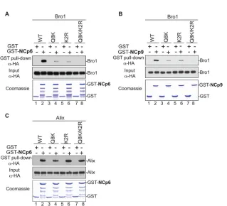
FIG 1Mapping of Alix Bro1 residues involved in the interaction with HIV-1 and EIAV NC domains. (A and B) GST, GST-NCp6, and GST-NCp9 fusion proteins were expressed inE. coli, captured on glutathione-conjugated beads, and subsequently incubated with lysates from 293T cells expressing WT HA-tagged Bro1 domain or the indicated Bro1 mutants (Q8K, K2R, and Q8/K2R). Captured proteins and cell lysates were analyzed by SDS-PAGE and Western blotting. GST fusion proteins were visualized by Coomassie blue staining. (C) Mutations that compromise the NC binding interface in Bro1 do not affect Alix V domain interaction with HIV-1 p6. Pulldown assays were performed as described above, with the only difference being that full-length HA-tagged Alix WT and the indicated mutants were used instead of the isolated Bro1 domain.
on November 7, 2019 by guest
http://jvi.asm.org/
Alix, the Bro1 domain, and the respective mutants were detected using anti-HA monoclonal antibodies (Sigma, St. Louis, MO).
Infectivity assay.Viral infectivity was quantified using TZM-bl cell assays (42). HeLa TZM-bl cells were obtained through the NIH AIDS Research and Reference Reagent Program, Division of AIDS, NIAID, NIH. They were seeded (2⫻104cells) in 96-well plates and the following
day infected in triplicate with HIV-1 PTAP-rescued virus stocks in the presence of 20g/ml DEAE-dextran (Sigma, St. Louis, MO). After 48 h, cells were assayed for luciferase activity using the Steady-Glo reagent kit (Promega, Madison, WI) according to the manufacturer’s instructions. Luminescence was quantified using a microplate reader (Turner BioSys-tems, Sunnyvale, CA).
Immunoprecipitation assays.The immunoprecipitation assays were conducted as previously described (36). Immunoprecipitation complexes and cell lysates (input fractions) were analyzed by SDS-PAGE and West-ern blotting using anti-HA and anti-Flag M2 antibodies (Sigma, St. Louis, MO). To perform the immunoprecipitation assays using the Strep-tagged proteins, cell lysates were incubated with Strep-Tactin Sepharose (IBA GmbH, Gottingen, Germany) for 2 h at 4°C. The matrix was washed five times in lysis buffer and eluted using SDS-PAGE loading buffer.
Pulldown assays and nuclease treatments.The empty pGEX vector or that carrying the coding sequences of HIV-1 NC-p6 and EIAV NC-p9 were expressed in BL21(DE3) pLysSE. coli(Stratagene), and their inter-actions with HA-Bro1and its mutants were examined in GST pulldown assays by following the protocol previously described (36). Where indi-cated, protein complexes captured on beads were incubated for 30 min at 37°C in the presence or absence of 50g/ml RNase A (EMD Chemicals, Inc., San Diego, CA) or 75 U (0.75 U/l) benzonase/nuclease (Novagen) in benzonase buffer (1.2 mM MgCl2, 50 mM Tris-HCl [pH 8.0]). Eluted
complexes and cell lysates (input fractions) were analyzed by SDS-PAGE and Western blotting using the indicated antibodies.
Alix knockdown and reconstitution.293T cells (2.5⫻106cells/ml)
were transfected with 250 pmol of a mixture of two RNA interference (RNAi) oligonucleotides (Invitrogen life Technologies, Grand Island, NY) against cellular Alix. After 36 h, cells were cotransfected with the same amount of RNAi, 500 ng of EIAVUKproviral DNA, and 150 ng of HA-Alix
or RNAi-resistant (RR) HA-Alix mutants. Cells and virus were harvested and processed as described above.
RESULTS
Identification of the NC-Bro1 binding interface.
The N-terminal
Alix Bro1 domain binds NC, while the central V domain binds the
short conserved sequence LYPXnL late (L) domain in the p6
re-gion (
10
–
12
,
31
). Whereas binding determinants and the role of
the latter have been extensively studied and characterized (
12
),
those of the former are not known. We sought to map residues in
the Alix Bro1 domain that mediate binding to HIV-1 and EIAV
NC and examined their role in virus release. Residues in Bro1 that
are accessible to solvent and therefore likely to be exposed and
engaged in protein-protein interactions have been selected using
SAS prediction for mutational analysis. More than 20 residues
displaying high SAS values (see Materials and Methods),
indicat-ing exposure, were found in the first 202 residues of the Alix Bro1
domain, a fragment sufficient to bind NC (
10
). Specifically,
resi-dues in this region were selected for mutational and further
anal-yses based on several criteria, including the following: (i) high SAS
values compared to residues known to be exposed in the Alix Bro1
domain, such as those belonging to the Phe105 loop (
36
), and (ii)
the ability to bind CHMP4. Mutants then were assessed for
cap-ture of NC, and the data obtained are summarized in
Table 1
. We
found that substitutions of either Q8 (glutamine residue in
posi-tion 8 in the Bro1 sequence) and K11 residues (Q8K mutant), or
altering K48, R51, and R56 residues (K2R mutant) to alanines,
caused a significant inhibition of the Bro1 domain interactions
with NC-p6 domains in GST pulldown assays (
Fig. 1A
, lanes 4 and
6). Binding became undetectable when both sets of mutations
were introduced in Bro1 (Q8K/K2R mutant) (
Fig. 1A
, lane 8;
Ta-ble 1
provides nomenclature for the mutants). Similar results were
obtained with EIAV NC-p9 protein (
Fig. 1B
). NC-Bro1
interac-tions had no effect on Alix V-p6 interacinterac-tions, since all defective
Alix Bro1 mutants retained interactions with the NC-p6 fragment
(
Fig. 1C
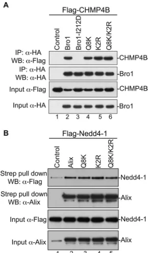
). Furthermore, NC-Bro1 interactions were inhibited
spe-cifically, since all Alix mutants retained binding to their natural
cellular partner CHMP4B, suggesting proper folding (
Fig. 2A
and
Table 1
). Since Alix also binds the ubiquitin ligase Nedd4-1, its
cellular partner that is important for function (
35
), we tested the
effect of mutations in the Bro1 domain on Alix interactions with
Nedd4-1. Q8K, K2R, and Q8K/K2R mutants displayed a Nedd4-1
interaction pattern comparable to their WT Alix counterpart (
Fig.
2B
). Together, these results indicated that residues Q8, K11, K48,
R51, R56, and K60 are part of the Bro1-NC binding interface.
Disruption of Bro1-NC binding inhibits Alix function in
vi-rus release.
To examine the functional significance of the
Bro1-NC interface, we disrupted its residues and assessed the
ef-fect on HIV-1 release using a virus rescue assay (
12
,
39
). Whereas
ectopic expression of WT Alix rescued HIV-1 lacking access to
ESCRT-I (HIV-1 PTAP
⫺mutant) (
Fig. 3A
, lanes 1 and 2), Alix
mutants with a compromised NC binding interface displayed
di-FIG 2Alix Bro1 mutants retain binding to their natural cellular partners. (A) Alix Bro1 mutants bind CHMP4B. 293T cells were cotransfected with Flag-tagged CHMP4B alone (lane 1), in combination with HA-Flag-tagged WT Bro1 (lane 2), or with the indicated mutant (lanes 3 to 6). Cells were lysed in RIPA buffer, and cleared lysates were incubated with anti-HA antibody-conjugated beads. Both input and immunoprecipitated (IP) complexes were analyzed by SDS-PAGE and Western blotting (WB) using the indicated antibody. (B) Alix Bro1 mutants bind Nedd4-1. 293T cells were cotransfected with Flag-tagged Nedd4-1 alone (lane 1), in combination with Strep-tagged WT Alix (lane 2), or with the indicated mutant (lanes 3 to 5). Cells were lysed in RIPA buffer, and cleared lysates were incubated with Strep-Tactin Sepharose. The input and the purified complexes were probed with the anti-Flag and anti-Alix antibodies.
on November 7, 2019 by guest
http://jvi.asm.org/
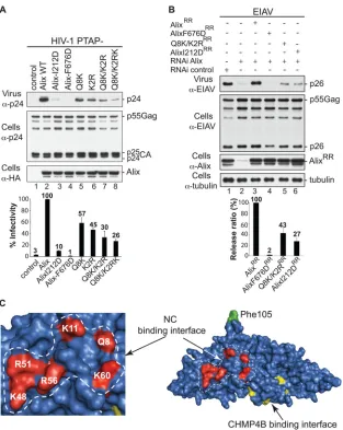
[image:3.585.349.494.66.314.2]minished virus stimulation abilities that were proportional to the
number of mutated residues (
Fig. 3A
, lanes 5 to 8). The effect of
the Q8K/K2RK mutation on viral release was comparable to that
of the I212D mutation, which disrupts Alix binding to the
essen-tial CHMP4 factors (
Fig. 3A
, compare lane 3 to lane 8). Similar
results were obtained when Alix mutants were used to functionally
replace cellular Alix and facilitate EIAV release (
Fig. 3B
). Placing
residues involved in NC-Bro1 interactions in the Bro1 crystal
structure (
12
) revealed a defined interface that exposes a cluster of
basic residues on one side of the boomerang (
Fig. 3C
). Together,
these data draw a direct correlation between the Bro1 domain’s
ability to bind NC and Alix function in virus release and suggest
the first direct functional link between Bro1-NC interactions and
virus release.
Bro1-NC binding involves RNA.
Mapping of the Bro1-NC
binding interface revealed that all residues involved are positively
charged (
Fig. 3C
). This finding was surprising because Bro1-NC
interactions were previously reported to be RNA independent
(
31
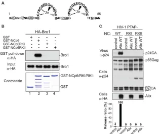
). Moreover, we previously showed that residues in NC
in-volved in association with the Alix Bro1 domain are also positively
charged. Indeed, NC mutants RKI and RKII, carrying alterations
of lysine and arginine (
Fig. 4A
) to alanine residues, ceased to bind
Bro1 and function in virus release (
Fig. 4B
and
C
and references 4,
10, 11, and 36), and these budding defects were alleviated by
pro-FIG 3Alix Bro1 interface involved in NC binding plays a critical role in virus release. (A) The Alix Bro1 mutants with a compromised NC binding interface show a decreased ability to stimulate the release of the HIV-1 PTAP⫺mutant. 293T cells were transfected with HIV-1 PTAP⫺proviral DNA alone (lane 1), with WT HA-tagged Alix (lane 2), or with the indicated Alix mutants (lanes 3 to 8). Cells and viruses were collected 24 h posttransfection, and their protein content was analyzed by SDS-PAGE and Western blotting using the indicated antibodies. Release of infectious viral particles was quantified using HeLa TZM-bl assays from five independent experiments and expressed relative to WT Alix (lane 2). Error bars represent standard deviations (SD). (B) An intact NC binding interface is required for Alix function in EIAV release. 293T cells were transfected twice with Alix RNAi oligonucleotides at 36-h intervals. At the second transfection, cells were cotransfected with EIAV provirus alone (lanes 1 and 2), with an RNAi-resistant (RR) version of Alix WT (lane 3), or with the indicated mutants (lanes 4 to 6). Cells and viruses were collected 24 h posttransfection, and their protein content was analyzed by SDS-PAGE and Western blotting using the indicated antibodies. Release ratios were calculated as described in Materials and Methods from 3 independent experiments and expressed relative to WT Alix (lane 3),
⫾SD. (C) Alix Bro1 residues engaged in NC interaction define a new functional interface. Residues Q8, K11, K48, R51, R56, and K60 placed in the Alix Bro1 domain crystal structure (2OEW) cluster within a positively charged exposed surface (shown in red) on one side of the Bro1 domain. For reference, the Phe105 residue (required for Alix function in HIV-1 release [36]) and the CHMP4B binding interface (27) are shown in green and yellow, respectively.
on November 7, 2019 by guest
http://jvi.asm.org/
[image:4.585.137.450.62.455.2]viding Gag with parallel access via Nedd4.2 expression to the
scis-sion-inducing members of the ESCRT pathway (
11
). This
indi-cated that mutations of basic residues in NC interfered with
NC-Bro1 interactions and interrupted Alix function in scission events.
Involvement of positively charged residues on both NC and Bro1
sides raised the possibility that their interaction involves a
nega-tively charged cellular factor. Since NC binds and incorporates
viral (as well as cellular) RNA into virions (
25
), we elected to test
its potential involvement in Bro1-NC interaction. GST-NC-p6
binding to the Alix Bro1 domain was tested in the presence or
absence of nuclease. Whereas GST-NC-p6 captured the Bro1
do-main as expected (
Fig. 5A
, left, lane 3), treatment of these
com-plexes with nuclease (lane 4) or RNase A (
Fig. 5B
, lanes 5 and 6)
abrogated binding. Similarly, GST-NC-p6 binding to APOBEC3G
(
34
,
44
), a host cell protein known to require RNA for interactions
with NC (
6
,
20
), was equally sensitive to nuclease treatment (
Fig.
5A
, right, lane 3) or RNase treatment (data not shown). Similar
results were obtained with the EIAV NC-p9 construct (
Fig. 5A
,
center). Collectively, these data indicate that RNA is involved and
important for NC-Bro1 interactions.
DISCUSSION
HIV-1 interactions with the ESCRT-associated protein Alix is
suf-ficient for virus production from T lymphocytes (
11
,
13
). Such
recruitment provides the only direct access to the cell membrane
fission machinery. Indeed, the Alix Bro1 domain binds CHMP4
isoforms, which are members of ESCRT-III and play a key role in
ringing and severing viral budding necks (
29
). Two interactions
have been identified between Gag and Alix. The interaction
be-tween the Gag p6 region and the Alix V domain was identified, and
its role in virus release was clearly established (
12
,
37
). The second
interaction that links NC to the Alix Bro1 domain has been
re-cently identified (
10
,
31
). Here, we identified residues in the Alix
Bro1 domain that define the binding interface with NC and found
that RNA is involved in such interactions.
Key determinants of NC-Bro1 interactions.
Since the
discov-ery of NC-Bro1 interactions, questions regarding their role in
vi-rus release arose. Several lines of evidence underscored the
impor-tance of NC-Bro1 interactions, including the findings that a
functional NC is required for Alix-mediated virus release (
4
,
10
,
11
,
31
), and mutant NC viruses that fail to bind the Bro1 domain
were also defective in virus budding (
4
,
10
,
11
). The role of
Bro1-NC interactions in virus release was further strengthened by
the identification of residues that mediate binding. Their
muta-tional analysis revealed that those critical for binding NC lie
within the first beta sheet (

1) and the second alpha helix (
␣
2) of
Bro1. Residues interacting with NC are positively charged and
cluster in a well-defined interface whose disruption was sufficient
to cause a dramatic loss of binding and Alix-mediated virus release
(
Fig. 1
and
3
). This provides direct evidence of a requirement for
contact between Bro1 and NC to achieve efficient virus release.
FIG 4Basic residues in NC are required for the interaction with the Alix Bro1 domain. (A) Schematic representation of the HIV-1 NC domain (amino acids 1 to 55). Lysine (K) and arginine (R) residues replaced with alanine in the RKI and RKII mutants are circled and underlined, respectively. (B) Mutation of basic residues at the N or C terminus of NC prevents NC-Bro1 interaction. GST, GST-NCp6, GST-NCp6RKI, or GST-NCp6RKII fusion protein was expressed inE. coli, captured on glutathione-conjugated beads, and subsequently incubated with lysates from 293T cells expressing the WT HA-Bro1 domain. Captured proteins and cell lysates were analyzed by SDS-PAGE and Western blotting using an anti-HA antibody. GST fusion proteins were visualized by Coomassie blue staining. (C) Alix function in HIV-1 release requires an intact NC domain. 293T cells were transfected with HIV-1 PTAP⫺proviral DNA harboring a WT NC (lanes 1 and 2) or the indicated NC mutant (lanes 3 to 6) either alone (lanes 1, 3, and 5) or with WT HA-tagged Alix (lanes 2, 4, and 6). Cells and viruses were collected 24 h posttransfection, and their protein content was analyzed by SDS-PAGE and Western blotting using the indicated antibodies. Release of viral particles was quantified from two independent experiments.
on November 7, 2019 by guest
http://jvi.asm.org/
[image:5.585.136.450.65.329.2]Why Alix requires binding to both NC and p6 to promote virus
exit is not clear (references
12
and
39
and
Fig. 2
). However, clues
into the necessity for interactions between NC and Bro1 come
from the latter’s ability to recruit CHMP4, which links Gag
di-rectly to membrane fission-inducing ESCRT-III members.
Con-sistent with this notion, Bro1 can function as the smallest unit of
Alix as its ectopic expression promoted virus release, provided it
retained binding to both NC and CHMP4 (
4
,
10
,
11
). Moreover,
an Alix mutant lacking binding to either NC or p6 failed to replace
cellular Alix and promote EIAV budding (
Fig. 3
), further
reaf-firming a key functional role for Bro1-NC interactions in virus
release.
The contribution of Bro1 versus V binding to Gag in Alix
func-tion during budding appears to differ. Indeed, disrupfunc-tion of
inter-actions with V was more detrimental to virus release than that of
Bro1, since the V binding-devoid AlixF676D exhibited less activity
in virus release than the NC binding-defective AlixQ8/K2RK (
Fig.
3
). Conversely, the latter’s defect mirrors interference with
CHMP4 recruitment (I212D mutant), as Alix mutants lacking
either determinants (
Fig. 3
) displayed comparable virus release
defects. Interestingly, AlixF676D lost the ability to locate to sites of
assembly at the membrane (
18
) despite retaining a functional NC
binding interface, suggesting NC-Bro1 interactions take place
only later in the viral egress process (i.e., in the budding neck),
a role in agreement with its functional importance (
Fig. 3
).
These findings suggest a model for Alix function in two steps
(
Fig. 6
). First, Alix is anchored to sites of assembly via p6-V
interactions and Bro1 seems to be unavailable to capture
CHMP4, possibly due to a structural masking (
45
). Next, Bro1
binds NC, possibly in the budding neck, and becomes available
to recruit CHMP4 during budding, a sequence of events that
fits with the recent visualization of CHMP4 at the membrane
once Gag assembly is complete (
18
).
RNA bridges interactions between NC and Bro1.
Identifica-tion of the NC binding interface in Bro1 revealed that residues
involved are positively charged and delineate a well-defined
inter-FIG 5RNA is involved in the Alix Bro1-NC interaction. (A) GST, GST-NCp6 (left), and GST-NCp9 (center) fusion proteins expressed inE. coliand captured on glutathione beads were incubated with lysates from 293T cells expressing HA-tagged WT Bro1 domain or APOBEC3G (right), followed by incubation with or without benzonase/nuclease. (B) GST and GST-NCp6 fusion proteins purified by glutathione beads were incubated with lysates from 293T cells expressing HA-tagged WT Bro1, followed by treatment with or without benzonase (lanes 3 and 4) or RNAse A (lanes 5 and 6). In both experiments, captured proteins and cell lysates were analyzed by SDS-PAGE and Western blotting. GST fusion proteins were visualized by Coomassie blue staining.
FIG 6Schematic representation of a two-step model for Alix function in virus assembly and budding. In the first step, Alix is recruited by Gag to the plasma membrane during assembly through interaction between its V domain and the (L)YPXnL motif in p6 and Alix is thus anchored at the membrane (left). Whether NC binds Bro1 during this step or not is unclear, since it was insufficient to locate Alix to the membrane when the LYPXnL motif was disrupted (18). In the second step, we propose that the NC-Bro1 interaction occurs in the budding neck and is followed (or accompanied) by the recruitment of CHMP4B (right). Basic residues found in both NC and Bro1 proteins mediate binding, and RNA (in red) plays the role of the negatively charged factor that bridges this interaction.
on November 7, 2019 by guest
http://jvi.asm.org/
[image:6.585.140.451.64.299.2] [image:6.585.140.451.588.675.2]face on one side of the Bro1 boomerang domain (
Fig. 3C
).
More-over, mutation of basic residues in NC also eliminated binding to
Bro1 (
4
,
10
,
36
), implying the involvement of a negatively charged
factor. RNA, however, was excluded from Bro1-NC interactions
in a previous report (
31
). This discrepancy is not clear but could be
due to the method employed to capture Bro1 (see Materials and
Methods for buffer composition in salt and detergent) and/or the
fragment used. Indeed, Popov et al. (
31
) used NC-p1 domains to
capture Alix, whereas we used NC-p1-p6 (
Fig. 5
). RNA
neverthe-less was the obvious candidate to bridge interactions between
Bro1 and NC, since the latter binds genomic RNA during
assem-bly (
17
,
22
). In agreement with this, RNA involvement was
con-firmed as nuclease and RNase treatments abrogated NC-Bro1
binding. Similarly, mutations of basic residues in NC that are
in-volved in RNA recruitment also eliminated NC-Bro1 interactions
(
Fig. 5
) and brought Alix function to a near halt (
10
,
35
). Recent
reports employing high-resolution imaging suggested that
genomic RNA localizes to the plasma membrane in the initial
steps of assembly before a Gag nascent complex became visible
(
17
,
22
), whereas recruitment of Alix accompanies Gag
accumu-lation during the early steps of assembly at the cell membrane
(
18
). These observations and our findings suggest that Gag-RNA
assembly complexes recruit Alix, which fits with the involvement
of RNA in interactions with the ESCRT-binding Alix Bro1
do-main and NC. The requirement for RNA in Alix Bro1-NC
inter-actions is not surprising, as NC binding of RNA early in viral
nascent particles is critical for its three-dimensional structure/
folding (
25
), a crucial step for particle assembly as well as the
subsequent steps of recruitment and utilization of ESCRT
com-ponents necessary for virus budding and exit (this study).
In summary, we identified the NC binding interface in Alix
Bro1. The nature of residues involved revealed a critical role for
RNA. Since interactions between Gag, genomic RNA, and Alix
precede CHMP4 recruitment to sites of assembly (
17
,
18
), we
propose a model (
Fig. 6
right) in which an NC-RNA-Bro1
nucleo-protein complex recruits CHMP4 during budding (in the budding
neck) in order for virus exit to proceed.
ACKNOWLEDGMENTS
We thank Sam T. Xiao and Jiansheng Jiang for help with SAS and Alicia Buckler-White and her team at the LMM core for sequencing.
This work was supported by the Intramural Research Program of the NIAID and in part by funds from the Office of AIDS Research (OAR), NIH.
REFERENCES
1.Adachi A, et al.1986. Production of acquired immunodeficiency syn-drome-associated retrovirus in human and nonhuman cells transfected with an infectious molecular clone. J. Virol.59:284 –291.
2.Babst M, Katzmann DJ, Estepa-Sabal EJ, Meerloo T, Emr SD.2002. Escrt-III: an endosome-associated heterooligomeric protein complex re-quired for mvb sorting. Dev. Cell3:271–282.
3.Babst M, Odorizzi G, Estepa EJ, Emr SD. 2000. Mammalian tumor susceptibility gene 101 (TSG101) and the yeast homologue, Vps23p, both function in late endosomal trafficking. Traffic1:248 –258.
4.Bello NF, et al.2012. Budding of retroviruses utilizing divergent L do-mains requires nucleocapsid. J. Virol.86:4182– 4193.
5.Bieniasz PD.2009. The cell biology of HIV-1 virion genesis. Cell Host Microbe5:550 –558.
6.Burnett A, Spearman P.2007. APOBEC3G multimers are recruited to the plasma membrane for packaging into human immunodeficiency virus type 1 virus-like particles in an RNA-dependent process requiring the NC basic linker. J. Virol.81:5000 –5013.
7.Carlton JG, Martin-Serrano J.2007. Parallels between cytokinesis and retroviral budding: a role for the ESCRT machinery. Science316:1908 – 1912.
8.Chen C, Li F, Montelaro RC.2001. Functional roles of equine infectious anemia virus Gag p9 in viral budding and infection. J. Virol.75:9762– 9770.
9.Demirov DG, Freed EO.2004. Retrovirus budding. Virus Res.106:87– 102.
10. Dussupt V, et al.2009. The nucleocapsid region of HIV-1 Gag cooperates with the PTAP and LYPXnL late domains to recruit the cellular machinery necessary for viral budding. PLoS Pathog.5:e1000339. doi:10.1371/ journal.ppat.1000339.
11. Dussupt V, et al.2011. Basic residues in the nucleocapsid domain of Gag are critical for late events of HIV-1 budding. J. Virol.85:2304 –2315. 12. Fisher RD, et al.2007. Structural and biochemical studies of ALIX/AIP1
and its role in retrovirus budding. Cell128:841– 852.
13. Fujii K, et al.2009. Functional role of Alix in HIV-1 replication. Virology 391:284 –292.
14. Garrus JE, et al.2001. Tsg101 and the vacuolar protein sorting pathway are essential for HIV-1 budding. Cell107:55– 65.
15. Huang M, Orenstein JM, Martin MA, Freed EO.1995. p6Gag is required for particle production from full-length human immunodeficiency virus type 1 molecular clones expressing protease. J. Virol.69:6810 – 6818. 16. Huthoff H, Malim MH.2007. Identification of amino acid residues in
APOBEC3G required for regulation by human immunodeficiency virus type 1 Vif and virion encapsidation. J. Virol.81:3807–3815.
17. Jouvenet N, Simon SM, Bieniasz PD.2009. Imaging the interaction of HIV-1 genomes and Gag during assembly of individual viral particles. Proc. Natl. Acad. Sci. U. S. A.106:19114 –19119.
18. Jouvenet N, Zhadina M, Bieniasz PD, Simon SM.2011. Dynamics of ESCRT protein recruitment during retroviral assembly. Nat. Cell Biol. 13:394 – 401.
19. Katoh K, et al.2003. The ALG-2-interacting protein Alix associates with CHMP4b, a human homologue of yeast Snf7 that is involved in multive-sicular body sorting. J. Biol. Chem.278:39104 –39113.
20. Khan MA, et al.2007. Analysis of the contribution of cellular and viral RNA to the packaging of APOBEC3G into HIV-1 virions. Retrovirology 4:48. doi:10.1186/1742-4690-4-48.
21. Kim J, et al.2005. Structural basis for endosomal targeting by the Bro1 domain. Dev. Cell8:937–947.
22. Kutluay SB, Bieniasz PD.2010. Analysis of the initiating events in HIV-1 particle assembly and genome packaging. PLoS Pathog.6:e1001200. doi: 10.1371/journal.ppat.1001200.
23. Lee B, Richards FM.1971. The interpretation of protein structures: esti-mation of static accessibility. J. Mol. Biol.55:379 – 400.
24. Li F, Chen C, Puffer BA, Montelaro RC.2002. Functional replacement and positional dependence of homologous and heterologous L domains in equine infectious anemia virus replication. J. Virol.76:1569 –1577. 25. Lu K, Heng X, Summers MF.2011. Structural determinants and
mech-anism of HIV-1 genome packaging. J. Mol. Biol.410:609 – 633. 26. Martin-Serrano J, Zang T, Bieniasz PD.2003. Role of ESCRT-I in
ret-roviral budding. J. Virol.77:4794 – 4804.
27. McCullough J, Fisher RD, Whitby FG, Sundquist WI, Hill CP.2008. ALIX-CHMP4 interactions in the human ESCRT pathway. Proc. Natl. Acad. Sci. U. S. A.105:7687–7691.
28. Montelaro RC, Parekh B, Orrego A, Issel CJ.1984. Antigenic variation during persistent infection by equine infectious anemia virus, a retrovirus. J. Biol. Chem.259:10539 –10544.
29. Morita E, et al.2011. ESCRT-III protein requirements for HIV-1 bud-ding. Cell Host Microbe9:235–242.
30. Morita E, Sundquist WI.2004. Retrovirus budding. Annu. Rev. Cell Dev. Biol.20:395– 425.
31. Popov S, Popova E, Inoue M, Gottlinger HG.2008. Human immuno-deficiency virus type 1 Gag engages the Bro1 domain of ALIX/AIP1 through the nucleocapsid. J. Virol.82:1389 –1398.
32. Rusten TE, Vaccari T, Stenmark H.2012. Shaping development with ESCRTs. Nat. Cell Biol.14:38 – 45.
33. Saff EB, Kuijlaars ABJ. 1997. Distributing many points on a sphere. Mathematical Intelligencer19:5–11.
34. Schafer A, Bogerd HP, Cullen BR. 2004. Specific packaging of APOBEC3G into HIV-1 virions is mediated by the nucleocapsid domain of the gag polyprotein precursor. Virology328:163–168.
35. Sette P, Jadwin JA, Dussupt V, Bello NF, Bouamr F.2010. The
on November 7, 2019 by guest
http://jvi.asm.org/
associated protein Alix recruits the ubiquitin ligase Nedd4-1 to facilitate HIV-1 release through the LYPXnL L domain motif. J. Virol.84:8181– 8192.
36. Sette P, et al.2011. The Phe105 loop of Alix Bro1 domain plays a key role in HIV-1 release. Structure19:1485–1495.
37. Strack B, Calistri A, Craig S, Popova E, Gottlinger HG.2003. AIP1/ALIX is a binding partner for HIV-1 p6 and EIAV p9 functioning in virus bud-ding. Cell114:689 – 699.
38. Stuchell-Brereton MD, et al. 2007. ESCRT-III recognition by VPS4 ATPases. Nature449:740 –744.
39. Usami Y, Popov S, Gottlinger HG.2007. Potent rescue of human im-munodeficiency virus type 1 late domain mutants by ALIX/AIP1 depends on its CHMP4 binding site. J. Virol.81:6614 – 6622.
40. VerPlank L, et al.2001. Tsg101, a homologue of ubiquitin-conjugating
(E2) enzymes, binds the L domain in HIV type 1 Pr55(Gag). Proc. Natl. Acad. Sci. U. S. A.98:7724 –7729.
41. von Schwedler UK, et al.2003. The protein network of HIV budding. Cell 114:701–713.
42. Wei X, et al.2002. Emergence of resistant human immunodeficiency virus type 1 in patients receiving fusion inhibitor (T-20) monotherapy. Antimicrob. Agents Chemother.46:1896 –1905.
43. Winn MD, et al.2011. Overview of the CCP4 suite and current develop-ments. Acta Crystallogr. D Biol. Crystallogr.67:235–242.
44. Zennou V, Perez-Caballero D, Gottlinger H, Bieniasz PD. 2004. APOBEC3G incorporation into human immunodeficiency virus type 1 particles. J. Virol.78:12058 –12061.
45. Zhai Q, et al.2011. Activation of the retroviral budding factor ALIX. J. Virol.85:9222–9226.
on November 7, 2019 by guest
http://jvi.asm.org/