Computer assisted diagnosis techniques (dermoscopy and spectroscopy-based)
for the diagnosis of skin cancer in adults
Review information
Review type: Diagnostic test accuracy
Review number: #164a
Authors
Lavinia Ferrante di Ruffano1, Yemisi Takwoingi1, Jacqueline Dinnes1, Naomi Chuchu1, Susan E Bayliss1, Clare Davenport1, Rubeta N Matin2, Kathie Godfrey3, Colette O'Sullivan3, Abha Gulati4, Sue Ann Chan5, Alana Durack6, Susan O'Connell7, Matthew D Gardiner8, Jeffrey Bamber9, Jonathan J Deeks1, Hywel C Williams10, Cochrane Skin Cancer Diagnostic Test Accuracy Group1
1Institute of Applied Health Research, University of Birmingham, Birmingham, UK 2Department of Dermatology, Churchill Hospital, Oxford, UK
3c/o Cochrane Skin Group, The University of Nottingham, Nottingham, UK 4Department of Dermatology, Barts Health NHS Trust, London, UK 5Birmingham Skin Centre, City Hospital, Birmingham, UK
6Dermatology, Addenbrooke’s Hospital, Cambridge University Hospitals NHS Foundation Trust, Cambridge, UK 7CEDAR Healthcare Technology Research Centre, Cardiff and Vale University Health Board, Cardiff, UK 8Kennedy Institute of Rheumatology, University of Oxford, Oxford, UK
9Joint Department of Physics, Institute of Cancer Research and The Royal Marsden NHS Foundation Trust, Sutton, UK 10Centre of Evidence Based Dermatology, University of Nottingham, Nottingham, UK
Citation example: Ferrante di Ruffano L, Takwoingi Y, Dinnes J, Chuchu N, Bayliss SE, Davenport C, Matin RN, Godfrey K, O'Sullivan C, Gulati A, Chan SA, Durack A, O'Connell S, Gardiner MD, Bamber J, Deeks JJ, Williams HC, Cochrane Skin Cancer Diagnostic Test Accuracy Group. Computer assisted diagnosis techniques (dermoscopy and spectroscopy-based) for the diagnosis of skin cancer in adults. Cochrane Database of Systematic Reviews , Issue . Art. No.: . DOI: .
Contact person
Jacqueline Dinnes
Institute of Applied Health Research University of Birmingham
Birmingham B15 2TT UK
E-mail: j.dinnes@bham.ac.uk
Dates
dermoscopic suspicion of malignancy, CAD may reduce unnecessary excisions without missing melanoma cases.
Objectives
To determine the accuracy of CAD systems for diagnosing cutaneous invasive melanoma and atypical intraepidermal melanocytic variants, BCC or cSCC in adults, and to compare its accuracy with that of dermoscopy when dermoscopy is also evaluated in CAD studies.
Search methods
We undertook a comprehensive search of the following databases from inception up to August 2016: Cochrane Central Register of Controlled Trials; MEDLINE; EMBASE; CINAHL; CPCI; Zetoc; Science Citation Index; US National Institutes of Health Ongoing Trials Register; NIHR Clinical Research Network Portfolio Database; and the World Health Organization International Clinical Trials Registry Platform. We studied reference lists and published systematic review articles.
Selection criteria
Studies of any design that evaluated CAD alone, or in comparison with dermoscopy, in adults with lesions suspicious for melanoma or BCC or cSCC, and compared with a reference standard of either histological confirmation or clinical follow-up.
Data collection and analysis
Two review authors independently extracted all data using a standardised data extraction and quality assessment form (based on QUADAS-2). We contacted authors of included studies where information related to the target condition or diagnostic threshold were missing. We estimated summary sensitivities and specificities separately by type of CAD system using the bivariate hierarchical model. Comparisons of CAD with dermoscopy were made using a) all available CAD data (indirect comparisons), and b) studies providing paired data for both tests (direct comparisons). The contribution of human decision making to the accuracy of CAD diagnoses was examined in a sensitivity analysis by removing studies that gave CAD results to clinicians to guide diagnostic decision-making.
Main results
In total 42 studies were included, 24 evaluating digital dermoscopy based CAD systems (Derm–CAD) in 23 study cohorts with 9602 lesions (1220 melanomas, at least 83 BCCs, 9 cSCCs), providing 32 datasets for Derm–CAD and 7 for dermoscopy. Eighteen studies evaluated spectroscopy based CAD (Spectro–CAD) in 16 study cohorts with 6336 lesions (934 melanomas, 163 BCC, 49 cSCCs), providing 32 datasets for Spectro–CAD and 6 for dermoscopy. These consisted of 15 studies using multispectral imaging (MSI), 2 studies using electrical impedance spectroscopy (EIS) and 1 study using diffuse reflectance spectroscopy. Studies were incompletely reported and of unclear to high risk of bias across all domains. Included studies inadequately address the review question due to an abundance of low quality studies, poor reporting, and recruitment of highly selected groups of participants.
Across all CAD systems, considerable variation was encountered in the hardware and software technologies used, the types of classification algorithm employed, methods used to train the algorithms, and which lesion morphological features were extracted and analysed across all CAD systems, and even between studies evaluating CAD systems. Meta–analysis found CAD systems had high sensitivity for correct identification of cutaneous invasive melanoma and atypical intraepidermal melanocytic variants in highly selected populations, but with low and very variable specificity, particularly for Spectro–CAD systems. Pooled data from 22 studies estimated the sensitivity of Derm–CAD for the detection of melanoma as 90.1% (95% CI: 84.0% to 94.0%) and specificity as 74.3% (95% CI: 63.6% to 82.7%). Pooled data from 8 studies estimated the sensitivity of multispectral imaging CAD (MSI–CAD) as 92.9% (95% CI: 83.7% to 97.1%) and specificity as 43.6% (95% CI: 24.8% to 64.5%). When applied to a hypothetical population of 1000 lesions at the mean observed melanoma prevalence of 20%, Derm–CAD would miss 20 melanomas and would lead to 206 false positive results for melanoma. MSI–CAD would miss 14 melanomas and would lead to 451 false diagnoses for melanoma. Preliminary findings suggest CAD systems are at least as sensitive as assessment of dermoscopic images for the diagnosis of invasive melanoma and atypical intraepidermal
melanocytic variants. It is not possible to make summary statements regarding the use of CAD in unreferred populations, or its accuracy in detecting keratinocyte cancers, or its use in any setting as a diagnostic aid, because of the paucity of studies.
Authors' conclusions
In highly selected patient populations all CAD types demonstrate high sensitivity, and could prove useful as a back-up for specialist diagnosis to assist in minimising the risk of missing melanomas. However, the evidence–base is currently too poor to understand whether CAD system outputs translate to different clinical decision–making in practice. Insufficient data are available on the use of CAD in community settings, or for the detection of keratinocyte cancers. The evidence–base for individual systems is too limited to draw conclusions on which might be preferred for practice. Prospective comparative studies are required that evaluate the use of already evaluated CAD systems as diagnostic aids, by comparison to
face–to–face dermoscopy, and in participant populations that are representative of those in which the test would be used in practice.
Plain language summary
What is the diagnostic accuracy of computer–assisted diagnosis techniques for the detection of skin cancer in
adults?
carcinoma (BCC). Melanoma is one of the most dangerous forms. If it is not recognised early treatment can be delayed and this risks the melanoma spreading to other organs in the body and may lead to eventual death. Cutaneous squamous cell carcinoma (cSCC) and basal cell carcinoma (BCC) are considered less dangerous as they are localised (less likely to spread to other parts of the body compared to melanoma). However, cSCC can spread to other parts of the body and BCC can cause disfigurement if not recognised early. Diagnosing a skin cancer when it is not actually present (a false positive result) might result in unnecessary surgery and other investigations and can cause stress and anxiety to the patient. Missing a diagnosis of skin cancer may result in the wrong treatment being used or lead to a delay in effective treatment.
What is the aim of the review?
The aim of this Cochrane Review was to find out how accurate computer–assisted diagnosis (CAD) is for diagnosing
melanoma, BCC or cSCC. The review also compared the accuracy of two different types of CAD and compared the accuracy of CAD with diagnosis by a doctor using a handheld illuminated microscope (a dermatoscope or ‘dermoscopy’). Researchers in Cochrane included 42 studies to answer these questions.
What was studied in the review?
A number of tools are available to skin cancer specialists which allow a more detailed examination of the skin compared to examination by the naked eye alone. Currently a dermatoscope which magnifies the skin lesion using a bright light source is used by most skin cancer specialists. CAD tests are computer systems that analyse information about skin lesions obtained from a dermatoscope or other techniques that use light to describe the features of a skin lesion (spectroscopy) to produce a result indicating whether skin cancer is likely to be present. CAD systems that get their information from dermoscopic images of lesions (Derm–CAD), or that use data from spectroscopy, were included in this review. Most of the spectroscopy studies used data from multispectral imaging (MSI–CAD) and are the main focus here. Results from CAD systems can be used alone to make a diagnosis of skin cancer (CAD–based diagnosis), or can be used by doctors in addition to their visual inspection examination of a skin lesion to help them reach a diagnosis (CAD–aided diagnosis). Researchers examined how useful CAD systems are to help diagnose skin cancers in addition to visual inspection and dermoscopy.
What are the main results of the review?
The review included 42 studies looking at CAD systems for the diagnosis of melanoma. There was not enough evidence to determine the accuracy of CAD systems for the diagnosis of BCC (3 studies) or cSCC (1 study).
Derm–CAD results for diagnosis of melanoma
The main results for Derm-CAD are based on 22 studies including 8992 lesions.
Applied to a group of 1000 skin lesions, of whom 200 (20%) actually do have melanoma, the results suggest that:
- An estimated 386 people will have a Derm–CAD result suggesting that a melanoma is present and of these 206 (53%) will not actually have a melanoma (false positive result)
- Of the 614 people with a Derm–CAD result indicating that no melanoma is present, 20 (3%) will in fact actually have a melanoma (false negative result)
There was no evidence to suggest that dermoscopy or Derm–CAD was different in its ability to detect or rule out melanoma. MSI–CAD results for diagnosis of melanoma
The main results for MSI–CAD are based on 8 studies including 2401 lesions. In a group of 1000 people, of whom 200 (20%) actually do have melanoma, then:
- An estimated 637 people will have an MSI–CAD result suggesting that a melanoma is present and of these 451 (71%) will not actually have a melanoma (false positive result)
- Of the 363 people with an MSI–CAD result indicating that no melanoma is present, 14 (4%) will in fact actually have a melanoma (false negative result)
melanomas than doctors using dermoscopy images. However, CAD systems also produced far more false positive diagnoses than dermoscopy and could lead to considerable increases in unnecessary surgery. The performance of CAD systems for detecting BCC and cSCC skin cancers is unclear. More studies are needed to evaluate the use of CAD by doctors for the diagnosis of skin cancer in comparison to face-to-face diagnosis using dermoscopy, in both primary care and in specialist skin cancer clinics.
How up-to-date is this review?
The review authors searched for and used studies published up to August 2016.
*In these studies, biopsy, clinical follow up, or specialist clinician diagnosis were the reference standards.
Background
This review is one of a series of Cochrane Diagnostic Test Accuracy (DTA) reviews on the diagnosis and staging of melanoma and keratinocyte skin cancers conducted for the National Institute for Health Research (NIHR) Cochrane Systematic Reviews Programme. Appendix 1 shows the content and structure of the programme. Table 1 provides a glossary of terms used and a table of acronyms used is provided in Appendix 2.
Target condition being diagnosed
There are three main forms of skin cancer. Melanoma is the most widely known amongst the general population, yet the commonest skin cancers in Caucasian populations are those arising from keratinocytes: basal cell carcinoma (BCC) and cutaneous squamous cell carcinoma (cSCC) (Gordon 2013; Madan 2010). In 2003, the World Health Organization estimated that between two and three million ‘non-melanoma’ skin cancers (of which BCC and cSCC are estimated to account for around 80% and 16% of cases, respectively) and 132,000 melanoma skin cancers occur globally each year (WHO 2003).
In this diagnostic test accuracy review there are three target conditions of interest (a) melanoma, (b) basal cell carcinoma (BCC), and (c) cutaneous squamous cell carcinoma (cSCC).
Melanoma
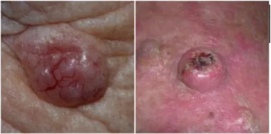
Melanoma arises from uncontrolled proliferation of melanocytes - the epidermal cells that produce pigment or melanin. Cutaneous melanoma refers to any skin lesion with malignant melanocytes present in the dermis, and includes superficial spreading, nodular, acral lentiginous, and lentigo maligna melanoma variants (see Figure 1). Melanoma in situ refers to malignant melanocytes that are contained within the epidermis and have not invaded the dermis, but are at risk of
progression to melanoma if left untreated. Lentigo maligna, a subtype of melanoma in situ in chronically sun-damaged skin, denotes another form of proliferation of abnormal melanocytes. Lentigo maligna can progress to invasive melanoma if its growth breaches the dermo-epidermal junction during a vertical growth phase (when it is a 'lentigo maligna melanoma'). However its malignant transformation is both lower and slower than for melanoma in situ (Kasprzak 2015). Melanoma in situ and lentigo maligna are both atypical intraepidermal melanocytic variants. Melanoma is one of the most serious forms of skin cancer, with the potential to metastasise to other parts of the body via the lymphatic system and bloodstream. It accounts for only a small proportion of skin cancer cases but is responsible for up to 75% deaths (Boring 1994; Cancer Research UK 2017).
The incidence of melanoma rose to over 200,000 newly diagnosed cases worldwide in 2012 (Erdmann 2013; Ferlay 2015
), with an estimated 55,000 deaths (Ferlay 2015). In the UK, melanoma has one of the fastest rising incidence rates of any cancer, and has the biggest projected increase in incidence between 2007 and 2030 (Mistry 2011). In the decade leading up to 2013, age standardised incidence increased by 46%, with 14,500 new cases in 2013 and 2,459 deaths in 2014 (Cancer Research UK 2017). Rates are higher in women than in men; however, the rate of incidence in men is increasing faster than in women (Arnold 2014).The rising incidence in melanoma is thought to be primarily related to an increase in recreational sun exposure and tanning bed use and an increasingly ageing population with higher lifetime recreational ultraviolet (UV) exposure, in conjunction with possible earlier detection (Belbasis 2016; Linos 2009
). Putative risk factors are reviewed in detail elsewhere (Belbasis 2016).
A database of over 40,000 US patients from 1998 onwards which assisted the development of the 8th American Joint Committee on Cancer (AJCC) Staging System indicated a five-year survival of 97% to 99% for stage I melanoma, dropping to between 32% and 93% in stage III disease depending on tumour thickness, the
presence of ulceration and number of involved nodes (Gershenwald 2017). While these are substantial increases relative to survival in 1975 (Cho 2014), mortality rates have remained static during the same period. This observation coupled with increasing incidence of localised disease, suggests that improvements in survival may be due to earlier detection and heightened vigilance (Cho 2014). Targeted therapies for stage IV melanoma (e.g. BRAF inhibitors) have
improved survival expectation and immunotherapies are evolving such that long term survival is being documented (e.g. using BRAF-inhibitors (Chapman 2012; Villanueva 2010) and MEK inhibitors (Dummer 2014; Larkin 2014), and immunomodulation (Chapman 2011; Hamid 2013; Hodi 2010).
Basal cell carcinoma
BCC can arise from multiple stem cell populations, including from the bulge and interfollicular epidermis (Grachtchouk 2011
slow-The diagnosis is often made incidentally rather than by people presenting with symptoms (Gordon 2013).
BCC most commonly occurs on sun-exposed sites on the head and neck (McCormack 1997) and are more common in men and in people over the age of 40. A rising incidence of BCC in younger people has been attributed to increased recreational sun exposure (Bath-Hextall 2007a; Gordon 2013; Musah 2013). Other risk factors include Fitzpatrick skin types I and II (Fitzpatrick 1975; Lear 1997; Maia 1995), previous skin cancer history, immunosuppression, arsenic exposure, and genetic predisposition such as in basal cell naevus (Gorlin) syndrome (Gorlin 2004; Zak-Prelich 2004). Annual incidence is increasing worldwide; Europe has experienced an average increase of 5.5% per year over the last four decades, the USA 2% per year, while estimates for the UK show incidence appears to be increasing more steeply at a rate of an additional 6 / 100,000 persons per year (Lomas 2012). The rising incidence has been attributed to an ageing population, changes in the distribution of known risk factors, particularly ultraviolet radiation, and
improved detection due to the increased awareness amongst both practitioners and the general population (Verkouteren 2017). Hoorens 2016 points to evidence for a gradual increase in the size of BCCs over time, with delays in diagnosis ranging from 19 to 25 months.
According to National Institute for Health and Care Excellence (NICE) guidance (NICE 2010), low risk BCCs that may be considered for excision are nodular lesions occurring in patients older than 24 years old who are not
immunosuppressed and do not have Gorlin syndrome. Furthermore, low risk lesions should be located below the clavicle, should be small (< 1 cm) with well-defined margins, not recurrent following incomplete excision and are not difficult to reach surgically or in highly visible locations (NICE 2010). Superficial BCCs are also typically low risk
and may be amenable to medical treatments such as photodynamic therapy or topical chemotherapy (Kelleners-Smeets 2017). Assigning BCCs as low or high risk influences the management options (Batra 2002; Randle 1996).
Advanced locally destructive BCC can arise from long-standing untreated lesions or from a recurrence of aggressive basal cell carcinoma after primary treatment (Lear 2012). Very rarely, BCC metastasises to regional and distant sites resulting in death, especially cases of large neglected lesions in those who are immunosuppressed or those with Gorlin syndrome (McCusker 2014). Rates of metastasis are reported at 0.0028% to 0.55% (Lo 1991), with very poor survival rates. It is recognised that basosquamous carcinoma (more like a high risk cSCC in behaviour and not considered a true BCC) is likely to have accounted for many cases of apparent metastases of BCC hence teh spuriously high reported incidence in some studies of up to 0.55% which is not seen in clinical practice (Garcia 2009).
Squamous cell carcinoma of the skin
Primary cSCC arises from the keratinocytes in the epidermis or its appendages. People with cSCC often present with an ulcer or firm (indurated) papule, plaque, or nodule (Griffin 2016) often with an adherent crust (Madan 2010). cSCC can arise in the absence of a precursor lesion or it can develop from pre-existing actinic keratosis (dysplastic epidermis) or Bowen's disease (considered by some to be cSCC in situ). The estimated annual risk of progression is <1% to 20% (Alam 2001) and 5% for lesions developing from pre–existing dysplasia (Kao 1986). It remains locally invasive for a
variable length of time, but has the potential to spread to the regional lymph nodes or via the bloodstream to distant sites, especially in immunosuppressed individuals (Lansbury 2010). High risk lesions are those arising on the lip or ear, recurrent cSCC, lesions arising on non-exposed sites, scars or chronic ulcers, tumours larger than 20mm in diameter or which have a histological depth of invasion greater than 4mm or poor differentiation status on histopathological examination (Motley 2009).
Chronic ultraviolet light exposure through recreation or occupation is strongly linked to cSCC occurrence (Alam 2001). It is particularly common in people with fair skin and in less common genetic disorders of pigmentation, such as albinism, xeroderma pigmentosum, and recessive dystrophic epidermolysis bullosa (RDEB) (Alam 2001). Other recognised risk factors include immunosuppression; chronic wounds; arsenic or radiation exposure; certain drug treatments, such as voriconazole and BRAF mutation inhibitors; and previous skin cancer history (Baldursson 1993; Chowdri 1996;
surrounding healthy skin might lead to considerable functional impairment (Bath-Hextall 2007b; Motley 2009; Lansbury 2010;
Stratigos 2015). Bath-Hextall and colleagues (Bath-Hextall 2007b) found a single trial comparing Mohs micrographic surgery with standard excision in BCC (Smeets 2004); the update at 10 years follow-up showed no statistically significant difference in recurrence with Mohs micrographic surgery (4.4% compared to 12.2% after surgical excision, P = 0.10) (van Loo 2014).
The main treatments for high risk BCC are wide local excision, Mohs micrographic surgery and radiotherapy. For low risk or superficial subtypes of BCC, or for small and or multiple BCCs at low risk sites (Marsden 2010), destructive techniques other than excisional surgery may be used (e.g. electrodesiccation and curettage or cryotherapy (Alam 2001;
Bath-Hextall 2007b)). Alternatively non-surgical (or non-destructive) treatments may be considered (Bath-Hextall 2007a;
Drew 2017; Kim 2014), including topical chemotherapy such as imiquimod (Williams 2017), 5-fluorouracil (Arits 2013), ingenol mebutate (Nart 2015) and photodynamic therapy (Bath-Hextall 2007b; Roozeboom 2016). Although non-surgical techniques are increasingly used, they do not allow histological confirmation of tumour clearance, and their use is dependent on accurate characterisation of the histological subtype and depth of tumour. The 2007 systematic review of BCC interventions found limited evidence from very small RCTs for these approaches (Bath-Hextall 2007b), which have only partially been addressed by subsequent studies (Bath-Hextall 2014; Kim 2014; Roozeboom 2012). Most BCC trials have compared interventions within the same treatment class, and few have compared medical versus surgical treatments (Kim 2014).
Vismodegib, a first-in-class Hedgehog signalling pathway inhibitor is now available for the treatment of metastatic or locally advanced BCC based on the pivotal study ERIVANCE BCC (Sekulic 2012). It is licensed for use in these patients where surgery or radiotherapy is inappropriate, e.g. for treating locally advanced periocular and orbital BCCs with orbital salvage of patients who otherwise would have required exenteration (Wong 2017). However, NICE has
recently recommended against the use of vismodegib based on cost effectiveness and uncertainty of evidence (NICE 2017). A systematic review of interventions for primary cSCC found only one RCT eligible for inclusion (Lansbury 2010). Current practice therefore relies on evidence from observational studies, as reviewed in Lansbury 2013, for example. Surgical excision with pre-determined margins is usually the first-line treatment (Motley 2009; Stratigos 2015). Estimates of recurrence after Mohs micrographic surgery, surgical excision, or radiotherapy, which are likely to have been evaluated in higher risk populations, have shown pooled recurrence rates of 3%, 5.4% and 6.4%, respectively with overlapping confidence intervals; the review authors advise caution when comparing results across treatments (Lansbury 2013).
Index test(s)
Computer–aided diagnosis (CAD) describes a range of artificial intelligence-based techniques that automate the diagnosis of skin cancer by using a computer to analyse lesion images, and determine the likelihood of malignancy, or need for excision. Each CAD system has a data collection component, which collects imaging or non-visual data (e.g. electrical impedance measurements) from the suspicious lesion and feeds it to the data processing component, which then performs a series of analyses to arrive at a diagnostic classification.
Images are acquired using a number of different techniques, though most commonly by digital dermoscopy (Derm–CAD) which creates digital subsurface images of the skin using a computer coupled with a dermatoscope, videocamera and digital television (Rajpara 2009; Esteva 2017). Commercially available systems include the DB–MIPS® (DB–Dermo MIPS) (Biomips Engineering SRL, Sienna Italy), MicroDERM (Visiomed AG, Germany), SolarScan
(Polartechnics Ltd, Australia) and MoleExpert (DermoScan GmbH, Germany), all of which are hand–held digital or video dermatoscopes that communicate with CAD analysis software (see Figure 3).
Other systems use spectroscopy (Spectro-CAD), whereby information on cell characteristics (such as cell shape or size) is gathered by measuring how electromagnetic waves pass through skin lesions. This information is most commonly acquired using multispectral imaging (MSI–CAD) that enable computer–generated graphic representations of lesion morphology to be produced from detecting light reflected at several wavelengths across the lesion. By far the most common of these is diffuse reflectance spectrophotometry imaging (DRSi), which uses light that diffusely penetrates the skin to a depth of 2–2.5mm beneath the surface to produce light reflectance images at a number of specific wavelengths across the visible – near infrared light spectrum (approximately 400–1000nm) to capture variations in light attenuation and scattering from melanin, collagen and blood vessel structures.
DRSi developed from diffuse reflectance spectroscopy, a non-visual spectroscopic technique which uses optical reflectance to distinguish between lesion types based on spectral shape and calibrated level of reflected light for wavelengths
continuously varying from the ultraviolet (320 nm) to the near infrared (1100 nm) with a high spectral resolution (4 nm) (e.g.
Marchesini 1992; Wallace 2000b). Commercially available DRSi computer aided diagnosis (CAD) systems include the SIAscope™ (MedX Health Corp, Canada), a hand–held unit that communicates with CAD analysis software (Figure 3). The MelaFind® system (Strata Skin Sciences (formerly Mela Sciences Inc), Horsham, PA, USA) was FDA approved; however, it no longer appears to be commercially available.
The Nevisense™ system (SciBase III, Sweden; Figure 3) is also commercially available, but is based on electrical
through the extracellular space. The spectral shape is therefore sensitive to cellular components and dimensions, internal structure and cellular arrangements. The Nevisense™ EIS system measures at 4 multiple depths and at 35 frequencies logarithmically distributed from 1.0 kHz to 2.5 MHz using a 5 x 5 mm area electrode covered in tiny pins that penetrate into the stratum corneum.
Other non-visual sources of lesion data include Raman spectroscopy, in which a laser is used to excite vibrations in molecules which then impart wavelength shifts to some of the scattered light waves, creating spectral patterns that are related to the molecular structure of lesions (Maglogiannis 2009), and fluorescence spectroscopy which uses a laser to excite electrons, causing molecules to absorb and then re-emit light in spectral patterns that are also related to the molecular structure of lesions (Rallan 2004).
All CAD systems use machine learning, where a classification algorithm learns features of groups of lesions (i.e. diagnostic types) by exposure to a ‘training set’ of lesions of known histological diagnosis. This process creates a model which is designed to distinguish between these lesion types in future observations. Examples of machine learning algorithms include discriminant analysis, decision trees, neural networks, fuzzy logic, nearest k-neighbours, logistic regression and support vector machines (SVMs), and all use different
mathematical equations to set out how observed features relate to a given diagnosis (Maglogiannis 2009; Masood 2013). Model outputs also vary, in part according to the type of data used to acquire lesion information, and can take the form of binary outputs indicating the presence of malignancy versus benignity (e.g. the Melafind® system), risk scores which can be used at varying thresholds (e.g. the DANAOS system used by MicroDerm), or graphical representations of the CAD pattern analysis which highlight areas of concern within a lesion (e.g. the SIAgraphs produced by SIAscope™). Artificial intelligence systems using continuous learning algorithms, where computer systems continuously develop their classification algorithm as each new case is examined, and do not stop learning at the end of a training period, are not addressed in this review.
Clinical Pathway
The diagnosis of skin lesions occurs in primary, secondary, and tertiary care settings by both generalist and specialist healthcare providers. In the UK, people with concerns about a new or changing lesion will present to their general practitioner rather than directly to a specialist in secondary care. A general practitioner with clinical concerns usually refers a patient to a specialist in secondary care – usually a dermatologist but sometimes to a surgical specialist such as a plastic surgeon or an ophthalmic surgeon. Suspicious skin lesions may also be identified in a referral setting, for
example by a general surgeon, and referred for a consultation with a skin cancer specialist (Figure 4). Skin cancers identified by other specialist surgeons (such as an ear, nose, and throat (ENT) specialist or maxillofacial surgeon will usually be diagnosed and treated without further referral.
Current UK guidelines recommend that all suspicious pigmented lesions presenting in primary care should be assessed by taking a clinical history and visual inspection using the seven-point checklist (MacKie 1990); lesions
suspected to be melanoma or cSCC should be referred for appropriate specialist assessment within two weeks (Chao 2013;
Marsden 2010; NICE 2015). Evidence is emerging, however, to suggest that excision of melanoma by GPs is not associated with increased risk compared with outcomes in secondary care (Murchie 2017). In the UK, low risk BCC are usually recommended for routine referral, with urgent referral for those in whom a delay could have a significant impact on outcomes, for example due to large lesion size or critical site (NICE 2015). Appropriately qualified generalist care providers increasingly undertake management of low risk BCC in the UK, such as by excision of low risk lesions (NICE 2010). Similar guidance is in place in Australia (CCAAC Network 2008).
For referred lesions, the specialist clinician will use history-taking, visual inspection of the lesion (in conjunction with other skin lesions), palpation of the lesion and associated lymph nodes in conjunction with dermoscopic examination to inform a clinical decision. If melanoma is suspected, then urgent 2mm excision biopsy is
using a hand-held microscope has become the most widely used tool for clinicians to improve diagnostic
accuracy of pigmented lesions, in particular for melanoma (Argenziano 1998; Argenziano 2012; Haenssle 2010; Kittler 2002), although it is less well established for the diagnosis of BCC or cSCC. Dermoscopy is frequently combined with visual inspection of a lesion in secondary care settings, and is also increasingly used in primary care, particularly in countries such as Australia (Youl 2007).The diagnostic accuracy, and comparative accuracy, of visual inspection and dermoscopy have been evaluated in a further three reviews in this series (Dinnes 2018a; Dinnes 2018b; Dinnes 2018c). Consideration of the degree of prior testing that study participants have undergone is key to interpretation of test accuracy indices, as these are known to vary according to the disease spectrum (or case-mix) of included participants (Lachs 1992; Moons 1997; Leeflang 2013; Usher-Smith 2016). Spectrum effects are often observed when tests that are developed further down the referral pathway have lower sensitivity and higher specificity when applied in settings with participants with limited prior testing (Usher-Smith 2016). Studies of individuals with suspicious lesions at the initial clinical presentation stage ('test naïve') are likely to have a wider range of differential diagnoses and include a higher proportion of people with benign diagnoses compared with studies of participants who have been referred for a specialist opinion on the basis of visual inspection (with or without dermoscopy) by a generalist practitioner. Furthermore, studies in more specialist settings may focus on equivocal or difficult to diagnose lesions rather than lesions with a more general level of clinical suspicion. However this direction of effect is not consistent across tests and diseases, the mechanisms in action often being more complex than prevalence alone and can be difficult to
identify (Leeflang 2013). A simple categorisation of studies according to primary, secondary or specialist setting therefore may not always adequately reflect these key differences in disease spectrum that can affect test performance.
Role of index test(s)
Skin cancer diagnosis, whether by visual inspection alone or with the use of dermoscopy is undertaken iteratively, using both implicit pattern recognition (non-analytical reasoning) and more explicit ‘rules’ based on conscious analytical reasoning (Norman 2009), the balance of which will vary according to experience and familiarity with the diagnostic question. In the hands of experienced dermatologists, dermoscopy has been shown to enhance the
accuracy of skin cancer detection (especially melanoma) when compared to unaided visual examination (Dinnes 2018b;
Dinnes 2018c). The subjectivity involved in interpreting lesion morphology is thought to underlie the decrease in accuracy that occurs when the dermatoscope is used by less experienced clinicians (Binder 1995).
The addition of computer–based diagnosis to these investigations has potential to increase the detection of melanomas by reducing the clinicians’ reliance on subjective information, which is necessarily interpreted using their experience of past cases. The additive value of CAD systems is also likely to vary with differences in setting, prior testing and
selection of participants, as previously discussed (Prior test(s)). CAD systems could therefore fulfil three different roles in clinical practice: 1) to help GPs, or other clinicians working in unreferred settings, to appropriately triage lesions for referral; 2) as part of a remote diagnostic service; or 3) as an expert–level second–opinion to specialists in referral settings. All three roles would rely on CAD being as sensitive for the diagnosis of melanoma as experienced dermatologists. On the other hand, the specificity required for CAD to add value differs for each of these three situations, as discussed below.
If sensitive enough, use of CAD in primary care could allow more appropriate triage of higher risk lesions to secondary care by increasing the early detection of potentially malignant lesions. However, although a relatively lower specificity (higher false positive rate) may be acceptable in a primary care setting, limiting false–positive diagnoses would create health service benefits by avoiding unnecessary referral, and alleviating patient anxiety more promptly. Similarly, the remote use of CAD could inform the need for referral, by sending images or other diagnostic data to specialist clinics, or even to commercial organisations, for remote interpretation, much as teledermatology is already used. In this circumstance, a relatively high specificity would be required in order to avoid unacceptable increases in rates of referral to specialist centres.
Finally, when used in referral settings as a complement to in-person diagnosis by a specialist, even if CAD could be shown pick up difficult to diagnose melanomas that might be missed on VI or dermoscopy, the specificity of the system would be need to be very high so as not to inordinately increase the burden of skin surgery. False-positive diagnoses not only cause unnecessary scarring from a biopsy or excision procedure, but also increase patient anxiety whilst they await the definitive histological results and increase healthcare costs as the number needed to remove to yield one melanoma diagnosis increases. Pigmented lesions are common, so the resource implication for even a small increase in the threshold to excise lesions in populations where melanoma rates are increasing, will avoid a considerable healthcare burden to both patient and healthcare provider, as long as lesions that are not excised turn out to be benign. The use of CAD to detect melanoma in specialist clinics would only be advantageous if it could be shown to detect skin cancers that would otherwise be missed, or to decrease unnecessary surgical intervention (i.e. removal of false–positive lesions) with no loss of sensitivity.
Delay in diagnosis of a BCC as a result of a false-negative test is not as serious as for melanoma because BCCs are usually slow-growing and very unlikely to metastasise, nevertheless delayed diagnosis can result in larger and more complex surgical procedures with consequent greater morbidity. Very sensitive diagnostic tests for BCC, however may compromise on lower specificity leading to a higher false positive rate and an increased burden of skin surgery such that a balance between sensitivity and specificity is needed. The greatest potential advantage of CAD in the management of BCC is likely to lie in its ability to perform rapid, non–invasive assessments of multiple lesions (common in BCC patients, Lear 1997).
Alternative test(s)
A number of other tests which may have a role in the diagnosis of skin cancer in a specialist setting have been reviewed as part of our series of systematic reviews, including reflectance confocal microscopy (Dinnes 2018d Dinnes 2018e), optical coherence tomography (Ferrante di Ruffano 2018a), high frequency ultrasound (Dinnes 2018f) and exfoliative cytology (Ferrante di Ruffano 2018b). Other tests with a role in earlier settings include teledermatology (Chuchu 2018a) and smart–phone applications (Chuchu 2018b). Reviews on the accuracy of gene expression testing and volatile organic compounds could not be performed as planned due to an absence of relevant studies. Evidence permitting, the accuracy of available tests will be compared in an overview review, exploiting within-study comparisons of tests and allowing the analysis and comparison of commonly used diagnostic strategies where tests may be used singly or in combination. We also considered and excluded a number of tests from this review such as tests used for screening (e.g. total body photography of those with large numbers of typical or atypical naevi) or monitoring (e.g. CAD systems used to monitor the progression of suspicious skin lesions).
Lastly, we did not assess the accuracy of histopathological confirmation following lesion excision because it is the
established reference standard for melanoma diagnosis and will be one of the standards against which the index tests are evaluated in these reviews.
Rationale
Our series of reviews of diagnostic tests used to assist clinical diagnosis of skin cancer aims to identify the most accurate approaches to diagnosis and provide clinical and policy decision-makers with the highest possible standard of evidence on which to base diagnostic and treatment decisions. With increasing rates of melanoma and basal cell carcinoma and a trend to adopt dermoscopy and other high resolution image analysis in primary care, the anxiety around missing early malignant lesions needs to be balanced against the risk of too many unnecessary referrals, and to avoid sending too many people with benign lesions for a specialist opinion. It is questionable whether all skin cancers identified by sophisticated techniques, even in specialist settings, help to reduce morbidity and mortality. It is also a concern that newer technologies incur the risk of increasing false-positive diagnoses. It is also possible that use of some technologies, e.g., widespread use of dermoscopy in primary care with little or no training, could actually result in harm by missing melanomas if they are used as replacement technologies for traditional history-taking and clinical examination of the entire skin. Many branches of medicine have noted the danger of such "gizmo idolatry" amongst doctors (Leff 2008). The central premise underlying CAD is that it uses quantitative, objective and expert–level assessments of lesion features, which lessens the need for specialist training and lengthy experience in test use. Given the reliance on specialist training and experience to make accurate skin cancer diagnosis using dermoscopy, CAD diagnosis has the potential to improve the health of patients by widening access to specialist diagnostic capabilities in primary and secondary care. If sensitive enough, introducing CAD could increase the early detection of skin cancers, which for melanoma and cSCC in particular, is critical to improving outcomes. As with any technology requiring significant investment, a full understanding of the benefits including patient acceptability and cost-effectiveness compared with usual practice should be obtained before such an approach can be recommended; establishing the accuracy of diagnosis and referral accuracy is one of the key components.
We identified four published systematic reviews focussing on the accuracy of CAD, two synthesising the performance of Derm–CAD systems (Ali 2012; Rajpara 2009), and two reviewing both Derm–CAD and Spectro–CAD systems (Rosado 2003;
Vestergaard 2008). All are limited by out–of–date search periods (Ali 2012 up to 2011, Rajpara 2009 and Vestergaard 2008
up to 2007, Rosado 2003 up to 2002), which is a key concern in the rapidly advancing field of machine learning. Another concern for Ali 2012, Rajpara 2009 and Rosado 2003 is their inclusion of studies which are ineligible for the current Cochrane review due to the absence of an independent validation set, a methodological feature likely to inflate the apparent accuracy of predictive models (Altman 2009). Rosado 2003 also selected datasets on the basis of highest performance, and pooled accuracy estimates for Derm–CAD with Spectro–CAD which we consider to be two different diagnostic tests. There is therefore a need for an up-to-date and rigorous review of the accuracy of dermoscopy–based CAD and of spectroscopy–based CAD which explicitly considers the following key characteristics .
dermoscopy when dermoscopy is also evaluated in CAD studies.
To determine the accuracy of CAD systems for diagnosing BCC in adults, and to compare the accuracy of CAD systems with that of clinician diagnosis using dermoscopy when dermoscopy is also evaluated in CAD studies.
To determine the accuracy of CAD systems for diagnosing cSCC in adults, and to compare the accuracy of CAD systems with that of clinician diagnosis using dermoscopy when dermoscopy is also evaluated in CAD studies.
Secondary objectives
i. To determine the accuracy of CAD systems for diagnosing invasive melanoma alone in adults, and to compare the accuracy of CAD systems with that of clinician diagnosis using dermoscopy
ii. To determine the accuracy of CAD systems for identifying any lesion requiring excision (due to any skin cancer or high–grade dysplasia) in adults, and to compare the accuracy of CAD systems with that of clinician diagnosis using dermoscopy
For each of the primary target conditions, to:
iii. To compare the diagnostic accuracy of CAD systems to clinician diagnosis using dermoscopy, where both tests have been evaluated in the same studies (direct comparisons);
iv. To determine the diagnostic accuracy of individual CAD systems;
v. To compare the accuracy of CAD–based diagnosis to CAD–assisted diagnosis (CAD results used by clinicians as a diagnostic aid)
vi. Where CAD systems are used as a diagnostic aid, to determine the effect of observer experience on diagnostic accuracy.
Investigation of sources of heterogeneity
We set out to investigate a range of potential sources of heterogeneity across our series of reviews, as outlined in our generic protocols (Dinnes 2015a; Dinnes 2015b) and described in Appendix 3; however, our ability to investigate these was prevented by the available data on each individual test reviewed.
Methods
Criteria for considering studies for this review
Types of studies
We included test accuracy studies that assessed the result of the index test against that of a reference standard, including the following:
studies where all participants received a single index test and a reference standard; studies where all participants received more than one index test and reference standard;
studies where participants were allocated (by any method) to receive different index tests or combinations of index tests and all receive a reference standard (between-person comparative studies (BPC));
studies that recruited series' of participants unselected by true disease status (referred to as case series for the purposes of this review);
diagnostic case-control studies that separately recruited diseased and non-diseased groups (see Rutjes 2005), however we did not include studies that compared results for malignant lesions to those for healthy skin (i.e. with no lesion present) both prospective and retrospective studies; and
studies where previously acquired clinical or dermoscopic images were retrieved and prospectively interpreted for study purposes.
We excluded studies from which we could not extract or derive 2x2 contingency data of the number of true positives, false positives, false negatives and true negatives, or if studies included fewer than five skin cancer cases or fewer than five benign lesions. Although the size threshold of five is arbitrary, such small studies are likely to give unreliable estimates of sensitivity or specificity, and may be biased like small randomised controlled trials of treatment effects.
Participants
We included studies in adults with pigmented or non–pigmented skin lesions considered to be suspicious for melanoma or an intraepidermal melanocytic variant or a keratinocyte skin cancer (BCC or cSCC). Studies examining adults at high risk of developing skin cancer, including those with a family history or previous history of skin cancer, atypical or dysplastic naevus syndrome, or genetic cancer syndromes were also eligible for inclusion.
We excluded studies that recruited only participants with malignant diagnoses.
We excluded studies conducted in children or which clearly reported inclusion of more than 50% of participants aged 16 and under.
Index tests
Studies reporting accuracy data for tests using automated diagnosis were eligible for inclusion, whether diagnosis was produced independently by the CAD system (system–based diagnosis), or by a clinician using a CAD system as a diagnostic aid (computer–assisted diagnosis). CAD systems using any type of data capture were eligible, including imaging and
approach using a separate 'test set' of participants or images. Studies were excluded if they:
evaluated a new statistical model or algorithm in the same participants or images as those used to train the model (i.e. absence of an independent test set);
used cross-validation approaches such as 'leave-one-out' cross-validation (Efron 1983); or
evaluated the accuracy of the presence or absence of individual lesion characteristics or morphological features, with no overall diagnosis of malignancy.
Although primary care clinicians can in practice be specialists in skin cancer, we considered primary care physicians as generalist practitioners and dermatologists as specialists. Within each group, we extracted any reporting of special interest or accreditation in skin cancer.
Target conditions
The primary target conditions were defined as the detection of:
any form of invasive cutaneous melanoma, or intraepidermal melanocytic variants (i.e., including melanoma in situ, or lentigo maligna, which has a risk of progression to invasive melanoma),
BCC cSCC
Two additional target conditions were considered in secondary analyses, namely the detection of: any form of invasive cutaneous melanoma alone
any skin lesion requiring excision: all forms of skin cancer listed above, as well as melanoma in situ, lentigo maligna, and lesions with severe melanocytic dysplasia.
Reference standards
The ideal reference standard is histopathological diagnosis in all eligible lesions. A qualified pathologist or
dermatopathologist should perform histopathology. Ideally, reporting should be standardised detailing a minimum dataset to include the histopathological features of melanoma to determine the American Joint Committee on Cancer (AJCC) Staging System (e.g. Slater 2014). We did not apply this as a necessary inclusion criterion, but extracted any pertinent information. Partial verification (applying the reference test only to a subset of those undergoing the index test) was of concern given that lesion excision or biopsy is unlikely to be carried out for all benign-appearing lesions within a representative population sample. Therefore to reflect what happens in reality, we accepted clinical follow-up of benign-appearing lesions as an eligible reference standard, whilst recognising the risk of differential verification bias (as misclassification rates of histopathology and follow-up will differ).
Additional eligible reference standards included cancer registry follow-up and 'expert opinion' with no histology or clinical follow-up. Cancer registry follow-up is considered less desirable than active clinical follow-up, as follow-up is not carried out within the control of the study investigators. Furthermore, if participant-based analyses as opposed to lesion-based analyses are presented, it may be difficult to determine whether the detection of a malignant lesion during follow-up is the same lesion that originally tested negative on the index test.
All of the above were considered eligible reference standards with the following caveats:
all study participants with a final diagnosis of the target skin cancer disorder must have a histological diagnosis, either subsequent to the application of the index test or after a period of clinical follow-up, and
at least 50% of all participants with benign lesions must have either a histological diagnosis or clinical follow-up to confirm benignity.
MEDLINE via OVID (from 1946);
MEDLINE In-Process & Other Non-Indexed Citations via OVID; and EMBASE via OVID (from 1980).
We searched the following bibliographic databases to 30 August 2016 for relevant published studies: the Cochrane Central Register of Controlled Trials (CENTRAL) Issue 7, 2016, in the Cochrane Library; the Cochrane Database of Systematic Reviews (CDSR) Issue 8, 2016 in the Cochrane Library; Cochrane Database of Abstracts of Reviews of Effects (DARE) Issue 2, 2015;
CRD HTA (Health Technology Assessment) database Issue 3, 2016;
CINAHL (Cumulative Index to Nursing and Allied Health Literature via EBSCO from 1960). We searched the following databases for relevant unpublished studies:
CPCI (Conference Proceedings Citation Index) via Web of Science™ (from 1990); Zetoc (from 1993)
SCI Science Citation Index Expanded™ via Web of Science™ (from 1900, using the "Proceedings and Meetings Abstracts" Limit function).
We searched the following trials registers:
The US National Institutes of Health Ongoing Trials Register (www.clinicaltrials.gov);
NIHR Clinical Research Network Portfolio Database ( http://www.nihr.ac.uk/research-and-impact/nihr-clinical-research-network-portfolio/);
The World Health Organization International Clinical Trials Registry Platform (apps.who.int/trialsearch/).
We aimed to identify all relevant studies regardless of language or publication status (published, unpublished, in press, or in progress). No date limits were applied.
Searching other resources
We have screened relevant systematic reviews identified by the searches for their included primary studies, and included any missed by our searches. We have checked the reference lists of all included papers, and subject experts within the author team have reviewed the final list of included studies. No citation searching was conducted.
Data collection and analysis
Selection of studies
Titles and abstracts were screened by at least one author (JDi or NC), with any queries discussed and resolved by consensus. A pilot screen of 539 MEDLINE references showed good agreement (89% with a kappa of 0.77) between screeners. Primary test accuracy studies and test accuracy reviews (for scanning of reference lists) of any test used to investigate suspected melanoma, BCC, or cSCC were included at initial screening. Inclusion criteria (Appendix 5) were applied independently by both a clinical reviewer (from one of a team of twelve clinician reviewers) and a
methodologist reviewer (JDi, NC or LFR) to all full text articles, disagreements were resolved by consensus or by a third party (JDe, CD, HW, and RM). Authors of eligible studies were contacted when insufficient data were presented to allow for the construction of 2x2 contingency tables.
Data extraction and management
One clinical (as detailed above) and one methodologist reviewer (JDi, NC or LFR) independently extracted data concerning details of the study design, participants, index test(s) or test combinations and criteria for index test positivity, reference standards, and data required to populate a 2x2 diagnostic contingency table for each index test using a piloted data
extraction form. Data were extracted at all available index test thresholds. Disagreements were resolved by consensus or by a third party (JDe, CD, HW, and RM).
Authors of included studies were contacted where information related to final lesion diagnoses or diagnostic threshold were missing. In particular, invasive cSCC (included as disease positive for one of our secondary objectives) is not always differentiated from ‘in situ’ variants such as Bowens disease (which we did not consider as disease positive for any of our definitions of the target condition).
Authors of conference abstracts published from 2013 to 2015 were contacted to ask whether full data were available. Conference abstracts were marked as 'pending' and we will revisit them in a future review update.
Dealing with multiple publications and companion papers
Where multiple reports of a primary study were identified, we maximised yield of information by collating all available data. Where there were inconsistencies in reporting or overlapping study populations, we contacted study authors for clarification in the first instance. If this contact with authors was unsuccessful, we used the most complete and up-to-date data source where possible.
Assessment of methodological quality
RM).
Statistical analysis and data synthesis
Our unit of analysis was the lesion rather than the person. This is because (i) in skin cancer initial treatment is directed to the lesion rather than systemically (thus it is important to be able to correctly identify cancerous lesions for each person), and (ii) it is the most common way in which the primary studies reported data. Although there is a theoretical possibility of
correlations of test errors when the same people contribute data for multiple lesions, most studies include very few people with multiple lesions and any potential impact on findings is likely to be very small, particularly in comparison with other concerns regarding risk of bias and applicability. For each analysis, only one dataset was included per study to avoid multiple counting of lesions. Where multiple CAD models or algorithms were assessed in an individual study, one was selected at random using a random number generator. Where studies evaluated CAD as a diagnostic aid by clinicians with varying degrees of experience, the dataset reporting the highest degree of clinical experience was selected. These selections were conducted without reference to the corresponding accuracy data.
Accuracy of dermoscopy was estimated separately according to whether the diagnosis recorded was based on a face–to–face (in-person) encounter or based on remote (image-based) assessment. Where multiple algorithms were assessed in an individual study, dermoscopy datasets were selected on the following preferential basis:
i. ‘no algorithm’ reported; data presented for clinician’s overall diagnosis or management decision ii. pattern analysis or pattern recognition
iii. ABCD algorithm (or derivatives of)
iv. 7-point checklist (also referred to as Glasgow/Mackie checklist) v. Menzies algorithm
vi. 3-point checklist
As for CAD, dermoscopy datasets reporting the highest degree of clinical experience were preferentially selected from studies reporting multiple results using clinicians of varying experience.
CAD study data were pooled for systems using similar methods of data acquisition; thus all studies using digital
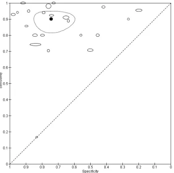
dermoscopy based CAD (Derm-CAD) were considered similar and pooled, however spectroscopy-based CAD (Spectro-CAD) systems analyse different data types and so were only pooled in these subgroups: multispectral imaging studies (MSI-CAD), electrical impedance spectroscopy (EIS-CAD), and diffuse reflectance spectroscopy (DRS-CAD). For each index test, algorithm or checklist under consideration, estimates of sensitivity and specificity were plotted on coupled forest plots and in receiver operating characteristic (ROC) space. CAD thresholds are created by complex statistical algorithms and a threshold is difficult to define. Therefore, we assumed results were binary for the purpose of pooling results across similar CAD systems. We estimated summary operating points (summary sensitivities and specificities) with 95% confidence and prediction regions using the bivariate model (Chu 2006; Reitsma 2005). Where
inadequate data were available for the analysis to converge, the model was simplified, first by assuming no correlation between estimates of sensitivity and specificity and secondly by setting variance terms to zero if little or no heterogeneity was observed on SROC plots (Takwoingi 2015).
Data on the accuracy of dermoscopy were extracted from all included studies that performed both CAD and dermoscopy in the same patients. We performed test comparisons using two analytic strategies. First we
performed indirect comparisons by using all studies of the two tests. Second we made direct comparisons of CAD and dermoscopy by including only comparative studies that assessed the accuracy of both tests in the same study population to enable a robust comparison (Takwoingi 2013). To minimize the risk of bias in the direct comparison, studies that performed either CAD or dermoscopy on a subsample of the total analysed population were excluded. In the
Assessment of reporting bias
Because of uncertainty about the determinants of publication bias for diagnostic accuracy studies and the inadequacy of tests for detecting funnel plot asymmetry (Deeks 2005), we did not perform tests to detect publication bias.
Results
Results of the search
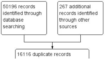
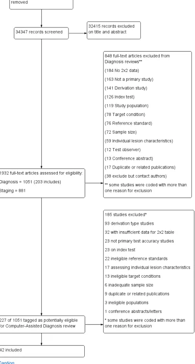
A total of 34,347 unique references were identified and screened for inclusion. Of these, 1051 full-text papers were reviewed for eligibility for any one of the suite of reviews of tests to assist in the diagnosis of melanoma or keratinocyte skin cancer. Of the 1051 full-text papers assessed, 848 were excluded from all reviews in our series (see Figure 5 PRISMA flow diagram of search and eligibility results).
Of the 227 studies tagged as potentially eligible for this review of CAD (166 for Derm–CAD, 61 for Spectro–CAD), 42 publications were included (24 Derm–CAD and 18 Spectro–CAD). Exclusions were mainly due to the absence of a ‘test’ set of lesions used to evaluate CAD's performance independently of the computer algorithm’s development (Derm–CAD n = 76, Spectro-CAD n = 17); inability to construct a 2x2 contingency table based on the data presented (Derm–CAD n = 24, Spectro-CAD n = 8); the use of ineligible index tests (Derm–CAD n = 18, Spectro-CAD n = 5) (for example: computers used to measure lesions but not to diagnose them, e.g. Seidenari 2012); or not meeting our requirements for an eligible reference standard (Derm–CAD n = 13, Spectro-CAD n = 9). Other reasons for exclusion included ineligible definition of the target condition (Derm–CAD n = 10, Spectro-CAD n = 3) and CAD systems based on evaluating the presence of a single lesion characteristic (Derm–CAD n = 17) (for example, a CAD system analysing the colour balance of a lesion). A list of the 185 publications excluded from this review with reasons for exclusion is provided in Characteristics of excluded studies, with a list of all studies excluded from the full series of reviews available as a separate pdf.
The authors of 10 publications were contacted to provide additional detail on published 2x2 data for the accuracy of CAD, with responses regarding four publications received to date. These did not result in the inclusion of any
additional studies, however did permit the inclusion of one additional dataset in an already–included study (Mollersen 2015
). One response highlighted an alternative publication that was independently ascertained by the project search, and was included (Serrao 2006); two replies were unable to provide the information requested in relation to two study publications, both of which were subsequently excluded due to incomplete 2x2 data. Attempts to contact authors of six publications failed, resulting in the exclusion of those six studies from review. In addition to these 10 attempted contacts, authors of one other publication (Walter 2012) were contacted as part of another review in this series, the
accuracy of visual inspection for the diagnosis of melanoma (Dinnes 2018a), to provided clarifications on methods used; the author response enabled it to be included.
The 42 included studies reported on 39 cohorts of lesions and provided 63 datasets with 13,445 lesions and 2452
malignancies. The majority of studies (n = 24, 57%) contributed data on the diagnostic accuracy of digital dermoscopy–based CAD systems (Derm–CAD), of which seven also compared the diagnostic accuracy of Derm–CAD with dermoscopic
diagnosis. The remaining 18 studies contributed data on the diagnostic accuracy of spectroscopy–based CAD (Spectro-CAD), of which five provided comparative accuracy data with dermoscopy. A cross-tabulation of studies by CAD type, reported comparisons and target conditions is provided in Table 2.
Studies were case series (n = 27, 64%), case control (n = 10, 24%), randomised controlled trial (n = 1, 2%), or of unclear design (n = 4, 10%). Lesion selection was most commonly retrospective (n = 22, 52%) or prospective (n = 15, 36%), though was unclear in five studies (12%). Studies included only pigmented (n = 29, 69%) or melanocytic lesions (n = 6, 14%), only suspected melanomas (n = 4, 10%), or any lesions suspected of malignancy (n = 2, 5%). Patient characteristics such as age and gender were reported by 15/42 studies.
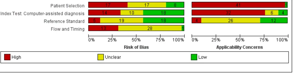
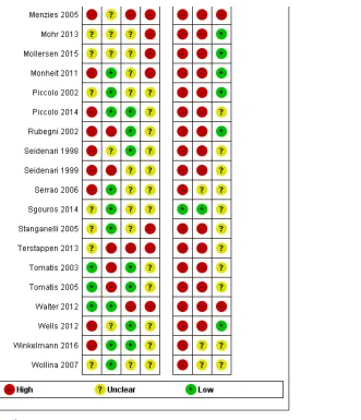
Methodological quality of included studies
The majority of included studies were of methodological concern primarily due to lack of applicability to the current review question, but also due to a high or unclear risk of bias in their design. Since there were no major differences in quality according to CAD type, we provide an overview of the quality and applicability of all included studies regardless of CAD type. The methodological quality of studies according to CAD type (Derm–CAD or spectroscopy–based CAD) is summarized in
Figure 6 and Figure 7.
The risk of participant selection bias was judged as high in 17 (40%) studies, due to the selection of lesions according to their final diagnosis (case–control studies: n = 10, 24%), and/or to the inappropriate exclusion of lesions with particular prognostic characteristics (n = 8, 19%), such as high-grade dysplastic lesions (Ferris 2015) or small/large
lesions (Malvehy 2014; Monheit 2011). Study eligibility criteria and participant exclusions were not reported clearly enough to ascertain the risk of selection bias in 17 studies (40%); this meant that we could not determine whether consecutive or random samples of lesions were recruited (n = 22, 52%), whether participants had been selected according to their final diagnoses (use of 'case–control' selection, n = 5, 12%), or whether participant exclusions were appropriate (n = 22, 52%). A single study (Sgouros 2014) was of low concern for the applicability of its participant sample to the current review question: it included unexcised lesions and did not recruit participants with multiple lesions. All others (n = 41, 98%) were of high concern due to the use of restricted participant groups and settings (n = 40, 95%), with study
at least 50% of benign cases (Boldrick 2007; Bono 1996; Sgouros 2014). Fourteen studies were also of concern due to their recruitment of participants with multiple lesions (including over 5% more lesions than participants). One of these, Dreiseitl 2009, provided patient–based 2x2 data as well as lesion–based data for the accuracy of a Derm–CAD system. These 2 analyses of the same population highlight the distortion that can occur in study populations that include multiple lesions per patient: lesion–based sensitivity was lower than patient based sensitivity (74% versus 89%), however lesion–based specificity was far higher (84% versus 48%) due to the inclusion of many disease negative lesions per patient.
Twenty–two (52%) studies did not report the number of participants included (precluding assessment of the inclusion of multiple lesions). Of the 18 studies including model derivation, 6 (33%) used a wide range of skin conditions to train the classification algorithm. Two (11%) used an inadequately narrow range (absence of non–dysplastic benign conditions), while the remaining 10 (56%) provided inadequate detail of diagnoses included in the training set.
Over half the studies were at high (n = 14, 33%) or unclear (n = 10, 24%) risk of bias due to the methods used to undertake the index test. Most studies (n = 40, 95%) blinded CAD results to the reference standard diagnosis though almost half (n = 20, 48%) failed to clearly pre–specify the diagnostic threshold, of which 13 (30%) were
threshold–finding studies that provided accuracy data for the best threshold possible once index test results had been examined. Most studies (n = 35, 83%) evaluated CAD in an independent population to that used to train the classification algorithm, either by external validation (n = 23, 55%) or internal validation (randomised division of a single study group into training and test sets: n = 12, 29%). An additional six (14%) studies used internal validation, but failed to specify whether division of the study group was made randomly (i.e. not selected according to
diagnosis), while one study was at risk of bias by selecting which diagnoses to place in the train and test sets (Tomatis 2003).
Of the 18 studies that included CAD model derivation (training of the classification algorithm), eight (44%) accounted for model overfitting by using a Support Vector Machine algorithm (Gilmore 2010; Mohr 2013; Stanganelli 2005),
performing a jack–knife calculation (Binder 1994; Burroni 2004), or another method (Rubegni 2002; Tomatis 2003; Tomatis 2005). One study specified that model optimisation was not incorporated (Garcia Uribe 2012) and 9 (50%) did not discuss overfitting. The majority of studies (n = 32, 76%) were of high concern regarding the applicability of the index test, due to their evaluation of an unestablished threshold (n = 23), lack of detail regarding the diagnostic threshold used (n = 16), and/or the use of non–expert clinicians (n = 2) in studies evaluating CAD as a diagnostic aid (n = 7).
Almost all studies reported use of an acceptable reference standard (n = 37, 88%), and around half (n = 19, 45%) clearly reported blinding of the reference standard to the CAD result. For the applicability of the reference standard, four reported using expert diagnosis for some lesions (high concern) and 30 (71%) were unclear as to whether histopathology had been interpreted by an experienced histopathologist or dermatopathologist.
Reporting of study flow and timing was generally poor with an unclear risk of bias in 28 (67%) studies, largely due to ambiguity regarding the interval between the application of the index test and reference standard (excision for histology or first follow–up visit) (n = 28). Thirteen (30%) studies were at a high risk of bias because they used different reference standards according to diagnosis (differential verification) (n = 6) and/or did not include all participants in the analysis (n = 10), primarily due to technical difficulties with the CAD system (n = 6).
Eleven of the 15 studies comparing CAD with dermoscopy were at high (n = 2) or unclear (n = 9) risk of bias. Six reported blinding between tests, two reported no blinding and seven were unclear. Half (n = 8) did not clearly report the interval between tests.
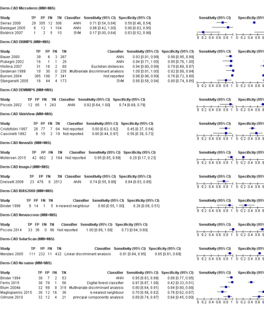
Findings
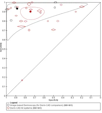
The 24 studies evaluating a Derm–CAD system reported 23 cohorts of lesions providing 32 CAD datasets with 9602 lesions including 1313 malignancies of which 1220 were melanomas, at least 83 BCCs (number not specified in one study,
Menzies 1996), and 9 cSCCs. The total number of study participants with suspicious lesions cannot be estimated due to lack of reporting in study publications (reported in only 10 studies (with 2400 participants). Two publications