Syndrome Coronavirus Vaccine Based on a Recombinant Measles
Virus Vaccine Platform
Anna H. Malczyk,a,bAlexandra Kupke,c,dSteffen Prüfer,aVivian A. Scheuplein,aStefan Hutzler,aDorothea Kreuz,eTim Beissert,f Stefanie Bauer,eStefanie Hubich-Rau,gChristiane Tondera,aHosam Shams Eldin,c,dJörg Schmidt,c,dJúlia Vergara-Alert,c,d Yasemin Süzer,aJanna Seifried,aKay-Martin Hanschmann,hUlrich Kalinke,i,jSusanne Herold,kUgur Sahin,f,gKlaus Cichutek,a,b Zoe Waibler,b,eMarkus Eickmann,c,dStephan Becker,c,dMichael D. Mühlebacha,b
Oncolytic Measles Viruses and Vaccine Vectors,a
Novel Vaccination Strategies and Early Immune Responses,e
and Biostatistics,h
Paul-Ehrlich-Institut, Langen, Germany; German Center for Infection Research, Langen, Germanyb
; Institut für Virologie, Philipps Universität Marburg, Marburg, Germanyc
; Universities Gießen & Marburg Lung Center, Department of Internal Medicine II, Section of Infectious Diseases, Giessen, Germanyk
; German Center for Infection Research, Marburg, Germanyd
; TRON gGmbH, Mainz, Germanyf
; Universitätsmedizin Mainz, Mainz, Germanyg
; Institute for Experimental Infection Research, TWINCORE, Centre for Experimental and Clinical Infection Research, Hannover, Germanyi
; German Center for Infection Research, Hannover, Germanyj
ABSTRACT
In 2012, the first cases of infection with the Middle East respiratory syndrome coronavirus (MERS-CoV) were identified. Since
then, more than 1,000 cases of MERS-CoV infection have been confirmed; infection is typically associated with considerable
morbidity and, in approximately 30% of cases, mortality. Currently, there is no protective vaccine available.
Replication-compe-tent recombinant measles virus (MV) expressing foreign antigens constitutes a promising tool to induce protective immunity
against corresponding pathogens. Therefore, we generated MVs expressing the spike glycoprotein of MERS-CoV in its
full-length (MERS-S) or a truncated, soluble variant of MERS-S (MERS-solS). The genes encoding MERS-S and MERS-solS were
cloned into the vaccine strain MV
vac2genome, and the respective viruses were rescued (MV
vac2-CoV-S and MV
vac2-CoV-solS).
These recombinant MVs were amplified and characterized at passages 3 and 10. The replication of MV
vac2-CoV-S in Vero cells
turned out to be comparable to that of the control virus MV
vac2-GFP (encoding green fluorescent protein), while titers of MV
vac2-CoV-solS were impaired approximately 3-fold. The genomic stability and expression of the inserted antigens were confirmed via
sequencing of viral cDNA and immunoblot analysis.
In vivo
, immunization of type I interferon receptor-deficient (IFNAR
ⴚ/ⴚ)-CD46Ge mice with 2
ⴛ
10
550% tissue culture infective doses of MV
vac2-CoV-S(H) or MV
vac2-CoV-solS(H) in a prime-boost
regi-men induced robust levels of both MV- and MERS-CoV-neutralizing antibodies. Additionally, induction of specific T cells was
demonstrated by T cell proliferation, antigen-specific T cell cytotoxicity, and gamma interferon secretion after stimulation of
splenocytes with MERS-CoV-S presented by murine dendritic cells. MERS-CoV challenge experiments indicated the protective
capacity of these immune responses in vaccinated mice.
IMPORTANCE
Although MERS-CoV has not yet acquired extensive distribution, being mainly confined to the Arabic and Korean peninsulas, it
could adapt to spread more readily among humans and thereby become pandemic. Therefore, the development of a vaccine is
mandatory. The integration of antigen-coding genes into recombinant MV resulting in coexpression of MV and foreign antigens
can efficiently be achieved. Thus, in combination with the excellent safety profile of the MV vaccine, recombinant MV seems to
constitute an ideal vaccine platform. The present study shows that a recombinant MV expressing MERS-S is genetically stable
and induces strong humoral and cellular immunity against MERS-CoV in vaccinated mice. Subsequent challenge experiments
indicated protection of vaccinated animals, illustrating the potential of MV as a vaccine platform with the potential to target
emerging infections, such as MERS-CoV.
I
n November 2012, a novel coronavirus was identified for the
first time in a patient from Saudi Arabia who presented with
severe respiratory disease. Later, this virus was termed Middle East
respiratory syndrome coronavirus (MERS-CoV) (
1
). By 26
De-cember 2014, 938 laboratory-confirmed cases of MERS-CoV,
mostly from Saudi Arabia and neighboring countries, had been
diagnosed and had resulted in 343 casualties (
2
). A few cases of
MERS-CoV were also detected in the United States, the United
Kingdom, Netherlands, Austria, France, Greece, Italy, and
Ger-many, indicating the virus’s principal potential for spread (
2
).
Fortunately, direct transmission upon contact with human
pa-tients seems to be limited, yet is still possible, as determined by
analysis of household contact infections among MERS patients’
families (
3
) and as evidenced by a recent cluster of MERS
infec-tions in South Korea, with 166 cases between 20 May and 19 June
2015, including 106 third-generation and 11 fourth-generation
cases (
4
,
5
). As a natural reservoir, dromedary camels have been
identified as the most likely source, based on partially identical
genomes detected in viruses isolated from humans or camels (
6
,
7
). Additionally, antibodies against the spike glycoprotein of
MERS-CoV with virus-neutralizing capacity were detected in
camels (
8–10
), and infection of humans with MERS-CoV have
been reported after contact with infected camels (
11
,
12
).
Inter-estingly, while all other members of the C lineage of the
Betacoro-navirus
genus have been found in different bat species (
13
,
14
),
only closely related, most likely precursor viruses of MERS-CoV
on November 7, 2019 by guest
http://jvi.asm.org/
have been identified in
Neoromicia capensis
bats (
15
). Thus,
MERS-CoV has a zoonotic origin, but sustained infections, the
severity of the disease, and the risk of virus adaption to gain
efficient human-to-human transmission mandates the
devel-opment of effective vaccines to combat local infections and to
be prepared for the eventual occurrence of a global pandemic,
as previously observed with severe acute respiratory syndrome
coronavirus (SARS-CoV) in 2003 (
16
).
Occurring 10 years before the current MERS-CoV epidemic,
SARS-CoV was the first
Betacoronavirus
of zoonotic origin with
potentially fatal outcomes in human patients (
1
). Experimental
vaccines protecting animal models against SARS have been
devel-oped (
17–19
), and the properties of such SARS vaccines may be
applicable to vaccines that should protect against MERS-CoV
in-fections. Both neutralizing antibodies and T cell responses are
essential for prevention of SARS-CoV infection (
17
,
18
). The spike
protein (S), a coronavirus class I fusion protein (
20
,
21
), has been
identified as the most immunogenic antigen of SARS-CoV, as it
induces a strong humoral as well as cellular immune response (
17
,
19
). Similarly, MERS-S constructs expressed by recombinant
modified vaccinia virus Ankara or recombinant adenoviral
vec-tors have already been demonstrated to induce neutralizing
anti-bodies (
22
,
23
). The detected neutralizing capacity of induced
antibodies is expected, since the receptor-binding domain (RBD)
in the S1 domain of both SARS-CoV and MERS-CoV S proteins
mediate host-cell receptor binding as a prerequisite for cell entry
(
24
,
25
). Thus, S1 is the main target of neutralizing antibodies
(
26
). Also the RBD of MERS-CoV-S alone has been demonstrated
to induce strong neutralizing antibody titers (
23
,
27–31
). In
com-bination with different adjuvants, even induction of T cell
re-sponses by the recombinant RBD has been described (
31
). Thus, a
prototypic MERS vaccine should be based on MERS-S expression,
since the induction of neutralizing antibodies has been shown to
be a direct correlate of protection in cases of SARS-CoV (
32
).
The measles vaccine is an efficient, live attenuated, replicating
virus that induces both humoral and cellular immune responses,
has an excellent safety record, and probably provides lifelong
pro-tection (
33
,
34
). The vaccine’s manufacturing process is extremely
well established (
35
), and millions of doses can be generated quite
easily and quickly. Generation of recombinant measles virus
(MV) from DNA via reverse genetics is feasible (
35
) and allows the
insertion of additional transcription units (ATU) by duplication
of sequences terminated by start and stop sequences (
36
). Hence,
genes expressing foreign antigens up to 6 kb can be cloned into the
MV backbone (
36
) and elicit coexpression of MV proteins and
inserted genes. Besides marker genes (
37
) or immune modulators
(
38
), expression of antigens from foreign pathogens like hepatitis
B or C virus (
39
,
40
), HIV (
41
), West Nile virus (WNV) (
42
,
43
),
dengue virus (
44
), Chikungunya virus (CHIKV) (
45
), or
SARS-CoV (
19
) by recombinant MVs has already been demonstrated.
Thereby, robust immune responses against vector and foreign
an-tigens are induced after vaccination of transgenic, MV-susceptible
type I interferon receptor-deficient (IFNAR
⫺/⫺)-CD46Ge mice
(
46
) or nonhuman primates with recombinant MVs, in general. In
particular, protection of vaccinated animals from lethal challenge
with WNV (
42
) or CHIKV (
45
) was demonstrated and indicated
the high efficacy of the system. Interestingly, prevaccinated
ani-mals with protective immunity against measles were still
amend-able to vaccination with the recombinant MV, since significant
immune responses against the foreign antigen(s) are consistently
induced (
41
,
45
), and the MV-based CHIKV vaccine
demon-strated efficacy in phase I trials irrespective of measles virus
im-munity (
47
).
Here, we aimed to utilize the efficacy of the MV vaccine
plat-form to generate a live attenuated vaccine against MERS-CoV
based on recombinant MV
vac2. This recombinant virus reflects the
MV vaccine strain Moraten (
48
), which has been authorized for
vaccination against measles. As the antigen, we choose the
MERS-CoV S glycoprotein to induce neutralizing antibodies and robust
cellular immunity. Two variants of the glycoprotein were analyzed
as antigen: the full-length, membrane-anchored MERS-S, and a
truncated, soluble form lacking the transmembrane domain
(MERS-solS). Both variants include the S1 domain as a target
structure. The soluble protein variant should be taken up better by
B cells (
49–51
) and thus should induce humoral immune
re-sponses more efficiently (
52
), potentially boosting
virus-neutral-izing antibody titers (VNTs). The respective genes were inserted
into two different positions of the MV genome to modulate
ex-pression of the antigens, and all recombinant MVs were
success-fully rescued. Cells infected with such viruses expressed the
de-sired antigens. Indeed, immunization of IFNAR
⫺/⫺-CD46Ge
mice induced strong humoral and cellular immune responses
di-rected against MV and MERS-CoV S which were sufficient to
protect vaccinated animals from MERS-CoV infection. Thus, MV
platform-based vaccines are a powerful option to develop a
prepandemic vaccine against MERS-CoV.
MATERIALS AND METHODS
Cells.Vero cell (African green monkey kidney cells; ATCC CCL-81), 293T cell (ATCC CRL-3216), and EL4 mouse T cell (ATCC TIB-39) lines were purchased from ATCC (Manassas, VA, USA) and cultured in Dulbecco’s modified Eagle’s medium (DMEM) supplemented with 10% fetal bovine serum (FBS; Biochrom, Berlin, Germany) and 2 mML-glutamine (L-Gln; Biochrom). JAWSII dendritic cells (ATCC CRL-11904) were purchased from ATCC and cultured in minimal essential medium alpha (MEM-␣) with ribonucleosides and deoxyribonucleosides (Gibco BRL, Eggenstein, Germany) supplemented with 20% FBS, 2 mML-Gln, 1 mM sodium pyruvate (Biochrom), and 5 ng/ml murine granulocyte-macrophage col-ony-stimulating factor (GM-CSF; Peprotech, Hamburg, Germany). DC2.4 and DC3.2 murine dendritic cell lines (53) were cultured in RPMI containing 10% FBS, 2 mML-Gln, 1% nonessential amino acids (Bio-chrom), 10 mM HEPES (pH 7,4), and 50M 2-mercaptoethanol (Sigma-Aldrich, Steinheim, Germany). All cells were cultured at 37°C in a humid-ified atmosphere containing 6% CO2for a maximum of 6 months of
culture after thawing of the original stock. Received16 July 2015 Accepted3 September 2015
Accepted manuscript posted online9 September 2015
CitationMalczyk AH, Kupke A, Prüfer S, Scheuplein VA, Hutzler S, Kreuz D, Beissert T, Bauer S, Hubich-Rau S, Tondera C, Eldin HS, Schmidt J, Vergara-Alert J, Süzer Y, Seifried J, Hanschmann K-M, Kalinke U, Herold S, Sahin U, Cichutek K, Waibler Z, Eickmann M, Becker S, Mühlebach MD. 2015. A highly immunogenic and protective Middle East respiratory syndrome coronavirus vaccine based on a recombinant measles virus vaccine platform. J Virol 89:11654 –11667.
doi:10.1128/JVI.01815-15.
Editor:S. Perlman
Address correspondence to Michael D. Mühlebach, Michael.Muehlebach@pei.de.
A.K. and S.P. contributed equally.
Copyright © 2015, American Society for Microbiology. All Rights Reserved.
on November 7, 2019 by guest
http://jvi.asm.org/
Plasmids. The codon-optimized gene encoding MERS-CoV-S (GenBank accession numberJX869059) flanked with AatII/MluI bind-ing sites in plasmid pMA-RQ-MERS-S was obtained by gene synthesis (Invitrogen Life Technologies, Regensburg, Germany). A truncated form of MERS-S lacking the transmembrane domain was amplified by PCR, flanked with AatII/MluI binding sites, and fully sequenced. Both antigens, as well as the immediate early cytomegalovirus (CMV) pro-moter (54), were inserted into p(⫹)BR-MVvac2-GFP(H) or p(⫹)
MVvac2-ATU(P) (48) via AatII/MluI or SfiI/SacII, respectively, to
gene-rate p(⫹)PolII-MVvac2-MERS-S(H), p(⫹)PolII-MVvac2-MERS-S(P),
p(⫹)PolII-MVvac2-MERS-solS(H), and p(⫹)PolII-MVvac2 -MERS-solS(P), respectively. For construction of lentiviral transfer vectors encod-ing the MERS-CoV antigens, the open readencod-ing frame (ORF) of MERS-S was amplified by PCR with primers encompassing flanking NheI/XhoI restriction sites and template pMA-RQ-MERS-S. Details on primers and PCR procedures are available upon request. PCR products were cloned into pCR2.1-TOPO (Invitrogen Life Technologies) and fully sequenced. Intact antigen ORF was cloned into pCSCW2gluc-IRES-GFP (55) by us-ing NheI/XhoI restriction sites to yield pCSCW2-MERS-S-IRES-GFP.
Production of lentiviral vectors.Viral vectors were produced using 293T cells and polyethylenimine (PEI; Sigma-Aldrich) transfection (56). A total of 1⫻107293T cells were seeded per 175-cm2cell culture flask and
cultured overnight. To produce lentivirus vectors pseudotyped with the G protein of vesicular stomatitis virus (VSV-G), these cells were transfected using a standard three-plasmid lentivirus vector system. Cells were trans-fected with 17.5g pCSCW2-MERS-S-IRES-GFP transfer vector, 6.23g pMD2.G, and 11.27g pCMV⌬R8.9 (57), as described previously (58). The medium was exchanged 1 day posttransfection, and HIVMERS-S-IRES-GFP
(VSV-G) vector particles were harvested 2 and 3 days after transfection. For harvest of vector particles, the supernatants of three culture flasks were fil-tered (0.45-m pore size), pooled, and concentrated by centrifugation (100,000⫻g, 3 h, 4°C). Pellets were resuspended in DMEM and stored at
⫺80°C.
Generation of antigen-expressing cell lines.Syngeneic target cells based on the C57BL/6-derived DC lines JAWSII, DC2.4, and DC3.2, as well as T cell line EL-4 were transduced with HIVMERS-S-IRES-GFP(VSV-G)
vector-containing supernatant to express MERS-S and the green marker protein GFP (JAWSIIgreen-MERS-S, EL-4green-MERS-S, DC2.4green
-MERS-S, and DC3.2green-MERS-S), thereby presenting respective pep-tides via major histocompatibility complex class I (MHC-I). EL-4 cells were alternatively transduced with HIVTurboFP635(VSV-G) vectors (59) to
express red fluorescent Katushka protein as a negative control (EL-4red).
For this purpose, 1⫻105target cells were seeded in 24-well plates and
transduced with 0.1, 1, or 10l of concentrated vector suspension. For analysis of transduction efficiencies, cells were fixed in 1% paraformalde-hyde (Merck Millipore, Darmstadt, Germany), and the percentages of GFP-positive or Katushka-positive cells were quantified by flow cytom-etry using an LSRII flow cytometer (BD, Heidelberg, Germany). Cell pop-ulations revealing a 1 to 10% fraction of GFP-positive cells were used for single-cell cloning by limiting dilution. For that purpose, cell dilutions with 50l conditioned medium statistically containing 0.3 cells were seeded per well in 96-well plates. Single-cell clones were cultured and analyzed by flow cytometry. GFP-positive clones were selected for further analysis.
Viruses.The viruses were rescued as described previously (54). In brief, 5g of MV genome plasmids with MERS-CoV antigen ORFs were cotransfected with plasmids pCA-MV-N (0.4g), pCA-MV-P (0.1g), and pCA-MV-L (0.4g) encoding MV proteins necessary for genome replication and expression. These plasmids were cotransfected into 293T cells cultured in 6-well plates by using Lipofectamine 2000 (Invitrogen Life Technologies). The transfected 293T cells were overlaid 2 days after transfection onto 50% confluent Vero cells seeded in 10 cm-dishes. Over-lay cultures were closely monitored for isolated syncytia, which indicated monoclonal replicative centers. Single syncytia were picked and overlaid onto 50% confluent Vero cells cultured in 6-well plates and harvested as
passage 0 (P0) by scraping and a freeze-thaw cycle of cells at the time of maximal infection. Subsequent passages were generated after titration to determine the 50% tissue culture infective dose (TCID50) of infectious
virus according to the method of Kaerber and Spaerman (60) and infec-tion of Vero cells at a multiplicity of infeciton (MOI) of 0.03. The viruses were passaged up to P10. MERS vaccine viruses and control viruses
MV-vac2-GFP(H) and MVvac2-GFP(P) in P3 were used for characterization,
and viruses in P4 were used for vaccination. MERS-CoV (isolate EMC/ 2012) (1) was used for neutralization assays, and challenge virus was prop-agated in Vero cells and titrated as described above for recombinant MV. All virus stocks were stored in aliquots at⫺80°C.
Measles virus genome sequence analysis.The RNA genomes of re-combinant MV in P3 or P10 were isolated using the QIAamp RNeasy kit (QIAgen, Hilden, Germany) according to the manufacturer’s instructions and resuspended in 50l RNase-free water. Viral cDNA was reverse tran-scribed using the SuperScript II reverse transcription (RT) kit (Invitro-gen) with 2l viral RNA (vRNA) as the template and random hexamer primers, according to manufacturer’s instructions. For specific amplifica-tion of antigen ORFs, the respective genomic regions of recombinant MVs were amplified by PCR with primers binding to sequences flanking the regions of interest and cDNA as the template. Detailed descriptions of primers and procedures are available upon request. The PCR products were directly sequenced (Eurofins Genomics, Ebersberg, Germany).
Western blot analysis.For Western blot analysis, cells were lysed and immunoblotted as previously described (61). A rabbit anti-MERS-CoV serum (1:1,000) was used as the primary antibody for MERS-CoV-S, and a rabbit anti-MV-N polyclonal antibody (1:25,000; Abcam, Cambridge, United Kingdom) was used for MV-N detection. A donkey horseradish peroxidase (HRP)-coupled anti-rabbit IgG (H&L) polyclonal antibody (1:10,000; Rockland, Gilbertsville, PA) served as the secondary antibody for both. Peroxidase activity was visualized with an enhanced chemilumi-nescence (ECL) detection kit (Thermo Scientific, Bremen, Germany) on Amersham ECL hyperfilm (GE Healthcare, Freiburg, Germany).
Production of recombinant soluble MERS-CoV spike protein.The S protein lacking the transmembrane domain was genetically tagged with six His residues at its carboxy terminus. The resulting construct was in-serted into a Semliki forest virus-derived self-replicating RNA vector (SFV replicon) downstream of the subgenomic promoter. These replicons were transcribedin vitroand purified as previously described (62,63). The integrity of purified replicon was assessed by on-chip electrophoresis (2100 BioAnalyzer; Agilent, Santa Clara, CA). To produce SFV vector particles, replicon RNA and helper RNA were coelectroporated into BHK21 cells by using a square-wave electroporator (one pulse, 750 V/cm for 16 ms; BTX ECM 830; Harvard Apparatus, Holliston, MA). Particles were harvested after 24 h, frozen in N2(liquid), and stored at⫺80°C. For
protein production, 2⫻107BHK21 cells were transduced with SFV
par-ticles (MOI, 40) and harvested after 24 h. Cell pellets were lysed (phos-phate-buffered saline [PBS], 0.2% Triton X-100, protease inhibitor cock-tail [Roche]) for 30 min at 4°C. Afterwards, cells were sonicated, and lysates were cleared by centrifugation (30 min, 21,000⫻g, 4°C). The supernatant was filtered (0.2m), loaded on a HisTrap high-performance (HP) column (17-5247-01; GE Healthcare), and washed with 10 volumes of binding buffer (20 mM Na2HPO4, 0.5 M NaCl, 10 mM imidazole). S
protein was eluted with a gradient of binding buffer containing 0.5 M imidazole followed by buffer exchange to PBS. Protein integrity was checked by Western blotting with a mouse anti-His monoclonal antibody (MAb; 1:50; Dianova, Germany).
Animal experiments.All animal experiments were carried out in com-pliance with the regulations of German animal protection laws and as autho-rized by the RP Darmstadt. Six- to 12-week-old IFNAR⫺/⫺-CD46Ge mice
expressing human CD46 were inoculated intraperitoneally (i.p.) with 1⫻ 105TCID
50of recombinant MV or 200l Opti-MEM on days 0 and 28
and bled via the retrobulbar route on days 7, 28, 32, and 49 postinfection (p.i.) under anesthesia. Serum samples were stored at⫺20°C. Mice were euthanized on day 32 or 49 p.i., and spleens were isolated. For challenge
on November 7, 2019 by guest
http://jvi.asm.org/
experiments, immunized mice were transduced intranasally (i.n.) on day 63 with 20 l of an adenovirus vector encoding human DPP4 and mCherry with a final titer of 2.5⫻108PFU per inoculum (AdV-hDPP4;
ViraQuest Inc.) and challenged i.n. with 20l of MERS-CoV at a final titer of 7⫻104TCID
50on day 68. The mice were euthanized 4 days after
challenge, and representative left lobe lung samples were prepared for RNA isolation.
Antibody ELISA.MV bulk antigens (10g/ml; Virion Serion, Würz-burg, Germany) or recombinant MERS-S protein (20g/ml) in 50l carbonate buffer (Na2CO3at 30 mM, NaHCO3at 70 mM; pH 9.6) was
used to coat wells of Nunc Maxisorp 96-well enzyme-linked immunosor-bent assay (ELISA) plates (eBioscience) and incubated overnight at 4°C. The plates were washed three times with 150l ELISA washing buffer (PBS, 0.1% [wt/vol]Tween 20) and blocked with 50l blocking buffer (PBS, 5% bovine serum albumin [BSA], 0.1% Tween 20) for 2 h at room temperature. Mice sera sampled on days⫺7 or 49 were serially diluted in ELISA dilution buffer (PBS, 1% BSA, 0.1% Tween 20), and 50l/well was used for the assay. The plates were incubated at 37°C for 2 h and washed again with ELISA washing buffer. Plates were incubated with 50l/well of HRP-conjugated rabbit anti-mouse IgG (Dako; 1:1,000 in ELISA dilution buffer) at room temperature for 1 h. Subsequently, the plates were washed and 100l tetramethylbenzidine substrate (eBioscience) was added per well. The reaction was stopped by addition of 50l/well H2SO4(1 N), and
the absorbance at 405 nm was measured.
Neutralization assays.For quantification of VNTs, mouse sera were serially diluted in 2-fold dilutions in DMEM. A total of 50 PFU of MVvac2
-GFP(P) or 200 TCID50of MERS-CoV was mixed with serum dilutions
and incubated at 37°C for 1 h. Virus suspensions were added to 1⫻104
Vero cells seeded 4 h prior to assay in 96-well plates and incubated for 4 days at 37°C. VNTs were calculated as the reciprocal of the highest dilu-tion that abolished infecdilu-tion.
ELISpot assays.Murine gamma interferon (IFN-␥) enzyme-linked immunosorbent spot (ELISpot) assays (eBioscience, Frankfurt, Ger-many) were performed according to the manufacturer’s instructions us-ing multiscreen immunoprecipitation (IP) ELISpot polyvinylidene diflu-oride 96-well plates (Millipore, Darmstadt, Germany). A total of 5⫻105
splenocytes isolated 4 days after boost immunization were cocultured with 5⫻104JAWSII
green-MERS-S, DC2.4green-MERS-S, or DC3.2green
-MERS-S, or untransduced DC cell lines for 36 h in 200l RPMI (10% fetal bovine serum [FBS], 2 nML-Gln, 1% penicillin-streptomycin). Medium alone served as the negative control. Concanavalin A (ConA; Sigma-Al-drich) at 10g/ml was used for demonstration of splenocyte reactivity. Recombinant MV bulk antigens (Virion Serion) at 10g/ml were used to analyze MV-specific immune responses in vaccinated animals. Cells were removed from the plates, and the plates were incubated with biotin-con-jugated anti-IFN-␥antibodies and avidin-HRP according to the manu-facturer’s instructions. 3-Amino-9-ethyl-carbazole (AEC; Sigma-Al-drich) substrate solution for development of spots was prepared according to the manufacturer’s instructions using AEC dissolved inN,N -dimethylformamide (Merck Millipore). Spots were counted using an Eli.Scan ELISpot scanner (AE.L.VIS, Hamburg, Germany) and ELISpot analysis software (AE.L.VIS).
T cell proliferation assay.Splenocytes isolated 3 weeks after booster immunization were labeled with 0.5M carboxyfluorescein succinimidyl ester (CFSE; eBioscience) as previously described (64). In brief, 5⫻105
labeled cells were seeded in RPMI 1640 supplemented with 10% mouse serum, 2 nML-glutamine, 1 mM HEPES, 1% penicillin-streptomycin, and
100M 2-mercaptoethanol in 96-well plates. Aliquots of 200l of me-dium containing ConA (10g/ml), MV bulk antigens (10g/ml), or 5⫻ 103JAWSII
green-MERS-S cells were added to each well, and cells were
cultured for 6 days. Medium and untransduced JAWSII cells served as controls. Stimulated cells were subsequently stained with CD3-PacBlue (1:50; clone 500A2; Invitrogen Life Technologies) and CD8-allophyco-cyanin (1:100; clone 53-6.7; eBioscience) antibodies and fixed with 1% paraformaldehyde (PFA) in PBS. Stained cells were analyzed by flow
cy-tometry using an LSR II flow cytometer (BD) and FACSDiva software (BD).
Cytotoxic T lymphocyte (CTL) killing assay.For restimulation of T cells isolated 4 days after booster immunization, 5⫻106splenocytes were
cocultured with 5⫻104JAWSII
green-MERS-S cells for 6 days in 12-well
plates in RPMI 1640 supplemented with 10% FBS, 2 nML-glutamine, 1
mM HEPES, 1% penicillin-streptomycin, 2-mercaptoethanol (100M), and 100 U/ml recombinant interleukin-2 (rIL-2; murine, Peprotech). A total of 2⫻103EL-4
redcells were labeled with 0.5M CFSE and mixed
with 8⫻103EL-4
green-MERS-S cells per well. Splenocytes were counted
and cocultured with EL-4 target cells at the indicated ratios for 4 h. After-wards, EL-4 cells were labeled with the fixable viability dye eFluor 780 (eBioscience), fixed with 1% PFA, and analyzed by flow cytometry using an LSR II flow cytometer (BD) and FACSDiva software (BD). For deter-mination of the antigen:NC EL-4 ratio, cell counts of living MERS-S-expressing cells were divided by the counts for living negative controls.
Determination of viral RNA copy numbers and infectious virus in mouse tissue.Samples of immunized and challenged mice (6- by 6-mm tissue slices of approximately 0.035⫾0.011 g [mean⫾standard devia-tion]) excised from the centers of left lung lobes were homogenized in 1 ml DMEM with ceramic beads (diameter, 1.4 mm) in a FastPrep SP120 instrument three times for 40 s at 6.5 m/s. The homogenate was centri-fuged for 3 min at 2,400 rpm in a Mikro 200R centrifuge (Hettich Lab Technology) to remove tissue debris. Live virus titers in supernatant (in TCID50per milliliter) were determined on Vero cells as described above.
Aliquots of 100l of the supernatants were used for RNA isolation with the RNeasy minikit (Qiagen) according to the manufacturer’s instruc-tions. The RNA amount was measured with the NanoDrop ND-100 spec-trophotometer. Total RNA was reverse transcribed and quantified by real-time PCR using the SuperScript III OneStep RT-PCR System (Invitrogen Life Technologies) as described previously (65) with the primer pair upE-Fwd and upE-Rev and the probe upE-Prb on an ABI7900 high-through-put fast real-time PCR system (Life Technologies Instruments).
Additionally, for every sample of the transduced and infected mice, evidence for successful hDPP4 transduction was determined by real-time RT-PCR for mCherry with the OneStep RT-PCR kit on a Rotor Gene Q apparatus (both from Qiagen). Primers and probe (Tib-Molbiol, Berlin, Germany) were as follows: mCherry forward, CATGGTAACGATGAGT TAG; mCherry reverse, GTTGCCTTCCTAATAAGG; mCherry probe, 6-carboxyfluorescein (FAM)–TACCACCTTACTTCCACCAATCGG–BBQ (BlackBerry quencher). Primers and probe were used at final concentra-tions of 0.4M and 0.2M, respectively. The quantitative reverse tran-scription-PCR (qRT-PCR) program was as follows: 50°C for 30 min; 95°C for 15 min; 40 cycles of 95°C for 15 s, 48°C for 30 s, and 72°C for 20 s. All samples for mCherry were evaluated in one run to exclude an impact of different conditions on the results in different runs. Quantification was carried out using a standard curve based on 10-fold serial dilutions of appropriate cloned RNA ranging from 102to 105copies. Briefly, PCR
fragments were generated using the primers described above. For cloning, the TOPO TA cloning kit with pCR2.1-TOPO plasmid (Invitrogen) and Escherichia coliwere used. Inserts were examined for correct orientation and length, amplified with plasmid-specific primers, purified, and tran-scribed into RNA by using the SP6/T7 transcription kit (Roche).
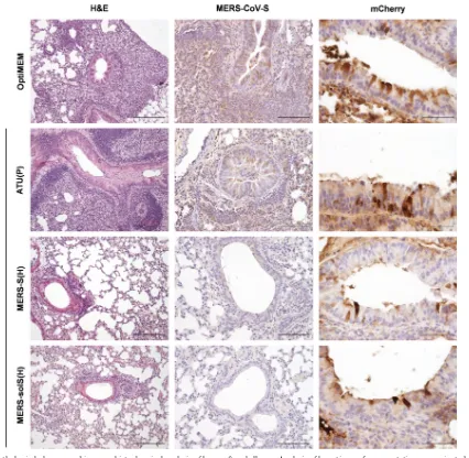
Histopathological and immunohistochemical examination of lung tissue.Lungs of vaccinated and mock-vaccinated mice transduced with AdV-hDPP4 were collected on day 4 postchallenge with MERS-CoV. Tis-sue was fixed in 4% PFA and embedded in paraffin. Sections were cut with a Leica RM2255 microtome (Leica Biosystems) and stained with hema-toxylin and eosin (H&E). For detection of MERS-CoV, a rabbit polyclonal antibody against MERS-CoV spike protein S1 (100208-RP; Sino Biologi-cal Inc., Beijing, China) diluted 1:50 was used. To monitor adenovirus transduction, a mouse monoclonal antibody against mCherry (ab125096; Abcam) diluted 1:250 was used after antigen retrieval with target retrieval solution (Dako) for 23 min at 97°C. To block unspecific binding, slides were incubated for 10 min with 20% nonimmune pig serum
on November 7, 2019 by guest
http://jvi.asm.org/
CoV) or for 30 min with 20% nonimmune horse serum (mCherry). Pri-mary antibodies were incubated overnight at 4°C. A pig anti-rabbit IgG and a biotinylated horse anti-mouse IgG served as secondary antibodies for MERS-CoV and mCherry, respectively. For detection of antigen-an-tibody complexes, the ABC method for mCherry and the rabbit PAP method for MERS-CoV were used in combination with diaminobenzi-dine for staining. Papanicolaou stain was used for counterstaining.
Statistical analysis.To compare the means of different groups from growth curves, neutralization assays, and ELISpot assays, a nonparametric one-way analysis of variance (one-way ANOVA) was performed. For the proliferation assay, the mean differences between control and vaccinated groups were calculated and analyzed by using an unpairedttest. To all three groups in the CTL killing assays, a linear curve was fitted for antigen versus the log-transformed effector-target ratio (E:T). ThePvalues for differences in slopes were calculated, and MVvac2-MERS-S(H) or MVvac2 -MERS-solS(H) was compared with the control, MVvac2-ATU(P). For analysis of challenge data, mean ratios and 95% confidence intervals were calculated based on log-transformed and back-transformed data. The ra-tio, instead of the difference, was chosen due to the rather log-normal distribution of the data. The widths of the confidence intervals caused high variability of the data, and limited sample sizes were used (n⫽10 observations each). For comparisons between groups, the Wilcoxon two-sample test was used.Pvalues were not adjusted for multiple comparisons due to the explorative character of the study.
RESULTS
Generation and expression of MERS-CoV-S by recombinant
MV
vac2.
Since the spike protein (S) of SARS-CoV has been shown
to potently induce humoral and cellular immune responses,
MERS-S was chosen as the appropriate antigen to be expressed by
the recombinant MV vaccine platform. In addition to full-length
MERS-S, a truncated form lacking the transmembrane and
cyto-plasmic domains (MERS-solS), was cloned into two different
ATUs either behind P (post-P) or H (post-H) cassettes of the
vaccine strain MVvac2
genome (
Fig. 1A
). Virus clones of all
recom-binant genomes were successfully rescued and amplified up to P10
in Vero cells, with titers of up to 6
⫻
10
7TCID50/ml. The stability
of the viral genomes was demonstrated via sequencing of viral
genomes after RT-PCR (data not shown). Besides the exclusion of
mutations or deletions of the antigen-encoding genes, the
verifi-cation of antigen expression is essential for vaccine function and,
thus, virus characterization. Western blot analysis of Vero cells
infected with the different MV
vac2-MERS vaccines revealed
ex-pression of the antigen (
Fig. 1B
). Interestingly, the expression of
both S and solS was higher when cells were infected with viruses
encoding antigens in post-H ATU compared to the post-P
con-structs. Therefore, growth kinetics were analyzed to check if the
insertion or expression of the S antigen variants into or by
recom-binant MV, respectively, impaired the vaccines’ replication (
Fig.
1C
and
D
). For that purpose, the vaccine viruses containing the
MERS-S or MERS-solS gene in post-H (
Fig. 1C
) or post-P (
Fig.
1D
) positions were analyzed in parallel to the corresponding
MV-vac2
-GFP control viruses. MV
vac2encoding full-length,
mem-brane-bound MERS-S grew comparably to the control viruses;
only MV
vac2-MERS-solS(P) (
Fig. 1D
) and MV
vac2-MERS-solS(H)
(
Fig. 1C
) revealed an approximately 3-fold-reduced maximal
vi-rus titer, albeit no statistical significance was observed [1.5
⫻
10
5TCID50/ml for MVvac2-MERS-solS(P) and 4.7
⫻
10
5TCID50/ml
for MVvac2-MERS-solS(H) versus 4.7
⫻
10
5for MVvac2-GFP(P)
FIG 1Generation and characterization of MVvac2-MERS-S and MVvac2-MERS-solS. (A) Schematic depiction of full-length MERS-S and a soluble variant
lacking the transmembrane and cytoplasmatic region (MERS-solS) (upper schemes) and recombinant MVvac2genomes used for their expression (lower
schemes). Antigen or antigen-encoding genes are depicted in dark gray; MV viral gene cassettes (light gray) are annotated. MluI and AatII restriction sites used for cloning of antigen-encoding genes into post-P or post-H ATU are highlighted (B) Immunoblot analysis of Vero cells infected at an MOI of 0.03 with MVvac2-MERS-S, MVvac2-MERS-solS, or MVvac2-GFP(H) (MVvac2), as depicted above the lanes. Uninfected cells served as mock controls. Blots were probed
using rabbit serum reactive against MERS-CoV (upper blot) or mAb reactive against MV-N (lower blot). Arrows indicate specific bands. (C and D) Growth kinetics of recombinant MV on Vero cells infected at an MOI of 0.02 with MVvac2-MERS-S (MERS-S), MVvac2-MERS-solS (MERS-solS), or MVvac2-GFP
encoding extra genes in post-H (C) or post-P (D) ATU. Titers of samples prepared at the indicated time points postinfection were determined on Vero cells. Means and standard deviations of three independent experiments are presented. ns, not significant.
on November 7, 2019 by guest
http://jvi.asm.org/
[image:5.585.80.506.65.295.2]and 1.2
⫻
10
6TCID50/ml for MVvac2-GFP(H)] (
Fig. 1C
). Thus,
cloning and rescue of MVs expressing MERS-CoV antigens, even
at the cost of an 4,049 bp additional genome length, were achieved
easily and relative quickly. All constructs expressed the inserted
antigens without significant impact on viral replication.
Antibodies with neutralizing capacity directed against MV
or MERS-CoV are induced by MV
vac2-MERS-S and MV
vac2-MERS-solS.
To test the efficacy of the MV
vac2-MERS vaccines
in
vivo
, genetically modified IFNAR
⫺/⫺-CD46Ge mice were chosen,
since they are the prime small animal model for analysis of
MV-derived vaccines (
46
). Based on the higher antigen expression of
MERS-S and MERS-solS if cloned into the post-H position of the
MV genome, the respective viruses were used for vaccination.
Thus, 6 mice per group were inoculated via the i.p. route on days
0 and 28, each time with 1
⫻
10
5TCID50
of MVvac2-MERS-S(H),
MV
vac2-MERS-solS(H), or MV
vac2-ATU(P), the latter a
recombi-nant control virus without an insertion of a foreign
antigen-en-coding gene cassette into an otherwise-empty additional
tran-scription unit. Medium-inoculated mice served as negative
controls. At 21 days after boost immunization, sera of immunized
mice were compared to prebleed sera by ELISA on antigen-coated
plates for antibodies binding to MV bulk antigens or MERS-S
(
Fig. 2A
and
B
). Indeed, sera of mice vaccinated with
MVvac2-MERS-S(H) or MV
vac2-MERS-solS(H) clearly encompassed IgG
binding to MERS-S (
Fig. 2B
), whereas no antibodies were found
in mice before vaccination (
Fig. 2A
) or in control mice. Moreover,
sera of mice vaccinated with any recombinant MV had IgG in the
serum that bound to MV bulk antigens, as expected, indicating
successful vaccination with MVs and general mouse reactivity. To
determine the neutralizing capacity of the induced antibodies, the
potential of serum dilutions to neutralize 200 TCID50
of
MERS-CoV or 50 PFU of MV
vac2-GFP(H) (
Fig. 3A
to
C
) was assayed. All
mice immunized with recombinant MV (including the control
virus) indeed developed MV VNTs already after the first
immu-nization (
Fig. 3B
). These titers were boosted approximately 6-fold
upon the second immunization (512 to 3,072 VNT) (
Fig. 3C
).
Evidence for induction of neutralizing antibodies against
MERS-CoV was only found in mice vaccinated with MVvac2-MERS-S(H)
or MV
vac2-solS(H), as expected. The VNT against
MERS-CoV reached a titer of 96 to 167 after the first immunization (
Fig.
3B
) and was boosted about 5- to 7-fold by the second
immuni-zation (
Fig. 3C
). Mice immunized with MVvac2-MERS-S(H)
in-duced slightly higher MERS-CoV VNTs than did MV
vac2express-ing the truncated form of the spike protein (167 versus 96 after the
first and 874 versus 640 after the second immunization) (
Fig. 3B
and
C
). However, this difference was not statistically significant.
No VNTs against MV or MERS-CoV were detected in control
mice inoculated with medium alone. In summary, both
recombi-nant MVs expressing MERS-S or MERS-solS specifically induced
significant amounts of antibodies in immunized mice capable of
neutralizing MV as well as MERS-CoV.
Splenocytes of animals vaccinated with MV
vac2-MERS-S or
MV
vac2-MERS-solS secrete IFN-
␥
upon MERS-S-specific
stimu-lation.
To analyze the ability of MV-based vaccine viruses to
in-FIG 2Induction of antibodies that specifically bind MERS-S (␣-MERS-S) or MV (␣-MV) antigens. Sera of mice vaccinated on days 0 and 28 with indicated viruses were sampled on days⫺7 (prebleed, A) and 49 (B) and analyzed for antibodies that bound MERS-S or MV bulk antigens by ELISA. Medium-inoculated mice served as mock controls. Antibodies binding to recombinant MERS-S or MV bulk antigens were detected at an optical density of 405 nm in the ELISA. Means and standard deviations of each group are depicted (n⫽6). Filled triangles, MVvac2-MERS-S(H); filled circles, MVvac2-MERS-solS(H);
open circles, mock controls; open squares, MVvac2-ATU(P).
FIG 3Analysis of neutralizing antibodies. VNTs for animals vaccinated on days 0 and 28 with the indicated viruses and sampled on day⫺7 (A and D), 28 (B and E), or 49 (C and F) for complete neutralization of 200 TCID50of
MERS-CoV or 50 PFU of MV. Medium-inoculated mice served as mock con-trols. VNTs were calculated as reciprocals of the highest dilution abolishing infectivity. Dots represent single animals (n⫽10); horizontal lines represents mean per group. Theyaxis starts at the detection limit; all mice with VNTs at the detection limit had no detectable VNT. ns, not significant; *,P⬍0.05; ***, P⬍0.0001.
on November 7, 2019 by guest
http://jvi.asm.org/
[image:6.585.231.537.63.407.2]duce MERS-CoV-specific cellular immune responses, splenocytes
of animals vaccinated with MERS-S(H) or
MVvac2-MERS-solS(H), or control animals inoculated with medium or
MVvac2-ATU(P), were analyzed for antigen-specific IFN-␥
secre-tion by ELISpot assay. For this purpose, mice were immunized
following the described prime-boost scheme, and splenocytes
were isolated 4 days after the second immunization. To
restimu-late the antigen-specific T cells
in vitro
, syngeneic murine DC cell
lines (JAWSII, DC2.4, and DC3.2) had been genetically modified
by lentiviral vector transduction to stably express MERS-S protein
and thereby presented the respective T cell MHC epitopes.
Single-cell clones were derived by flow cytometric sorting of single
GFP-positive cells. Antigen expression by transduced DCs was verified
by Western blot analysis (data not shown).
ELISpot assays using splenocytes of vaccinated animals in
co-culture with JAWSII-MERS-S revealed about 2,400
IFN-␥-secret-ing cells per 1
⫻
10
6splenocytes after immunization with MV
vac2
-MERS-S or MVvac2-MERS-solS (
Fig. 4A
). In contrast, control
mice revealed a background response of about 200 IFN-
␥
-pro-ducing cells per 1
⫻
10
6splenocytes. As expected, restimulation of
T cells by JAWSII presenting no exogenous antigen revealed only
reactivity in the background range (
Fig. 4A
). To rule out clonal or
cell line-associated artifacts, antigen-specific IFN-
␥
secretion by
splenocytes of MVvac2-MERS-S- or
MVvac2-MERS-solS-vacci-nated mice was confirmed by stimulation with transgenic DC2.4
(
Fig. 4B
) or DC3.2 (
Fig. 4C
) cell clones expressing MERS-S. These
cell lines stimulated 1,200 to 2,300 IFN-
␥
-secreting cells per 1
⫻
10
6splenocytes in animals receiving the recombinant MERS
vac-cines, whereas no background stimulation of respective controls
was observed. The differences between MV control and
MVvac2-MERS-S- or MV
vac2-MERS-solS-vaccinated mice were significant
for all cell lines. Additionally, cellular immune responses targeting
MV antigens were detected upon stimulation with MV bulk
anti-gens in vaccinated mice that had received any recombinant virus,
as expected. However, MV bulk antigens stimulated only about
930 to 1,500 IFN-␥-secreting cells per 1
⫻
10
6splenocytes of
MV-vaccinated animals. Finally, splenocytes of all mice revealed a
sim-ilar basic reactivity to unspecific T cell stimulation, as confirmed
by similar numbers of IFN-
␥
-secreting cells upon ConA treatment
(
Fig. 4D
). Remarkably, both stimulation by ConA or MV bulk
antigens resulted in lower numbers of IFN-
␥
⫹cells than
stimula-tion by DCs expressing MERS-S, indicating an extremely robust
induction of cellular immunity against this antigen. Thus, the
gen-erated MV-based vaccine platform expressing S or
MERS-solS not only induces humoral but also strong MERS S-specific
cellular immune responses.
MV
vac2-MERS-S(H) or MV
vac2-MERS-solS(H) induce
anti-gen-specific CD8
ⴙCTLs.
While ELISpot analyses revealed
anti-gen-specific IFN-
␥
secretion by vaccinated mice’s T cells, we next
aimed at detecting antigen-specific CD8
⫹CTLs, which would be
important for clearance of virus-infected cells. For that purpose,
proliferation of CD8
⫹T cells upon stimulation with MERS-S was
analyzed 3 weeks after the boost via a flow cytometric assay. Mice
were immunized as described above, and splenocytes were
iso-lated 21 days after the boost. JAWSII cells expressing MERS-S
were used for restimulation of MERS-S-specific T cells. The
splenocytes were labeled with CFSE and subsequently cocultured
with JAWSII-MERS-S cells or, as a control, with parental JAWSII
cells for 6 days and finally stained for CD3 and CD8 before being
analyzed by fluorescence-activated cell sampling for proliferation,
detectable by the dilution of the CFSE stain due to cell division.
T cells of mice vaccinated with MERS-S or
MVvac2-MERS-solS revealed an increase in the population of CD3
⫹CD8
⫹CFSE
lowcells after restimulation with JAWSII-MERS-S cells
com-pared to restimulation with parental JAWSII without MERS
anti-gens (
Fig. 5A
). In contrast, T cells of control mice did not reveal
this pattern, but the CFSE
lowpopulation remained rather
con-stant, as expected. This specific increase in CD3
⫹CD8
⫹CFSE
lowcells, which was significant for MV
vac2-MERS-S-vaccinated and
nearly significant (
P
⫽
0.0505) for MVvac2-MERS-solS-vaccinated
mice, indicated that CD3
⫹CD8
⫹CTLs specific for MERS-S
pro-liferated upon respective stimulation. Thus, MERS-specific
cyto-toxic memory T cells are induced in mice after vaccination with
MVvac2-MERS-S(H) or MVvac2-MERS-solS(H).
Induced T cells revealed antigen-specific cytotoxicity.
To
demonstrate the effector ability of induced CTLs, a killing assay
was performed to directly analyze antigen-specific cytotoxicity
(
Fig. 5B
). Splenocytes of immunized mice isolated 4 days
post-booster vaccination were cocultured with JAWSII-MERS-S or the
nontransduced control JAWSII cells for 6 days to restimulate
an-tigen-specific T cells. When these restimulated T cells were
coin-cubated with a defined mixture of EL-4green-MERS-S target and
EL-4
redcontrol cells (ratio, 4:1), only T cells from MV
vac2-MERS-S(H)-vaccinated or MVvac2-MERS-sol-MERS-S(H)-vaccinated mice
sig-nificantly shifted the ratio of live MERS-S-expressing target cells
to control cells in a dose-dependent manner (
Fig. 5B
). This
anti-gen-dependent killing was also dependent on restimulation with
JAWSII-CoV-S cells, since naive T cells did not shift significantly
the ratios of target to nontarget cells.
These results indicated that CTLs isolated from
MVvac2-MERS-S(H)- or MV
vac2-MERS-solS(H)-vaccinated mice are
ca-pable of lysing cells expressing MERS-S. Neither splenocytes of
FIG 4Secretion of IFN-␥after antigen-specific restimulation of splenocytes. (A to C) IFN-␥ELISpot analysis results with splenocytes of mice vaccinated on days 0 and 28 with indicated viruses, isolated 4 days after boost immunization and after coculture with JAWSII (A), DC2.4 (B), or DC3.2 (C) dendritic cell lines transgenic for MERS-S or untransduced controls (NC). (D) To analyze cellular responses directed against MV, splenocytes were stimulated with 10
g/ml MV bulk antigens (MV-N) or left unstimulated (unst.). The reactivity of splenocytes was confirmed by ConA treatment (10g/ml). Presented are means and standard deviation per group (n⫽6). ns, not significant; *,P⬍ 0.05; **,P⬍0.01.
on November 7, 2019 by guest
http://jvi.asm.org/
[image:7.585.40.285.63.256.2]control mice restimulated with JAWSII-MERS-S nor splenocytes
of MERS-S-vaccinated mice restimulated with control JAWSII
cells showed such an antigen-specific killing activity. These results
demonstrated that the MV-based vaccine platform induces fully
functional antigen-specific CD8
⫹CTLs in vaccinated mice when
applied as a MERS-CoV vaccine.
Vaccination of mice with MV
vac2-MERS-S(H) or MV
vac2-solS(H) rescues animals from challenge with
MERS-CoV.
The induction of strong humoral and cellular immune
re-sponses directed against MERS-CoV in mice vaccinated with
MVvac2-MERS-S(H) or MVvac2-MERS-solS(H) indicated that
those animals are possibly protected against a challenge with
MERS-CoV. To investigate the efficacy of the candidate vaccines,
two independent experiments were performed in which groups of
five mice were either vaccinated with MVvac2-MERS-S(H),
MV
vac2-MERS-solS(H), or control MV [MV
vac2-ATU(P)] or left
untreated. All mice immunized with MVvac2-MERS-S(H) or
MV-vac2-MERS-solS(H) showed VNTs directed against MERS-CoV,
with titers up to 1,280 for MERS-S and up to 960 for MERS-solS.
No MERS-CoV-neutralizing antibodies were detected in control
mice (data not shown). Since the murine DPP4 does not serve as a
functional MERS-CoV entry receptor (
66
) and mice are therefore
not susceptible to MERS-CoV infection, the vaccinated mice were
i.n. transduced with a recombinant adenoviral vector to express
human DPP4 (AdV-hDPP4) in murine airways. At 5 days after
airway transduction with AdV-hDPP4, mice were infected i.n.
with 7
⫻
10
4TCID50
MERS-CoV. Four days later, animals were
euthanized, lungs were isolated, the tissue was homogenized, and
homogenates were used for purification of total RNA and virus
titration. In the lungs of mock-infected control mice, MERS-CoV
RNA was detected by qRT-PCR (9,649
⫾
3,045 MERS-CoV
ge-nome copies/ng RNA) (
Fig. 6A
). Mice vaccinated with control
MVvac2-ATU(P) showed slightly lower copy numbers of viral
RNA (5,923
⫾
3,045 MERS-CoV genome copies/ng RNA) (
Fig.
6A
). Vaccination with S(H) or
MVvac2-MERS-solS(H) resulted in near-complete reduction of viral loads to 74
⫾
60 genome copies/ng RNA or 51
⫾
32 genome copies/ng RNA,
respectively (
Fig. 6A
). Next, titers of infectious virus were
deter-mined in the lung tissue. While the titers were generally low, they
corresponded to the qRT-PCR data. In mock-treated control mice,
titers of up to 5,000 TCID50/ml were determined (mean, 868
⫾
692
TCID
50/ml) and in lungs of mice vaccinated with the vaccine
back-bone without MERS antigen [MVvac2-ATU(P)], 1,673
⫾
866
TCID
50/ml were detected. A considerable albeit statistically not
sig-nificant reduction of infectious virus titers was found in mice
vacci-nated with MV
vac2-MERS-S(H) or MVvac2-MERS-solS(H)
com-pared to mock control mice (
Fig. 6B
). These results revealed that,
indeed, vaccination with the recombinant measles viruses was able to
protect mice against a challenge with MERS-CoV.
MERS-CoV infection of transduced mice was not always
suc-cessful, which was indicated by a completely negative PCR result
for viral genomes in about 40% of all animals. In approximately
30% of MERS-CoV-negative animals, PCR for the mCherry gene
was negative, indicating that transduction was not successful and
explaining why these mice were not susceptible. Why the
remain-ing transduced mice were not infected is currently unclear.
How-ever, even when the dropout animals were included in statistical
analysis, the difference between mean viral loads of the medium
control group and MVvac2-MERS-solS(H)-treated mice (ratio,
278.2; 95% confidence interval, 1.52 to 50,904) stayed significant
(
P
⫽
0.0329). Protection of the MVvac2-MERS-S(H)-vaccinated
group was close to significance (
P
⫽
0.057) compared to
mock-treated animals (ratio, 149.2; 95% CI, 0.82 to 27,301).
Histological analyses were performed to analyze if the reduced
viral load in mice vaccinated with MVvac2-MERS-solS(H) or
MV
vac2-MERS-S(H) was matched by decreased pathological
changes in mouse lungs (
Fig. 7
). For this purpose, lungs were
examined with H&E staining to visualize inflammation.
Addi-tionally, MERS-S and mCherry expression was determined by
im-munohistochemistry using antigen-specific antibodies.
Consis-tent with the qRT-PCR results, all mice that were positive in the
FIG 5Induction of MERS-S-specific CTLs. (A) Proliferation assay using splenocytes of mice vaccinated on days 0 and 28 with MVvac2-MERS-S(H) or
MVvac2-MERS-solS(H) and isolated 21 days after boost immunization, after
coculture with JAWSII dendritic cell lines transgenic for MERS-S (right, filled triangles), or untransduced controls (left, filled circles). Depicted are the per-centages of CD8⫹T cells with low CFSE staiing, indicating proliferation in the samples. Results for splenocytes of vaccinated mice are displayed individually and the trend between paired unstimulated and restimulated samples is out-lined. Splenocytes of control vaccinated mice [open circles, mock; open squares, MVvac2-ATU(P)] were pooled. (B and C) Killing assay using
spleno-cytes of mice vaccinated on days 0 and 28 and isolated 4 days after boost immunization. Splenocytes were cocultured with untransduced JAWSII (B) or with antigen-presenting JAWSII-MERS-S (C) or for 6 days. Activated CTLs were then cocultured with EL-4-MERS-S target cells (antigen) and EL-4red
control cells (NC) at the indicated effector:target (E:T) ratios for 4 h. Ratios of living target versus nontarget cells (antigen:NC) were determined by flow cy-tometry. Filled triangles, MVvac2-MERS-S(H); filled circles, MVvac2
-MERS-solS(H); open circles, mock; open squares, MVvac2-ATU(P). Results shown are
means and standard deviation of each group (n⫽6). ns, not significant; *,P⬍ 0.05; ***,P⬍0.0001.
on November 7, 2019 by guest
http://jvi.asm.org/
[image:8.585.40.286.64.390.2]qRT-PCR for the mCherry gene expressed mCherry in epithelia of
the lungs, demonstrating successful transduction (
Fig. 7
, right
col-umn). The histopathological examination of H&E-stained lung
tissues clearly showed differences between the vaccinated mice
and controls (
Fig. 7
, left column). In the mock (Opti-MEM) as
well as vector control [MV
vac2-ATU(P)] groups, large areas of
inflamed tissue were observed to be densely packed with
lympho-cytes, macrophages and, to a lesser extent, neutrophils and
eosin-ophils. Moreover, hyperplasia of the bronchus-associated
lym-phoid tissue was present to various degrees. These inflamed areas
colocalized with expression of MERS-CoV spike protein (
Fig. 7
,
middle column). Mice that were vaccinated with recombinant
MV expressing MERS-S showed fewer signs of inflammation and
consistently lower MERS-S expression after challenge with MERS.
These differences were most obvious in lungs of MV
vac2-MERS-solS(H)-vaccinated animals, where only small foci of
inflamma-tion could be observed. These results revealed that vaccinainflamma-tion
with recombinant MV expressing MERS S reduced pathological
changes in the lungs of MERS-CoV-infected mice.
DISCUSSION
In this study, we have demonstrated the capacity of recombinant
MV encoding different forms of the MERS-CoV S glycoprotein to
induce both strong humoral and cellular immune responses that
revealed a protective capacity in a challenge model of mice
vacci-nated with these stable live-attenuated vaccines. So far, different
strategies to develop vaccines against MERS-CoV have been
pro-posed, including recombinant full-length S protein (
67
) or the
receptor-binding domain (RBD) of MERS-S (
27
,
28
,
30
,
31
,
68
), as
well as platform-based approaches using modified vaccinia virus
Ankara (MVA) (
22
) or adenoviral vectors (AdV) (
23
) encoding
MERS-S. Similar to our MV-based vaccine, these experimental
vaccines induced humoral immune responses with
virus-neutral-izing capacities. Among vectored vaccines, immunization with
MVA- or AdV-expressing MERS-S resulted in VNTs of
approxi-mately 1,800 or 1,024, respectively, when used to immunize
BALB/c mice. Vaccination with MV
vac2-MERS-S or MV
vac2-MERS-solS induced somewhat lower VNTs of about 840, which is
an extremely robust titer if it is taken into account that mice were
immunized with 10
3-fold fewer virus particles than with MVA and
10
6-fold lower particles than with replication-deficient AdV.
Moreover, transgenic IFNAR
⫺/⫺-CD46Ge mice have been used in
our study with defects in type I IFN receptor signaling. Knockout
of the type I IFN receptor results in reduced adaptive immune
responses (
68–70
), since type I IFNs are an important link
be-tween the innate and adaptive immunity via, among others
fac-tors, activation of DCs (
71
), leading to a disadvantage for the
mouse adaptive immune system. Nevertheless, these mice have to
be used routinely to analyze efficacy of MV-based vaccines in a
small animal model (
46
), since wild-type mice are not susceptible
to MV infection for mainly two reasons. First, murine
homo-logues of MV receptors cannot be used for cell entry (
69
), with the
exception of nectin-4 (
70
). Second, MV replication is strongly
impaired by type I IFN responses (
71
,
72
), and mice with an intact
IFNAR feedback loop failed to be susceptible to MV infection
(
46
). Therefore, the IFNAR
⫺/⫺-CD46Ge mouse strain transgenic
for human MV vaccine receptor CD46 and with a knockout of the
IFNAR is used to analyze MV-based vaccines. Additionally, the
mouse strain backgrounds (BALB/c versus C57BL/6) differ in T
helper cell responses (BALB/c, predominantly Th2; C57BL/6,
pre-dominantly Th1 responses [
73
]), which reflects the different
bal-ance of cellular versus humoral immunity (
74
,
75
). Thus, the
mouse model which had to be used in this study certainly is
dis-advantageous with respect to VNTs. To directly compare efficacy
of the different vector systems, all vectors should ideally be used
side by side in the same animal model. This may be a focus of
future studies. The VNTs of about 1,000 induced by three
immu-nizations with recombinant RBD are hardly comparable to our
results, since other protocols for determination of VNTs were
used in the other studies (
27
,
31
). Interestingly, the expression of
the soluble version of S by MV did not enhance VNTs. This is
consistent with humoral immunity induced by DNA vaccines
tar-geting SARS-CoV. Plasmids encoding soluble SARS-S lacking the
transmembrane domain provoked lower VNTs than
membrane-bound variants (
32
). An altered, less physiological conformation
of the S protein has been proposed to result from deletion of the
transmembrane domain, which should be responsible for worse
immune recognition and lower antibody titers binding to the
na-tive, correctly folded S proteins in virus particles. In contrast, the
soluble S1 domain of MERS-S expressed by AdV actually induced
FIG 6Viral loads after MERS-CoV challengein vivo. (A and B) Viral loads, determined as genome copies per nanogram of RNA (A) or infectious virus titers (B) in the lungs of prevaccinated mice after transduction with DPP4-encoding AdV 21 days after boost and challenge with MERS-CoV 25 days after boost. Two independent experiments (n⫽4 to 5 per group). Error bars, standard errors of the means (SEM). Dotted line, limit of detection (LOD of qPCR,⬍1.7 copies/ng RNA). ns, not significant; *,P⬍0.05. (C) AdV transduction control mCherry mRNA results (in copies per nanogram of RNA). Error bars, SEM.
on November 7, 2019 by guest
http://jvi.asm.org/
[image:9.585.49.542.64.218.2]slightly higher VNTs than did full-length S (
23
). However, soluble
constructs consisting of the MERS S1 and S2 domains have not
been compared to the soluble S1 domain yet. Interestingly,
re-combinant MV expressing soluble MERS-S revealed slightly
im-paired replication in comparison to control MV, in contrast to
MV expressing full-length MERS-S. This impaired viral
replica-tion might be based on cytotoxicity of MERS-solS, probably as a
result of an altered folding or the solubility of the S protein.
Cyto-toxic effects of the S protein have already been observed for the S2
domain of SARS-S (
76–80
), but not for other coronaviruses, like
mouse hepatitis coronavirus (
81
). However, both MV-based
vac-cines encoding either the soluble or the full-length variant of
MERS-S did induce strong VNTs and cellular immune responses.
The protective capacity of humoral immune responses against
CoV infection is controversial. Neutralizing antibodies have been
identified as correlates of protection against SARS-CoV challenge,
since passive serum transfer was sufficient to rescue animals from
challenge (
32
,
82
) and T cell depletion did not impair protection
(
32
). In contrast, immunization with the nucleocapsid protein
resulted in protection against the coronavirus infectious
bronchi-tis virus (IBV) without induction of neutralizing antibodies (
83
,
84
), indicating the capacity of cellular immune responses for IBV
protection. Anyway, the antigenic potential of S for induction of
CD4
⫹or CD8
⫹T cell immunity has already been demonstrated
for SARS-CoV (
32
,
85
) by using recombinant protein or DNA
vaccines. Also for MERS-CoV, application of RBD protein
to-gether with adjuvants has been shown to induce cellular immunity
(
27
,
31
). We demonstrated in this study that induction of cellular
immunity by a vectored vaccine works independently from
adju-vants or the application strategy. The MV-based vaccine induced
very strong MERS-S-specific CD8
⫹T cell responses, revealed by
ELISpot, killing, and proliferation assays. The broad repertoire of
FIG 7Histopathological changes and immunohistochemical analysis of lungs after challenge. Analysis of lung tissue of representative prevaccinated mice (as indicated) after transduction with hDPP4-encoding AdV and challenge with MERS-CoV. Pictures arranged in a row were from samples of the same individual mouse. Paraffin-fixed tissue was stained with H&E (first column; scale bar, 200m), with Ab against MERS-CoV-S antigen (middle column; scale bar, 100m), and as a control of AdV transduction against mCherry (right column; scale bar, 50m).
on November 7, 2019 by guest
http://jvi.asm.org/
[image:10.585.85.511.63.479.2]reactivity, in the case of antigen-specific proliferation also 21 days
after the booster immunization, indicated induction of both a
functional effector and memory T cell repertoire by MV
vac2-MERS-S and MVvac2-MERS-solS. Thereby, the extraordinary high
number of IFN-
␥
-secreting T cells in vaccinated mice both
stresses the potential of the vaccine platform and underlines the
immunogenicity of MERS-S.
On top, the present study tested whether the induced immune
responses protected mice against a challenge infection with
MERS-CoV. Indeed, vaccination with MVvac2-MERS-S or
MV
vac2-MERS-solS significantly reduced viral loads in the lungs
of vaccinated mice after challenge with MERS-CoV. As expected,
this reduction of viral load correlated with reduced pathological
alterations in the lung, indicating that MV-derived MERS
vac-cines were able to confer protection against MERS-CoV infection.
At least 4 mice out of each group did not reveal any MERS-CoV
infection nor any pathological lung alterations indicating failure
of infection in these individuals. In 30% of those mice,
transduc-tion with the recombinant adenovirus expressing human DPP4
did not appear to be successful. However, the majority of mice
with no signs of MERS-CoV infection at all showed expression of
mCherry, indicating that transduction was successful. Currently,
the reason for the failure to infect these animals is unclear.
The direct correlates of protection in the vaccinated mice
re-main to be determined in future studies. Most recently, mice
transgenic for human DPP4 have been developed that allow
anal-ysis of MERS-CoV infection on a more robust and physiologic
basis (
86
). These could only be used for analysis of MV-based
vaccines after intercrossing them with IFNAR
⫺/⫺-CD46Ge or
similar mouse strains to obtain mice simultaneously susceptible to
MV and MERS-CoV, which may also be a focus of future work.
Efficacy of MVvac2-based MERS vaccines has been
demon-strated in MV-naive mice. Theoretically, preexisting antivector
immunity against the MV backbone may be considered a potential
limitation both for the specific MERS vaccines tested in this study
and for use of recombinant MV as a vaccine platform, in general,
for MV-immunized patients (
87
). However, it has been clearly
demonstrated both in mice (
41
,
45
) and nonhuman primates (
41
)
with humoral immune responses regarded to be protective against
measles that vaccination with recombinant MVs encoding
anti-gens of HIV-1 (
41
) or Chikungunya virus (
45
) still induced
sur-prisingly robust antigen-specific immune responses. Most
inter-estingly, when the efficacy of recombinant MV-CHIKV vaccine in
a phase I trial in human volunteers was analyzed, the vaccine was
recently shown to be effective in inducing anti-CHIKV immune
responses irrespective of preexisting antimeasles immunity (
47
).
These data question the “sterilizing” character of measles
immu-nity and clearly indicate the potential of recombinant MV as a
promising vaccine platform for vaccination against MERS-CoV
or other infectious agents, in general. Indeed, the efficacy of
MV-based recombinant vaccines has been demonstrated preclinically
with quite a range of different pathogen antigens, e.g., HBV (
39
),
dengue virus (
44
), WNV (
42
), and CHIKV (
45
). Additionally, the
efficacy of MV to induce immune responses against coronaviruses
has been shown for the S protein and nucleocapsid protein of
SARS-CoV (
19
). All these recombinant vaccines have in common
that they are based on a very-well-known platform: MV vaccines
have been shown to exhibit an extremely beneficial safety profile
in light of the millions of applied doses over the last 40 years. Only
heavily immune-suppressed patients are excluded from measles
vaccination campaigns, but the protection holds over decades and
is thought to be most likely for life (
33
,
34
).
Most interestingly, a quite similar recombinant vaccine based
on a rhabdovirus, a member of another family within the
Monon-egavirales
order, is currently being tested in the clinic as an
exper-imental vaccine against Ebola virus (EBOV) infection.
Recombi-nant VSV encoding the Ebola Zaire strain glycoprotein. replacing
VSV-G (VSV-ZEBOV) was shown to be effective in animal
mod-els (
88
,
89
) and is now being tested in phase I trials for safety in
human patients (
90
), in preparation to being moved to the field to
combat current EBOV epidemics. Thereby, the potential interest
in such platform-based vaccines to combat emerging or
reemerg-ing infections is impressively highlighted.
Taken together, MV vaccine strain Moraten-derived
recombi-nant MV
vac2vaccines are effective vaccines against MERS-CoV,
inducing both humoral and cellular immune responses protective
for vaccinated animals. Thereby, the capacity of the recombinant
MV-based vaccine platform for fast generation of effective
vac-cines has been demonstrated also with a more general view to
future emerging or reemerging infections, but also with a view on
MERS-CoV. MV-MERS-S provides an opportunity for further
development of this experimental vaccine to be prepared
espe-cially to reduce the risk of pandemic spread of this disease.
ACKNOWLEDGMENTS
We thank Vivian Koch, Daniela Müller, Michael Schmidt, and Gotthard Ludwig for excellent technical assistance, Sonja Witzel, Manuel Gimmel, Tina Hempel, Aileen Laubenheimer, and Tobias Blödel for excellent tech-nical support in production of recombinant MERS-S protein, and Roland Plesker for assistance in animal experiments. We thank Lucie Sauerhering and Erik Dietzel for help with animal studies under biosafety level 4 con-ditions. We are indebted to Ron Fouchier for providing MERS-CoV strain EMC/2012, to Kenneth Rock for DC2.4 and DC3.2 cells, and to Christian Drosten and Doreen Muth for PCR protocols. We further thank Bakhos Tannous for providing pCSCW2gluc-IRES-GFP and Kenneth Lundström for the SFV vector system.
TWINCORE is a joint venture between the Hannover Medical School and the Helmholtz Centre for Infection Research, in Hannover, Germany. This work was supported by the German Center for Infection Research (DZIF; TTU 01.802, TTU 01.904) and the Deutsche Forschungsgemein-schaft (SFB1021, SFB593).
REFERENCES
1.Zaki AM, van Boheemen S, Bestebroer TM, Osterhaus AD, Fouchier RM.2012. Isolation of a novel coronavirus from a man with pneumonia in Saudi Arabia. N Engl J Med367:1814 –1820.http://dx.doi.org/10.1056 /NEJMoa1211721.
2.World Health Organization.2014. Middle East respiratory syndrome coronavirus (MERS-CoV)—Saudi Arabia. World Health Organization, Geneva, Switzerland. http://www.who.int/csr/don/17-december-2014 -mers/en/.
3.Drosten C, Meyer B, Müller MA, Corman VM, Al-Masri M, Hossain R, Madani H, Sieberg A, Bosch BJ, Lattwein E, Alhakeem RF, Assiri AM, Hajomar W, Albarrak AM, Al-Tawfiq JA, Zumla AI, Memish ZA.2014. Transmission of MERS-coronavirus in household contacts. N Engl J Med 371:828 – 835.http://dx.doi.org/10.1056/NEJMoa1405858.
4.Petersen E, Hui DS, Perlman S, Zumla A.2015. Middle East respiratory syndrome: advancing the public health and research agenda on MERS. Lessons from the South Korea outbreak. Int J Infect Dis36:54 –55.http: //dx.doi.org/10.1016/j.ijid.2015.06.004.
5.Cowling BJ, Park M, Fang VJ, Wu P, Leung GM, Wu JT. 2015. Preliminary epidemiological assessment of MERS-CoV outbreak in South Korea, May to June. Euro Surveill20:pii⫽2113.http://dx