Coat Protein Regulation by CK2, CPIP,
HSP70, and CHIP Is Required for Potato
Virus A Replication and Coat Protein
Accumulation
Andres Lõhmus, Anders Hafrén,* Kristiina Mäkinen
Department of Food and Environmental Sciences, University of Helsinki, Helsinki, Finland
ABSTRACT We demonstrate here that both coat protein (CP) phosphorylation by protein kinase CK2 and a chaperone system formed by two heat shock proteins, CP-interacting protein (CPIP) and heat shock protein 70 (HSP70), are essential for potato virus A (PVA; genusPotyvirus) replication and that all these host proteins have the capacity to contribute to the level of PVA CP accumulation. An E3 ubiquitin li-gase called carboxyl terminus Hsc70-interacting protein (CHIP), which may partic-ipate in the CPIP-HSP70-mediated CP degradation, is also needed for robust PVA gene expression. Residue Thr243 within the CK2 consensus sequence of PVA CP was found to be essential for viral replication and to regulate CP protein stabil-ity. Substitution of Thr243 either with a phosphorylation-mimicking Asp (CPADA) or with a phosphorylation-deficient Ala (CPAAA) residue in CP expressed from vi-ral RNA limited PVA gene expression to the level of nonreplicating PVA. We found that both the CPAAA mutant and CK2 silencing inhibited, whereas CPADA mutant and overexpression of CK2 increased, PVA translation. From our previous studies, we know that phosphorylation reduces the RNA binding capacity of PVA CP and an excess of CP fully blocks viral RNA translation. Together, these find-ings suggest that binding by nonphosphorylated PVA CP represses viral RNA translation, involving further CP phosphorylation and CPIP-HSP70 chaperone ac-tivities as prerequisites for PVA replication. We propose that this mechanism contributes to shifting potyvirus RNA from translation to replication.
IMPORTANCE Host protein kinase CK2, two host chaperones, CPIP and HSP70, and viral coat protein (CP) phosphorylation at Thr243are needed for potato virus A (PVA) replication. Our results show that nonphosphorylated CP blocks viral translation, likely via binding to viral RNA. We propose that this translational block is needed to allow time and space for the formation of potyviral replication complex around the 3= end of viral RNA. Progression into replication involves CP regulation by both CK2 phosphorylation and chaperones CPIP and HSP70.
KEYWORDS E3 ubiquitin ligase CHIP, HSP70, potato virus A, potyvirus, coat protein, heat shock protein HSP40, protein kinase CK2, translation, viral replication
T
he main function of the capsid and coat proteins (CPs) of positive-strand RNA viruses is to package viral genomes into virions. An increasing body of evidence shows that viral CPs act as important regulators of other viral functions during the infection process as well. CPs modulate translation of viral RNA; binding of a few molecules of alfalfa mosaic virus (AMV) CP to its genomic RNAs is required for efficient translation of viral transcripts (1). The hepatitis C virus core protein modulates trans-lation from the viral 5=internal ribosome entry site (IRES) (2). CPs can also regulate viral RNA replication since, for example, the AMV CP has been shown to enhance RNA synthesis by bridging the viral RdRp and RNA, possibly by inducing a highly orderedReceived5 July 2016Accepted10 November 2016
Accepted manuscript posted online16 November 2016
CitationLõhmus A, Hafrén A, Mäkinen K. 2017. Coat protein regulation by CK2, CPIP, HSP70, and CHIP is required for potato virus A replication and coat protein accumulation. J Virol 91:e01316-16. https://doi.org/10.1128/ JVI.01316-16.
EditorAnne E. Simon, University of Maryland
Copyright© 2017 American Society for Microbiology. All Rights Reserved. Address correspondence to Kristiina Mäkinen, kristiina.makinen@helsinki.fi.
*Present address: Anders Hafrén, Swedish University of Agricultural Sciences, Uppsala, Sweden.
crossm
on November 7, 2019 by guest
http://jvi.asm.org/
structure at the 3=end of viral RNA (3, 4). The CP of brome mosaic virus (BMV) enhances synthesis of viral RNA in early infection stages by binding to a 3= end RNA element called SLC present in BMV genomic RNAs. Later in infection, as the level of BMV CP rises, it starts to inhibit both viral replication and RNA translation (5, 6). CPs also regulate the formation of viral replication complexes (VRCs). BMV CP induces membrane rearrange-ments visually similar to the structures observed in infected cells (7). The CP of tobacco mosaic virus (TMV), although not essential for replication, enhances the formation of VRCs, making them appear sooner, and increases their size (8). In our earlier work, we found that heat shock protein 70 (HSP70) together with its cochaperone CPIP regulates gene expression-associated functions of potato virus A (PVA; genusPotyvirus) CP (9). Furthermore, we showed that PVA CP suppresses viral translation via cotranslational CP-CP interactions in a concentration-dependent manner (9, 10).
As the CP often regulates processes during viral infection in a concentration-dependent manner (2, 5), tight control over CP production is required. CP expression from subgenomic RNAs (sgRNAs) can be one way to regulate its levels, allowing the expression of CP in large amounts only after successful replication (11). This mechanism is not used in the case of potyviruses. They exploit a polyprotein expression strategy for CP production. Without additional means of regulation, this strategy results in CP being produced simultaneously and in an equimolar ratio with the replication proteins. How the regulation of CP levels during potyvirus infection is achieved is not yet fully understood, but some mechanisms have been proposed (12). One possibility to down-regulate CP accumulation early in the infection is by directing CP to targeted degra-dation via the HSP70 and CP-interacting protein (CPIP) chaperone system (9). Another possibility is regulation of CP functions by phosphorylation. Capsid protein phosphor-ylation has been found to regulate the early events of virus infection and replication in many cases (13–18). For example, point mutations at the phosphorylation sites of cauliflower mosaic virus CP were shown to abolish virus infectivity (15). We have shown that CP phosphorylation by protein kinase CK2 is essential for PVA infection and downregulates CP binding to RNA (19, 20), suggesting that a CP phosphorylation-mediated mechanism controls potyviral CP functions. In the current study, we sought to understand in further mechanistic detail how PVA CP regulates viral gene expression. On the basis of the results obtained, we propose that PVA CP regulation by CK2, HSP70, and CPIP is an essential component of PVA infection, possibly shifting PVA RNA from translation to replication.
RESULTS
The CK2 phosphorylation site in CP is important for PVA replication.Our earlier study showed that mutations both mimicking and prohibiting CK2-mediated phos-phorylation of CP impair the development of systemic PVA infection (19). The preferred site for thein vitrophosphorylation of PVA CP by protein kinase CK2 is Thr242at the CK2 consensus site242TTSEED247(19). However, both the threonine residues and the serine residue within this site fulfill the criteria of being a CK2 phosphorylation site. When they were all mutated to alanine residues (CPAAA), the virus did not move either from cell to cell or systemically. We could nevertheless detect a CPATAreversion rendering the virus capable of systemic infection at 20 days postinfection (dpi) (19), showing that Thr243 alone is essential for PVA infection.
As a next step in characterizing the importance of CP regulation by CK2, we aimed at distinguishing whether disruption of this mechanism causes the single-cell infection phenotype due to impaired replication or cell-to-cell movement. For this, we intro-duced mutations into the CK2 phosphorylation consensus site-encoding sequence in theRluc-containing PVA infectious cDNA (icDNA) and quantified viral gene expres-sion. The introduced mutations were 242AAAEED247 (phosphorylation-deficient PVAAAA), 242ADAEED247 (phosphorylation-mimicking PVAADA), 242ATAEED247 (natural revertant PVAATA), and finally a fourth one with nucleotide substitutions affecting the encoding RNA but not the242TTSEED247protein sequence (PVAsilent). Gene expression levels from PVAAAA, PVAADA, PVAATA, and PVAsilent were compared to that of the
Lõhmus et al. Journal of Virology
on November 7, 2019 by guest
http://jvi.asm.org/
replication-deficient PVAΔGDD, the movement- and assembly-deficient PVACPmutvirus, and wild-type PVA (PVAWT). All these constructs are schematically presented in Fig. 1A. When these PVA icDNAs were expressed inNicotiana benthamianaplants via Agrobac-teriuminfiltration, we found equal expression levels of PVAsilent, PVAATA, and PVAWT. This reinforced that Thr243, not Thr242, is the essential residue to be phosphorylated during PVA infection in planta. Gene expressions of both the phosphorylation-mimicking PVAADAand the phosphorylation-deficient mutant PVAAAA stayed at the level of nonreplicating PVAΔGDD and significantly below PVACPmut expression levels (Fig. 1B), which suggests that they have primary defects in replication rather than cell-to-cell movement. Interestingly, PVAADAexpression level is higher whereas PVAAAA expression is lower than that of PVAΔGDDin a statistically significant manner (Fig. 1C). These data suggest that CK2-mediated phosphorylation of CP is important for PVA replication and that PVAAAARNA is defective in translation whereas PVAADARNA is translated more efficiently than PVAΔGDD. A comparison of 18 potyviral CP se-quences indicates that the CK2 consensus sequence and either Thr or Ser at the position corresponding to the Thr243of PVA CP are fully conserved among these viruses (Fig. 1D).
CK2 is found in membrane-associated viral RNP complexes.In our earlier work, we identified HSP70 and CPIP as components of a membrane-associated viral RNP complex in PVA-infected plants with functions in regulating CP to support replication (9). In that study, we identified HSP70 and CPIP as crucial components of viral translation associated with potyvirus replication. As the results presented above sug-gested that CP is regulated by CK2 and this is essential for replication, we analyzed whether the CK2␣-subunit was associated with these viral complexes. For this purpose, PVANIbSIIIand PVAVPgSIIIicDNAs and Strep-tag purification were used, as described in reference 9, to purify viral replication proteins VPg and NIb from membrane fractions of infected plants. PVANIbSIIIand PVAVPgSIIIicDNAs infect plants similarly to PVAWTas judged by the expression of virus-encoded green fluorescent protein (GFP) (9). Western blot analysis revealed specific copurification of CK2␣-subunit with VPg and NIb from the membranes that harbor potyviral replication complexes and polysomes on which active potyviral RNA translation occurs (Fig. 2) (21–24). These results place CK2 in close association with PVA replication proteins, which is consistent with the hypothesis that CK2 phosphorylates CP in connection to PVA translation and/or replication.
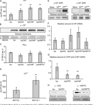
CPIP, HSP70, CK2, CHIP, and the proteasome regulate CP accumulation. Ex-pression of a dominant negative CPIP mutant (CPIPΔ66) leads to strong accumulation of CP in the absence of PVA infection (9). CPIPΔ66lacks the J domain through which native CPIP recruits HSP70 to promote CP ubiquitination and degradation. The carboxyl terminus Hsc70 interacting protein (CHIP) is an established E3 ubiquitin ligase that interacts with HSP70 and HSP90 to catalyze polyubiquitination of their target proteins (see references 25 and 26 and references therein). We therefore regarded CHIP as the most likely candidate E3 ubiquitin ligase for HSP70-CPIP-driven CP ubiquitination. To test whether CK2, HSP70, or CHIP can influence CP accumulation, we coexpressed CPWT with RNA hairpin constructs that initiate CK2, HSP70, and CHIP silencing. After 4 days, enzyme-linked immunosorbent assay (ELISA) and Western blot analyses showed that silencing of these host factors led to an approximately 2-fold increase in CP accumu-lation, suggesting that CK2, HSP70, and CHIP promote CP degradation (Fig. 3A and B). The origin of the double band in Fig. 3B is not clear, although it is possible to conclude that the two forms are not due to phosphorylation of the CK2 site, as also CPAAA migrates similarly. Fluc was also coexpressed in the analysis to function as an internal control, and its unaltered levels underscore that increased protein accumulation was CP specific (Fig. 3C). We also examined in the CK2-, HSP70-, and CHIP-silenced background the level of CP mRNA accumulation. While CK2 and HSP70 silencing did not alter CP mRNA amounts in comparison to that of the empty silencing vector (hp⫺) used as a control, CHIP silencing was found to do so (Fig. 3F). Therefore, the role of CHIP remains elusive in CP turnover, as an increased CP mRNA amount likely accounts for the
on November 7, 2019 by guest
http://jvi.asm.org/
FIG 1The CK2 phosphorylation consensus sequence is necessary for PVA replication. (A) Full-length PVA icDNA constructs. The amino acid mutations in the CK2 phosphorylation consensus region are shown under the CP coding sequence, and the nucleotide sequence corresponding to the mutated region is shown on the left. (B) Plants were infiltrated with various PVA icDNA constructs carryingRenillaluciferase gene as a reporter.Renillaluciferase activity values derived from PVA mutants were measured at 3 dpi. (C)Renillaluciferase activity values were measured at 3 dpi from the nonreplicating PVA mutant PVAΔGDDand two CK2 consensus sequence mutants, PVAAAAand PVAADA(n⫽9). (D) Alignment of 18 potyviral CP sequences showing
the CK2 phosphorylation consensus site (boxed area). Conservation of either Thr or Ser residue at the location corresponding to T243of PVA CP is
marked in bold. Amino acid numbering is according to the PVA sequence.**,P⬍0.01.
Lõhmus et al. Journal of Virology
on November 7, 2019 by guest
http://jvi.asm.org/
[image:4.585.46.517.89.669.2]increased CP amounts to some extent. Silencing of the host proteins was confirmed either by reverse transcription-quantitative PCR (RT-qPCR) or by Western blotting (Fig. 3G and H). However, inhibition of the proteasome by MG132 resulted in a 2-fold CP accumulation similar to that seen with silencing CK2, CHIP, and HSP70 (Fig. 3D). These FIG 2CK2␣-subunit was copurified with NIb and VPg from a crude membrane fraction. Western blot analysis of samples pulled down with StrepIII-tagged PVA protein NIb (NIbSIII) or VPg (VPgSIII) from the
membrane fractions probed with anti-CK2 ␣antibody shows that protein kinase CK2 ␣-subunit is copurified with PVA replication proteins.
FIG 3Regulation of CP accumulation by CK2, HSP70, CHIP, proteasome, and CK2 phosphorylation site within CP. (A and B) The host factors CK2, CHIP, and HSP70 were transiently silenced by RNA hairpin constructs, and the effect on the accumulation of CPWTwas measured in a noninfection context as shown by ELISA-based
quantification (A) and Western blotting with anti-CP antibody (B) at 4 dpi. CPWTwasAgrobacteriuminfiltrated
at an OD600of 0.5, and silencing constructs were infiltrated at an OD600of 0.3. (C) Transiently expressed Fluc
enzymatic activity was not affected by the silencing of CK2, CHIP, and HSP70. (D) The 26S proteasome activity was inhibited with MG132 compound, and the accumulation of CPWTwas measured by ELISA in a
noninfec-tion context. (E) Accumulanoninfec-tion of PVA CP phosphorylanoninfec-tion-deficient and phosphorylanoninfec-tion-mimicking mutants is increased compared to that of CPWTat 3 dpi. (F) RT-qPCR was used to quantitate the mRNA levels of
transiently expressed PVA CP in CK2, CPIP, HSP70, and CHIP silencing backgrounds at 4 dpi. (G and H) RT-qPCR of CPIP and CHIP mRNA levels (G) and Western blotting with anti-CK2 and anti-HSP70 antibodies (H) were used to verify silencing at 4 dpi after initiation of silencing. hp⫺is an empty silencing vector used as a control. The following significance code is used:*,P⬍0.05;**,P⬍0.01).
on November 7, 2019 by guest
http://jvi.asm.org/
[image:5.585.104.308.71.123.2] [image:5.585.49.365.261.619.2]results support the involvement of CK2, the proteasome, and putatively CHIP in regulation of PVA CP in addition to the previously established CPIP and HSP70 (9).
As silencing of CK2 caused CP accumulation, a logical interpretation was that the phosphorylation status of the CK2 consensus site in CP affects its turnover. Therefore, we studied the accumulation of CPs mutated in the CK2 site by Western blotting (Fig. 3E). Both the phosphorylation-deficient (CPAAA) and phosphorylation mimic (CPADA) mutants accumulated to higher levels than CPWT. Accumulation of both CPAAA and CPADAcompared to that of CPWTsuggested that the lack of dynamic phosphorylation reduces CP turnover.
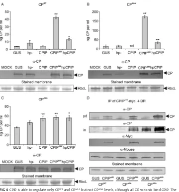
CPIP contributes to the suppression of the accumulation of CPWTand CPAAA,
but not CPADA. We next analyzed how overexpression of CPIP or CPIPΔ66 affected CPWT, CPAAA, and CPADAaccumulation levels. The RNA hairpin construct targeting CPIP was also included in the experiment. As expected on the basis of our earlier results (9), the overexpression of CPIP reduced the CPWTamount well below the control level and rendered it close to undetectable (Fig. 4A). Although hairpin-mediated silencing is not FIG 4CPIP is able to regulate only CPWTand CPAAAbut not CPADAlevels, although all CP variants bind CPIP. The
accumulation of CPWT(A), the phosphorylation-deficient mutant CPAAA(B), and the phosphorylation-mimicking
mutant CPADA(C) was monitored by ELISA and Western blotting when coexpressed with CPIP or CPIPΔ66or during
CPIP silencing (hpCPIP) at 4 dpi. The following significance codes are used:**,P⬍0.01;*,P⬍0.05; nd, not detectable. (D) CPIPΔ66or GUS as a control was coexpressed with PVA CP mutants in a noninfection context. CPIPΔ66
was immunoprecipitated at 4 dpi, and the eluates were probed for the presence of PVA CP by Western blotting (top [pd]). The total amounts (prior to purification) of CP are shown in the second panel (in). The presence of CPIPΔ66was verified by probing with␣-myc antibody and an equal amount of antibodies with anti-mouse antibody
and loading by staining the membranes with Ponceau S solution. CPs were infiltrated at an OD600of 0.5 and all
other constructs at an OD600of 0.3.
Lõhmus et al. Journal of Virology
on November 7, 2019 by guest
http://jvi.asm.org/
[image:6.585.42.402.67.453.2]complete and some endogenous CPIP may have remained as in Fig. 3G, accumulation of CPWTincreased in the presence of hpCPIP compared to the controls. Still, a substan-tially higher accumulation of CPWTwas observed when the dominant negative mutant CPIPΔ66 was overexpressed. The phosphorylation-deficient mutant CPAAA behaved similarly to CPWTin its response to CPIP silencing and CPIPΔ66overexpression (Fig. 4B). However, the phosphorylation-mimicking mutant CPADAdid not respond to CPIP, as it accumulated to high levels under all the tested conditions (Fig. 4C). These data suggest that phosphorylated CP may escape degradation via the CPIP pathway, whereas the nonphosphorylated CP is capable to enter the pathway provided that CPIP is present in sufficient amounts.
CP phosphorylation prevents CP degradation downstream of CPIP.To under-stand further the link between CK2-mediated phosphorylation of CP and CPIP-mediated regulation of CP functions, we wanted to find out whether CK2 phosphory-lation plays a role in the CP-CPIP interaction. The dominant negative J-domain mutant CPIPΔ66still binds potyviral CP (27). Because CPIP functions as a cochaperone of HSP70 via interaction of the J domain (28), the CPIPΔ66mutant is unable to deliver CP to HSP70 and the degradation pathway. Therefore, to allow the formation of a more stable and detectable CPIP-CP complex, we coexpressed PVA CP and the phosphorylation site mutants with a myc-tagged CPIPΔ66. This was followed by immunoprecipitation of CPIPΔ66, and subsequent detection of CPs showed that CPWT, CPAAA, and CPADAall bind CPIP (Fig. 4D). This result showed that CK2-mediated phosphorylation of Thr243in CP is not regulating binding of CPIP to CP.
The phosphomimic CPADA does not, whereas the phosphorylation deficient
CPAAAdoes, inhibit PVA translation.When CPWTis present in high concentrations, it inhibits PVA RNA translation (9, 10). The results reported in reference 10 suggest that the inhibition occurs through cotranslational interactions between CP accumulated in transand CP translated from viral RNA incis. Our next question was whether CPAAAand CPADAhave the capacity to suppress PVA translation when expressedin trans. CPWT, which strongly inhibits, and CPmut, which does not inhibit, PVA gene expression were used as controls for comparison. To this end, PVAWTand PVAΔGDDwere coexpressed together with different CPs, and viral gene expression was determined by Rluc activity at 3 dpi (Fig. 5). CPAAAinhibited PVA gene expression as strongly as the CPWT, resulting in almost complete inhibition of PVA translation. However, similarly to CPmut, CPADA had only a low inhibitory effect on PVA translation. The effect of CPs on PVAΔGDDgene expression was similar to the effect on PVAWTgene expression (compare left and right panels in Fig. 5), indicating that the inhibition happens at the translational level. Our conclusion is that the capacity of CP to inhibit PVA translation can be downregulated via phosphorylation of Thr243 of CP. In support of this, CK2 overexpression together with nonreplicating PVA led to significantly increased translation whereas CK2 silencing led to significantly reduced translation of viral RNA (Fig. 5). PVAWTgene expression was also increased by CK2 overexpression (Fig. 5), indicating that CK2 promotes PVA infection, possibly by increasing viral translation.
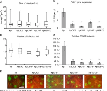
Host proteins CK2, CPIP, HSP70, and CHIP are all required to achieve a normal level of gene expression during PVA infection. Our results show that mutations altering the CP CK2 phosphorylation site cause defects in PVA replication, that CK2, CPIP, HSP70, and CHIP regulate CP accumulation and that phosphorylation of Thr243 regulates the ability of CP to inhibit PVA translation. Altogether, we hypothesized that CK2, CPIP, HSP70, and CHIP are all involved in regulating the CP primarily during translation/replication rather than particle assembly and virus cell-to-cell movement. To test whether the effect on viral gene expression is related to viral movement, we set up an experiment in which PVA-GFP was infiltrated at a lowAgrobacteriumconcentration (optical density at 600 nm [OD600], 0.0005) during transient silencing of the host proteins CPIP, CHIP, CK2, and HSP70. Accordingly, the low concentration of Agrobac-teriumcarrying PVA-GFP icDNA allowed for visualization of individual infection foci. The silencing was initiated with hairpin constructs 1 day before infiltration of virus icDNA to
on November 7, 2019 by guest
http://jvi.asm.org/
ensure strong silencing when evaluating possible effects on cell-to-cell movement. PVA infection focus sizes and their amount were measured at 3 days after viral icDNA infiltration. Despite the size of infection foci being somewhat variable within treat-ments, no significant difference in focus size appeared between the treatments (Fig. 6A). There was also no difference in the number of initiated infections between the different treatments (Fig. 6B). Moreover, PVA gene expression and RNA amount was significantly lower due to the silencing (Fig. 6C and D). Representative figures showing infection foci in various silencing backgrounds are presented in Fig. 6E.
These data suggest that silencing of CK2, CPIP, HSP70, and CHIP does not impair cell-to-cell movement but instead reduces the level of PVA gene expression at the single-cell level. This shared phenotype of the host factors supports their importance in regulating CP during replication-associated translation of PVA.
DISCUSSION
The conventional role of the potyviral CP is to encapsidate the viral genome in particles and aid in transportation of the virus within and between plants. However, as shown for several plant viruses, CPs can have many more functions during infection (reviewed in references 12, 29, and 30). These include CP-mediated regulation of viral RNA replication and translation, for which, e.g., brome mosaic virus represents a well-studied example (see reference 31 and references therein). In the present study, we aimed to deepen our understanding of how CP regulates potyvirus infection. Our previous studies have established that the phosphorylation of CP by CK2 has an essential role in PVA infection, the importance of which could be in regulating the interaction between CP and viral RNA (19, 20). We have additionally found that the host chaperones CPIP and HSP70 are important factors of potyvirus replication and mediate CP degradation (9). Degradation of CP by the CPIP/HSP70 pathway is important to maintain replication and thereby enables much stronger infection by potyviruses (9, 27). In addition, we have established that CP inhibits viral gene expression via cotrans-lational interactions between CPs accumulated intransand CP produced from PVA RNA incis(10). The current study revealed that similarly to CPIP and HSP70, both CK2 and the CK2 phosphorylation site within PVA CP are important for PVA replication and that FIG 5PVA RNA translation is regulated by CP in a CK2 phosphorylation-dependent manner. The gene expression, reported byRenillaluciferase activity, of wild-type PVA (PVAWT) (left) and replication-deficient PVA (PVAΔGDD) (right)
was measured at 3 dpi in the context of overexpressing the wild-type PVA CP (CPWT), nonphosphorylatable CP
(CPAAA), and phosphorylation-mimicking CP (CPADA). As a control, a CP mutant debilitated in viral RNA binding was
used (CPmut) (leftmost bar charts on both panels). Additionally, the gene expression of PVAWTwas measured during
overexpression of tCK2␣(PVAWT, right bar chart) and the gene expression of PVAΔGDDduring overexpression of
tCK2␣ and silencing of CK2␣ (PVAΔGDD, middle and rightmost bar charts, respectively) at 3 dpi. Anti-CK2␣
immunoblots indicate the level of CK2␣when overexpressed and silenced as a comparison to the control samples.
Lõhmus et al. Journal of Virology
on November 7, 2019 by guest
http://jvi.asm.org/
[image:8.585.41.402.70.265.2]CK2 together with CPIP, HSP70, and putatively CHIP regulate CP accumulation. In addition, we found that CP phosphorylation by CK2 regulates PVA translation, and this is likely linked to the different RNA binding capacities of the nonphosphorylated and phosphorylated CP. Our interpretation of the results is represented by a hypothetical model for discussion (Fig. 7).
Our earlier studies have established that CK2-mediated phosphorylation of CP is important for PVA infection, as CP phosphorylation site mutants failed to spread from cell to cell (19). However, it remained unclear whether this defect was at the single-cell level or movement related. In the present study, we found that CP-phosphorylation mutants had gene expression levels comparable to those of the nonreplicating PVAΔGDD rather than nonmoving PVACPmut, suggesting that CK2-mediated CP phos-phorylation is important for replication. Moreover, because replication is impaired for both the phosphomimic (PVAADA) and the phosphodeficient (PVAAAA) mutants, dynamic regulation between the two forms of CP appears to be important in the process.
The CK2 phosphorylation consensus site resides at a putative RNA binding domain according to a PVA CP structural model (32). In agreement with this, CK2 affects the capacity of CP to bind RNA (20) and altogether supports that the RNA binding capacity of potyviral CP is needed for replication. A silent CK2 site mutant, in which only the RNA sequence was changed, as well as PVAATA, carrying T242A and S244A mutations in CP, replicated to levels comparable with those of PVAWTand caused full infection. This FIG 6Host factors CK2, CPIP, CHIP, and HSP70 are required for robust PVA expression at a single-cell level. The host factors CK2, CPIP, CHIP, and HSP70 were transiently silenced by RNA hairpin constructs, and the effects on the size of infection foci (A) and the infection initiation efficiency of PVAGFP(B) were measured. (C and D) PVA gene
expression (C) and RNA levels (D) were measured in these silencing backgrounds. RNA hairpin (hp) silencing constructs were infiltrated at an OD600of 0.3 1 day before the virus, which was infiltrated at an OD600of 0.05 for
PVAWTand an OD
600of 0.0005 for PVAGFP. Viral movement and gene expression were measured at 3 dpi. (E)
Representative images of PVA-GFP infection foci in the CK2, CPIP, HSP70, and CHIP silencing backgrounds at 3 dpi. Asterisks denote statistical significance:**,P⬍0.01;*,P⬍0.05.
on November 7, 2019 by guest
http://jvi.asm.org/
[image:9.585.40.406.72.385.2]supports that the CK2 consensus site of PVA CP and PVA replication have a link at the protein level rather than at the encoding RNA level. Conservation of the CK2 consensus site in potyviruses also suggests a functional importance for CP at the protein level. For comparison, it is interesting that tobacco etch virus (TEV) replication tolerates large deletions in its CP (33). More specifically, it was established that a CP protein fragment located after residue 189 needs to be translated for successful replication. At the same time, viruses carrying C-terminal CP deletions were functional in replication only if the deletion started after residue 245. Intriguingly, the putative CK2 phosphorylation site of TEV CP (236VGTAEED242, where boldface type indicates the Thr conservation site) lies within the peptide needed for replication (33). However, acis-active RNA element was proposed to overlap the minimal essential TEV CP protein fragment and it was also proposed to be required for TEV replication.
In our previous study (10), we proposed that the site for the CP-RNA interaction on viral RNA is at the CP-encoding region and the binding to this site occurs in the context of ongoing translation. To initiate potyviral replication complex assembly, 6K2 tethers NIa to the endoplasmic reticulum (ER) membranes while NIb is directed via the NIa interaction and by the secondary structures in the 3=untranslated region (UTR) and the 3=part of the CP-encoding region to the 3=end of the viral RNA (34, 35). It has been shown that the RNA polymerase, NIb, of tobacco vein mottling virus (TVMV; genus Potyvirus) interacts with CP (36). Interestingly, this interaction was weakened in a catalytically inactive NIb mutant. We found that CK2␣associates with the VPg- and NIb-containing protein complexes purified from the crude cellular membranes and showed previously that the similarly purified complexes also contain HSP70, CPIP, viral RNA, and CP (9). On the basis of these data, it is possible that CP interacts directly with FIG 7Proposed model of CP-regulated shift from translation to PVA replication. The nonphosphorylated CP binds to viral RNA and blocks translation. The translational block by CP enables removal of ribosomes and recruiting of NIb to the 3=UTR of PVA RNA for the assembly of the viral replication complex (VRC). CK2-mediated phosphorylation of CP assists CPIP and HSP70 in detaching CP from viral RNA, and E3 ubiquitin ligase CHIP is recruited by HSP70 for CP ubiquitination and degradation. Removal of CP from PVA RNA allows initiation of the replication reactions.
Lõhmus et al. Journal of Virology
on November 7, 2019 by guest
http://jvi.asm.org/
[image:10.585.83.329.70.359.2]NIb on viral RNA or that CP and VPg/NIb are interconnected via viral RNA. Collectively, these results place CK2 and CP in association with the replication complex consisting of NIb and NIa and reinforce the link between CP phosphorylation and PVA replication. When PVA replication complexes were purified via the membrane-spanning 6K2, only a minor amount of CP compared to that of replication proteins NIa and NIb was found in these membranous structures (24). This would speak for a transient presence of CP on the viral RNA to be replicated whereas it may not be required for the replication reactionsper se.
CPIP is an important regulator of potyviral CP required for replication and acts via binding CP and delivering it to HSP70. This is likely followed by CP ubiquitination and degradation and is essential for potyviruses to avoid premature shutdown of replication (9, 27). CHIP is the anticipated E3 ubiquitin ligase to associate with HSP70 and mediate polyubiquitination of CP to promote its degradation by the proteasome (37, 38). CHIP downregulation affected CP mRNA accumulation to some extent, and therefore the role of CHIP in CP degradation remains still putative. Proteasome inhibition caused CP accumulation. Similarly to HSP70, CPIP, and CK2, we also established that CHIP was important for a robust PVA infection unrelated to intercellular movement. Based on our previous study linking CPIP and HSP70 to CP regulation (9), we now propose that both CHIP and CK2 are likewise involved in regulating CP during PVA RNA translation and replication cycle and not in cell-to-cell movement.
Global proteomic analyses of protein ubiquitination show that phosphorylatable amino acid residues flank ubiquitinated Lys residues with high propensity (39, 40). Protein phosphorylation has been linked both to activation (41) and to inhibition (42, 43) of proteasome-mediated degradation of the target protein. One of the questions of the current study was to address if CK2-mediated phosphorylation affects degradation of CP via the CPIP pathway. The CK2 phosphorylation-mimicking mutant CPADAcan bind CPIP but did not undergo CPIP-mediated degradation, suggesting that CP phos-phorylation could protect from degradation. However, our data also support that the interactions between CK2, CPIP, HSP70, and CP may occur on viral RNA, and therefore it is possible that CPADAdoes not localize to a context where the necessary interactions to enter the degradation pathway take place. We do not exclude the alternative or even parallel possibility that CPs are phosphorylated prior to binding to viral RNA. In such case, it is feasible to speculate that phosphorylation could promote sufficient CP accumulation by protecting it from degradation and simultaneously keep a certain proportion of CP away from inhibiting viral RNA. Production of CPAAAin turn may lead to a situation where most CP molecules bind viral RNA. In the absence of CP phos-phorylation, the CPIP/HSP70 system either is dysfunctional or gets overpowered sooner than in the presence of CPWT. In our previous work (19), we have shown that CPAAA produced inEscherichia coliis able to form viruslike particles (VLPs). The basis for the stability of the CPAAAmutant when produced from monocistronic mRNA could there-fore be the formation of VLPs via CP-CP interactions. To this end, a conceivable role of CPWTphosphorylation would be to aid CPIP and HSP70 functions by downregulating either the RNA binding affinity or the proportion of CP molecules binding to RNA.
Although many important questions remain, in conclusion of this study a plausible scenario could be that binding of nonphosphorylated CP to viral RNA in the vicinity of the 3=UTR of PVA RNA keeps that region free from ribosomes and allows time for the assembly of the replication complex. CP phosphorylation and the CPIP and HSP70 chaperone system remove CP from PVA RNA and open viral RNA for the replication reactions.
MATERIALS AND METHODS
Plants and virus and plant expression constructs.Nicotiana benthamianaplants were kept under greenhouse conditions and infiltrated withAgrobacterium tumefaciensstrain C58C1 pGV2260 at the four-to six-leaf stage. Viral constructs and viral protein expression constructs used in this study were based on the full-length infectious cDNA (icDNA) copies of PVA strain B11 (GenBank accession number AJ296311).
on November 7, 2019 by guest
http://jvi.asm.org/
icDNA constructs for PVAWT, PVA-GFP, PVACPmut, PVAΔGDD, PVAAAA, PVANIbSIII, and PVAVPgSIIIare
described in references 9 and 19. icDNA constructs for PVAATA, PVAADA, and PVAsilentwere done using
site-directed mutagenesis using primers CP-ATA-Fwd (5=GCA ACT GCA GAA GAG GAC ACA GAA AG 3=) for CPATA, CP-ADA-Fwd (5=GCA GAT GCA GAA GAG GAC ACA GAA AG 3=) for CPADA, and CP-silent-Fwd
(5=ACG ACG AGC GAA GAG GAC ACA GAA AG 3=) for CPsilentto modify the plasmid pKJE4 carrying the
PVA CP-coding region. From pKJE4, the mutated CP constructs were cut out with restriction enzymes MluI and AgeI and inserted into pKJE23 using the same restriction enzyme sites to replace the CP in PVA icDNA. The mutated PVA icDNA was transferred to the binary agro vector pRD400 using KpnI and SalI. The final icDNA constructs in pRD400 are under the control of CaMV 35S promoter andA. tumefaciens
nopaline synthase transcriptional terminator (nos).
Transient expression constructs for individual CPs were done as follows: CP-coding regions were amplified from their respective icDNA using primers CP_N-termATG_F (5=CCG GTA CCA TGG CCG AAA CTC TTG ATG CAA G 3=) and CP_C-term_R (5=GGT CTA GAT TAC ACC CCC TTC ACG CCT A 3=) and Phusion DNA polymerase (Thermo Scientific). The forward primer adds a KpnI restriction site and an in-frame ATG codon, and the reverse primer adds an XbaI restriction site directly after the TAA stop codon. The CP PCR fragments were digested with KpnI and XbaI restriction enzymes and inserted into the same sites in pHTT690, which contains CaMV 35S promoter and nos terminator. From pHTT690, the 35S-CP-nos
fragments were transferred to the binary agro vector pRD400 using HindIII restriction sites.
To produce the transient expression construct of the tCK2␣, the tCK2␣gene was amplified by PCR from pQE30::tCK2 (19) using primers 5=ATC GCT CGA GAT GTC AAA AGC TCG TGT TTA CA 3=and 5=CGA TGG ATC CTT ACT GCG TCC TCA TCC T 3=carrying XhoI and BamHI restriction sites, respectively. After XhoI and BamHI restriction, the PCR fragment was inserted into the same restriction sites in the multicloning site of pRT101, providing CaMV 35S promoter and terminator. From pRT101, the 35S-tCK2-T-35Scassette was cleaved out with HindIII and inserted into pRD400 binary vector.
To make hairpin silencing constructs targeting CK2, CPIP, CHIP, or HSP70, around 300- to 400-bp fragments of these genes were PCR amplified fromN. benthamianacDNA. The PCR fragments were inserted into pDONRZEOplasmid and finally to pHellsGate8 plasmid using the Gateway cloning system.
To amplify CK2, primers CK2sil-Fwd (5=CAG AAC CTC TGT GGG GGA CCA AC 3=) and CK2sil-Rev (5=CAT TCC TGC AAA CAT GCA TCC AAG 3=), for CPIP, primers CPIPsil-Fwd (5=AGT GAA GAA GAT TTG AAA AAA TCG 3=) and CPIPsil-Rev (5=TTT GTG CCT TTC TTC CAA C 3=), for CHIP, CHIPsil-Fwd (5=CTG CCA TTG ATG CTT ATA CCG AGG C 3=) and CHIPsil-Rev (5=TTT GCT TTA GCA AGC TCT TCC CT 3=), and for HSP70, primers HSP70sil-Fwd (5=CCG GAT CCA TGG CTG GTA AAG GAG AAG 3=) and HSP70sil-Rev (5=CCC GAT CGG CAT GCT TAG TCG ACT TCC TCA ATC 3=) were used.
DAS-ELISA.For double-antibody sandwich ELISA (DAS-ELISA), samples were collected 4 days after infiltrating N. benthamiana plants with Agrobacterium carrying protein expression constructs. The
Agrobacteriumconcentrations used for infiltration are given in the figure legends. Infiltrated plant tissue was homogenized in sampling buffer (Tris-buffered saline [TBS], 0.5% Tween 20, 2% polyvinyl pyrrolidine, and 0.2% bovine serum albumin [BSA], pH 7.4) at a ratio of 1 g of plant material per 3 ml of buffer and centrifuged at 1,000⫻ gfor 5 min to remove cell debris. The undiluted supernatant was used in subsequent steps. Ninety-six-well plates (Costar 3590, high binding; Corning Incorporated, USA) were coated with PVA CP-specific monoclonal antibody (1041-07 PVA, 58/0, MAB; Adgen Phytodiagnostics) using a 1:1,000 dilution in coating buffer (0.05 M carbonate-bicarbonate, pH 9.6) and 100l of buffer per well and incubated at 37°C for 3 h. After washing the plate 3 times with 300l of washing buffer (50 mM Tris-Cl, 150 mM NaCl, 0.5% Tween 20 [pH 7.4]) per well, 100l of sample supernatant per well was added and the plate was incubated overnight at 4°C. The next day, the plate was washed as previously and 100 l per well of 1:4,000 diluted alkaline phosphatase-conjugated anti-PVA-CP antibody (1041-07 PVA, 58/0, MAB-AP; Adgen Phytodiagnostics) was added, and the plate was incubated at 37°C for 3 h. After another wash, detection of CP was done by using 0.5 mg/ml phosphatase substrate (5-mg tablets; Sigma-Aldrich) in substrate buffer (0.1 M diethanolamine [pH 9.8]), 100l per well. Absorbance was recorded with the Bio-Rad 680 microplate reader at 405 nm. Different concentrations of heterologously produced PVA CP were used as protein concentration standards.
Immunoprecipitation of CPIP⌬66.Samples were collected 4 days after infiltratingN. benthamiana
plants withAgrobacteriumcontaining protein expression constructs (CPs at an OD600of 0.5, GUS and
CPIPΔ66at an OD
600of 0.3). Lysates were prepared using 3 ml sampling buffer (TBS, 0.5% Tween 20, 1 mM
phenylmethylsulfonyl fluoride [PMSF]) per 1 g of plant tissue. The lysates were cleared by centrifugation at 17,000 ⫻ g for 5 min at 4°C. Five microliters (1 ng) of anti-c-Myc antibody (9E10; Santa Cruz Biotechnology) was added to 300l of lysates, and the mixture was incubated at 4°C for 1 h. Then, 20 l of Dynabeads protein G (Life Technologies), equilibrated in the sampling buffer, was added to the samples, which were incubated at 4°C for 18 h. Finally, the beads were washed four times with 1 ml of sampling buffer to remove nonspecifically bound proteins. Specifically bound proteins were eluted by heating the beads with 1⫻Laemmli sample buffer at 95°C for 5 min.
StrepIII-tag specific affinity purification.The purification was done as described in reference 9. Briefly,N. benthamianaleaves systemically infected with PVANIbSIIIand PVAVPgSIIIwere harvested at 8 days
postinfection (dpi) and homogenized at the ratio of 1 g of leaves per 2 ml of buffer (100 mM Tris-HCl, 150 mM NaCl, 5 mM EDTA, 1 mM PMSF, and 13% sucrose [pH 8.0]). A heavy membrane fraction was prepared from the homogenized leaf material by centrifugation. The membrane fraction was treated with 1% Triton X-100 and then subjected to Strep-tag specific affinity purification through Strep-tactin Sepharose (IBA). Bound proteins were eluted with buffer containing 2.5 mM desthiobiotin (100 mM Tris-HCl, 150 mM NaCl, 5 mM EDTA, 2.5 mM desthiobiotin [pH 8.0]).
Lõhmus et al. Journal of Virology
on November 7, 2019 by guest
http://jvi.asm.org/
Agrobacterium-mediated delivery of constructs, Rluc-assay, and transient silencing of host genes.Agrobacteriumisolates carrying different protein expression constructs and PVA icDNAs were infiltrated to the abaxial side of N. benthamianaleaves using a needleless syringe. The infiltration concentrations ofAgrobacteriumwere as follows: OD600of 0.3 for silencing constructs of CK2, CPIP, CHIP,
and HSP70; OD600of 0.5 for CP expression constructs during silencing and OD600of 0.1 when together
with viral icDNA; OD600 of 0.05 for PVAWT::RluciicDNA. The viral icDNA-carrying agrobacteria were
infiltrated 1 day after the protein expression or silencing constructs. The control firefly luciferase construct (Fluc) was always infiltrated together with the Rluc-carrying viral icDNAs at OD600of 0.01. PVA
gene expression was quantified by measuringRenillaluciferase (Rluc) activity 3 days after agroinfiltration of the virus. Rluc values were normalized against Fluc values using the following formula: normalized Rluc activity ⫽ (average Fluc activity/Fluc activity per sample) ⫻ Rluc activity per sample. Each experiment was done using at least four samples, and experiments were repeated at least twice. Error bars indicate standard deviations, and Student’s t test was used to calculate the significance of differences between samples (*,P⬍0.05;**,P⬍0.01).
Western blotting.For the detection of CP mutant accumulation, theAgrobacteriumisolates carrying CPWT, CPAAA, and CPADAexpression constructs were infiltrated at an OD
600of 1 and sampled 3 days
postinfiltration. Standard Western blotting procedure was followed, and anti-CP antibody raised in rabbits was used for CP detection. For HSP70 and CK2 detection, a commercial polyclonal rabbit antibody against HSP70/HSC70 (9) and a polyclonal antibody raised in rabbits againstNicotiana tabacumCK2 ␣-subunit (19) were used.
Proteasome inhibition assay.Agrobacteria carrying PVA CPWTexpression construct were infiltrated
into N. benthamiana leaves at an OD600of 0.3. To the same areas, a 50 M solution of MG132
(Sigma-Aldrich) in water or water as a control was infiltrated 3 days later. After 24 h, samples were collected from the infiltrated leaves and a DAS-ELISA was performed to quantify PVA CP.
Quantitative RT-PCR.Total RNA was extracted from 100 mg of plant tissue using the GeneJET Plant RNA Purification minikit (Thermo Scientific). One microgram of extracted RNA was treated with 1 unit of RQ1 RNase-free DNase (Promega) at 37°C for 30 min. A reverse transcription reaction was performed on the DNase-treated RNA using the RevertAid H Minus First Strand cDNA synthesis kit (Thermo Scientific) according to the manufacturers’ recommendations. Five units of RNase H (New England BioLabs) was used to digest the remaining RNA in the cDNA at 37°C for 20 min. RNase H-treated cDNA was used in quantitative PCR using the DyNAmo Flash SYBR green qPCR mastermix (Thermo Scientific) and the CFX96 Touch Real-Time PCR detection system (Bio-Rad). To verify silencing of CHIP and CPIP by qPCR primers for CHIP (5=GAG TGG GAG CAT GAA TCT AC 3=and 5=CAG GAA CCT CAG TTG GAG T 3=) and CPIP (5=
CAC AGA TGT TGT TAA GCC 3=and 5=TAG TCA ACA GTC CTG CC 3=) were used. Protein phosphatase 2A (PP2A) gene with PP2A Forward primer (5=GAC CCT GAT GTT GAT GTT CG 3=) and PP2A Reverse primer (5=GAG GGA TTT GAA GAG AGA TTT C 3=) and F-box protein gene with F-box Forward primer (5=GGC ACT CAC AAA CGT CTA TTT C 3=) and F-box Reverse primer (5=TGG GAG GCA TCC TGC TTA T 3=) were used as internal controls (44).
Infection focus amount and size determination.PVA-GFP was infiltrated at an OD600of 0.0005 to
initiate infection in widely spaced individual cells. Silencing and control constructs were infiltrated 1 day before at an OD600of 0.3. The infection initiation efficiency was assessed 3 dpi from virus infiltration by
counting the visible infection foci reported by GFP fluorescence from digital micrographs of the infected plant leaves with the approximate area of 10.5 mm2. The infection foci were measured from the same
images. At least 10 areas were used. Images were recorded using an Axioskop 2 plus microscope (Carl Zeiss) with an AxioCam HRc microscope camera and AxioVision 4.8 software (Carl Zeiss). Student’sttest was used to calculate the significance values, which are indicated as follows in the figures:*,P⬍0.05;
**,P⬍0.01; no asterisk indicates that there is no statistical significance.
ACKNOWLEDGMENTS
We thank Swarnalok De for the help with potyvirus CP multiple alignment and Säde Virkki for excellent assistance in the greenhouse. We thank the CSIRO Division of Plant Industry for providing the pHG8 silencing plasmid.
Financial support from the Academy of Finland (1138329 and 1298254) and the Jane and Aatos Erkko Foundation for K.M. and from the Integrative Life Sciences doctoral program, Niemi Foundation, and Vilho, Yrjö ja Kalle Väisälä Foundation for A.L. is gratefully acknowledged.
REFERENCES
1. Neeleman L, Linthorst HJ, Bol JF. 2004. Efficient translation of alfamovi-rus RNAs requires the binding of coat protein dimers to the 3=termini of the viral RNAs. J Gen Virol 85:231–240. https://doi.org/10.1099/ vir.0.19581-0.
2. Boni S, Lavergne JP, Boulant S, Cahour A. 2005. Hepatitis C virus core protein acts as a trans-modulating factor on internal translation initia-tion of the viral RNA. J Biol Chem 280:17737–17748. https://doi.org/ 10.1074/jbc.M501826200.
3. Guogas LM, Filman DJ, Hogle JM, Gehrke L. 2004. Cofolding organizes
alfalfa mosaic virus RNA and coat protein for replication. Science 306: 2108 –2111. https://doi.org/10.1126/science.1103399.
4. Reichert VL, Choi M, Petrillo JE, Gehrke L. 2007. Alfalfa mosaic virus coat protein bridges RNA and RNA-dependent RNA polymerase in vitro. Virology 364:214 –226. https://doi.org/10.1016/j.virol.2007.02.026. 5. Yi G, Letteney E, Kim CH, Kao CC. 2009. Brome mosaic virus capsid
protein regulates accumulation of viral replication proteins by binding to the replicase assembly RNA element. RNA 15:615– 626. https:// doi.org/10.1261/rna.1375509.
on November 7, 2019 by guest
http://jvi.asm.org/
6. Yi G, Vaughan RC, Yarbrough I, Dharmaiah S, Kao CC. 2009. RNA binding by the brome mosaic virus capsid protein and the regulation of viral RNA accumulation. J Mol Biol 391:314 –326. https://doi.org/ 10.1016/j.jmb.2009.05.065.
7. Bamunusinghe D, Seo JK, Rao AL. 2011. Subcellular localization and rearrangement of endoplasmic reticulum by brome mosaic virus capsid protein. J Virol 85:2953–2963. https://doi.org/10.1128/JVI .02020-10.
8. Asurmendi S, Berg RH, Koo JC, Beachy RN. 2004. Coat protein regulates formation of replication complexes during tobacco mosaic virus infec-tion. Proc Natl Acad Sci U S A 101:1415–1420. https://doi.org/10.1073/ pnas.0307778101.
9. Hafrén A, Hofius D, Rönnholm G, Sonnewald U, Mäkinen K. 2010. HSP70 and its cochaperone CPIP promote potyvirus infection in Nicotiana benthamiana by regulating viral coat protein functions. Plant Cell 22: 523–535. https://doi.org/10.1105/tpc.109.072413.
10. Besong-Ndika J, Ivanov KI, Hafren A, Michon T, Makinen K. 2015. Cotrans-lational coat protein-mediated inhibition of potyviral RNA translation. J Virol 89:4237– 4248. https://doi.org/10.1128/JVI.02915-14.
11. Annamalai P, Rao AL. 2006. Packaging of brome mosaic virus sub-genomic RNA is functionally coupled to replication-dependent tran-scription and translation of coat protein. J Virol 80:10096 –10108. https:// doi.org/10.1128/JVI.01186-06.
12. Ivanov KI, Mäkinen K. 2012. Coat proteins, host factors and plant viral replication. Curr Opin Virol 2:712–718. https://doi.org/10.1016/ j.coviro.2012.10.001.
13. Yu M, Summers J. 1994. Multiple functions of capsid protein phosphor-ylation in duck hepatitis B virus replication. J Virol 68:4341– 4348. 14. Cartier C, Sivard P, Tranchat C, Decimo D, Desgranges C, Boyer V. 1999.
Identification of three major phosphorylation sites within HIV-1 capsid. Role of phosphorylation during the early steps of infection. J Biol Chem 274:19434 –19440.
15. Chapdelaine Y, Kirk D, Karsies A, Hohn T, Leclerc D. 2002. Mutation of capsid protein phosphorylation sites abolishes cauliflower mosaic virus infectivity. J Virol 76:11748 –11752. https://doi.org/10.1128/ JVI.76.22.11748-11752.2002.
16. Lan YT, Li J, Liao W, Ou J. 1999. Roles of the three major phosphorylation sites of hepatitis B virus core protein in viral replication. Virology 259: 342–348. https://doi.org/10.1006/viro.1999.9798.
17. Law LJ, Ilkow CS, Tzeng W-P, Rawluk M, Stuart DT, Frey TK, Hobman TC. 2006. Analyses of phosphorylation events in the rubella virus capsid protein: role in early replication events. J Virol 80:6917– 6925. https:// doi.org/10.1128/JVI.01152-05.
18. Law LM, Everitt JC, Beatch MD, Holmes CF, Hobman TC. 2003. Phosphor-ylation of rubella virus capsid regulates its RNA binding activity and virus replication. J Virol 77:1764 –1771. https://doi.org/10.1128/JVI.77.3.1764 -1771.2003.
19. Ivanov KI, Puustinen P, Gabrenaite R, Vihinen H, Rönnstrand L, Valmu L, Kalkkinen N, Mäkinen K. 2003. Phosphorylation of the potyvirus capsid protein by protein kinase CK2 and its relevance for virus infection. Plant Cell 15:2124 –2139. https://doi.org/10.1105/tpc.012567.
20. Ivanov KI, Puustinen P, Merits A, Saarma M, Mäkinen KM. 2001. Phos-phorylation down-regulates the RNA-binding function of the coat pro-tein of potato virus A. J Biol Chem 276:13530 –13540. https://doi.org/ 10.1074/jbc.M009551200.
21. Martin MT, Garcia JA. 1991. Plum pox potyvirus RNA replication in a crude membrane fraction from infected Nicotiana clevelandii leaves. J Gen Virol 72(Part 4):785–790. https://doi.org/10.1099/0022-1317-72-4 -785.
22. Schaad MC, Jensen PE, Carrington JC. 1997. Formation of plant RNA virus replication complexes on membranes: role of an endoplasmic reticulum-targeted viral protein. EMBO J 16:4049 – 4059. https://doi.org/10.1093/ emboj/16.13.4049.
23. Wei T, Wang A. 2008. Biogenesis of cytoplasmic membranous vesicles for plant potyvirus replication occurs at endoplasmic reticulum exit sites in a COPI- and COPII-dependent manner. J Virol 82:12252–12264. https:// doi.org/10.1128/JVI.01329-08.
24. Lohmus A, Varjosalo M, Makinen K. 2016. Protein composition of 6K2-induced membrane structures formed during Potato virus A infection. Mol Plant Pathol 17:943–958. https://doi.org/10.1111/mpp.12341. 25. Jiang J, Ballinger CA, Wu Y, Dai Q, Cyr DM, Hohfeld J, Patterson C. 2001.
CHIP is a U-box-dependent E3 ubiquitin ligase: identification of Hsc70 as a target for ubiquitylation. J Biol Chem 276:42938 – 42944. https:// doi.org/10.1074/jbc.M101968200.
26. Paul I, Ghosh MK. 2014. The E3 ligase CHIP: insights into its structure and regulation. Biomed Res Int 2014:918183. https://doi.org/10.1155/2014/ 918183.
27. Hofius D, Maier AT, Dietrich C, Jungkunz I, Bornke F, Maiss E, Sonnewald U. 2007. Capsid protein-mediated recruitment of host DnaJ-like proteins is required for Potato virus Y infection in tobacco plants. J Virol 81: 11870 –11880. https://doi.org/10.1128/JVI.01525-07.
28. Kelley WL. 1998. The J-domain family and the recruitment of chaperone power. Trends Biochem Sci 23:222–227. https://doi.org/10.1016/S0968 -0004(98)01215-8.
29. Weber PH, Bujarski JJ. 2015. Multiple functions of capsid proteins in (⫹) stranded RNA viruses during plant-virus interactions. Virus Res 196: 140 –149. https://doi.org/10.1016/j.virusres.2014.11.014.
30. Makarov VV, Kalinina NO. 2016. Structure and noncanonical activities of coat proteins of helical plant viruses. Biochemistry (Mosc) 81:1–18. https://doi.org/10.1134/S0006297916010016.
31. Kao CC, Ni P, Hema M, Huang X, Dragnea B. 2011. The coat protein leads the way: an update on basic and applied studies with the Brome mosaic virus coat protein. Mol Plant Pathol 12:403– 412. https://doi.org/10.1111/ j.1364-3703.2010.00678.x.
32. Baratova LA, Efimov AV, Dobrov EN, Fedorova NV, Hunt R, Badun GA, Ksenofontov AL, Torrance L, Jarvekulg L. 2001. In situ spatial organiza-tion of Potato virus A coat protein subunits as assessed by tritium bombardment. J Virol 75:9696 –9702. https://doi.org/10.1128/ JVI.75.20.9696-9702.2001.
33. Mahajan S, Dolja VV, Carrington JC. 1996. Roles of the sequence encod-ing tobacco etch virus capsid protein in genome amplification: require-ments for the translation process and a cis-active element. J Virol 70:4370 – 4379.
34. Haldeman-Cahill R, Daros JA, Carrington JC. 1998. Secondary structures in the capsid protein coding sequence and 3=nontranslated region involved in amplification of the tobacco etch virus genome. J Virol 72:4072– 4079.
35. Wang X, Ullah Z, Grumet R. 2000. Interaction between zucchini yellow mosaic potyvirus RNA-dependent RNA polymerase and host poly-(A) binding protein. Virology 275:433– 443. https://doi.org/10.1006/ viro.2000.0509.
36. Hong Y, Levay K, Murphy JF, Klein PG, Shaw JG, Hunt AG. 1995. A potyvirus polymerase interacts with the viral coat protein and VPg in yeast cells. Virology 214:159 –166. https://doi.org/10.1006/ viro.1995.9944.
37. Connell P, Ballinger CA, Jiang J, Wu Y, Thompson LJ, Hohfeld J, Patterson C. 2001. The co-chaperone CHIP regulates protein triage decisions me-diated by heat-shock proteins. Nat Cell Biol 3:93–96. https://doi.org/ 10.1038/35050618.
38. Demand J, Alberti S, Patterson C, Hohfeld J. 2001. Cooperation of a ubiquitin domain protein and an E3 ubiquitin ligase during chaperone/ proteasome coupling. Curr Biol 11:1569 –1577. https://doi.org/10.1016/ S0960-9822(01)00487-0.
39. Xu G, Paige JS, Jaffrey SR. 2010. Global analysis of lysine ubiquitination by ubiquitin remnant immunoaffinity profiling. Nat Biotechnol 28: 868 – 873. https://doi.org/10.1038/nbt.1654.
40. Kim W, Bennett EJ, Huttlin EL, Guo A, Li J, Possemato A, Sowa ME, Rad R, Rush J, Comb MJ, Harper JW, Gygi SP. 2011. Systematic and quanti-tative assessment of the ubiquitin-modified proteome. Mol Cell 44: 325–340. https://doi.org/10.1016/j.molcel.2011.08.025.
41. Wang Y, Guan S, Acharya P, Koop DR, Liu Y, Liao M, Burlingame AL, Correia MA. 2011. Ubiquitin-dependent proteasomal degradation of human liver cytochrome P450 2E1: identification of sites targeted for phosphorylation and ubiquitination. J Biol Chem 286:9443–9456. https://doi.org/10.1074/jbc.M110.176685.
42. Dimmeler S, Breitschopf K, Haendeler J, Zeiher AM. 1999. Dephosphor-ylation targets Bcl-2 for ubiquitin-dependent degradation: a link be-tween the apoptosome and the proteasome pathway. J Exp Med 189: 1815–1822. https://doi.org/10.1084/jem.189.11.1815.
43. Nalavadi VC, Muddashetty RS, Gross C, Bassell GJ. 2012. Dephosphorylation-induced ubiquitination and degradation of FMRP in dendrites: a role in immediate early mGluR-stimulated translation. J Neurosci 32:2582–2587. https://doi.org/10.1523/JNEUROSCI.5057 -11.2012.
44. Liu D, Shi L, Han C, Yu J, Li D, Zhang Y. 2012. Validation of reference genes for gene expression studies in virus-infected Nicotiana benthami-ana using quantitative real-time PCR. PLoS One 7:e46451. https:// doi.org/10.1371/journal.pone.0046451.
Lõhmus et al. Journal of Virology