PEARLS
Helminth infection–induced malignancy
Paul J. Brindley1,2*, Alex Loukas3*
1 Department of Microbiology, Immunology and Tropical Medicine, School of Medicine & Health Sciences, The George Washington University, Washington DC, United States of America, 2 Research Center for Neglected Tropical Diseases of Poverty, School of Medicine & Health Sciences, The George Washington University, Washington DC, United States of America, 3 Centre for Biodiscovery and Molecular Development of Therapeutics, Australian Institute of Tropical Health & Medicine, James Cook University, Cairns,
Queensland, Australia
*[email protected](PJB);[email protected](AL)
Infection with some helminth pathogens represents a biological
carcinogen
Infectious diseases cause more than 20% of cancers in the developing world [1]. About a dozen pathogens including Epstein-Barr virus and human T cell lymphocytotropic virus 1 are among the well-known examples. In addition, infection with several trematodes, which are eukaryotes, can cause malignancy. The International Agency for Research on Cancer cat-egorizes infection with the fish-borne trematodesOpisthorchis viverriniandClonorchis sinen-sisand the blood flukeSchistosoma haematobiumas Group 1 biological carcinogens [2]. In addition to parasitism directly damaging development, health, and prosperity of infected populations, infection with these helminths leads to cholangiocarcinoma (CCA) (bile duct cancer) and squamous cell carcinoma (SCC) of the urinary bladder, respectively [2]. By con-trast, infection with phylogenetic relatives, also trematodes of the phylum Platyhelminthes and also major pathogens, is not carcinogenic. These irregularities suggest that either hel-minth-specific metabolites contribute to tumorigenesis and/or that certain tissues or organs are particularly susceptible to infection-induced malignancy. Moreover, each of these hel-minth infections must be viewed holistically in the context of a perfect storm of risk for can-cer (see [3]).
Helminth infection–induced cancers
O. viverrini and C. sinensis infection–induced cholangiocarcinoma
Infection is accomplished by ingestion of undercooked freshwater fish infected with the meta-cercaria stage of these species of liver flukes. Human infection leads to hepatobiliary disease, including cholangitis and periductal fibrosis. In liver fluke–endemic regions, infection is the major risk factor for CCA [2,4,5]. CCAs are slow-growing adenocarcinomas that metastasize to distant sites due to proximity to lymphatic vessels. Liver fluke–related CCA is often diag-nosed at an advanced stage, when the primary cancer is no longer amenable to curative surgery [5]. The mechanism(s) by which infection initiates genetic lesions that eventually culminate in CCA is not understood, although it is likely to be multifactorial, involving biliary tract and sys-temic chronic inflammation and associated endogenous nitrosation [6,7], secretion of mito-gens and other mediators by the parasite, and cofactors including dietary preferences for nitrosamines-rich foods [4,8,9] (Fig 1).
a1111111111 a1111111111 a1111111111 a1111111111 a1111111111 OPEN ACCESS
Citation: Brindley PJ, Loukas A (2017) Helminth infection–induced malignancy. PLoS Pathog 13(7): e1006393.https://doi.org/10.1371/journal. ppat.1006393
Editor: Laura J Knoll, University of Wisconsin Medical School, UNITED STATES
Published: July 27, 2017
Copyright:©2017 Brindley, Loukas. This is an open access article distributed under the terms of theCreative Commons Attribution License, which permits unrestricted use, distribution, and reproduction in any medium, provided the original author and source are credited.
Funding: AL acknowledges support from NHMRC via a Senior Principal Research Fellowship, project and program grants. Support from the National Institute of Allergy and Infectious Diseases (NIAID), Tropical Medicine Research Center award number P50AI098639, and the National Cancer Institute (NCI) award number R01CA164719 is gratefully acknowledged. The funders had no role in study design, data collection and analysis, decision to publish, or preparation of the manuscript.
Urogenital schistosomiasis and bladder cancer
Three major species of schistosomes are the agents of schistosomiasis:S.japonicumandS. mansonicause intestinal schistosomiasis whereasS.haematobiumcauses urogenital schistoso-miasis. Of about 112 million cases ofS.haematobiuminfection in sub-Saharan Africa, 70 mil-lion are associated with hematuria, 18 milmil-lion with major bladder wall pathology, and 10 million with hydronephrosis leading to kidney damage. Deposition of ova ofS.haematobium in the bladder wall can lead to SCC of the bladder [2,10].
Initiation, promotion, and progression of tumorigenesis
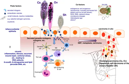
[image:2.612.60.574.80.414.2]Where opisthorchiasis is endemic, people can remain infected for decades. Opisthorchiasis provokes inflammation of the biliary tree, with hyperplasia and metaplasia of the cholangio-cytes that line the biliary tract adjacent to the flukes. Opisthorchiasis-induced fibrosis engulfs the proliferating cells, manifesting as biliary periductal fibrosis [4,11]. Chronic inflammation in response to parasite metabolites and growth factors is implicated in the inflammatory Fig 1. Schematic representation of hypothesized processes of carcinogenesis of the biliary tract and urinary bladder during chronic infection with fish-borne liver flukes Opisthorchis viverrini (Ov) and Clonorchis sinensis (Cs) and the blood fluke Schistosoma haematobium (Sh). Photomicrographs: adult developmental stages of Cs and Ov and the egg stage of Sh. During infection, mutations initiate carcinogenesis, perhaps as the consequence of interaction of epithelial cell chromosomal DNA with inflammation-associated reactive oxygen species (ROS) and reactive nitrogen species (RNS) and lipid peroxidation and/or metabolites released by the worms, such as catechol estrogen quinones and oxysterols. Subsequently, mediators of helminth origin such as granulin from Ov promote epidermal to mesenchymal transition (EMT), transformed cell growth, complementary angiogenesis, down-regulation of apoptosis, and other hallmarks of cancer.
response linked with infection [11]. Additional factors including carriage ofHelicobacterand other microbiome changes within the biliary tract might participate [8,12]. Elevated plasma interleukin-6 (IL-6) is associated with marked increase in risk of periductal fibrosis during opisthorchiasis and compounds pathogenesis by promoting a fibrogenic inflammatory milieu. The cell of origin of CCA is the cholangiocyte, a specialized epithelial cell that lines the bile duct. Following initiation, for example by oxidation of cholangiocyte chromosomal DNA by oxysterols generated by reactive oxygen species (ROS) and reactive nitrogen species (RNS) arising during opisthorchiasis-induced oxidative stress [13] and/or oxysterols released by the liver fluke [8,14], oncogenesis appears to be promoted by cholestasis and chronic inflamma-tion. The release and downstream consequences of IL-6, platelet-derived growth factor, tumor necrosis factor-alpha (TNF-α), and transforming growth factor-beta (TGF-β) are pivotal to the proliferation of cholangiocytes. Autonomous proliferation, evasion of apoptosis, and angio-genesis sustain the incipient neoplasm [11].
Bladder cancer is the most common tumor of the urinary system and consists of 2 main forms—urothelial carcinoma (UCC) and SCC. The bladder is lined by a specialized epithelium termed the urothelium, which is exposed routinely to potential carcinogens and hence is at particular risk of cancer. The urothelium is a stratified epithelium composed of keratin 5 (K5)-expressing basal cells, intermediate cells, and umbrella cells. K5-positive basal cells likely give rise to SCC [10,15–17]. UCC accounts for around 90% of bladder cancers, and major risk fac-tors include tobacco smoking, occupational exposure to aromatic amines and polycyclic hydrocarbons, and bladder stones, among others [10,15]. In regions endemic for urogenital schistosomiasis, SCC is more common than UCC and may be the cancer of highest incidence [2,10].
Schistosome eggs entrapped in the bladder wall release metabolites, presumably to facilitate egress of the egg to the lumen and to the external environment. The process leads to hematuria and to chronic inflammation, in turn increasing the risk of urothelial hyperplasia, dysplasia, and SCC (Fig 1). Urogenital schistosomiasis (UGS) is a chronic infection, often interrupted by drug treatment and often followed in turn by reinfection. Fibrosis induced by entrapped schis-tosome eggs may promote cellular proliferation, hyperplasia, and metaplasia that eventually induce carcinogenesis [10]. Mass spectrometric analysis of urine during UGS reveals estrogen-like metabolites including catechol estrogen quinones (CEQ), estrogen-likely of schistosome origin, CEQ-DNA adducts, and novel metabolites derived from 8-oxo-7, 8-dihydro-20 -deoxyguano-sine (8-oxodG) [18]. Nitrosamines and increased levels of beta-glucuronidase and cyclooxy-genase-2 derived from schistosomes also represent potential bladder carcinogens. These helminth infection–derived carcinogens may damage DNA, leading to somatic mutations in oncogenes such asp53, retinoblastoma protein, epidermal growth factor receptor, and erbB2 receptor tyrosine kinase. In like fashion, chromosomal adducts including 8-oxodG and 8-nitroguanine and lipid peroxide-DNA increase during opisthorchiasis (see [19]).
Investigation of UGS-induced bladder cancer is challenging given that rodent models do not exhibit urogenital disease; infection withS.haematobiumcauses hepatointestinal disease in rodents. However, in a recently developed rodent model, eggs ofS.haematobiuminjected into the bladder wall of mice provoke egg-associated pathogenesis more reflective of the human condition [20,21]. Preneoplastic lesions involving epithelial to mesenchymal (EMT)-like profiles have been described in this model following coadministration of exogenous nitro-samines [22]. These approaches might lead to deeper understanding of the carcinogenesis of UGS-induced SCC. A hamster model involving coadministration of nitroso compounds and liver fluke infection has long been employed to study infection-induced bile duct cancer [2,4,
not produced byS.mansoniandS.japonicumand/or the hepatointestinal niche of these intes-tinal disease–causing schistosomes is less disposed to schistosome infection–induced
malignancy.
Mutations, mutational signatures, rearrangements, epigenetics
Cancer arises when mutations occur in the DNA of the genome of the target cell and the muta-tions lead to uncontrolled cellular proliferation, invasion, and metastasis. The landscape of mutations that accumulate within the tumor record the mutagenic processes that have taken place over the life span of the malignancy. Each mutation endows an imprint on the genome of the tumor, documenting the types of DNA lesion and repair processes that lead to base sub-stitutions, insertions, deletions, and structural variations. These mutational profiles have implications for diagnosis, therapy, and public health interventions.
Metabolites of helminth origin, including oxysterols, catechol estrogens, heme from ingested blood, and others, all of which are reactive species, may depurinate host cell DNA, leading to error-prone repair that results in mutations of cancer driver genes [8,18]. At the genomic level, analysis of the mutation profiles ofO.viverrini–related versus nonliver fluke– induced CCA reveals marked variation in mutation patterns [24]. Somatic mutations occur frequently in the tumor suppressor genesp53andsmad4inO.viverrini–induced CCA. By con-trast, somatic mutations in the genes encoding BRCA1 associated protein-1 and isocitrate dehydrogenases 1 and 2 are more common in non-O.viverrini–associated CCA [24,25]. Mutations inp53andsmad4directly affect the p53- and TGF-signaling pathways, both of which are involved in tumorigenesis. Thus, distinct causes of CCA induce discrete somatic alterations, even within the same cancer type [25]. Emphasizing this point, liver fluke infec-tion–induced CCA exhibits altered DNA methylation and transcriptional profiles reflective of xenobiotic metabolism and pro-inflammatory responses in comparison to nonliver fluke infection–induced CAA and to healthy biliary duct [24–26].
Details also have emerged from whole exome sequences of UCC, although the specific mutational landscape of SCC and hence UGS-induced SCC have not been reported [2,15]. Mutation of sonic hedgehog can initiate carcinogenesis in bladder cancer [16]. Recurrent mutations occur in>30 other genes involved in cell proliferation, differentiation, genetic sta-bility, and specifically cell-cycle regulation, chromatin regulation, and kinase signaling path-ways [27]. Bladder cancer frequently exhibits C to T or C to G mutations at TC dinucleotides, which may reflect hyperactive DNA editing by apolipoprotein B mRNA-editing enzyme, cata-lytic polypeptide-like (APOBEC) cytidine deaminases [28].
Host–parasite interactions
surface ofO.viverriniEVs block the internalization by cholangiocytes of liver fluke EVs and suppress the proliferation and secretion of IL-6 by cholangiocytes [33]. Other helminth teins induce changes that reflect the hallmarks of cancer, including EMT phenotypes, pro-inflammatory cytokines, and repression of apoptosis [32,35]. Type 2 T helper cell (Th2) responses induced by schistosome proteins including interleukin 4-inducing principle of schistosome eggs (IPSE) not only facilitate egress of the schistosome egg to the bladder lumen by modulating the granuloma but also appear to modulate vasculogenic and cellular responses conducive to neoplasia [10,36]. Targeting these or other parasite–host cell communicating proteins with vaccines may not only block helminth infections but deliver novel anticancer vaccines [11].
Acknowledgments
We thank Javier Sotillo, Michael Smout, Thewarach Laha, Victoria Mann, Nuno Value, Jose´ M. Costa and Banchob Sripa for fruitful discussions including assistance with illustrations. We acknowledge collaboration and support of colleagues in TOPIC, the Tomsk Opisthorchiasis Consortium,http://www.topic-global.org/
References
1. de Martel C, Ferlay J, Franceschi S, Vignat J, Bray F, Forman D, et al. Global burden of cancers attribut-able to infections in 2008: a review and synthetic analysis. Lancet Oncol. 2012; 13(6):607–15.https:// doi.org/10.1016/S1470-2045(12)70137-7PMID:22575588.
[image:5.612.202.574.72.323.2]2. Humans IWGotEoCRt. Biological agents. Volume 100 B. A review of human carcinogens. IARC Monogr Eval Carcinog Risks Hum. 2012; 100(Pt B):1–441. PMID:23189750.
Fig 2. Liver fluke granulin promotes wound repair and angiogenesis. Opisthorchis viverrini secretes a growth factor termed liver fluke granulin, Ov-GRN-1, an orthologue of mammalian granulin. Ov-GRN-1 stimulates cholangiocytes to proliferate and promotes wound healing and angiogenesis. A recent report on
Ov-GRN-1 [32] illustrates some of these carcinogenic-conducive properties of the growth factor, as follows:
(A) rate of wound closure on a cutaneous lesion on mice, (B) the angiogenic nature of Ov-GRN-1 revealed using the chorioallantoic membrane assay in fertilized quail eggs.
3. Zhu L, Finkelstein D, Gao C, Shi L, Wang Y, Lopez-Terrada D, et al. Multi-organ Mapping of Cancer Risk. Cell. 2016; 166(5):1132–46 e7.https://doi.org/10.1016/j.cell.2016.07.045PMID:27565343. 4. Sripa B, Kaewkes S, Sithithaworn P, Mairiang E, Laha T, Smout M, et al. Liver fluke induces
cholangio-carcinoma. PLoS Med. 2007; 4(7):e201.https://doi.org/10.1371/journal.pmed.0040201PMID:
17622191.
5. Khuntikeo N, Loilome W, Thinkhamrop B, Chamadol N, Yongvanit P. A Comprehensive Public Health Conceptual Framework and Strategy to Effectively Combat Cholangiocarcinoma in Thailand. PLoS Negl Trop Dis. 2016; 10(1):e0004293.https://doi.org/10.1371/journal.pntd.0004293PMID:26797527. 6. Haswell-Elkins MR, Satarug S, Tsuda M, Mairiang E, Esumi H, Sithithaworn P, et al. Liver fluke infection
and cholangiocarcinoma: model of endogenous nitric oxide and extragastric nitrosation in human carci-nogenesis. Mutat Res. 1994; 305(2):241–52. PMID:7510035.
7. Satarug S, Lang MA, Yongvanit P, Sithithaworn P, Mairiang E, Mairiang P, et al. Induction of cyto-chrome P450 2A6 expression in humans by the carcinogenic parasite infection, opisthorchiasis viver-rini. Cancer Epidemiol Biomarkers Prev. 1996; 5(10):795–800. PMID:8896890.
8. Brindley PJ, da Costa JM, Sripa B. Why does infection with some helminths cause cancer? Trends Can-cer. 2015; 1(3):174–82.https://doi.org/10.1016/j.trecan.2015.08.011PMID:26618199.
9. Mitacek EJ, Brunnemann KD, Suttajit M, Martin N, Limsila T, Ohshima H, et al. Exposure to N-nitroso compounds in a population of high liver cancer regions in Thailand: volatile nitrosamine (VNA) levels in Thai food. Food Chem Toxicol. 1999; 37(4):297–305. PMID:10418946.
10. Honeycutt J, Hammam O, Fu CL, Hsieh MH. Controversies and challenges in research on urogenital schistosomiasis-associated bladder cancer. Trends Parasitol. 2014; 30(7):324–32.https://doi.org/10. 1016/j.pt.2014.05.004PMID:24913983.
11. Sripa B, Brindley PJ, Mulvenna J, Laha T, Smout MJ, Mairiang E, et al. The tumorigenic liver fluke Opisthorchis viverrini—multiple pathways to cancer. Trends Parasitol. 2012; 28(10):395–407.https:// doi.org/10.1016/j.pt.2012.07.006PMID:22947297.
12. Deenonpoe R, Mairiang E, Mairiang P, Pairojkul C, Chamgramol Y, Rinaldi G, et al. Elevated preva-lence of Helicobacter species and virupreva-lence factors in opisthorchiasis and associated hepatobiliary dis-ease. Sci Rep. 2017; 7:42744.https://doi.org/10.1038/srep42744PMID:28198451.
13. Jusakul A, Yongvanit P, Loilome W, Namwat N, Kuver R. Mechanisms of oxysterol-induced carcinogen-esis. Lipids Health Dis. 2011; 10:44.https://doi.org/10.1186/1476-511X-10-44PMID:21388551. 14. Vale N, Gouveia MJ, Botelho M, Sripa B, Suttiprapa S, Rinaldi G, et al. Carcinogenic liver fluke
Opisthorchis viverrini oxysterols detected by LC-MS/MS survey of soluble fraction parasite extract. Parasitol Int. 2013; 62(6):535–42. PMID:23973383.
15. Knowles MA, Hurst CD. Molecular biology of bladder cancer: new insights into pathogenesis and clinical diversity. Nat Rev Cancer. 2015; 15(1):25–41.https://doi.org/10.1038/nrc3817PMID:25533674. 16. Shin K, Lim A, Odegaard JI, Honeycutt JD, Kawano S, Hsieh MH, et al. Cellular origin of bladder
neopla-sia and tissue dynamics of its progression to invasive carcinoma. Nat Cell Biol. 2014; 16(5):469–78.
https://doi.org/10.1038/ncb2956PMID:24747439.
17. Van Batavia J, Yamany T, Molotkov A, Dan H, Mansukhani M, Batourina E, et al. Bladder cancers arise from distinct urothelial sub-populations. Nat Cell Biol. 2014; 16(10):982–91, 1–5.https://doi.org/10. 1038/ncb3038PMID:25218638.
18. Gouveia MJ, Santos J, Brindley PJ, Rinaldi G, Lopes C, Santos LL, et al. Estrogen-like metabolites and DNA-adducts in urogenital schistosomiasis-associated bladder cancer. Cancer Lett. 2015; 359(2):226– 32.https://doi.org/10.1016/j.canlet.2015.01.018PMID:25615421.
19. Yongvanit P, Pinlaor S, Bartsch H. Oxidative and nitrative DNA damage: key events in opisthorchiasis-induced carcinogenesis. Parasitol Int. 2012; 61(1):130–5.https://doi.org/10.1016/j.parint.2011.06.011
PMID:21704729.
20. Fu CL, Odegaard JI, Herbert DR, Hsieh MH. A novel mouse model of Schistosoma haematobium egg-induced immunopathology. PLoS Pathog. 2012; 8(3):e1002605.https://doi.org/10.1371/journal.ppat. 1002605PMID:22479181.
21. Honeycutt J, Hammam O, Hsieh MH. Schistosoma haematobium egg-induced bladder urothelial abnor-malities dependent on p53 are modulated by host sex. Exp Parasitol. 2015; 158:55–60.https://doi.org/ 10.1016/j.exppara.2015.07.002PMID:26160678.
22. Chala B, Choi MH, Moon KC, Kim HS, Kwak C, Hong ST. Development of Urinary Bladder Pre-Neopla-sia by Schistosoma haematobium Eggs and Chemical Carcinogen in Mice. Korean J Parasitol. 2017; 55(1):21–9.https://doi.org/10.3347/kjp.2017.55.1.21PMID:28285503.
24. Chan-On W, Nairismagi ML, Ong CK, Lim WK, Dima S, Pairojkul C, et al. Exome sequencing identifies distinct mutational patterns in liver fluke-related and non-infection-related bile duct cancers. Nat Genet. 2013; 45(12):1474–8.https://doi.org/10.1038/ng.2806PMID:24185513.
25. Jusakul A, Kongpetch S, Teh BT. Genetics of Opisthorchis viverrini-related cholangiocarcinoma. Curr Opin Gastroenterol. 2015; 31(3):258–63.https://doi.org/10.1097/MOG.0000000000000162PMID:
25693006.
26. Andresen K, Boberg KM, Vedeld HM, Honne H, Jebsen P, Hektoen M, et al. Four DNA methylation bio-markers in biliary brush samples accurately identify the presence of cholangiocarcinoma. Hepatology. 2015; 61(5):1651–9.https://doi.org/10.1002/hep.27707PMID:25644509.
27. Cancer Genome Atlas Research N. Comprehensive molecular characterization of urothelial bladder carcinoma. Nature. 2014; 507(7492):315–22.https://doi.org/10.1038/nature12965PMID:24476821. 28. Alexandrov LB, Ju YS, Haase K, Van Loo P, Martincorena I, Nik-Zainal S, et al. Mutational signatures associated with tobacco smoking in human cancer. Science. 2016; 354(6312):618–22.https://doi.org/ 10.1126/science.aag0299PMID:27811275.
29. van Tong H, Brindley PJ, Meyer CG, Velavan TP. Parasite Infection, Carcinogenesis and Human Malig-nancy. EBioMedicine. 2016.https://doi.org/10.1016/j.ebiom.2016.11.034PMID:27956028.
30. Smout MJ, Laha T, Mulvenna J, Sripa B, Suttiprapa S, Jones A, et al. A granulin-like growth factor secreted by the carcinogenic liver fluke, Opisthorchis viverrini, promotes proliferation of host cells. PLoS Pathog. 2009; 5(10):e1000611.https://doi.org/10.1371/journal.ppat.1000611PMID:19816559. 31. Li J, Razumilava N, Gores GJ, Walters S, Mizuochi T, Mourya R, et al. Biliary repair and carcinogenesis
are mediated by IL-33-dependent cholangiocyte proliferation. J Clin Invest. 2014; 124(7):3241–51.
https://doi.org/10.1172/JCI73742PMID:24892809.
32. Smout MJ, Sotillo J, Laha T, Papatpremsiri A, Rinaldi G, Pimenta RN, et al. Carcinogenic Parasite Secretes Growth Factor That Accelerates Wound Healing and Potentially Promotes Neoplasia. PLoS Pathog. 2015; 11(10):e1005209.https://doi.org/10.1371/journal.ppat.1005209PMID:26485648. 33. Chaiyadet S, Sotillo J, Smout M, Cantacessi C, Jones MK, Johnson MS, et al. Carcinogenic Liver Fluke
Secretes Extracellular Vesicles That Promote Cholangiocytes to Adopt a Tumorigenic Phenotype. J Infect Dis. 2015; 212(10):1636–45.https://doi.org/10.1093/infdis/jiv291PMID:25985904.
34. Hemler ME. Tetraspanin functions and associated microdomains. Nat Rev Mol Cell Biol. 2005; 6 (10):801–11.https://doi.org/10.1038/nrm1736PMID:16314869.
35. Matchimakul P, Rinaldi G, Suttiprapa S, Mann VH, Popratiloff A, Laha T, et al. Apoptosis of cholangio-cytes modulated by thioredoxin of carcinogenic liver fluke. Int J Biochem Cell Biol. 2015; 65:72–80.
https://doi.org/10.1016/j.biocel.2015.05.014PMID:26007234.