1
A PROSPECTIVE STUDY ON FUNCTIONAL OUTCOME OF COMMINUTED FRACTURE SHAFT OF HUMERUS OPERATED BY
MINIMALLY INVASIVE ANTERIOR PLATE OSTEOSYNTHESIS
DISSERTATION SUBMITTED FOR MS (ORTHOPAEDICS)
MADURAI MEDICAL COLLEGE MADURAI
2018
THE TAMIL NADU
DR. MGR MEDICAL UNIVERSITY CHENNAI, TAMIL NADU
2
This is to certify that the work “A STUDY ON THE ANALYSIS OF FUNCTIONAL OUTCOME OF COMMINUTED FRACTURE SHAFT OF HUMERUS BY MINIMALLY INVASIVE ANTERIOR PLATE OSTEOSYNTHESIS" which is being submitted for M.S. Orthopaedics, is a bonafide work of Dr. D. SANJAY KANTH, Post Graduate Student at Department of Orthopaedics, Madurai Medical College, Madurai.
The Dean ,
3
CERTIFICATE
This is to certify that the work “A STUDY ON THE ANALYSIS OF FUNCTIONAL OUTCOME OF COMMINUTED FRACTURE SHAFT OF HUMERUS BY MINIMALLY INVASIVE ANTERIOR PLATE OSTEOSYNTHESIS" which is being submitted for M.S. Orthopaedics, is a bonafide work of Dr. D. SANJAY KANTH, Post Graduate Student at Department of Orthopaedics, Madurai Medical College, Madurai.
Prof. Dr. R. ARIVASAN, M.S Ortho, D.Ortho., Professor and Head,
Department of Orthopaedics & Traumatology Madurai Medical College,
4
CERTIFICATE
This is to certify that this dissertation “A STUDY ON THE ANALYSIS OF FUNCTIONAL OUTCOME OF COMMINUTED FRACTURE SHAFT OF HUMERUS BY MINIMALLY INVASIVE ANTERIOR PLATE
OSTEOSYNTHESIS"is the bonafide work done by Dr. D. SANJAY KANTH under my direct guidance and supervision in the Department of Orthopaedic
Surgery, Madurai Medical College, Madurai-20
Prof. Dr. B. SIVAKUMAR, M.S Ortho., .Ortho., Professor and Chief Ortho unit-IV
Department of Orthopaedics & Traumatology Madurai Medical College,
5
ACKNOWLEDGEMENT
I am grateful to Prof. Dr. R. ARIVASAN, M.S.,Ortho, D. Ortho., Professor and Head, Department of Orthopaedic Surgery and Traumatology,
Madurai Medical College in guiding me to prepare this dissertation.
I am greatly indebted and thankful to my beloved Chief,
Prof. Dr. B. Sivakumar, M.S.,Ortho, D.Ortho., Ortho-IV unit, Department of Orthopaedic Surgery and Traumatology, Madurai Medical College for his
invaluable help, encouragement and guidance rendered to me in preparing this
dissertation.
I am most indebted and take immense pleasure in expressing my deep sense
of gratitude to Prof.Dr.V.R.Ganesan M.S.Ortho., D.Ortho, and Dr. N. Thanappan M.S.Ortho for their easy accessibility and timely suggestion, which enabled me to bring out this dissertation.
At the very outset I would like to thank Dr. S, Maruthu Pandiyan M.S, the Dean, Madurai Medical College and Govt. Rajaji Hospital, Madurai for permitting
me to carry out this study in this hospital.
6
I also take this opportunity to thank Dr.J.MaheswaranM.S.Ortho., Dr.T.C.PremkumarM.S.Ortho.,Dr.T.SaravanaMuthuM.S.Ortho.,
Dr.V.A.PrabhuM.S.Ortho., Dr.R.Karthick Raja M.S.Ortho., Dr.Senthil Kumar M.S.Ortho., Dr. Gopi Manohar DNB ortho, Dr. S.Anbarasan M.S.Ortho., Dr. R.Gokulnath M.S.Ortho., Dr. S.Karthikeyan M.S.Ortho., Dr. K.Singaravel M.S.Ortho., Assistant Professors, Department of Orthopaedics, Madurai Medical College, for their timely help and guidance given
to me during all stages of the study.
7
DECLARATION
I, Dr. D. SANJAY KANTH, solemnly declare that the
dissertation titled“A STUDY ON THE ANALYSIS OF FUNCTIONAL
OUTCOME OF COMMINUTED FRACTURE SHAFT OF HUMERUS BY MINIMALLY INVASIVE ANTERIOR PLATE OSTEOSYNTHESIS ", has been prepared by me. This is submitted to “The Tamil Nadu Dr. M.G.R. Medical University, Chennai, in partial fulfilment of the regulations for the award of M S degree branch II Orthopaedics.
8 PART A
ACKNOWLEDGMENT
CONTENTS Page No.
Introduction 10
Aim and Objective 13
Review of Literature 14
Functional Anatomy 19
Classification 21
Treatment 22
9 PART -B
ANNEXURES :
a. BIBLIOGRAPHY
b. PATIENT PROFORMA
c. CONSENT FORM
d. MASTER CHART
e. ETHICAL COMMITTEE APPROVAL
f. PLAGIARISM FIRST PAGE & DIGITAL RECEIPT
CONTENTS Page No.
Methodology 35
Cases 45
Observation & Results 69
Discussion 85
10
INTRODUCTION
Humeral shaft fractures make up approximately 1% of all fractures. Typically, they are the result of direct trauma but also occur in sports where rotational forces are greater, for example, baseball or arm wrestling . Fractures of the middle or distal third of the shaft put the radial nerve at risk. In a small percentage of cases humeral shaft fractures are associated with a vascular injury. Open fractures are uncommon but can represent serious injuries particularly if associated with crushing in industrial injuries.
Nonoperative treatment of diaphyseal humeral fractures can be
accomplished with various techniques such as velpeau bandage, a sling and
body bandage, abduction cast or splint, coaptation splint or u-slab, hanging
arm cast, and functional bracing. Functional bracing, as described by sarmiento
et al is widely used by orthopedic practitioners for the management of acute
diaphyseal humeral fractures. sarmiento et al. have also presented the largest
series of 620 patients treated with functional bracing with adequate follow -up .
Indications for operative reduction and fixation of diaphyseal humeral
11
unacceptable position after conservative treatment, open fractures, transverse
fractures, comminuted fractures with radial nerve palsy and pseudoarthrosis.
By 1996 the previous list was enriched with segmental fractures, pathological
fractures, bilateral fractures, floating elbow, polytrauma cases, neurologic loss
after penetrating injury, associated vascular injury, and intra-articular fracture
extension while some of the previous indications, such as open fractures or
fractures associated with radial nerve palsy, were reassessed.
Over the last 10 to 20 years surgeons have paid attention to the details
and secondary characteristics of fracture patterns and although the basic list of
indications for operative treatment has not changed, more “relative”
indications have been added. Inability to maintain satisfactory reduction by
closed means is one of the main indications for surgical treatment.
Plating enables the surgeon to reduce and hold the critical articular or
periarticular fragments. Although plating can be technically demanding, the
results are predictable. Associated shoulder or elbow stiffness is infrequent,
unless there is periarticular or intraarticular extension of the fracture planes.
Plating is also best for holding corrected malunion cases following osteotomy
12
Another option for managing humeral fractures is intramedullary
nailing. Recent designs include nails with smaller diameters, which are more
flexible, have multiple locking options, and can compress the fracture.
Humeral nails can be inserted either antegrade or retrograde in a reamed or
unreamed manner.
Minimally invasive approaches should be considered to plate a
multifragmentary humeral shaft fracture and are usually performed with a pair
of incisions, one distal and one proximal. Minimally invasive plate
osteosynthesis techniques are challenging and have the beneft of reducing
13
AIM OF THE STUDY
To Analyse and Evaluate the functional outcome Of surgical
management of shaft of humerus by minimally invasive anterior plate Osteosynthesis
OBJECTIVES :
1) Clinical and Radiological assessment of patients with comminuted
diaphyseal fracture of humerus treated by Minimally Invasive Anterior
Plate Osteosynthesis
2) To assess the time of union of these fractures
3) To assess the functional outcome of fracture fixation in these patients by
14
REVIEW OF LITERATURE
Pospula et al and Abu Noor et al 2006 reported twelve patients (11 males and 1 female) with an average age of 29.8 years (range 17-46 years) with comminuted
diaphyseal fractures of the humerus treated by minimal access surgery using
standard AO/ASIF implants. One case of transient neurological deficit was
reported.
Zhiquan et al 2007 , reported thirteen patients from May 2004 to October 2005 with an average age of 38.1 years (range, 25 to 60 years) obtained from a surgical
database of 1 surgeon. All fractures united with a mean healing time of 16.2 weeks
(range, 12 to 32 years). There were no nonunions, radial nerve palsies, or implant
failures. The UCLA scoring system showed excellent results in 7 cases (53.8%)
and good results in 6 cases (46.2%). Thirteen patients had excellent results of their
elbow function when assessed with the Mayo elbow performance scoring system.
Nikolaus Schwarz et al, 2009 in a study analysed 25 patients treated with minimal invasive anterior plate osteosynthesis. There were no intraoperative
complications, no infections and no iatrogenic injuries of the radial or axillary
15
plate that was too short; another fracture probably healed but the distal screws
broke; and one patient was lost to follow-up
Livani et al and Belangero et al 2009, reported the use of MIPO in the treatment of 15 patients with humeral shaft fractures, 8 of which were polytraumatized. They
used 4.5 mm DC plates with two screws in each main fragment and they reported
only one screw loosening. They also encountered one superficial infection and one
nonunion. The healing time for the united fractures ranged from 8 to 12 weeks and
all but two patients regained full range of elbow motion.
Apivatthakakul et al 2009 reported, 23 patients operated using the less invasive plate osteosynthesis technique between January 2003 and January 2006. The mean
healing time was 14.6 weeks. In one patient with delayed union, healing was
observed after 28 weeks. 19 patients had good to excellent elbow function with a
mean HSS Score of 93.5 points. All patients achieved satisfactory shoulder
function with a mean Constant Score of 85.8 points compared to 90.6 on the
healthy side. Complications observed were one paresthesia of lateral cutaneous
nerve of forearm (no radial nerve injury) and one loosening of the LCP (Locking
16
Apivatthakakul et al 2010 reported that “when a plate is placed on the anterior side of the humeral shaft, the mean distance from the closest part of the plate to
the radial nerve is 3.2 mm. and on pronation of the forearm, the radial nerve was
noted to move medially closer to the distal end of the plate and was at risk of
iatrogenic injury”. For this reason, the supination position of the forearm should be
maintained during the entire procedure.
Concha et al. 2010 presented the largest series of 35 patients, 15 of which were polytraumatized. They used MIPO with two screws on each side of the fracture.
Union rate was 91.5% (32/35) at an average of 12 weeks (8 to 16). The authors
reported some complications that included two infections and three cases of varus
malunion of more than 15 degrees. They also reported that only 20/32 patients had
full extension of the elbow and 20/32 patients obtained 130 degrees of flexion
M.Shetty et al.2011, in a study concluded that Excellent shoulder scores in 27 (84.3%) of the cases in this series, could be because of the level of fracture. The
remaining cases had good outcome. The elbow function gauged by the MEPI
score showed excellent outcome in 26 cases (81.2%), good outcome in five cases
(15.6%), and a fair result in one case (3.1%) (Who had an associated fracture in
17
Hadhoud MM. et al 2015, reported no infection occurred in patients treated with minimal invasive plate osteosyntesis. Union was observed at a mean period of
12.87weeks (range: 12-14weeks). Two incidences of delayed union (13.33%)
which happened to unite between about 16 and 18 weeks after fixation without
any intervention.
Gareth Davies et al in a study, 2016 compared the risk of major complications after either minimally invasive plate osteosynthesis (MIPO) or intramedullary
nailing (IMN) of humeral shaft fractures. This study suggests that humeral MIPO
results in a significantly lower pooled major complication rate( nonunion,
infection, and iatrogenic radial nerve injury) than that of IMN, and it should
therefore be considered an attractive alternative to IMN in those patients requiring
surgical stabilization of a traumatic humeral shaft fracture.
Bin-feng Yu et al 2016, compared the efficacy and safety of minimally invasive plate osteosynthesis (MIPO) and conventional plate osteosynthesis for humeral
shaft fracture.. There was a lower incidence of iatrogenic radial nerve palsy in
patients with MIPO. There was no statistically significant difference in in the risk
of developing nonunion, delay union, malformation, screw loosening, infection,
18
Deepak s et al, 2016 in his study reported that the mean radiological fracture union time was 12.87 weeks (range: 12-18 weeks). Shoulder function was
excellent in 26 cases (86.67%) and good in remaining 4 cases (13.33%) based on
the UCLA score. Elbow function was excellent in 24 cases (80%), good in 6 cases
(20%) as determined by MEPI score
Fabio Teruo Matsunaga et al , 2017 in a study compared the clinical and radiographic outcomes between patients who had been treated with bridge plate
osteosynthesis and those who had been managed nonoperatively with a functional
brace. The mean DASH score of the bridge plate group was statistically superior
to that of the functional brace group at 6 months. The bridge plate group also had a
significantly more favorable nonunion rate (0% versus 15%) and less mean
residual angular displacement seen on the anteroposterior radiograph (2.0° versus
10.5°). No difference between the groups was detected with regard to the SF-36
score, pain level, Constant-Murley score, or angular displacement seen on the
19
FUNCTIONAL ANATOMY
The humeral shaft extends from the surgical neck proximally to the humeral
condyles distally. It has a cylindrical shape proximally, is conical in its middle
section, and in the distal third becomes dramatically flattened in the coronal
plane. The humeral head is just proximal to and in line with the medullary canal.
The humeral condyles are not in line with the distal end of the canal but angled
45° anteriorly. Distally, the triangular dorsal surface is bound by the medial and
lateral supracondylar crests and the olecranon fossa.
The arm muscles are divided into anterior flexor and posterior extensor
compartments. If the fracture is situated between the rotator cuff and the pectoralis
major muscle, the humeral head will be abducted, flexed, and externally
rotated relative to the glenoid and the shaft pulled into extension, abduction,
anterior and medial translation relative to the head. If the fracture lies between the
pectoralis muscle and the deltoid insertion, the proximal fragment
will be adducted and the distal fragment laterally displaced. In fractures distal to
the deltoid insertion, the proximal fragment will be abducted. In the case of a
fracture proximal to the brachioradialis and extensor muscles, the distal fragment
20
The brachial artery and vein as well as the median and ulnar nerves traverse
the anterior compartment medial to the coracobrachialis muscle proximally and
the brachialis muscle distally. The axillary nerve and the posterior circumflex
humeral artery originate posteriorly and wind round the surgical neck about 5–6
cm below the lateral edge of the acromion. The radial nerve runs posteriorly
through the triceps brachii muscle, occupying the radial groove in the midshaft
area.
At the junction of the middle and distal third of the humerus ,about a hand
breadth above the lateral epicondyle, the radial nerve perforates the lateral
intermuscular septum. Here the nerve is less mobile and more vulnerable when
displacement of fragments occurs.
At this level the radial nerve may also have split into a leash
of fibres. The division of the radial nerve into posterior interosseous and
superficial radial nerves can occur high in the spiral groove with the two nerves
running together. Care must be taken to ensure all parts of the nerve are under the
21
22 TREATMENT MODALITIES
Non- Operative management
Operative management
Conventional Plate Osteosynthesis(Anterior approach, posterior
approach, medial approach)
Intramedullary Nailing
Minimal Invasive plate Osteosynthesis(MIPO)
NON- OPERATIVE MANAGEMENT
Nonoperative treatment of diaphyseal humeral fractures can be
accomplished with various techniques such as velpeau bandage, a sling and
body bandage, abduction cast or splint, coaptation splint or u-slab, hanging
arm cast, and functional bracing. Functional bracing, as described by sarmiento
et al is widely used by orthopedic practitioners for the management of acute
diaphyseal humeral fractures. sarmiento et al. have also presented the largest
23 OPERATIVE MANAGEMENT
INDICATIONS
Preoperative planning Timing of surgery
There are rarely any indications for emergency surgery in
humeral shaft fractures other than with an associated vascular injury. Open
fractures should be managed expeditiously. Otherwise, these fractures are best
24 IMPLANT OF CHOICE
Plating enables the surgeon to reduce and hold the critical
articular or periarticular fragments. Although plating can be technically
demanding, the results are predictable. Associated shoulder or elbow stiffness is
infrequent, unless there is periarticular or intraarticular extension of the fracture
planes. Plating is also best for holding corrected malunion cases following
osteotomy and remains the treatment of choice for nonunion of the humerus.
Implants have to be patient-specific. Dynamic compression plate (DCP) or locking
compression plate (LCP) is preferred depending the bone quality and fracture
type. In all cases, selected implants should be 8 holes long or longer.
The upper extremity has a large rotational excursion and because of this, it is wise
to use a long plate to maximize the length of the moment arm.
Another option for managing humeral fractures is Intra Medullary
nailing. Recent designs include nails with smaller diameters, which are more
flexible, have multiple locking options, and can compress the fracture. Humeral
nails can be inserted either antegrade or retrograde in a reamed or unreamed
25
surgical neck and the transition between shaft and distal metaphysis. More recent
nail designs allow treatment of fractures that extend into the proximal humerus.
Intramedullary nailing plays a particular role for pathological fractures and
segmental fractures. With good technique, IM nailing permits good fracture
alignment and adequate stability with good functional results. Closed nailing does
not allow intraoperative visualization of the radial nerve. Most IM devices are
inserted in an antegrade fashion. Retrograde nailing from distally, via the superior
aspect of the olecranon fossa, is possible but is technically
demanding with a significant risk of iatrogenic distal humeral fracture.
External fixation is rarely used to treat humeral shaft fractures and is mainly
limited to initial treatment of cases with extensive soft-tissue injury, bone loss,
gross contamination, vascular loss, or infection.
SURGICAL APPROACHES
Anterior approach
Plating of proximal humeral shaft fractures may be performed through the
anterolateral approach. This approach, which is a distal extension of the
26
patient is positioned supine or in a beach chair position, ideally on a radiolucent
side table. In the distal part of the exposure, the brachialis muscle is split and the
radial nerve is protected by using the lateral portion of the brachialis as
a barrier. The radial nerve penetrates the lateral intramuscular septum and can be
surprisingly close to the end of the plate. Care must be taken to ensure that the
nerve is not trapped beneath the plate, and if the surgeon chooses to visualize and
protect the nerve, dissection and handling must be gentle to avoid neuropraxia .
Posterior approach
This is most commonly used for fractures involving the distal third of the
humerus. However, it is easily extended for more proximal fractures once the
radial nerve has been identifed. Care needs to be taken more proximally, as the
axillary nerve will be encountered approximately 5 cm below the lateral margin
of the acromion. The patient is positioned in a prone or lateral decubitus position.
For prone positioning, the fractured upper arm rests on a radiolucent side table
with the forearm hanging down. For the lateral decubitus position the patient is
supported with bolsters or a vacuum pack. It is important to be able to fully
radiologically image the bone in two planes, preferably without moving the arm,
throughout the procedure. This imaging is usually easier in the full prone position.
27
carefully split and the nerve can be identifed by gentle, blunt dissection and
palpation.
Minimally Invasive Plate Osteosynthesis
Minimally invasive approaches should be considered to plate
a multifragmentary humeral shaft fracture and are usually performed with a pair of
incisions, one distal and one proximal. The distal incision is usually anterior:
splitting brachialis and avoiding the radial nerve as it penetrates the lateral
intermuscular septum. A more lateral distal incision can be used for a plate coming
down the lateral side, but great care must be taken to avoid damaging the radial
nerve. In both cases, the incision must be long enough for the surgeon to be certain
that the radial nerve (laterally) and median nerve and brachial artery (medially) are
safe. Proximal incisions are either anterior, with a plate running along the
anterior surface, or lateral with a plate running laterally or spiraling around to lie
anteriorly in its distal extent. Minimally invasive plate osteosynthesis techniques
are challenging and have the beneft of reducing soft-tissue damage but are not
28 Medial approach
This is not commonly used but is an option when posterior and
anterolateral soft tissues are poor or when there is an associated vascular injury. It
has also been advocated for obese patients and for nonunions or double plating.
The ulnar nerve is retracted posteriorly and the median nerve and vascular
structures are protected anteriorly.
INTRAMEDULLARYNAILING:
Antegrade nailing is performed with the patient supine or
semi-seated/beach chair, with the chest elevated to approximately 30°. Exposure is
through a small anterolateral deltoid splitting approach, which starts from the
anterolateral corner of the acromion and extends about 3 cm. The anterior fibers of
the deltoid may need to be detached from the front of the acromion and later
reattached. The rotator cuff is visualized and incised in line with its fibers.
The articular cartilage of the humeral head is visible between the retracted
supraspinatus tendon fibres. Good radiological visualization is essential
throughout the nailing procedure of all parts of the bone.
Retrograde nailing is performed with the patient prone. Access for
retrograde nailing requires an incision about 8 cm in length over the distal portion
29
supracondylar area is exposed through a triceps-splitting approach. The entry
point is just at the proximal edge of the olecranon fossa. The entry point must be
identifed withcare and is started with a 3.2 drill followed by a 4.5 mm drill
and then carefully widened with a burr to avoid supracondylar fractures, caused by
the nail flexing the distal segment. Iatrogenic transverse flexion-type
supracondylar fractures are a real risk with retrograde nailing, and under no
circumstances should the nail be forced or hammered in.
REDUCTION
Reduction for plating should be atraumatic, with minimal disruption of the
soft tissues. It is achieved by careful traction to restore length, alignment, and
rotation. In oblique or spiral fractures this can be maintained with pointed
reduction forceps or cerclage wire. Transverse fractures are often best reduced
using the plate. The plate is placed extraperiosteally to protect periosteal blood
supply. Temporary external fxation is a helpful tool to obtain and maintain
reduction for multifragmentary fractures. Minimally invasive techniques may also
be used but the surgeon must have good knowledge of the anatomy and be aware
of the risk to neurovascular structures, such as the radial nerve. In closed nailing,
30 PRINCIPLES OF FIXATION
Plate
To achieve adequate fixation of the plate, the screws should engage six to
eight cortices (usually three to four holes) with screw spreading both above and
below the fracture. The aim, wherever possible, should be interfragmentary
compression, either by placing a lag screw (ideally placed through the plate) or by
applying axial preload using the dynamic compression holes or the articulated
tension device. No periosteal stripping should be done for either plate fixation or
screw placement. It is mandatory to make sure the radial nerve is not trapped
under the end of the plate. This is done through direct observation.
Intramedullary nail
The IM nail is inserted, without force, while its progress is monitored by
image intensification. If there is significant resistance to nail insertion, there are
three options: enlarge the entry point, widen the IM canal with reamers, or choose
a smaller nail diameter. With careful rotational movements and without using a
hammer, the nail is advanced across the fracture gap by hand after fracture
reduction. Various proximal and distal locking combinations are possible. Double
locking is advised both proximally and distally to enhance nail stability. To add
31
compression device is used in transverse or short oblique fractures; it cannot be
used in longitudinally unstable fractures.
External fixator
For external fxation, a unilateral, half-pin frame is sufficient for fracture
stabilization. Because the courses of nerves and vessels vary, limited open
placement of the pins is recommended. A small incision is made and bluntly
dissected to bone and the guide placed through this incision to protect all nerves.
Aftercare
Range of motion of the elbow and shoulder is gradually increased by
active-assisted mobilization until the incision has healed. Active motion can then
begin and with a stable humeral reconstruction, the patient can safely move the
extremity even against resistance. After IM nailing, shoulder and elbow exercises
can start immediately but rotational movements against resistance
should be minimized. With plating or nailing of the humerus, under good
32 COMPLICATIONS
Early complications
•Iatrogenic fracture with nailing
•Radial nerve palsy with initial injury, with closed reduction or with operative
intervention
•Delayed union
•Malunion
•Infection
A feared complication is radial nerve palsy resulting in a
wrist drop. When patients present initially with a radial nerve
defcit and a closed injury, it is almost always a neuropraxia; primary exploration
of the nerve is not absolutely indicated. More than 95% of nerve injuries will
recover spontaneously. The patients can be followed up clinically and with serial
electrodiagnostic studies. Radial nerve palsy in open injuries requires exploration,
as there is a signifcant rate of traumatic laceration. If the nerve is lacerated, early
microsurgical repair or nerve graft gives the best chance of a good outcome.
Postprocedural nerve palsy should be explored when the nerve has not been seen
33
To prevent axillary nerve damage when performing antegrade IM nailing, it
is advisable to make small skin incisions, perform blunt dissection to the bone, and
use protective cannulas; interlocking screws must not protrude more than
2 mm from the far cortex.
Late complications
•Nonunion
•Malunion
•Implant failure, especially in osteoporotic bone
Inadequate fixation, poor soft-tissue handling, and circumferential
periosteal dissection may all contribute to the development of a nonunion. In
plating, the principles of careful soft-tissue management should be followed
closely. Implant failure is uncommon other than in osteoporotic bone or in
combination with poor implant selection or operative technique.
Early and late infection can occur although it is less common
in the upper limb compared with lower limb fractures. The risks of infection must
34
individual patient, carefully considering comorbidities and risk factors, such as
diabetes.
Prognosis and outcome
Nonoperative treatment is still the method of choice for
most humeral shaft fractures using some form of fracture bracing. It produces
good and reliable results without the risks of operative treatment. Fracture gap,
smoking, and female gender all independently increase the healing time
in fractures managed in this manner. Plate fixation achieves consistently good
results when used for both open and closed fractures. According to published
reports of 600 humeral platings, there is a 92–98% union rate and primary bone
grafting is only used for complex, multifragmentary fractures or bone loss. The
infection rate is less than 1% and iatrogenic radial nerve palsy is 3%. More than
97% of these patients achieve good functional results.
In a prospective multicenter study, 104 patients were
treated with the unreamed humeral nail for humeral shaft
fractures. Surgeons evaluated the procedure and graded 90%
as excellent or good in patients and as excellent or good in
35
METHODOLOGY Aim:
To Analyse and Evaluate the functional outcome Of surgical
management of shaft of humerus by minimally invasive anterior plate Osteosynthesis
Objective:
1) Clinical and Radiological assessment of patients with comminuted
diaphyseal fracture of humerus treated by Minimally Invasive Anterior
Plate Osteosynthesis
2) To assess the time of union of these fractures
3) To assess the functional outcome of fracture fixation in these patients by
36 Design: Prospective
Period:November 2016 to October 2018
Inclusion criteria
fractures of the shaft of humerus
age more than 18 years.
Simple injury
Mid 3rd comminuted fractures
Spiral fractures
Osteoporotic fractures
Exclusion criteria
Patients who not fit for surgery.
Patients below 18 years of age.
Compound fractures
37
Timing Of Surgery: 7 to 18 days from the time of injury.
Materials and Methods:
Source of DATA:
Patients attending Department of Orthopaedics in GOVT RAJAJI HOSPITAL & MADURAI MEDICAL COLLEGE from Nov 2016 to Oct 2018
who are diagnosed with comminuted shaft of humerus fracture and willing for
surgery.
Pre-Operative Assessment:
X-ray of the affected arm including one joint above and one joint below;
including the ipsilateral shoulder and elbow joints
Minimum two views are necessary: Antero-posterior and Lateral Views.
Anteroposterior and lateral radiographs were used to template
the exact length of implant
The Fracture pattern was classified according to Orthopaedic Trauma
38 Procedure :
Surgical approach
With the arm and forearm fully supinated and supported on a surgical table, two small windows must be made on
the anterior surface of the arm. The most proximal window
is made between the lateral border of the proximal part of
the biceps and the medial border of the deltoid.
INCISION: A 3 cm longitudinal incision is made proximally starting approximately 6 cm distal to the anterior part of the acromion process. The
dissection is carried down to the humerus using the intermuscular interval
described above. Distally, a 3 cm longitudinal incision is made on the anterior
aspect of the arm in the midline 3 cm proximal to the flexion crease of the elbow.
EXPOSURE: The interval between the biceps brachii and the brachialis is identified. The biceps is retracted medially with the lateral cutaneous branch of
musculocutaneous nerve which lies on the anterior surface of the brachialis. The
brachialis is then split longitudinally along its midline to reach the periosteum of
the anterior cortex of the distal humerus . The lateral cutaneous branch of the
musculocutaneous nerve is retracted together with the medial half of the split
brachialis muscle using Army Navy retractors. The lateral half of the brachialis
39
pierced the lateral intermuscular septum and is lying between the brachioradialis
and brachialis muscles.
PREPARATION AND INTRODUCTION OF THE PLATE
The critical steps to take before introducing the plate are to
prepare adequate space for the tunnel through the tight
musculotendinous section between the brachialis and the
deltoid muscles, and ensure that the tunnel is in the correct
plane and direction. Before insertion of the plate the fracture must be initially reduced to achieve correct alignment and rotation. Once the plate is placed in the tight tunnel and a screw is inserted in one fragment, rotation cannot be altered.
The plate can be introduced directly from the proximal window to the distal
window manually, keeping the elbow at 90° with the forearm supinated to protect
40
plate under the brachialis in the middle portion of the arm.
It is important to slide in the plate with contact on the bone until it reaches the distal window. During this procedure the elbow must be kept in traction and aligned by an assistant. The LCP can be introduced using two drill sleeves attached to one end to act like a handle.
Another technique to introduce the plate uses a tunneling instrument introduced
deep to the brachialis from the distal to the proximal incision. Some difficulty may
be encountered at the proximal part of the tunnel during passage of the tunneling
instrument due to the intricate blending of the fibers of the brachialis and deltoid
muscles along the lateral aspect of the tunnel at this point. To avoid injury to the
radial nerve at the lateral aspect of the distal humerus, the tunneling instrument
should be passed along the anterior, or slightly anteromedial aspect of the
humerus. The selected narrow LCP is then tied with a suture to a hole at the tip of
the tunneling instrument and pulled back with it along the track that was created.
41 Reduction and fixation
When using the LCP, an LCP drill sleeve attached to each end of the plate is helpful to manipulate the plate into the
correct position. These drill sleeves are used as a guide for
correctly placing the plate on the anterior surface of the humerus by putting the
sleeve perpendicular to the bicondylar plane of the elbow. After positioning the
plate over the center of the anterior surface of the distal humerus, it is fixed with
one cortex screw distally which is not completely tightened. Reduction
of the fracture is usually achieved by traction to restore
length, abduction, and correct varus. The intercondylar axis is kept perpendicular
to the long head of the biceps to correct rotational deformities. The assistant
maintains this position and alignment is checked with image intensification. In the
proximal window the plate is maintained in place using the drill guide and the drill
hole is made. The screw is inserted proximally and both screws
are tightened. The alignment is verified with image intensification. If it is correct
one or two more screws are inserted into each fragment . It is preferable to fix the
screws in a divergent direction to catch more of the cortex. The divergent screw
42 POST OPERATIVE PROTOCOL :
All patients are immobilized with arm sling
At the end of 48 hrs – pendular exercise and elbow ROM started.
When Pain reduces – Active assisted Shoulder & elbow ROM exercises
were started.
Wound inspection was done on 3rd, 6th & 9th POD
Suture removal was done on 11thPost operative day.
Union was assessed by absence of pain & tenderness at fracture site and
presence of bridging callus in 3 out of 4 cortices
Patients were followed up Clinically and Radiologically at 6wks, 3 months,
and 6 months & yearly intervals until the fracture heal completely
At the time of admission fractures were classified according to the
Orthopaedic Trauma Association classification. Nature of the injury was
also noted.
In the post operative radiographs humerus malalignment was measured. The
degree of the angulation (varus or valgus), (Antero-posterior), (rotational) and
43 Postoperative Scoring system :-
1. Clinical Assessment :-
CONSTANT MURLEY SCORE FOR SHOULDER
All patients were assessed postoperatively at 3 months, 6 months and 2
months followup and score calculated at each visit. The score is calculated
for 100 points with the following 4 parameters,
PAIN: 15 POINTS
ACTIVITY OF DAILY LIVING: 20
STRENGTH: 25
RANGE OF MOTION: 40
MAYO ELBOW PERFORMANCE SCORE FOR ELBOW
All patients were assessed postoperatively at 3 months, 6 months and 2
months followup and score calculated at each visit. The score is calculated
for 100 points with the following 4 parameters
PAIN: 45 POINTS
ACTIVITY OF DAILY LIVING: 25
44
RANGE OF MOTION: 20
2. Radiological Assessment :
Degree of angulation at the fracture site
45 CASE 1:
Name : SELVALAKSHMI
Age/Sex : 57/F
Occupation: HOUSEWIFE
Diagnosis : SIMPLE #SHAFT OF HUMERUS LT MID 3RD COMMINUTED
Associated injuries : NIL
Procedure : MINIMAL INVASIVE ANTERIOR PLATE OSTEOSYNTHESIS
Complications : NIL
Secondary procedure : NIL
46
PRE
-
OP
INTRA
-
OP
47 CASE 1
MEPS SCORE: 100 CONSTANT SCORE: 85
48 CASE 2:
Name : SAKTHIGANESH
Age/Sex : 24/M
Occupation: SALES REP
Diagnosis : SIMPLE #SHAFT OF HUMERUS LT MID 3RD COMMNUTED
Associated injuries : NIL
Procedure :MINIMAL INVASIVE ANTERIOR PLATE OSTEOSYNTHESIS
Complications : NIL
Secondary procedure : NIL
49
PREOP XRAY
INTRA-OP PICTURE
50 MEPS SCORE: 100
CONSTANT SCORE: 99
51 CASE 3:
Name : ROSHINI
Age/Sex : 16/F
Occupation: STUDENT
Diagnosis : SIMPLE #SHAFT OF HUMERUS RT MID 3RD COMMINUTED
Associated injuries : # ISOLATED RADIUS RT
PROX 3RD
Procedure : MINIMAL INVASIVE ANTERIOR PLATE
OSTEOSYNTHESIS FOR HUMERUS & CLOSED REDUCTION / TENS NAILING FOR RADIUS
Complications : NIL
Secondary procedure : NIL
52
INTRA
-
OP
PRE
-
OP
XRAY
POST
-
OP
53
6 months
post op
MEPS SCORE: 100
54 CASE 4:
Name : SRINIVASAN
Age/Sex : 51/M
Occupation: BROKER
Diagnosis : SIMPLE #SHAFT OF HUMERUS LT
MID 3RD COMMNUTED
Associated injuries : NIL
Procedure : MINIMAL INVASIVE ANTERIOR PLATE
OSTEOSYNTHESIS
Complications : Delayed union by 20 weeks. Secondary procedure : NIL
55
PRE
-
OP
XRAY
INTRA
-
OP
56
MEPS SCORE: 100
CONSTANT SCORE: 87
57 CASE 5:
Name : MACHATHAI
Age/Sex : 58/F
Occupation: HOUSE WIFE
Diagnosis : SIMPLE #SHAFT OF HUMERUS RT
MID 3RD COMMNUTED Associated injuries : NIL
Procedure : MINIMAL INVASIVE ANTERIOR PLATE
OSTEOSYNTHESIS
Complications : RADIAL NERVE PALSY ,
recovered at 6 months followup Secondary procedure : NIL
58
PRE
-
OP
INTRA
-
OP
INTRA-OP
59
12 months
post op
MEPS SCORE: 100
60 CASE 6:
Name : SATHISH KUMAR
Age/Sex : 24/M
Occupation: SHOP VENDOR
Diagnosis : SIMPLE #SHAFT OF HUMERUS LT
MID 3RD COMMNUTED Associated injuries : NIL
Procedure : MINIMAL INVASIVE ANTERIOR PLATE
OSTEOSYNTHESIS Complications : NIL
Secondary procedure : NIL
61
PRE
-
OP
62
12 months
post op
MEPS SCORE: 100
63 CASE 7:
Name : ARUMUGAM
Age/Sex : 30/M
Occupation: SHOP VENDOR
Diagnosis : SIMPLE #SHAFT OF HUMERUS RT
MID 3RD COMMNUTED Associated injuries : NIL
Procedure : MINIMAL INVASIVE ANTERIOR PLATE
OSTEOSYNTHESIS Complications : NIL
Secondary procedure : NIL
64
65
12 months
post op
MEPS SCORE: 100
66 CASE 8:
Name : RAMASAMY
Age/Sex : 48/M
Occupation: FARMER
Diagnosis : SIMPLE #SHAFT OF HUMERUS RT
MID 3RD COMMNUTED Associated injuries : NIL
Procedure : MINIMAL INVASIVE ANTERIOR PLATE
OSTEOSYNTHESIS Complications : NIL
Secondary procedure : NIL
67
PREOP
68
12 months
post op
MEPS SCORE: 100
69 OBSERVATION AND RESULTS :
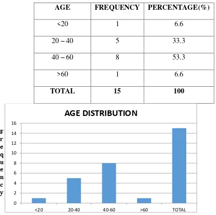
1. AGE DISTRIBUTION:
Among the 15 patients studied, highest number of patients were seen in 40-60
[image:69.595.47.469.260.679.2]years (53.3%) age group. The average was 42.7 years.
TABLE 1 –AGE DISTRIBUTION
AGE FREQUENCY PERCENTAGE(%)
<20 1 6.6
20 – 40 5 33.3
40 – 60 8 53.3
>60 1 6.6
TOTAL 15 100
AGE ( in years)
0 2 4 6 8 10 12 14 16
<20 20-40 40-60 >60 TOTAL
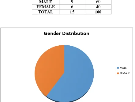
70 2. GENDER DISTRIBUTION:
Among the 15 cases there were 9 Male and 6 Female patients with predominant
[image:70.595.55.496.271.606.2]Male distribution;
TABLE 2 – GENDER DISTRIBUTION
Gender Distribution
MALE FEMALE
Gender Frequency Percentage (%)
MALE 9 60
FEMALE 6 40
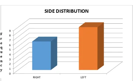
71 3. SIDE DISTRIBUTION:
Among the patients studied most of the patients had affected left side compared
[image:71.595.54.500.331.598.2]with right.
TABLE : 3- SIDE DISTRIBUTION
0 1 2 3 4 5 6 7 8 9 RIGHT LEFT
SIDE DISTRIBUTION
Frequency Percentage (%)
RIGHT 6 40
LEFT 9 60
TOTAL 15 100
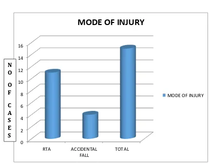
72 4. MODE OF INJURY :
Table 4 – Mode of Injury
Mode Frequency Percentage (%)
RTA 11 73.3
Accidental Fall 4 26.6
Total 15 100
0 2 4 6 8 10 12 14 16 RTA ACCIDENTAL FALL TOTAL
MODE OF INJURY
MODE OF INJURY
73
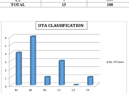
[image:73.595.69.513.293.614.2]5. FRACTURE CLASSIFICATION (OTA Classification)
TABLE : 5 – CLASSIFICATION (OTA Classification)
Classification Frequency Percentage (%)
B1 4 26.67
B2 6 40
B3 1 6.67
C1 3 20
C2 0 0
C3 1 6.67
TOTAL 15 100
0 1 2 3 4 5 6
B1 B2 B3 C1 C2 C3
No. Of Cases
No. Of Cases
74
6. RADIOLOGICAL VALGUS/VARUS ANGULATION :
Out of the 15 cases, 11 cases had angulation 0-10 degrees. 3 cases had reported varus angulation >10 degree, but without functional impairment. No cases reported valgus angulation. 1 case reported posterior angulation
Table 7
0 2 4 6 8 10 12
0-10 degree >10 degree
VARUS ANGULATION VALGUS
ANTERIOR ANGULATION POSTERIOR ANGULATION
Frequency Percentage
0-10 degree Varus 11 73.3
>10 degree varus 3 20
Valgus 0 0
Antero-posterior angulation 1 6.7
75
[image:75.595.50.531.100.388.2]8. SHOULDER/ELBOW RANGE OF MOTION : Table 8
SHOULDER Range Of Motion
Range Of Motion Frequency Percentage (%)
Excellent ( 100% ) 9 60
Good ( 75 – 100% ) 4 26.7
Fair ( 50 – 75% ) 2 13.3
Poor ( <50% ) - -
Total 15 100
ELBOW Range Of Motion
Range Of Motion Frequency Percentage (%)
Excellent ( 100% ) 12 80
Good ( 75 – 100% ) 3 20
Fair ( 50 – 75% ) - -
Poor ( <50% ) - -
Total 15 100
0 2 4 6 8 10 12 Excellent ( 100% )
Good ( 75
– 100% )
Fair ( 50 – 75% )
Poor ( <50% )
76
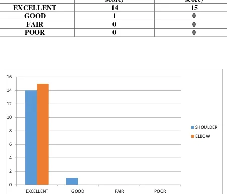
[image:76.595.61.513.195.582.2]9. SHOULDER FUNCTION EVALUATION (CLINICAL) : Table 9 SHOULDER FUNCTION(constant score) ELBOW FUNCTION(MEPS score)
EXCELLENT 14 15
GOOD 1 0
FAIR 0 0
POOR 0 0
0 2 4 6 8 10 12 14 16
EXCELLENT GOOD FAIR POOR
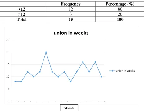
77 10. TIMEOF UNION :
[image:77.595.60.525.213.571.2]The average union time is 11.9 weeks, ranging from 8-20 weeks.
Table 10
Pattern of Time of Union
Frequency Percentage (%)
<12 12 80
>12 3 20
Total 15 100
0 5 10 15 20 25
union in weeks
union in weeks
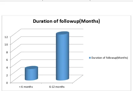
78 11. DURATION OF FOLLOW UP :
Table 11
12. COMPLICATIONS :
Table 12
Complications Frequency Percentage (%)
Radial Nerve Palsy 2 13.3
INFECTION 0 0
DELAYED UNION 1 6.6
NONUNION 0 0
Total 15 100
0 2 4 6 8 10 12
< 6 months 6-12 months
Duration of followup(Months)
Duration of followup(Months)
Frequency Percentage
< 6 months 3 20
6 – 12 months 12 80
[image:78.595.67.512.207.511.2]79 RESULTS :
1. 15 patients who had comminuted diaphyseal humerus fractures who were
treated in Department Of Orthopaedics and Traumatology, Govt. Rajaji
Hospital, Madurai were followed up in the study.
2. The longest follow up was One year; The shortest duration being three months. The mean duration was found to be 9.4 months.
3. Age incidence ranged from 16 to 62 years with average age being 42.7 years
4. Side of the fracture :
The left side was more commonly involved [9 in number] than the
right side [6 in number].
5. Nature of the injury:
Most cases were due to road traffic accidents (73.3%). The Other
80
6. Assessment of radiological valgus/varus angulation:
Among the 15 cases, 6 cases had no angulation & 5 cases did have
Minimum angulation of less than 10° of varus or valgus angulation were
accepted which remodeled to correct alignment over due course of time. 3 cases had varus angulation (>10 degree) which showed no significant
functional impairment and no cases had valgus angulation.
7. Antero-Posterior Malalignment:
1 case had reported posterior angulation because of excessive plate
contouring.
8. Rotational malalignment and Shortening :
None of the patients had any amount of rotational malalignment or
shortening.
9. Time of union :
81
10. Range of movements at the shoulder & elbow [expressed as a percentage] :
The mean range of movements To assess the range of movements ,
the patients were divided in to 4 groups :
o Excellent : 100% Range of Motion
o Good : >75% Range of Motion
o Fair : 50 – 75% Range of Motion
o Poor : <50% Range of Motion
With respect to shoulder Range of Motion, Among the 15 patients ; 9
patients(60%) had excellent results,4 patients(26.7%) had good result,
2(13.3%) had fair result and no poor result.
With respect to Elbow Range of Motion, Among the 15 patients ; 12
patients(80%) had excellent results,3 patients(20%) had good result,
82 11.SHOULDER FUNCTIONAL SCORE:
Shoulder function is assessed by CONSTANT MURLEY SCORE.
The mean constant score was 87 on the affected side and 90.67 on the
unaffected side.
Grading of the constant score- the difference between the score on the
normal side and affected side is calculated for each patients and
graded based on the difference as
o EXCELLENT: <10
o GOOD: 11-20
o FAIR:21-30
o POOR: >30
Shoulder function was assessed by CONSTANT MURLEY SCORE.
Among the 15 patients, 14 patients had excellent results and 1 patient
83
12. ELBOW FUNCTIONAL SCORE
ELBOW FUNCTION is assessesed by MAYO ELBOW
PERFORMANCE SCORE.
The mean MEPS score was 97.33 ranging from 85-100.
Grading of the MEPS SCORE
o EXCELLENT: >90
o GOOD: >81-90
o FAIR: 71-80
o POOR: <80
Elbow function score was assessed by MAYO ELBOW PERFORMANCE
SCORE. Among the 15 patients, All patients had excellent elbow function
84
13.The mean surgical time with MIPO was 69 minutes (range: 60–90 minutes). The average blood loss with MIPO was 109ml(range : 75-150 minutes)
14. Complications:
2 out of 15 had Radial nerve palsy post operatively. Postoperatively, these
cases are given with cockup splints, preferably dynamic cockup splits.
Nerve conduction study was done in these 2 cases by 6 weeks. Recovery
was assessed at every followup by sensory and motor examination. 1 case
had full recovery by the end of 6 months and the other case showed no
recovery by the end of 1 year for which tendon transfer to be planned.
One case showed delayed union by 20 weeks. The fracture was fixed in
85 DISCUSSION :
Minimally invasive surgical treatment of skeletal injuries
aims to preserve the biology of soft tissue and bone. The
rationale for performing mechanical stabilization through
fracture fixation is the obvious need to restore anatomy and
mechanical function of the bone. Optimal bone healing requires a balance between mechanics and biology and is aided by modern osteosynthesis. In ORIF, The problem was that, all too often, precise reduction and absolute stable fixation were achieved at the expense of extensive soft-tissue trauma caused by the surgery.
Minimally invasive surgery is not determined by the length of the incisions
but more by the reduction technique and soft-tissue handling, a defnition of MIO
includes the following recommendations:
• Small soft-tissue windows are used to allow the insertion
of implants and instruments remote from the fracture site.
• Minimal additional trauma to the soft tissue and fractured
86
when it is necessary to achieve fracture alignment.
• Special instruments are designed to be used at the fracture
site that cause minimal additional trauma.
MIPO scores over open reduction and plate fixation of humerus
fractures by decreasing the surgical trauma to the soft tissue and
maintaining the periosteal circulation. Application of the plate on the bone
by an open technique interferes with the local vascularization, leading to
osteonecrosis beneath the implant, which can cause delayed healing or
non-healing (the reported rate of nonunion being 5.8%). The primary bone
healing without callus formation is not very strong and there exists a real
risk for refracture after removal of the implant in the open technique.
MIPO is that it is devoid of the entry-point problems of
intramedullary nailing such as rotator cuff impingment.
The average union time for fractures in our study was 11.9 weeks
(range: 8–20 weeks) and union rate was 93.7 %. One case showed delayed
87
to excessive traction after initial proximal screw placement. The results
were good compared to CONCHA ET AL STUDY where Union rate was
91.5% (32/35) at an average of 12 weeks. All the cases showed union
without primary or secondary bone grafting.
ORIF for comminuted fractures draws the need for lag screw fixation or
bone grafting which prolongs the surgery time , blood loss and
postoperative morbidity. Nevertheless ,the risk of nonunion rate is higher
than MIPO due to extensive soft tissue stripping according to literature
around 5.8 %. MIPO gains advantage over ORIF in these issues.
Esmailiejah, et al. found better results with MIPO when compared to
open reduction and plating as regard to the time of surgery and
iatrogenic radial nerve injury (3% versus 12%) and the rate of infection
(0% versus 6%), patients managed with the MIPO technique had also
shorter time for union and earlier return to their previous level of
88
Out of the 15 cases, 4 cases had more than 10 degree angulation which does
not show any functional impairment. So near normal biological reduction in
MIPO does not compromise on functional outcome of the patient.
The mean surgical time with MIPO was 69 minutes (range: 60–90 minutes)
which was less compared to M Shantharam Shetty et al study which was 91.5.
Shoulder function was assessed by CONTSANT MURLEY SCORE which
was 87 on affected side and 90.67 on healthy side and better compared to
Apivatthakakul et al study which reported 85.8 on affected side and 90.6 on the healthy side.
No incidence of postoperative shoulder pain and stiffness which is a relative
complication in Intramedullary group due to nail impingement.
The mean MEPS score for elbow is 97.66 which was comparable to other
89
No cases reported infection postoperatively which was better compared to
concha et al study which reported 2 cases of infection.
Postoperative iatrogenic radial nerve palsy was reported in 2 cases which
was higher compared to Deepak S etal study and Hadhoud MM. et al. one case recovered by 6 months followup & one case did not show recovery at 1
year for which tendon transfer to be planned subsequently. These nerve
injury occurred earlier in the study probably due to plate offset and unicortical drilling with chance of drill bit slippage into the neural structures posteriorly. Hence plate position should be visualized digitally
and radiologically before drilling. Take care to be in the proper
intermuscular plain and the plate advanced gently in close contact to bone over the anterior surface in a proximal to distal direction to protect deltoid insertion. The forearm must be positioned in supination; Pronation brings the radial nerve closer to plate according to
Apivatthakakul et al study. Taking in mind the danger zone for musculocutaneous and radial nerves.
90
The average blood loss was less compared to ORIF and all the patients
showed early return of activities due to decreased postoperative morbidity.
LIMITATIONS OF THE STUDY :
1. Sample size is small compared with other similar studies.
2. Not a comparative study
SUGGESTIONS :
1. A larger sample size will improve the quality of the study.
2. Comparison with intramedullary nailing and ORIF needs to be studied for
91 CONCLUSION :
Minimal invasive plate osteosynthesis offers excellent functional outcome
for comminuted shaft of humerus with better union rate and decreased risk of non union compared to ORIF.
Near normal biological reduction in MIPO offers equally good functional outcome with better union rate compared to Anatomical reduction in ORIF, more so for comminuted fractures.
There is decreased postoperative morbidity with early return to function.
The operating time and blood loss are less compared to ORIF. The chance of infection is negligible due to decreased surgical exposure.
Risk of radial nerve palsy is there to start with, but with experience can be
neglected.
92
None of the studies had so far compared MIPO , ORIF & IM NAILING
for COMMINUTED DIAPHYSEAL FRACTURES
BIBLIOGRAPHY :
Apivatthakakul T, Phornphutkul C, Laohapoonrungsee A, et al (2009) Less
invasive plate osteosynthesis in humeral shaft fractures. Oper Orthop
Traumatol;21(6):602–13.
An Z, Zeng B, He X, Chen Q, Hu S. Plating osteosynthesis of mid-distal humeral
shaft fractures:minimally invasive versus conventional open reduction technique
Int Orthop. 2010; 34:131-135
Apivatthakakul T, Patiyasikan S,Luevitoonvechkit S (2010) Danger zone for
locking screw placement in minimally invasive plate osteosynthesis (MIPO) of
93
Pospula W, Abu Noor T. Percutaneous fixation of comminuted fractures of the
humerus: initial experience at Al Razi hospital, Kuwait Med Princ Pract. 2006;
1:423-426
Hadhoud MM, Darwish AE, Mesriga MM. Minimally invasive plate
osteosynthesis versus open reduction and plate fixation of humeral shaft fractures.
Menoufia Med J.[serial online] 2015; 28:154-61.
Zhiquan A, Bingfang Z, Yeming W, Chi Z, Peiyan H. Minimally invasive plating
osteosynthesis (MIPO) of middle and distal third humeral shaft fractures, J Orthop
Trauma. 2007; 21:628-33
Bin-feng Yu; Liang-le Liu; Guo-jing Yang; Lei Zhang; Xi-peng Lin Comparison
of minimally invasive plate osteosynthesis and conventional plate osteosynthesis
for humeral shaft fracture: A metaanalysis Medicine. 95(39):e4955, SEP 2016
Gareth Davies; Gerald Yeo; Mahendrakumar Meta; David Miller; Erik Hohmann;
Kevin Tetsworth Case-Match Controlled Comparison of Minimally Invasive Plate
Osteosynthesis and Intramedullary Nailing for the Stabilization of Humeral Shaft
94
Fabio Teruo Matsunaga; Marcel Jun Sugawara Tamaoki; Marcelo Hide
Matsumoto; Nicola Archetti Netto; Flavio Faloppa; Joao Carlos Belloti Minimally
Invasive Osteosynthesis w