Varicella-Zoster Virus ORF9p Binding to Cellular Adaptor
Protein Complex 1 Is Important for Viral Infectivity
Marielle Lebrun,
aJulien Lambert,
aLaura Riva,
a*
Nicolas Thelen,
bXavier Rambout,
c*
Caroline Blondeau,
aMarc Thiry,
bRobert Snoeck,
dJean-Claude Twizere,
cFranck Dequiedt,
cGraciela Andrei,
dCatherine Sadzot-Delvaux
aaVirology and Immunology Unit, GIGA-Infection, Immunity and Inflammation, University of Liege, Liege,
Belgium
bCellular Biology Unit, GIGA-Neurosciences, University of Liege, Liege, Belgium
cProtein Signaling and Interactions Laboratory, GIGA-Molecular Biology of Diseases, University of Liege, Liege,
Belgium
dRega Institute, KUL, Leuven, Belgium
ABSTRACT
ORF9p (homologous to herpes simplex virus 1 [HSV-1] VP22) is a
varicella-zoster virus (VZV) tegument protein essential for viral replication. Even though its
precise functions are far from being fully described, a role in the secondary
envelop-ment of the virus has long been suggested. We performed a yeast two-hybrid
screen to identify cellular proteins interacting with ORF9p that might be important
for this function. We found 31 ORF9p interaction partners, among which was AP1M1,
the
subunit of the adaptor protein complex 1 (AP-1). AP-1 is a heterotetramer
in-volved in intracellular vesicle-mediated transport and regulates the shuttling of
cargo proteins between endosomes and the
trans
-Golgi network via clathrin-coated
vesicles. We confirmed that AP-1 interacts with ORF9p in infected cells and mapped
potential interaction motifs within ORF9p. We generated VZV mutants in which each
of these motifs was individually impaired and identified leucine 231 in ORF9p to be
critical for the interaction with AP-1. Disrupting ORF9p binding to AP-1 by mutating
leu-cine 231 to alanine in ORF9p strongly impaired viral growth, most likely by preventing
efficient secondary envelopment of the virus. Leucine 231 is part of a dileucine motif
conserved among alphaherpesviruses, and we showed that VP22 of Marek’s disease
vi-rus and HSV-2 also interacts with AP-1. This indicates that the function of this interaction
in secondary envelopment might be conserved as well.
IMPORTANCE
Herpesviruses are responsible for infections that, especially in
immu-nocompromised patients, can lead to severe complications, including neurological
symptoms and strokes. The constant emergence of viral strains resistant to classical
antivirals (mainly acyclovir and its derivatives) pleads for the identification of new
targets for future antiviral treatments. Cellular adaptor protein (AP) complexes have
been implicated in the correct addressing of herpesvirus glycoproteins in infected
cells, and the discovery that a major constituent of the varicella-zoster virus
tegu-ment interacts with AP-1 reveals a previously unsuspected role of this tegutegu-ment
protein. Unraveling the complex mechanisms leading to virion production will
cer-tainly be an important step in the discovery of future therapeutic targets.
KEYWORDS
adaptin, ORF9p, VP22, adaptor proteins, dileucine, herpesviruses,
secondary envelopment, tegument, varicella-zoster virus, viral assembly
A
ll viruses are cellular parasites and subvert the host machinery to replicate and/or
to interfere with cellular pathways. Herpesviruses, with their complex infectious
cycle, do not escape this rule and use subcellular trafficking pathways to assemble and
generate new virions.
Received22 February 2018Accepted14 May 2018
Accepted manuscript posted online23 May 2018
CitationLebrun M, Lambert J, Riva L, Thelen N, Rambout X, Blondeau C, Thiry M, Snoeck R, Twizere J-C, Dequiedt F, Andrei G, Sadzot-Delvaux C. 2018. Varicella-zoster virus ORF9p binding to cellular adaptor protein complex 1 is important for viral infectivity. J Virol 92:e00295-18.https://doi.org/10.1128/JVI .00295-18.
EditorRozanne M. Sandri-Goldin, University of California, Irvine
Copyright© 2018 American Society for Microbiology.All Rights Reserved. Address correspondence to Catherine Sadzot-Delvaux, [email protected]. *Present address: Laura Riva, Immunity and Pathogenesis Program, Infectious and Inflammatory Diseases Center, Sanford Burnham Prebys Medical Discovery Institute, La Jolla, California, USA; Xavier Rambout, Center for RNA Biology, Department of Biochemistry and Biophysics, School of Medicine and Dentistry, University of Rochester, Rochester, New York, USA.
M.L., J.L., and L.R. contributed equally to this article.
VIRUS-CELL INTERACTIONS
crossm
on November 6, 2019 by guest
http://jvi.asm.org/
Varicella-zoster virus (VZV) is a human alphaherpesvirus responsible for two
pathol-ogies. Primary infection generally occurs during childhood and leads to varicella, whose
symptomatic phase is characterized by a generalized cutaneous rash during which the
virus establishes a latent infection in sensory ganglia. When the host immune response
is impaired or decreased, the virus may reactivate and reach back to the skin, where it
causes a localized cutaneous rash usually associated with pain, known as zoster.
Herpesvirus particles are made of a nucleocapsid containing the DNA genome,
surrounded by the tegument, a complex protein layer. The tegument is contained in a
lipid envelope, in which is inserted a series of viral glycoproteins mediating viral entry
into the host cell. Capsids are assembled in the nucleus, where they acquire a viral
genome copy before crossing the nuclear envelope to reach the cytoplasm, where a
secondary envelopment takes place. Therefore, the definitive envelope results from a
very complex process of acquisition of a primary envelope at the inner nuclear
membrane, deenvelopment at the outer nuclear membrane, and reenvelopment in
association with intracellular membranes (1). While
trans
-Golgi network (TGN)-derived
vesicles were long thought to be the site of the secondary envelopment, more recent
data have challenged this old dogma (2, 3). The labeling of endocytic tubules freshly
retrieved from the cell surface has shown many herpes simplex virus 1 (HSV-1) capsids
budding through these labeled vesicles and demonstrated that endocytosis from the
plasma membrane into endocytic tubules could actually provide the viral envelope (4).
This idea is further strengthened by some old but also new data demonstrating the
importance of endocytosis for the correct cellular targeting and virion incorporation of
some herpesvirus glycoproteins (5–8).
VZV ORF9p, homologous to HSV-1 VP22, is a major tegument phosphoprotein
conserved among the alphaherpesviruses. It is one of the most abundant tegument
proteins, with a high number of copies being embedded in each virion (9, 10). HSV
VP22 has been shown to shuttle between the nucleus and the cytoplasm and to play
various roles in infected cells. In particular, it is required for the cytoplasmic
redistri-bution of some proteins, among which are VP16, ICP4, ICP27, and ICP0 (11), and has
been shown to directly interact with VP16, ICP0, gE, gM, and gD (12). Thanks to its
interaction with gE, VP22 can be recruited to the Golgi apparatus/
trans
-Golgi network.
The deletion of the C-terminal part of VP22, which is responsible for its interaction with
gE, impairs its recruitment into the complexes described above and its packaging into
virions, leading to poor growth in epithelial cells. These observations suggest a role in
viral assembly (13). The deletion of
UL49
, coding for VP22 in pseudorabies virus (PRV),
has only a minor impact on viral growth (14, 15), whereas in HSV-1, the lack of VP22 is
rapidly compensated for by mutations in
UL41
(16–18). However, HSV-1 null mutants
replicate less efficiently and do not accumulate at the cell surface, and the absence of
VP22 affects the virion composition and indirectly modulates viral fitness (19–21).
VZV ORF9p has been less characterized, but contrary to HSV-1 or PRV VP22, it has
been shown to be essential (22, 23). In transfected cells, it shuttles between the nucleus
and the cytoplasm, and in infected cells, it partially localizes to the endomembrane
network, including the TGN (22, 24). In infected cells, ORF9p interacts with the major
glycoprotein gE and the major transactivator IE62, and mutation of the ORF9p
inter-action motif in IE62 has a strong impact on viral growth (25–27). In yeast two-hybrid
(Y2H) experiments, ORF9p has been shown to interact with many other viral proteins,
including glycoproteins, tegument proteins, and capsid proteins (28, 29). Recently, we
have also shown that ORF9p interacts with and is phosphorylated by ORF47p, one of
the two VZV protein kinases, and that this phosphorylation is crucial for both nuclear
egress and secondary envelopment (30, 31). All these observations suggest that ORF9p,
like VP22, could be central in viral assembly and particularly in orchestrating the
secondary envelopment process.
Cellular adaptor protein (adaptin or AP) complexes are heterotetramers important
for the intracellular trafficking of membrane-bound proteins. Five AP complexes have
been described so far (32, 33). Both AP-1 and AP-2 bind to clathrin and are involved in
clathrin-dependent transport, whereas AP-3, even though it is able to interact with
August 2018 Volume 92 Issue 15 e00295-18 jvi.asm.org 2
on November 6, 2019 by guest
http://jvi.asm.org/
clathrin, together with AP-4 and AP-5, mediates clathrin-independent transport (34, 35).
The AP-1 complex is specifically implicated in the vesicular transport between
endo-somes and the TGN (36). Like all AP family members, it is composed of two large
subunits (
␥
and

1), one medium subunit (
1), and one small subunit (
1). It is
recruited from the cytosol by its cargo and allows subsequent binding to clathrin,
followed by membrane curvature and vesicle formation (33, 37).
To dissect more precisely the role of ORF9p in the VZV infectious cycle, we searched
for new cellular partners through a Y2H screen and identified 31 distinct candidates,
among which was AP1M1, the
subunit of the adaptor protein complex 1 (AP-1
1),
known to play a role in protein trafficking. The interaction between AP-1
1 and VZV
ORF9p was confirmed
in vitro
by glutathione
S
-transferase (GST) pulldown as well as by
coimmunoprecipitation (co-IP) using infected cell extracts. We identified five potential
interaction motifs (one acidic region, two tyrosine-based motifs, and two dileucine
motifs) within the ORF9p primary sequence. Viruses were generated by mutation of
these potential interaction motifs within the
orf9
coding sequence in the pOka genome.
The characterization of these mutants revealed that the mutation of leucine 231, which
is conserved among alphaherpesviruses, completely abolishes the interaction between
ORF9p and the AP-1 complex and strongly impairs the infectivity of the virus. In cells
infected with this mutant strain, only a limited number of viral particles were found at
the cell surface, transport vesicles containing complete virions or light particles were
very rare compared to their occurrence in wild-type (WT) VZV-infected cells, and
abnormal features were observed by electron microscopy. This suggests that, in the
absence of an ORF9p/AP-1 interaction, the viral components are not properly
ad-dressed within the cell and/or that virions could be somehow retargeted for
degrada-tion by either the lysosomal or the autophagy pathway, or by both pathways.
To our knowledge, this is the first time that an interaction between a herpesvirus
tegument protein and the AP-1 complex has been described, and altogether, our
results suggest that this interaction is important for the formation of infectious viral
particles and, thus, VZV pathogenicity.
RESULTS
ORF9p interacts with the adaptor protein complex 1.
In order to identify cellular
partners for ORF9p, we performed a yeast two-hybrid (Y2H) screen against the human
ORFeome version 5.1 (hORFeome 5.1). Thirty-one distinct candidates were identified by
sequencing clones growing on the selection medium. These interactions were then
verified in a pairwise retest. For this experiment, not only the full-length ORF9p but also
N-terminal ORF9p deletion mutants (ORF9p amino acids [aa] 50 to 302, 100 to 302, and
150 to 302) and C-terminal deletion mutants (ORF9p aa 1 to 250, 1 to 200, and 1 to 150)
were used as baits. Based on the literature, four additional proteins interacting with
HSV-1 VP22, namely, SET, ANP32B, HIST1H4I, and HIST1H3E, were included as positive
controls. All 31 candidates and the four controls were found to be positive in the Y2H
pairwise retest. Fifteen were found to be positive in both orientations (Gal4-activation
domain ORF9p [AD-ORF9p] with the Gal4 DNA binding domain candidate
[DB-candidate] and DB-ORF9p with the AD-candidate) and are highlighted in bold in
Fig. 1A. All interactions were maintained when the 50 first amino acids (aa 50 to 302
construct) or the 50 last amino acids (aa 1 to 250 construct) of ORF9p were deleted but
lost with larger deletions (aa 100 to 302, 150 to 302, 1 to 200, and 1 to 150), suggesting
that the region of interaction is likely located between amino acids 50 and 250 of
ORF9p (data not shown).
We performed gene ontology enrichment analyses using both the DAVID (38) and
TOPPGENE (39) platforms to classify the Y2H-identified interaction partners based on
their cellular functions (Fig. 1A and B). Interestingly, 12 interaction partners were
involved in organelle organization, a process that is certainly required for the infectious
cycle and, more precisely, for viral assembly. Among them, we focused on AP1M1, the
subunit of adaptor protein complex 1 (AP-1), which mediates the bidirectional
transport between TGN and endosomes (32). The interaction between ORF9p and AP-1
ORF9p/AP-1 Interaction Is Important for VZV Infectivity Journal of Virology
on November 6, 2019 by guest
http://jvi.asm.org/
was first confirmed by a GST pulldown assay in which an ORF9p-GST fusion protein or
GST alone was incubated with total cell extracts from uninfected or pOka VZV-infected
MeWo cells. Both AP-1 subunits were pulled down from uninfected and infected cell
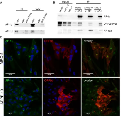
extracts (Fig. 2A). To confirm the ORF9p/AP-1 interaction in an infectious context, we
performed coimmunoprecipitation (co-IP) experiments from infected cell extracts.
Be-cause there was no antibody against the
subunit suitable for immunoprecipitation
(IP), the AP-1 complex was immunoprecipitated via the
␥
subunit from infected MeWo,
ARPE-19, and MRC-5 cells (at 48 h postinfection [hpi]), three cell lines frequently used
for VZV production. VZV-ORF9-V5 (pOka genomic background), expressing ORF9p
fused to a V5 epitope, was used as the wild-type (WT) strain. This virus has been
previously characterized and replicates with the same efficacy as the wild-type pOka
control (30). Western blotting on the immunoprecipitated complex revealed the
pres-ence of both ORF9p and AP-1
1 in the three cell lines (Fig. 2B). We next studied the
subcellular localization of ORF9p and AP-1 in MRC-5 and ARPE-19 cells. Contrary to
MeWo cells, MRC-5 cells do not form syncytia and ARPE-19 cells fuse moderately upon
VZV infection, facilitating immunofluorescence colocalization analyses. MRC-5 and
ARPE-19 cells were infected with VZV-ORF9-V5 and fixed at 48 hpi. Consistent with
ORF9p binding to AP-1, a substantial amount of ORF9p colocalized with the AP-1
complex in cytoplasmic structures close to the nucleus (Fig. 2C).
ORF9p leucine 231 is important for ORF9p interaction with the AP-1 complex.
Three types of motifs are known to mediate the binding of cargo proteins to the AP-1
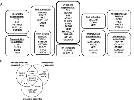
FIG 1ORF9p interacts with cellular proteins involved in various processes. A yeast two-hybrid screen against human ORFeome 5.1 was performed and identified 31 potential cellular partners of ORF9p. The 31 candidates, along with 4 controls (SET, ANP32N, HIST1H4I, HIST1H3E) known to interact with VP22, were then confirmed in a pairwise retest and classified based on a Gene Ontology analysis. (A) Interactions confirmed in both directions in the retest are highlighted in bold. (B) Twelve out of the 35 interacting proteins are involved in the organelle organization category, which can be subdivided into several subclasses.
August 2018 Volume 92 Issue 15 e00295-18 jvi.asm.org 4
on November 6, 2019 by guest
http://jvi.asm.org/
[image:4.585.41.508.71.421.2]complex: acidic clusters, tyrosine-based motifs (NPXY or YXX
⌽
), and dileucine motifs
[(D/E)XXXL(L/I)] (40). ORF9p primary sequence analysis revealed an acidic cluster
(
85EDDFEDIDE
93), two tyrosine-based motifs (
61YADL
64and
268YAQV
271), and two
dileu-cine motifs (
211ELDRLL
216and
227EGLNLI
232) (Fig. 3A), all of which were found between
amino acids 50 and 250, a region that we showed to be required for binding to AP-1
1.
The VZV-ORF9p-ΔAC-V5 strain, in which the acidic region is deleted, was already
available (31). We generated four additional VZV mutants in which the tyrosine-based
or the dileucine motifs were independently mutated.
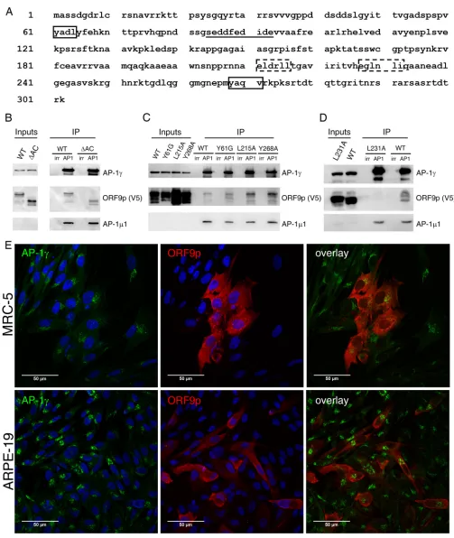
In the co-IP experiment, neither the deletion of the acidic region nor the mutation
of tyrosine 61 to glycine or tyrosine 268 and leucine 215 to alanine had an impact on
the interaction between ORF9p and the AP-1 complex (Fig. 3B and C), while the
mutation of leucine 231 to alanine completely abolished this interaction (Fig. 3D). In
addition, ORF9p did not colocalize with AP-1 in MRC-5 or ARPE-19 cells infected with
VZV-ORF9-L231A-V5 (compare Fig. 3E to Fig. 2C).
FIG 2ORF9p interacts and colocalizes with the AP-1 complex. (A) The interaction of ORF9p and the AP-1 complex was verified by GST pulldown using ORF9p-GST and total extracts of MeWo cells infected or not infected by pOka VZV. NI, noninfected. (B) Coimmuno-precipitation of the ORF9p/AP-1 complex from VZV-ORF9-V5 (WT)-infected MeWo, ARPE-19, and MRC-5 cells (48 hpi). An antibody against AP-1␥was used for immunoprecipitation, and the presence of ORF9p-V5, AP-1␥, and AP-11 was verified by Western blotting. Normal mouse IgG was used as the IP control (irr). (C) MRC-5 and ARPE-19 cells were infected with VZV-ORF9-V5 for 48 h and immunostained with a mouse anti-V5 and a rabbit anti-AP-1␥. Appropriate secondary antibodies were used, and nuclei were counterstained with TO-PRO-3. Images were recorded with a 63⫻oil objective.
ORF9p/AP-1 Interaction Is Important for VZV Infectivity Journal of Virology
on November 6, 2019 by guest
http://jvi.asm.org/
[image:5.585.41.466.71.479.2]FIG 3ORF9p leucine 231 is important for ORF9p interaction with the AP-1 complex. (A) The ORF9p primary sequence harbors several potential AP-1 interaction motifs, two tyrosine-based motifs (boxes with solid lines) and two dileucine motifs (boxes with dashed lines), as well as an acidic domain (underlined). (B, C, D) The AP-1␥subunit was immunoprecipitated from total extracts of MeWo cells infected for 48 h by VZV-ORF9-V5 (WT) (B, C and D), VZV-ORF9-ΔAC-V5 (B), VZV-ORF9-Y61G-V5, -L215A-V5, and-Y268A-V5 (C), and VZV-ORF9-L231A-V5 (D). A control immunoprecipitation with normal mouse IgG was performed in parallel (irr). The presence of ORF9p, AP-1␥, and AP-11 in the immunoprecipitated complex was verified by Western blotting. AC, acidic region. (E) MRC-5 and ARPE-19 cells were infected with VZV-ORF9-L231A-V5 for 48 h and immunostained with a mouse anti-V5 and a rabbit anti-AP-1␥. Appropriate secondary antibodies were used, and nuclei were counterstained with TO-PRO-3. Images were recorded with a 63⫻oil objective.
August 2018 Volume 92 Issue 15 e00295-18 jvi.asm.org 6
on November 6, 2019 by guest
http://jvi.asm.org/
[image:6.585.39.542.66.662.2]The dileucine motif important for ORF9p interaction with AP-1 is conserved
among alphaherpesviruses.
Although the ORF9p acidic region, which overlaps the
ORF47p-binding site, is not strictly conserved among alphaherpesviruses, many VP22
homologs possess, in their N-terminal part, an acidic cluster downstream of serine
residues. None of the above-described tyrosine-based motifs are conserved among
alphaherpesviruses, even though HSV-1 harbors a motif that resembles the VZV
61YADL
64motif and a real tyrosine-based motif in an upstream region (
18YEDL
22).
However, both the
211ELDRLL
216and
227EGLNLI
232dileucine motifs were highly
con-served among the 27 alphaherpesvirus genomes that we analyzed (Fig. 4A). In
partic-ular, the glutamic acid as well as the first and second (iso)leucines of the
227EGLNLI
232motif were conserved in almost all viruses (93%, 96%, and 78%, respectively) (Fig. 4A).
To verify whether the interaction with AP-1 is shared by other alphaherpesviruses,
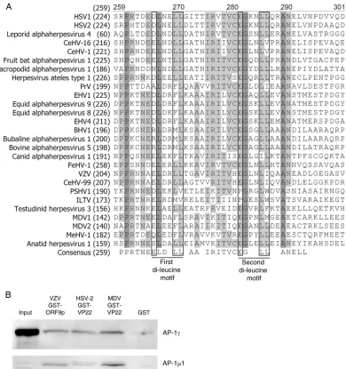
FIG 4The ORF9p (VP22) interaction with the AP-1 complex is conserved among alphaherpesviruses. (A) The primary sequences of 27 homologous VP22 proteins were aligned with the Vector NTi program (Invitrogen). Only the region containing the two dileucine motifs is shown. Dark gray, identical residues; light gray, similar residues. The first dileucine motif is relatively well conserved (present in 11 homologs); the second is highly conserved (present in 20 homologs). HSV-1, herpes simplex virus 1; CeHV-16, cercopithecine herpesvirus 16; PrV, pseudorabies virus; EHV1, equine herpesvirus 1; BHV1, bovine herpesvirus 1; FeHV-1, feline herpesvirus 1; PSHV1, psittacine herpesvirus 1; ILTV, infectious laryngotracheitis virus; MDV1, Marek’s disease herpesvirus 1; MeHV-1, meleagrid herpesvirus 1. (B) The VZV ORF9p homolog (HSV-2 or MDV VP22) interaction with the AP-1␥ and1 subunits was analyzed by GST pulldown using the GST-ORF9p or GST-VP22 fusion protein and MeWo cell total cell extracts.
ORF9p/AP-1 Interaction Is Important for VZV Infectivity Journal of Virology
on November 6, 2019 by guest
http://jvi.asm.org/
[image:7.585.48.438.70.485.2]HSV-2 and Marek’s disease herpesvirus (MDV) VP22 was cloned into pGEX 5.1. It is worth
noting that while HSV-2 VP22 harbors a well-conserved dileucine motif (
250EGKNLL
255),
the MDV VP22 motif (
264EGPNLM
269) is not perfectly conserved, with the second leucine
being replaced by a methionine. The fusion proteins were purified on glutathione
agarose beads in parallel with GST alone as a control and used in a GST pulldown assay
in the presence of total cell extracts from uninfected MeWo cells. In both cases, the
1
and
␥
subunits of the AP-1 complex were detected, reflecting that ORF9p homologs
can also interact with this adaptor protein complex (Fig. 4B).
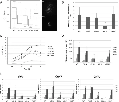
ORF9p leucine 231 is important for viral infectivity in both MeWo and MRC-5
cells.
The infectivity of all mutants was determined in both MeWo cells (Fig. 5A and B)
and MRC-5 cells (Fig. 5C to E). The size of the infection foci in MeWo cells was assessed
at 48 hpi and showed that all mutants, except VZV-ORF9-Y268A-V5, present a slight to
moderate growth defect compared to the WT strain. However, the mutation of leucine
231 had the greatest impact on viral growth (Fig. 5A). In parallel, a known number of
individual infected cells was used to infect MeWo cells seeded in a 24-well plate, and
the number of infection foci was determined at 72 hpi. The number of infection foci
present in the well corresponds to the number of infectious cells present in the
inoculum. The mean ratio of infectious cells/infected cells was significantly reduced for
VZV-ORF9-L231A-V5 compared to the wild type or the other mutant strains (less than
10% compared to 30 to 40% in Fig. 5B). This suggests that the entry and maybe the
expression of viral genes are not affected by the L231A mutation, while the production
and/or egress of the progeny virion might be affected.
In MRC-5 cells, minor to moderate differences in the growth properties of
VZV-ORF9-Y61G-V5, -L215A-V5, and -Y286A-V5 were observed, while the infectivity of
VZV-ORF9-L231A-V5 was severely impaired, with the number of PFU at 72 hpi being
more than five times lower than the number of VZV-ORF9-V5 PFU (Fig. 5C). To get
additional information regarding the different steps of the infectious cycle that may be
impacted by the mutation, MRC-5 cell RNA and genomic DNA were extracted at each
time point to determine the expression level of the three classes of viral genes, i.e.,
immediate early (IE;
orf4
), early (E;
orf47
), and late (L;
orf40
) genes, and the amount of
viral genomic DNA. The amount of viral genomes was expressed per nanogram of total
DNA (Fig. 5D), and RNA levels were normalized to the 18S rRNA level and expressed as
a fold induction relative to that in the VZV-ORF9-V5-infected cells at 24 hpi (Fig. 5E).
Both analyses confirmed a global growth defect of the VZV-ORF9-L231A-V5 mutant,
although a slight decrease in IE, E, and L gene expression was also observed for
VZV-ORF9-Y61G-V5, -Y268A-V5, and -L215-V5 (Fig. 5E).
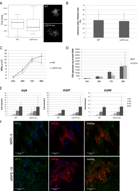
To confirm that the growth defect of the L231A mutant was solely due to the leucine
mutation, a revertant virus (VZV-ORF9-L231A-rev-V5) was generated by reintroducing
the wild-type
orf9
instead of the mutant copy into VZV-ORF9-L231A-V5. The infectivity
of VZV-ORF9-L231A-rev-V5 in MeWo and MRC-5 cells was then compared to that of
VZV-ORF9-V5. No major differences could be observed between the two viral strains,
demonstrating that the growth defect of VZV-ORF9-L231A-V5 is attributable only to the
leucine 231 mutation (Fig. 6A to E). In addition, immunofluorescence on infected MRC-5
and ARPE-19 cells shows that the colocalization of ORF9p and AP-1 is restored when the
L231 mutation is repaired (Fig. 6F).
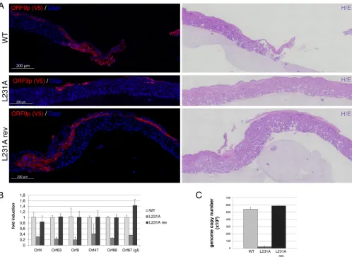
ORF9p leucine 231 is important for viral infectivity in a 3D skin model.
VZV-ORF9-L231A-V5 infectivity was then evaluated in a previously described human
three-dimensional (3D) skin model (41). Five thousand VZV-ORF9-V5-, -L231A-V5-, and
-L231A-rev-V5-infected MRC-5 cells were layered on skin rafts and maintained in culture
for 6 days. Immunohistochemistry with an anti-V5 antibody to detect ORF9p-V5 was
performed on five series of 6 sections (Fig. 7A). Large infection foci were present in all
series for VZV-ORF9-V5 and -L231A-rev-V5, whereas very small foci were observed only
in one set of VZV-ORF9-L231A-V5 sections. In addition, VZV-ORF9-L231A-V5 foci
re-mained limited to the upper layers of keratinocytes, in contrast to the spreading
throughout the thickness of the skin rafts observed with VZV-ORF9-V5 and -L231-rev-V5
(Fig. 7A). It is worth noting that a similar result was obtained by labeling the sections
August 2018 Volume 92 Issue 15 e00295-18 jvi.asm.org 8
on November 6, 2019 by guest
http://jvi.asm.org/
with an antibody against IE63, reflecting a global growth defect rather than a particular
issue with the expression or detection of L231A-V5 (data not shown). The massive
reduction of infectivity was also observed when the viral genome copy number and
viral gene expression were measured, respectively, by quantitative PCR (qPCR) or
quantitative reverse transcription-PCR (qRT-PCR) (Fig. 7B and C).
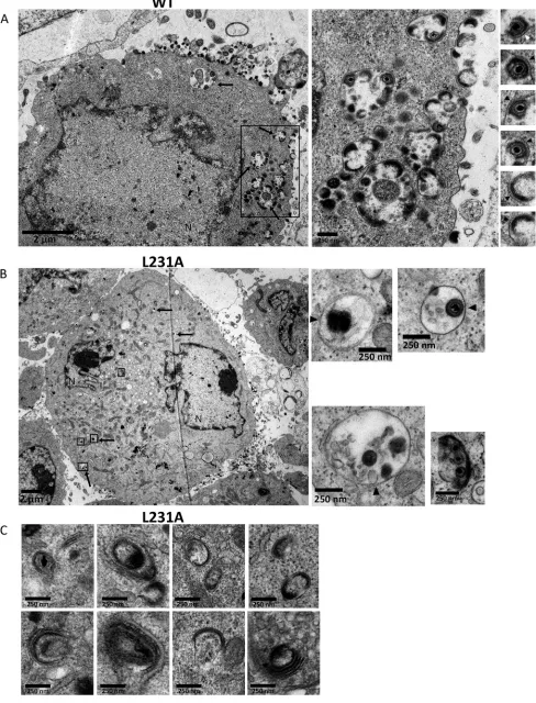
The L231A mutant exhibits assembly and egress defects.
MeWo cells were
analyzed at the ultrastructural level using a transmission electron microscope (TEM) to
FIG 5The L231A mutation strongly impacts infectivity in MeWo and MRC-5 cells. VZV-ORF9-V5 (WT) and mutant strain infectivity was assessed in MeWo cells (A and B) or MRC-5 cells (C to E). (A) Infection focus measurement at 48 hpi. MeWo cells were infected for 48 h, and the size of the infection foci was determined using CellProfiler software and expressed as the number of pixels present in each infection focus. The box plot depicts the 1st and 3rd quantiles (the lower and upper limits of the boxes, respectively) and the median (heavy black lines). Error bars represent minimum and maximum values. (B) Infectivity of infected cells. A known number of infected cells was used to infect MeWo cells seeded in a 24-well plate, and the number of infectious foci was determined in each well 3 days later. The graph shows the mean ratio of infectious cells/infected cells; error bars represent the standard deviation (SD). (C) Growth curves in MRC-5 cells. The graph shows the results of one representative experiment out of four; error bars represent the standard error of the mean (SEM). (D) The amount of the VZV genome over time was quantified by qPCR on DNA extracts. Primers in the humanp21promoter were used for normalization. Serial dilutions of a BAC-VZV of a known concentration were used to build a standard curve. Results are expressed as the absolute number of VZV genomes per nanogram of total DNA. Means from three independent experiments are depicted; error bars represent the SD. (E) In parallel, qRT-PCR was performed on RNA extracts to quantify the expression of IE (orf4), E (orf47), and L (orf40) genes. The expression of 18S rRNA was used to calculate the change in the threshold cycle number, and for each gene, relative expression levels were calculated, with the expression level for the WT at 24 hpi being used as a control. Means from three independent experiments are depicted; error bars represent the SD. (A, B, D, and E) A two-tailedttest was used to compare, at each time point, each mutant to the WT strain.*,P⬍0.05;**,P⬍0.01.
ORF9p/AP-1 Interaction Is Important for VZV Infectivity Journal of Virology
on November 6, 2019 by guest
http://jvi.asm.org/
[image:9.585.43.548.72.506.2]FIG 6The replacement of ORF9-L231A-V5 by a WT ORF9-V5 copy restores the infectivity and the colocalization with AP-1. (A to E) VZV-ORF9-V5 (WT) and VZV-ORF9-L231A-rev-V5 (L231A rev) infectivity was assessed in MeWo cells (A and B) or MRC-5 cells (C to E). (A) Infection focus measurement at
(Continued on next page)
August 2018 Volume 92 Issue 15 e00295-18 jvi.asm.org 10
on November 6, 2019 by guest
http://jvi.asm.org/
[image:10.585.42.504.68.708.2]search for abnormal phenotypes. About 20 ORF9-V5-infected cells and 20
VZV-ORF9-L231A-V5-infected cells were carefully analyzed. While many complete virions or
light particles were observed at the periphery of VZV-ORF9-V5-infected cells (Fig. 8A),
they were scarce in VZV-ORF9-L231A-V5-infected cells (Fig. 8B). Transport vesicles
containing enveloped virions and light particles were abundant and large in
VZV-ORF9-V5-infected cells (Fig. 8A, middle) but extremely rare in VZV-ORF9-L231A-VZV-ORF9-V5-infected
cells, and when present, they were small and contained only a few viral particles (Fig.
8B). Interestingly, we noticed that while the particles of the ORF9-V5 virus (complete
virions and light particles) seemed to be tightly bound to the membrane of the
transport vesicles and to the cell membrane (Fig. 8A, right), this association appeared
to be much looser for the L231A-V5 strain (Fig. 8B, right). In addition, L231A-V5-infected
cells frequently showed dense material, likely the viral tegument, in association with
curved membranes (endoplasmic reticulum, Golgi cisternae, and/or endocytic tubules)
without being associated with viral particles or light particles (Fig. 8C). Surprisingly,
vesicles resembling lysosomes and autophagosomes, some of them in the close vicinity
of transport vesicles, were frequently observed in VZV-ORF9-L231A-V5-infected cells
(Fig. 9A). Such figures were only rarely observed in WT-infected cells, and when
observed, they were usually not in the vicinity of transport vesicles (Fig. 9B).
DISCUSSION
Although it is admitted that the tegument proteins, in general, and ORF9p or its
homologs, in particular, play a role in envelopment, the molecular mechanisms and the
interactions supporting these crucial steps are still poorly understood. The Y2H
exper-iment described in this paper is the first attempt to identify putative cellular partners
of ORF9p. The screening of hORFeome 5.1 with ORF9p, used as bait, together with a
pairwise retest identified 35 cellular proteins directly interacting with ORF9p.
Interest-ingly, the ORF9p region mapping to amino acids (aa) 50 to 250 containing the
homology region (HR; aa 136 to 250) conserved in all alphaherpesviruses is important
for these interactions. A gene ontology analysis to classify the partners based on their
cellular functions highlighted several cellular processes, among which were
microtu-bule cytoskeleton, chromatin organization, RNA metabolic processes, and transcription,
suggesting that ORF9p could play various roles in infected cells. This is in agreement
with the fact that both ORF9p and VP22 have been reported to interact with the
cytoskeleton (26, 42), while HSV VP22 is also known to bind to the chromatin in dividing
cells and to mRNA, enhancing thereby the accumulation of mRNA at early times of
infection and protein synthesis at late times of infection (42–44).
Remarkably, 12 candidates out of the 35 were related to organelle organization.
Among these proteins, four, namely, ANXA2, GOLGA2, CHMP5, and AP1M1, were
associated with both intracellular transport and vesicle-mediated transport. ANXA2
(annexin A2) is a calcium-dependent, anionic phospholipid-binding protein that has
pleiotropic functions, among which is the capacity to interact with endosomes
following organelle destabilization (45). GOLGA2 (golgin A2) is a
cis
-Golgi matrix
FIG 6Legend (Continued)
48 hpi. MeWo cells were infected for 48 h, and the size of the infection foci was determined using CellProfiler software and expressed as the number of pixels present in each infection focus. The box plot depicts the 1st and 3rd quantiles (the lower and upper limits of the boxes, respectively) and the median (heavy black lines). Error bars represent minimum and maximum values. (B) Infectivity of infected cells. A known number of infected cells was used to infect MeWo cells seeded in a 24-well plate, and the number of infectious foci was determined in each well 3 days later. The graph shows the mean ratio of infectious cells/infected cells; error bars represent the standard deviation (SD). (C) Growth curves in MRC-5 cells. The graph shows the results of one representative experiment out of four; error bars represent the standard error of the mean (SEM). (D) The amount of the VZV genome over time was quantified by qPCR on DNA extracts. Primers in the humanp21promoter were used for normalization. Serial dilutions of a BAC-VZV of a known concentration were used to build a standard curve. Results are expressed as the absolute number of VZV genomes per nanogram of total DNA. Means from three independent experiments are depicted; error bars represent the SD. (E) In parallel, qRT-PCR was performed on RNA extracts to quantify the expression of IE (orf4), E (orf47), and L (orf40) genes. The expression of 18S rRNA was used to calculate the change in the threshold cycle number, and for each gene, relative expression levels were calculated, with the expression level for the WT at 24 hpi being used as a control. Means from three independent experiments are depicted; error bars represent the SD. (A, B, D, and E) A two-tailedttest was used to compare, at each time point, each mutant to the WT strain.*,P⬍ 0.05. (F) MRC-5 and ARPE-19 cells were infected with VZV-ORF9-L231A-rev-V5 for 48 h and immunostained with both a mouse anti-V5 and a rabbit anti-AP-1␥. Appropriate secondary antibodies were used, and nuclei were counterstained with TO-PRO-3. Images were recorded with a 63⫻oil objective.
ORF9p/AP-1 Interaction Is Important for VZV Infectivity Journal of Virology
on November 6, 2019 by guest
http://jvi.asm.org/
protein that plays a major role in the stacking of Golgi cisternae and maintenance of the
Golgi apparatus structure and participates in the glycosylation and transport of proteins
and lipids in the secretory pathway (46, 47). CHMP5 (charged multivesicular body
protein 5) is a component of ESCRT-III (endosomal sorting complex required for
transport III), a complex involved in both the degradation of surface receptor proteins
and the formation of endocytic multivesicular bodies (MVBs) (48). Knowing the
poten-tial implication of MVBs in the secondary envelopment process of herpesviruses, this
could be of a particular interest (49, 50). Finally, AP1M1 is the
subunit of the adaptor
protein complex 1 (AP-1), which mediates the bidirectional clathrin-dependent
traffick-ing of cargo proteins between the TGN and the endosomal network. The interaction
with the cargo protein is mediated by the
subunit or by the junction between the
␥
and
subunits, while the clathrin and accessory molecules interact with the

and
␥
subunits (34).
The interaction of the most abundant tegument protein with the AP-1 complex
appears to be highly relevant in the context of viral egress. Indeed, the site of secondary
FIG 7ORF9p leucine 231 is important for viral infectivity in a 3D skin model. Human primary keratinocytes were allowed to divide and differentiate at the air-liquid interface on top of a collagen matrix for 4 days. VZV-ORF9-V5 (WT)-, VZV-L213A-V5-, and VZV-L231A-rev-V5-infected MRC-5 cells were then layered on the epithelial cells and skin rafts were processed 6 days later. Three skin rafts were prepared for each infection. (A) For each infection, one raft was embedded in paraffin and a series of successive sections was prepared. (Left) Immunostaining against ORF9p (anti-V5 primary antibody). Nuclei were counterstained with DAPI. (Right) Control hematoxylin-eosin (H/E) staining. (B) Total RNA was extracted from a second skin raft, and the expression of IE (orf4andorf63), E (orf9,
orf47andorf66), and L (orf67) genes was analyzed by qRT-PCR. The expression of 18S rRNA was used to calculate the change in the threshold cycle value, and for each gene, relative expression levels were calculated, with the VZV-ORF9-V5-infected raft being used as a control. Means for internal replicates are shown, and error bars represent the SD. (C) Genomic DNA was extracted from a third raft, and qPCR was performed to evaluate the number of VZV genome copies per microgram of total DNA. Primers in the humanp21promoter were used for normalization. Serial dilutions of a BAC-VZV of a known concentration were used to build a standard curve. Means for internal replicates are shown, and error bars represent the SD.
August 2018 Volume 92 Issue 15 e00295-18 jvi.asm.org 12
on November 6, 2019 by guest
http://jvi.asm.org/
[image:12.585.44.542.70.433.2]FIG 8The VZV-ORF9-L231A-V5 mutant exhibits assembly and egress defects. Transmission electron microscopy was used to compare VZV-ORF9-V5-infected cells to VZV-ORF9-L231A-V5-infected cells. (A) VZV-ORF9-V5-infected MeWo cells are characterized by many particles at the cell surface as well as many
(Continued on next page)
ORF9p/AP-1 Interaction Is Important for VZV Infectivity Journal of Virology
on November 6, 2019 by guest
http://jvi.asm.org/
[image:13.585.41.529.77.717.2]envelopment and the precise nature of the transport vesicles are still debated. The
importance of the TGN in the secondary envelopment process was widely recognized
not only because most tegument proteins and glycoproteins present in extracellular
particles accumulate in this cellular compartment before being incorporated into the
final particles but also because of the lipid composition of the viral envelope (2, 3).
Additionally, ultrastructural studies have identified capsids budding through vesicles
positively stained for TGN markers (51–54). More recent work on HSV-1 has brought the
idea that endocytic tubules instead might be the major site of secondary envelopment
(4). In addition, some glycoproteins are first transported to the plasma membrane and
secondarily endocytosed and transported to the TGN, supposedly the site of final
envelopment, suggesting that endocytosis might represent an essential step in viral
assembly (5, 55). We confirmed the interaction between ORF9p and AP-1 in a GST
pulldown assay and by coimmunoprecipitation from infected MeWo, MRC-5, or
ARPE-19 cell lysates. This was further supported by the colocalization of the ORF9p and
AP-1
␥
proteins in the infectious context. The mutation of ORF9p leucine 231 led to a
complete loss of interaction between ORF9p and AP-1, which was confirmed by a lack
of colocalization, indicating that
227EGLNLI
232is crucial for this interaction. The
infec-tivity of the VZV-ORF9-L231A-V5 mutant was dramatically impaired in both MeWo and
MRC-5 cells as well as in a human 3D skin model. The interaction between AP members
and viral components has been shown to be important for the infectivity of various
viruses. For example, AP-1 interacts with HIV Nef (56) and with African swine fever virus
CD2v (57), while AP-2 interacts with the hepatitis B virus large envelope protein (58).
Interactions with AP-1 generally require cargo proteins to be inserted in the
mem-brane in the vicinity of AP-1, usually thanks to lipid modifications. A bioinformatic
analysis of ORF9p reveals that glycine 6 and cysteine 10 might be myristoylated or
palmitoylated, respectively. We are currently investigating this hypothesis.
Neverthe-less, some cytoplasmic proteins are known to bind adaptor protein complexes in order
to bring cargo to the sorting machinery. For example, PACS-1, a cytoplasmic protein, is
required to mediate the interaction between furin and AP-1 for the subsequent
addressing to the TGN (59). PACS-1 is also described to bind the mannose 6-phosphate
receptor (MPR) or VZV gE (59) and to mediate the interaction between HIV Nef, AP-1,
and the major histocompatibility complex (MHC) (60). It is thus possible that, even in
the absence of a lipid modification, ORF9p, like PACS-1, ensures the proper localization
of viral components necessary for assembly. Any mutation of ORF9p impairing its
interaction with AP-1 would then affect the localization of other viral components and,
consequently, viral assembly. Mass spectrometry analyses on the immunoprecipitated
AP-1 complex are ongoing and will allow characterization, more broadly, of this
complex in VZV-ORF9-V5- or VZV-ORF9-L231A-V5-infected cells.
Of note, the mutation of the conserved dileucine motifs in HSV-1 VP22 has
dem-onstrated their importance for the expression or proper redistribution of VP22 itself and
of ICP0, ICP4, ICP27, and VP26 and their importance for neurovirulence (11). In addition,
the mutation of either dileucine motif in HSV-1 VP22 impedes its interaction with VP16,
while the mutation of the first dileucine motif impairs its interaction with gE and,
consequently, the incorporation of VP22 into the virion (61). Immunostaining and
Western blotting experiments to detect possible modifications of the expression and/or
localization of several VZV proteins, among which were some of the homologs of the
HSV proteins described above, revealed only a minor nuclear retention of the kinase
ORF47p (data not shown). Besides, preliminary results suggest that ORF9p interaction
with gE, ORF10p (VP16 homolog), or IE62, the major VZV transactivator (which is an
FIG 8Legend (Continued)
transport vesicles (arrows and middle). Enveloped virions or light particles were tightly associated with membranes (right, arrowheads). The boxed region in the left panel is shown as an enlargement in the middle panel. (B) VZV-ORF9-L231A-V5-infected MeWo cells are characterized by very few particles at the cell surface and very few transport vesicles (arrows). When present, viral particles are not tightly associated with the membranes (arrowheads). Specific regions of the left panel are boxed and enlarged in the right panels. N, nucleus. (C) A dense material bound to cisternae from the Golgi apparatus or the endoplasmic reticulum is frequently observed in VZV-ORF9-L231A-V5-infected MeWo cells.
August 2018 Volume 92 Issue 15 e00295-18 jvi.asm.org 14
on November 6, 2019 by guest
http://jvi.asm.org/
FIG 9VZV-ORF9-L231A-V5-infected MeWo cells present peculiar features. (A) TEM micrographs of VZV-ORF9-L231A-V5-infected (48 hpi) MeWo cells showing the presence of many lysosomes (black arrows) and autophagosomes (white arrows). VZV particles in transport vesicles are present in close vicinity (asterisks). (B) TEM micrographs of VZV-ORF9-V5 infected MeWo cells (48 hpi) display few lysosomes (black arrows) and autophagosomes (white arrows). N, nucleus.
ORF9p/AP-1 Interaction Is Important for VZV Infectivity Journal of Virology
on November 6, 2019 by guest
http://jvi.asm.org/
[image:15.585.41.524.73.697.2]HSV-1 ICP4 homolog but which shares some functional properties with VP16) (27), is
maintained despite the L231A mutation (data not shown). Additional experiments
would be required to verify whether some other viral proteins are mislocalized or
incorrectly expressed in VZV-ORF9-L231-V5-infected cells and whether the final
com-position of the released virions is affected. A very elegant flow virometry analysis of the
extracellular HSV-1 particles indicates that the particles sorted for their high VP22
content show a modest but reproducible increase in infectivity compared to the
particles containing smaller amounts of VP22, although the authors concluded that the
VP22 level acts only indirectly on viral fitness (20). Although technically difficult due to
the small amount of particles produced, a mass spectrometry analysis of
VZV-ORF9-L231A-V5 particles would certainly be informative. Alternatively, immunostaining on
ultrathin sections with antibodies against the main glycoproteins might give some clue
to provide an understanding of why the infectivity of this virus is so low.
Electron microscopy analyses clearly reflected the growth defect of
VZV-ORF9-L231A-V5: (i) VZV-ORF9-L231A-V5-infected cells showed very few enveloped virions or
light particles at the cell surface and sparse small transport vesicles containing only a
few particles, (ii) the particles seemed not to be tightly bound to the cell membrane or
transport vesicle membrane, contrary to what is observed with the wild-type strain, and
(iii) a dense material resembling the viral tegument associated with curved vesicles
most likely belonging to the Golgi apparatus, the endoplasmic reticulum, and/or the
endocytic tubule network was often observed. These observations suggest that ORF9p/
AP-1 interaction might be necessary to recruit and/or stabilize other viral components
at the appropriate localization to ensure the final assembly.
On the other hand, TEM analyses did not reveal any accumulation of capsids in
the cytoplasm of VZV-ORF9-L231-V5-infected cells, which would be expected with
a defect only at a secondary egress step. Moreover, in VZV-ORF9-L231A-V5-infected
MRC-5 cells sorted by flow cytometry according to their expression of green
fluorescent protein (GFP), TEM analyses revealed the presence of capsids only in a
low percentage of cells (data not shown). This might reflect either a nucleocapsid
assembly default or a targeting of newly produced virions for degradation. In the
cell culture systems used to produce and study VZV, the majority of particles
reaching the cell surface are actually noninfectious and aberrant at the
ultrastruc-tural level (62, 63). It was postulated that after secondary envelopment, VZV
particles are redirected to the late secretory pathway, where they are partially
degraded before reaching the cell surface (52). More recently, the transport vesicles
containing VZV virions were shown to be single walled and positive for both Rab11
(endocytic pathway) and LC3B (autophagic pathway), and exocytosis of VZV
parti-cles was shown to rely, at least partially, on a convergence between the autophagy
and endosomal pathways (64, 65). Interestingly, vesicles resembling lysosomes and
autophagosomes, some of them being in the close vicinity of transport vesicles,
were frequently observed in VZV-ORF9-L231A-V5-infected cells, while they were
rarer in WT-infected cells. Immunostaining on ultrathin sections is required to draw
conclusions on the precise nature of these vesicles. Nevertheless, it is tempting to
speculate that it might reflect that the diversion of the autophagic pathways to the
virus’s own benefit is impaired in the mutant virus, leading to virion degradation.
It is interesting to note that among the ORF9p interaction partners identified in the
Y2H experiment, we found MAP1LC3A, also known as LC3.
In conclusion, we have shown that ORF9p, the major tegument protein, interacts
with the AP-1 complex through the
227EGLNLI
232dileucine motif and that the
mutation of this motif dramatically impairs the infectious cycle. Like all viruses, VZV
exploits the cellular machinery to its own profit, including the mechanisms allowing
the transport of cargo to the appropriate localization. The fact that this interaction
is conserved in HSV-2 and MDV and that the dileucine motif is conserved in almost
all alphaherpesviruses suggests that the interaction with AP-1 is important for the
infectious cycle.
More broadly, it is possible that this interaction perturbs the cell physiology by
August 2018 Volume 92 Issue 15 e00295-18 jvi.asm.org 16
on November 6, 2019 by guest
http://jvi.asm.org/
interfering with the transport of cellular proteins, as it has been described for Nef of HIV,
which leads to a decrease of MHC class I or CD4 at the cell surface (66). This possibility
is under investigation.
MATERIALS AND METHODS
Cell culture.MeWo (a human melanoma cell line; ATCC HTB-65), MRC-5 (human primary embryonic lung fibroblasts), and ARPE-19 (human retinal pigmented epithelium; ATCC CRL-2302) cells were cultured in Eagle minimal essential medium (MeWo and MRC-5 cells) or Dulbecco’s modified Eagle’s medium– Ham’s F-12 medium (ARPE-19 cells) supplemented with 1% nonessential amino acids, 1%L-glutamine, 1% antibiotic mix (penicillin-streptomycin), and 10% fetal bovine serum (Fisher Scientific, Gibco).
Recombinant virus production.To reconstitute recombinant viruses, MeWo cells were transfected with bacterial artificial chromosomes (BACs) containing the entire WT or mutated pOka genome (3g per well of 6-well plates) using 4.5l of the JetPEI transfection reagent (Polyplus transfection). All BACs contained a gene coding for GFP under the control of the cytomegalovirus promoter, allowing the detection of infected cells by confocal microscopy or flow cytometry. At 3 days after transfection, the cells were transferred into a 25-cm2flask and passaged every 2 to 3 days until typical infection foci appeared. MRC-5 or ARPE-19 cells were infected by coculture with infected MeWo cells at a 24/1 ratio. Antibodies. The following commercial antibodies were used: mouse anti-V5 (catalog number R960-25; Life Technologies), mouse anti-␥-adaptin (catalog number A4200; Sigma), and rabbit anti-␥ -adaptin (catalog number ab220251; Abcam). Antibody against AP-11 was a kind gift of L. Traub (67). VZV anti-IE63 (9D12) was previously described (68). Alexa Fluor 405- and Alexa Fluor 568-conjugated secondary antibodies were obtained from Invitrogen.
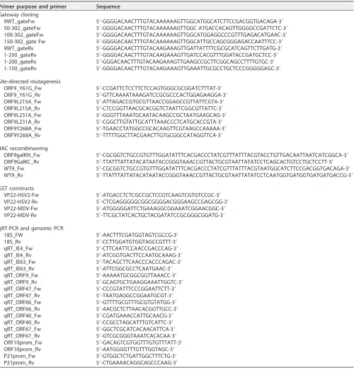
High-throughput yeast two-hybrid assay.orf9was amplified by PCR with the 9WT_gateFw and 9WT_gateRv primers (Table 1) and cloned by Gateway cloning (Invitrogen) in pAD-dest-CYH and pDB-dest, which were transformed, respectively, intoMATaY8800 and MAT␣Y8930 Saccharomyces cerevisiaestrains (as previously described [69, 70]). The AD-ORF9p Y8800 was mated to each of the 15,483 DB ORFs Y8930 of the human ORFeome 5.1 (CCSB; Dana-Farber Cancer Institute; described in reference 71), the 165 pools of 94 AD ORFs of the hORFeome 5.1 were mated to DB ORF9 Y8930, and interactions were identified strictly as described in reference 72. Colonies positive for the GAL1::HIS3 and GAL1::ADE2 selective markers but negative for autoactivation were selected for PCR amplification (with Zymolyase 20T [Seikagaku Biobusiness] and PlatinumTaqDNA polymerase [Invitrogen]). Interacting proteins were identified by sequencing of the respective AD and DB ORFs. The interaction of partners of interest with WT and sixorf9truncated constructs (expressing aa 50 to 302, 100 to 302, 150 to 302, 1 to 250, 1 to 200, and 1 to 150) was validated/tested similarly using isolated AD and DB clones.
GST pulldown.pGex-ORF9 has been described previously (30). The MDV and HSV-2UL49genes were amplified by PCR, using pcDNA3-UL49 (a kind gift from C. Denesvre [73]) or genomic DNA purified from HSV-2-infected cells as the template and the primers described in Table 1. Amplified sequences were inserted into pGEX-5X-1, which had previously been digested with SmaI. pOka VZV-infected and noninfected MeWo cells were lysed with GST buffer (50 mM Tris-HCl [pH 8], 5 mM EDTA, 150 mM NaCl, 10 mM MgCl2, 1% Triton X-100, cOmplete protease inhibitor cocktail [1:50; Roche]) and centrifuged for 3 min at maximum speed. Cleared lysates were incubated for 2 h at 4°C with GST-, GST-ORF9p-, or GST-VP22-coated agarose beads (GE Healthcare). After three washes, the pulled-down proteins were eluted and subjected to Western blotting.
Immunofluorescence.MRC-5 or ARPE-19 cells were seeded on glass coverslips and infected by coculture with infected MeWo cells. At 48 h postinfection (hpi), cells were washed with phosphate-buffered saline (PBS), fixed for 10 min in 4% paraformaldehyde–PBS, permeabilized for 10 min in 0.3% Triton X-100 –PBS, and blocked with 10% bovine serum albumin (BSA)–PBS for 45 min. The coverslips were then incubated overnight at 4°C with the primary antibodies diluted in 5% BSA–PBS (anti-V5, 1/250; anti-AP-1␥, 1/250). After 3 washes in 5% BSA–PBS, the coverslips were incubated for 1 h with the appropriate Alexa Fluor 405- or Alexa Fluor 568-conjugated secondary antibody (1/400 in 5% BSA–PBS at room temperature). Nuclei were stained with TO-PRO-3 (1/2,000), and images were recorded with an Olympus FV1000 confocal microscope using a 63⫻oil objective.
Site-directed mutagenesis.pcDNA-3.1Y61G, L215A, L231A, and -ORF9p-Y268A were generated by directional mutagenesis using TurboPFU polymerase (Stratagene), the appro-priate primers (Table 1), and pcDNA-3.1-ORF9-V5 as a template (30).
Construction of BAC-VZV carrying the ORF9p mutation.All constructs were created by modifi-cation of the BAC-VZV-pOka WT (a kind gift from H. Zhu [74]) using the GalK positive/negative selection technique described by Warming et al. (75) and materials (pGalK plasmid and SW102 bacterial strain) obtained from the Biology Research Branch (BRB) at NCI, Bethesda, MD.
V5 and VZV-ORF9-ΔAC-V5 (ΔAC-V5) were already described (30, 31). BAC-VZV-ORF9-Y61G-V5, -L215A-V5, -L231A-V5, and -Y268A-V5 were generated by replacing theorf9coding sequence by thegalK-expressing cassette, which was subsequently replaced by the mutated copy oforf9. The primers listed in Table 1 and the four mutated pcDNA-3.1 vectors were used to obtain the recombination cassettes. To create the L231A revertant BAC, the ΔORF9 GalK cassette was reintroduced into BAC-VZV-ORF9-L231A-V5 and subsequently replaced by a WT ORF9-V5 copy.
Coimmunoprecipitation.Infected MeWo cells (48 hpi) were lysed, and the immunoprecipitation step was performed as described by Iijima et al. (66), except that protein A/G magnetic beads (Pierce) were used and a phosphatase inhibitor cocktail (25 mM-glycine, 1 mM Na3VO4, 1.5 mM NaF) was added in the lysis buffer. After four washes of the beads (1% Triton X-100, 150 mM NaCl, 50 mM Tris-HCl [pH
ORF9p/AP-1 Interaction Is Important for VZV Infectivity Journal of Virology
on November 6, 2019 by guest
http://jvi.asm.org/
7.0], 1 mM CaCl2, 1 mM MgCl2), proteins were eluted with 2% SDS at 37°C for 10 min, followed by the addition of an equal volume of SDS loading buffer and another incubation at 37°C for 10 min.
Viral growth curve.To assess the growth properties of the ORF9p mutant in MeWo cells, infected cells were first filtered through a cell strainer to remove syncytia and obtain a single-cell suspension. The proportion of GFP-positive cells in the suspension, corresponding to infected cells, was assessed using a FACSCanto II flow cytometer, and a known number of infected cells was then used to infect MeWo cells. At 48 hpi, pictures of the infection foci were recorded (range, 16 to 92) using an inverted fluorescence microscope (Olympus FSX-100). To determine the size of the infection foci, the pictures were processed with CellProfiler software (www.cellprofiler.org) (76). In parallel, at 48 and 72 hpi, the number of infection foci was determined in each well (n⫽8) and divided by the number of infected cells used to infect the cells at day 0, to obtain the ratio of infectious cells/infected cells.
[image:18.585.44.544.81.604.2]For the growth curve in MRC-5 cells, infected MeWo cells were trypsinized on day 1 and the proportion of GFP-positive cells, corresponding to infected cells, was assessed using a FACSCanto II flow TABLE 1Primers used in this study
Primer purpose and primer Sequence Gateway cloning
9WT_gateFw 5=-GGGGACAACTTTGTACAAAAAAGTTGGCATGGCATCTTCCGACGGTGACAGA-3=
50-302_gateFw 5=-GGGGACAACTTTGTACAAAAAAGTTGGC ATGACCACAGTTGGGGCCGATTCTC-3=
100-302_gateFw 5=-GGGGACAACTTTGTACAAAAAAGTTGGCATGGAGGCCCGTTTGAGACATGAAC-3=
150-302_gate Fw 5=-GGGGACAACTTTGTACAAAAAAGTTGGCATTGCCAGCGGGAGACCAATTTCC-3=
9WT_gateRv 5=-GGGGACAACTTTGTACAAGAAAGTTGATTATTTTCGCGCATCAGTTCTTGATG-3=
1-250_gateRv 5=-GGGGACAACTTTGTACAAGAAAGTTGATCCACGTTTGGATACCGATGCTCC-3=
1-200_gateRv 5=-GGGACAACTTTGTACAAGAAAGTTGAAGCCGCTTCGGCAGCCTTTTGTGC-3=
1-150_gateRv 5=-GGGGACAACTTTGTACAAGAAAGTTGAAATTGCGCCTGCTCCCGGGGGAGC-3=
Site-directed mutagenesis
ORF9_Y61G_Fw 5=-CCGATTCTCCTTCTCCAGTGGGCGCGGATCTTTAT-3=
ORF9_Y61G_Rv 5=-GTTCAAAATAAAGATCCGCGCCCACTGGAGAAGGA-3=
ORF9L215A_Fw 5=-ATTAGACCGTGCGTTAACCGGAGCCGTTATTCGTA-3=
ORF9L215A_Rv 5=-CTCCGGTTAACGCACGGTCTAATTCGGCGTTATTC-3=
ORF9L231A_Fw 5=-GGGTTTAAATGCAATACAAGCCGCTAATGAAGCAG-3=
ORF9L231A_Rv 5=-CGGCTTGTATTGCATTTAAACCCTCATGCACCGTA-3=
ORF9Y268A_Fw 5=-TGAACCTATGGCCGCACAAGTTCGTAAGCCAAAAA-3=
ORF9Y268A_Rv 5=-TTTTTGGCTTACGAACTTGTGCGGCCATAGGTTCA-3=
BAC recombineering
ORF9galKN_Fw 5=-CGCGGTCTGCCGTGTTTGGATATTTCACGACCCTATCGTTTATTTACGTACCTGTTGACAATTAATCATCGGCA-3=
ORF9GalKC_Rv 5=-TTATTTATTATACATAATACCGGGTAAACCGTTACTGCGTAATTATATCCTCAGCACTGTCCTGCTCCTT-3=
WT9_Fw 5=-CGCGGTCTGCCGTGTTTGGATATTTCACGACCCTATCGTTTATTTACGTAATGGCATCTTCCGACGGTGACAGA-3=
WT9_Rv 5=-TTATTTATTATACATAATACCGGGTAAACCGTTACTGCGTAATTATATCCTCAATGGTGATGGTGATGATGACCG-3=
GST constructs
VP22-HSV2-Fw 5=-ATGACCTCTCGCCGCTCCGTCAAGTCGTGTCCGC-3=
VP22-HSV2-Rv 5=-CTCGAGGGGGCGGCGGGGACGGGAAGCCGAGCGG-3=
VP22-MDV-Fw 5=-ATGGGGGATTCTGAAAGGCGGAAATCGGAACGGC-3=
VP22-MDV-Rv 5=-TTCGCTATCACTGCTACGATATCCGCGGGCGGATG-3=
qRT-PCR and genomic PCR
18S_FW 5=-AACTTTCGATGGTAGTCGCCG-3=
18S_Rv 5=-CCTTGGATGTGGTAGCCGTTT-3=
qRT_IE4_Fw 5=-CTTCAATTCCAACCGACCCAG-3=
qRT_IE4_Rv 5=-ATCGGTGACTTCCAATGCAAAG-3=
qRT_IE63_Fw 5=-TACAGCTTCAACCCACCCAGAC-3=
qRT_IE63_Rv 5=-ATTCGGCGCCTCAATGAAC-3=
qRT_ORF9_Fw 5=-AAAAATGCGGCGGTTAAACC-3=
qRT_ORF9_Rv 5=-GCAGTGCTGAAGGAAATTGGTC-3=
qRT_ORF47_Fw 5=-CCCGTATTTCCCGGAATTCTT-3=
qRT_ORF47_Rv 5=-TAATGAGGCCGGAATGCGT-3=
qRT_ORF66_Fw 5=-GTTTTGCGTTTGCGTGTATGG-3=
qRT_ORF66_Rv 5=-AACGCTCTTAACACGGTTGCC-3=
qRT_ORF40_Fw 5=-CGATGAAACCATTGCAACG-3=
qRT_ORF40_Rv 5=-CCGCCTAGCATTTGTCATTC-3=
qRT_ORF67_Fw 5=-GGCTCGCATCACAACATTCA-3=
qRT_ORF67_Rv 5=-GTCGCGGGTAAATCACACAA-3=
ORF10prom_Fw 5=-GACAGTCGTGGTTTGTGTTTATT-3=
ORF10prom_Rv 5=-AATGGGGTTTGTTTGGTAGC-3=
P21prom_Fw 5=-GTGGCTCTGATTGGCTTTCTG-3=
P21prom_Rv 5=-CTGAAAACAGGCAGCCCAAG-3=
August 2018 Volume 92 Issue 15 e00295-18 jvi.asm.org 18
on November 6, 2019 by guest
http://jvi.asm.org/
cytometer. The cells in four 25-cm2flasks of MRC-5 cells for each viral strain were then infected with 500 infected MeWo cells. On days 1 to 4, a 25-cm2flask for each virus was trypsinized and an aliquot was serially diluted (range, 1/62.5 to 1/2,000) and used to infect MRC-5 cells in 24-well plates. Each 24-well plate was analyzed twice (48 hpi and 72 hpi) with an inverted fluorescence microscope to determine the number of infection foci. The cells that were not used for the serial dilution were divided into three tubes, two of which were frozen for subsequent DNA and RNA extraction, while the third was centrifuged; the cells were fixed in 4% platelet-activating factor for 20 min; and the proportion of infected cells was determined by fluorescence-activated cell sorting (FACS).
qPCR and qRT-PCR.RNA was extracted from infected cells with the TriPure reagent (Roche) and treated with RNase-free DNase for an hour at 37°C. One microgram of RNA was used to produce cDNA with a RevertAid H Minus First Strand cDNA synthesis kit (Thermo Scientific). Twenty nanograms of cDNA was used for the subsequent PCR, except for 18S rRNA primers, for which 20 pg was used. The expression of 18S rRNA was used for normalization.
For genomic DNA extraction, cells were resuspended in Tris-SDS buffer (10 mM Tris, pH 7.4, 10 mM EDTA, 150 mM NaCl, 0.4% SDS) containing 0.2 mg/ml of proteinase K and incubated for 3 h at 50°C. Genomic DNA was subsequently isolated via a classical phenol-chloroform extraction. For viral genome quantification, 10 ng of DNA was used for the subsequent PCR. Primers in the VZVorf10promoter were used for viral genome detection, and normalization was performed with primers against the humanp21
promoter. In order to obtain the absolute number of VZV genomes present in the samples, a standard curve was built in parallel via serial dilution of a BAC-VZV WT DNA preparation of a known concentration. Quantitative PCR or RT-PCR were performed with a Roche LightCycler 480 apparatus in 384-well plates, in which triplicates of 2l of genomic DNA or cDNA were mixed with 8l of a mix containing FastStart Universal Sybr green master mix (Roche) and specific primers (Table 1).
3D skin model.For the preparation of epidermal equivalents, a collagen matrix solution was made with collagen mixed on ice with 10-fold-concentrated Ham’s F-12 medium, 10-fold reconstitution buffer, and Swiss 3T3 J2 fibroblasts. One milliliter of the collagen matrix solution was poured into 24-well plates. After gel equilibration with 1 ml of growth medium overnight at 37°C, 2.5⫻105PHK cells were seeded on top of the gels and maintained submerged for 24 to 48 h. The collagen rafts were raised and placed onto stainless steel grids at the interface between the air and the liquid culture medium. The growth medium was a mixture of Ham’s F-12 medium and Dulbecco’s modified Eagle’s medium (1:2) supple-mented with 0.5g/ml hydrocortisone, 10 ng/ml epidermal growth factor, 10% fetal calf serum, 2 mM
L-glutamine, 10 mM HEPES, 1 mM sodium pyruvate, 10⫺10mM cholera toxin, 5g/ml insulin, 5g/ml transferrin, and 15⫻10⫺4mg/ml 3,3=,5=-triiodo-L-thyronine. Epithelial cells were allowed to stratify for 4 days, with the medium being replaced every other day. Five thousand infected MRC-5 cells were then placed on top of the epithelium, which was maintained in culture for 6 additional days. The cultures were then harvested and either fixed in 10% buffered formalin and embedded in paraffin for subsequent immunohistohemistry or frozen for further RNA or DNA extraction.
Immunohistochemistry.Skin raft sections (5m) were incubated for an hour with the primary antibodies (anti-V5 [1/500] and anti-IE63 [1/250] in a Dako Real detection system), washed three times with PBS, and incubated for 30 min with the secondary antibody (Alexa Fluor 568-conjugated anti-mouse immunoglobulin [1/500] in a Dako Real detection system). A DAPI (4= ,6-diamidino-2-phenylindole)-containing mounting medium (catalog number S3023; Dako) was used for nuclear staining and sample fixation.
Alternatively, sections were washed, incubated in hematoxylin-eosin, washed again, and mounted with Leica CV Mount mounting medium. Observations were made with a Zeiss LSM880 Airy scan confocal microscope with a 40⫻oil objective or with an Olympus FSX-100 microscope with a 40⫻dry objective. Transmission electron microscopy.MeWo cells infected with VZV-ORF9-V5 or VZV-L231A-V5 were sorted by FACS, fixed for 90 min at 4°C with 2.5% glutaraldehyde in Sörensen 0.1 M phosphate buffer (pH 7.4), and postfixed for 30 min with 2% osmium tetroxide. After dehydration in graded ethanol, samples were embedded in Epon. Ultrathin sections obtained with a Reichert Ultracut S ultramicrotome were contrasted with uranyl acetate and lead citrate. Observations were made with a JEOL JEM-1400 transmission electron microscope at 80 kV.
ACKNOWLEDGMENTS
This work was supported by the University of Liege, Fonds National pour la
Recherche Scientifique (F.R.S.-FNRS, Belgium), and by the Fonds Leon Fredericq. J.L.
and L.R. were supported by the Fonds pour la Recherche dans l’Industrie et
l’Agriculture (FRIA, Brussels, Belgium). C.B. was supported by the
FP7-People-COFUND 2013-2019 program.
We are grateful to L. Traub for the generous gift of anti-AP-1
1 antibodies and
S. Jonjic for anti-ORF10p antibodies. We also thank H. Zhu for the BAC-VZV-pOka
WT, C. Denesvre for pcDNA3-UL49, and the Biology Research Branch (BRB) at NCI,
Bethesda, MD, for the GalK plasmid and for the SW102 bacterial strain. Finally, we
thank C. Lassence for technical assistance; P. Piscicelli for TEM preparation; S.
Ormenese, J. J. Goval, and S. Freeman for technical support with confocal
micros-copy (Imaging Technological Platform, GIGA-R); T. DiSalvo, A. Marquet, and C.
ORF9p/AP-1 Interaction Is Important for VZV Infectivity Journal of Virology
on November 6, 2019 by guest
http://jvi.asm.org/
Humblet for immunohistochemistry; R. Stephan for cell sorting; and E. De
Waegen-aere for organotypic assays.
REFERENCES
1. Mettenleiter TC, Klupp BG, Granzow H. 2009. Herpesvirus assembly: an update. Virus Res 143:222–234. https://doi.org/10.1016/j.virusres.2009 .03.018.
2. Hambleton S, Gershon MD, Gershon AA. 2004. The role of the trans-Golgi network in varicella zoster virus biology. Cell Mol Life Sci 61:3047–3056. https://doi.org/10.1007/s00018-004-4269-7.
3. Johnson DC, Baines JD. 2011. Herpesviruses remodel host membranes for virus egress. Nat Rev Microbiol 9:382–394.https://doi.org/10.1038/ nrmicro2559.
4. Hollinshead M, Johns HL, Sayers CL, Gonzalez-Lopez C, Smith GL, Elliott G. 2012. Endocytic tubules regulated by Rab GTPases 5 and 11 are used for envelopment of herpes simplex virus. EMBO J 31:4204 – 4220.https:// doi.org/10.1038/emboj.2012.262.
5. Maresova L, Pasieka TJ, Homan E, Gerday E, Grose C. 2005. Incorporation of three endocytosed varicella-zoster virus glycoproteins, gE, gH, and gB, into the virion envelope. J Virol 79:997–1007.https://doi.org/10.1128/ JVI.79.2.997-1007.2005.
6. Beitia Ortiz de Zarate I, Cantero-Aguilar L, Longo M, Berlioz-Torrent C, Rozenberg F. 2007. Contribution of endocytic motifs in the cytoplas-mic tail of herpes simplex virus type 1 glycoprotein B to virus replication and cell-cell fusion. J Virol 81:13889 –13903.https://doi .org/10.1128/JVI.01231-07.
7. Albecka A, Laine RF, Janssen AFJ, Kaminski CF, Crump CM. 2016. HSV-1 glycoproteins are delivered to virus assembly sites through dynamin-dependent endocytosis. Traffic 17:21–39.https://doi.org/10 .1111/tra.12340.
8. Nixdorf R, Mettenleiter TC, Klupp BG. 2001. Role of the cytoplasmic tails of pseudorabies virus glycoproteins B, E and M in intracellular localiza-tion and virion incorporalocaliza-tion. J Gen Virol 82:215–226.https://doi.org/10 .1099/0022-1317-82-1-215.
9. Elliott GD, Meredith DM. 1992. The herpes simplex virus type 1 tegument protein VP22 is encoded by gene UL49. J Gen Virol 73:723–726.https:// doi.org/10.1099/0022-1317-73-3-723.
10. Spengler M, Niesen N, Grose C, Ruyechan WT, Hay J. 2001. Interactions among structural proteins of varicella zoster virus. Arch Virol Suppl 2001:71–79.
11. Tanaka M, Kato A, Satoh Y, Ide T, Sagou K, Kimura K, Hasegawa H, Kawaguchi Y. 2012. Herpes simplex virus 1 VP22 regulates translocation of multiple viral and cellular proteins and promotes neurovirulence. J Virol 86:5264 –5277.https://doi.org/10.1128/JVI.06913-11.
12. Maringer K, Stylianou J, Elliott G. 2012. A network of protein interactions around the herpes simplex virus tegument protein VP22. J Virol 86: 12971–12982.https://doi.org/10.1128/JVI.01913-12.
13. Stylianou J, Maringer K, Cook R, Bernard E, Elliott G. 2009. Virion incor-poration of the herpes simplex virus type 1 tegument protein VP22 occurs via glycoprotein E-specific recruitment to the late secretory pathway. J Virol 83:5204 –5218.https://doi.org/10.1128/JVI.00069-09. 14. del Rio T, Werner HC, Enquist LW. 2002. The pseudorabies virus VP22
homologue (UL49) is dispensable for virus growth in vitro and has no effect on virulence and neuronal spread in rodents. J Virol 76:774 –782. https://doi.org/10.1128/JVI.76.2.774-782.2002.
15. Fuchs W, Klupp BG, Granzow H, Hengartner C, Brack A, Mundt A, Enquist LW, Mettenleiter TC. 2002. Physical interaction between envelope gly-coproteins E and M of pseudorabies virus and the major tegument protein UL49. J Virol 76:8208 – 8217. https://doi.org/10.1128/JVI.76.16 .8208-8217.2002.
16. Sciortino MT, Taddeo B, Giuffrè-Cuculletto M, Medici MA, Mastino A, Roizman B. 2007. Replication-competent herpes simplex virus 1 isolates selected from cells transfected with a bacterial artificial chromosome DNA lacking only the UL49 gene vary with respect to the defect in the UL41 gene encoding host shutoff RNase. J Virol 81:10924 –10932. https://doi.org/10.1128/JVI.01239-07.
17. Ebert K, Depledge DP, Breuer J, Harman L, Elliott G. 2013. Mode of virus rescue determines the acquisition of VHS mutations in VP22-negative herpes simplex virus 1. J Virol 87:10389 –10393.https://doi.org/10.1128/ JVI.01654-13.
18. Mbong EF, Woodley L, Dunkerley E, Schrimpf JE, Morrison LA, Duffy C.
2012. Deletion of the herpes simplex virus 1 UL49 gene results in mRNA and protein translation defects that are complemented by secondary mutations in UL41. J Virol 86:12351–12361.https://doi.org/10.1128/JVI .01975-12.
19. Duffy C, Lavail JH, Tauscher AN, Wills EG, Blaho JA, Baines JD. 2006. Characterization of a UL49-null mutant: VP22 of herpes simplex virus type 1 facilitates viral spread in cultured cells and the mouse cornea. J Virol 80:8664 – 8675.https://doi.org/10.1128/JVI.00498-06.
20. El Bilali N, Duron J, Gingras D, Lippé R. 2017. Quantitative evaluation of protein heterogeneity within herpes simplex virus 1 particles. J Virol 91:e00320-17.https://doi.org/10.1128/JVI.00320-17.
21. Elliott G, Hafezi W, Whiteley A, Bernard E. 2005. Deletion of the herpes simplex virus VP22-encoding gene (UL49) alters the expression, local-ization, and virion incorporation of ICP0. J Virol 79:9735–9745.https:// doi.org/10.1128/JVI.79.15.9735-9745.2005.
22. Che X, Reichelt M, Sommer MH, Rajamani J, Zerboni L, Arvin AM. 2008. Functions of the ORF9-to-ORF12 gene cluster in varicella-zoster virus replication and in the pathogenesis of skin infection. J Virol 82: 5825–5834.https://doi.org/10.1128/JVI.00303-08.
23. Tischer BK, Kaufer BB, Sommer M, Wussow F, Arvin AM, Osterrieder N. 2007. A self-excisable infectious bacterial artificial chromosome clone of varicella-zoster virus allows analysis of the essential tegument protein encoded by ORF9. J Virol 81:13200 –13208.https://doi.org/10.1128/JV