B. V. Venkataram Prasad,
cTerence S. Dermody
b,eaDepartment of Pathology, Microbiology, and Immunology, Vanderbilt University School of Medicine,
Nashville, Tennessee, USA
bDepartment of Pediatrics, University of Pittsburgh School of Medicine, Pittsburgh, Pennsylvania, USA
cVerna and Marrs McLean Department of Biochemistry and Molecular Biology, Baylor College of Medicine,
Houston, Texas, USA
dDepartment of Biological Sciences, University of Pittsburgh, Pittsburgh, Pennsylvania, USA
eDepartment of Microbiology and Molecular Genetics, University of Pittsburgh School of Medicine, Pittsburgh,
Pennsylvania, USA
ABSTRACT
Viral nonstructural proteins, which are not packaged into virions, are
essen-tial for the replication of most viruses. Reovirus, a nonenveloped, double-stranded RNA
(dsRNA) virus, encodes three nonstructural proteins that are required for viral replication
and dissemination in the host. The reovirus nonstructural protein
NS is a
single-stranded RNA (ssRNA)-binding protein that must be expressed in infected cells for
pro-duction of viral progeny. However, the activities of
NS during individual steps of the
re-ovirus replication cycle are poorly understood. We explored the function of
NS by
disrupting its expression during infection using cells expressing a small interfering RNA
(siRNA) targeting the
NS-encoding S3 gene and found that
NS is required for viral
ge-nome replication. Using complementary biochemical assays, we determined that
NS
forms complexes with viral and nonviral RNAs. We also discovered, using
in vitro
and
cell-based RNA degradation experiments, that
NS increases the RNA half-life.
Cryo-electron microscopy revealed that
NS and ssRNAs organize into long, filamentous
structures. Collectively, our findings indicate that
NS functions as an RNA-binding
pro-tein that increases the viral RNA half-life. These results suggest that
NS forms
RNA-protein complexes in preparation for genome replication.
IMPORTANCE
Following infection, viruses synthesize nonstructural proteins that
me-diate viral replication and promote dissemination. Viruses from the family
Reoviridae
encode nonstructural proteins that are required for the formation of progeny
vi-ruses. Although nonstructural proteins of different viruses in the family
Reoviridae
di-verge in primary sequence, they are functionally homologous and appear to
facili-tate conserved mechanisms of dsRNA virus replication. Using
in vitro
and cell culture
approaches, we found that the mammalian reovirus nonstructural protein
NS binds
and stabilizes viral RNA and is required for genome synthesis. This work contributes
new knowledge about basic mechanisms of dsRNA virus replication and provides a
foundation for future studies to determine how viruses in the family
Reoviridae
as-sort and replicate their genomes.
KEYWORDS
RNA-binding proteins, dsRNA replication, reovirus,
NS
V
iruses contain the minimal components required to enter host cells and deliver
their genetic payloads into the cell interior. For productive infection of a host, viral
genomes must be replicated at sites of primary infection and packaged into new virions
for dissemination to susceptible cells at local or distant sites. Viruses encode proteins
that are not packaged into virions to aid in these postentry replication steps. These
Received4 April 2018Accepted7 May 2018 Accepted manuscript posted online16 May 2018
CitationZamora PF, Hu L, Knowlton JJ, Lahr RM, Moreno RA, Berman AJ, Prasad BVV, Dermody TS. 2018. Reovirus nonstructural protein σNS acts as an RNA stability factor promoting viral genome replication. J Virol 92:e00563-18.https://doi.org/10.1128/JVI .00563-18.
EditorSusana López, Instituto de Biotecnologia/UNAM
Copyright© 2018 American Society for Microbiology.All Rights Reserved.
Address correspondence to Terence S. Dermody, terence.dermody@chp.edu.
on November 6, 2019 by guest
http://jvi.asm.org/
proteins, called nonstructural proteins, participate in multiple viral replication steps,
including translation of viral mRNAs (1), replication of viral genomes (2), construction of
sites for particle assembly (3), and inhibition of innate immune responses (4). Due to
constraints on viral genome size (5), most nonstructural proteins play multiple roles
during infection (6). Consequently, teasing out individual activities of viral nonstructural
proteins is challenging.
Mammalian orthoreoviruses (called reoviruses here) are nonenveloped viruses that
infect virtually all mammalian species but cause disease only in the very young (7, 8).
The reovirus virion is an
⬃
80-nm particle composed of two concentric protein shells
that encapsidate 10 segments of double-stranded RNA (dsRNA) (9). Reovirus replication
begins with recognition of glycan attachment factors (10–12) and cell surface receptors
(13, 14). Following internalization, reoviruses traffic within endosomes (15, 16) and
undergo acid-dependent proteolytic disassembly (17), which allows core particles to
penetrate the endosomes and gain access to the cytoplasm (18). Within the cytoplasm,
reovirus cores are transcriptionally active and release viral mRNAs (19, 20). These
mRNAs are translated by cellular ribosomes, yielding eight structural and three
non-structural proteins (9). The nonnon-structural proteins reorganize membranes from the
endoplasmic reticulum (21) to form neo-organelles called inclusions or factories (22–
24), which serve as sites of viral genome replication and particle assembly (25, 26). By
a mechanism that is not understood and is likely aided by nonstructural proteins (27,
28), viral mRNAs are sorted and assembled into a replicase or precore complex (29, 30),
followed by dsRNA synthesis and formation of nascent cores (31, 32). These newly
synthesized cores are transcriptionally active and release viral mRNAs until transcription
is silenced by the coalescence of outer-capsid proteins onto nascent cores (33). Mature
reovirus particles are released from cells through an unknown mechanism.
Understand-ing how reoviruses replicate is important, as the viruses have oncolytic potential (34)
and may trigger celiac disease (35, 36).
The reovirus nonstructural protein
NS is a 41-kDa protein highly conserved among
different reovirus strains (37, 38). Initially incorrectly identified as a poly(C)-dependent
RNA polymerase (39),
NS is a single-stranded RNA (ssRNA)-binding protein with a
reported specificity for certain regions of reovirus mRNA (40, 41), although it appears
to show comparable binding to viral and nonviral RNAs (42).
NS has been defined as
a nonclassical helicase, exhibiting strand displacement activity in the absence of ATP
hydrolysis and MgCl
2(42). Following reovirus infection of host cells,
NS and
NS,
another nonstructural protein that is responsible for nucleation of reovirus inclusions
(22), are among the initial translation products (9).
NS and
NS interact with each
other in reovirus inclusions (43–45) using a mechanism that partially depends on the
presence of RNA (23). Throughout the course of infection, reovirus inclusions mature
(46), recruit cellular membranes (21), and attract the host translational machinery (47),
probably to localize the synthesis of reovirus proteins to sites of genome replication
and particle assembly. These studies give valuable insight into
NS, but its function
during reovirus replication remains poorly understood.
To enhance knowledge of the activities of
NS during reovirus replication, we used
cells expressing a small interfering RNA (siRNA) targeting the reovirus
NS-encoding S3
gene and quantified the effect of
NS knockdown on various steps of viral replication.
We found that
NS is dispensable for primary, but not secondary, rounds of
transcrip-tion and functranscrip-tions prior to genome replicatranscrip-tion. We hypothesized that
NS promotes
genome replication by stabilizing viral mRNAs. Using complementary
in vitro
and
cell-based assays, we discovered that
NS and RNAs organize into long filamentous
structures and that
NS increases the half-life of viral mRNAs. These results define
NS
as an RNA stability factor required for genome replication during reovirus infection.
RESULTS
Developing a system to study
NS function.
In a previous study (48), we
engineered HEK293T cells to stably express an siRNA targeting the reovirus
NS-encoding S3 gene. We sorted the cells by flow cytometry and selected clones that
Zamora et al. Journal of Virology
on November 6, 2019 by guest
http://jvi.asm.org/
showed the lowest expression of
NS after infection with reovirus strain type 3 Dearing
(T3D) (data not shown). These cells were termed
NS-siRNA cells (Fig. 1A). Using the
reovirus plasmid-based reverse genetics system (49), we engineered a viral mutant that
contained four synonymous mutations within the S3 gene siRNA target site of T3D (Fig.
1B). We termed this virus T3D resistant (T3D-R), as we hypothesized that it would be
resistant to the siRNA expressed by
NS-siRNA cells. To determine whether
NS is
expressed in T3D-R-infected
NS-siRNA cells, we allowed either T3D or T3D-R to be
adsorbed onto
NS-siRNA cells and quantified
NS protein abundance by
immuno-blotting (Fig. 1C and D). Infection of
NS-siRNA cells with T3D resulted in a 96%
reduction of
NS protein levels relative to infection with T3D-R, indicating that T3D-R
effectively escapes RNA interference (RNAi) targeting in
NS-siRNA cells. To confirm
that
NS knockdown in T3D-infected
NS-siRNA cells is specific, we allowed either T3D
or T3D-R to be adsorbed onto HEK293T cells expressing a nontargeting siRNA (green
fluorescent protein [GFP]-siRNA cells) and quantified
NS protein abundance by
im-munoblotting (Fig. 1C and D). T3D- and T3D-R-infected GFP-siRNA cells expressed
comparable amounts of
NS, indicating that the
NS protein of T3D is produced in
293T cells expressing a nontargeting siRNA. To test whether the synonymous mutations
introduced in the S3 gene of T3D-R virus alter the production of viral progeny, we
allowed either T3D or T3D-R to be adsorbed onto GFP-siRNA and
NS-siRNA cells and
quantified viral yields at 24 h postadsorption by plaque assay (Fig. 1E). T3D and T3D-R
viruses produced comparable yields at 24 h postadsorption in GFP-siRNA cells,
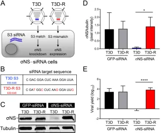
sug-FIG 1(A) Schematic of viruses and cells used to studyNS function. HEK293T cells engineered to stably express an siRNA against theNS-encoding S3 gene (NS-siRNA cells) were infected with either T3D or T3D-R virus. Representations of the S3 gene segments for T3D (blue) and T3D-R (red) are highlighted. The S3 gene segment of T3D contains target sequences complementary to the S3 siRNA ofNS-siRNA cells, whereas the S3 gene segment of T3D-R does not. (B) Sequences corresponding to nucleotides 630 to 648 of T3D and T3D-R S3 genes. Synonymous mutations are shown in red. (C) T3D or T3D-R virus was allowed to adsorb ontoNS-siRNA cells or control cells expressing a nontargeting siRNA (GFP-siRNA cells) at an MOI of 10 PFU per cell and incubated for 24 h. The cell lysates were resolved by SDS-PAGE and immunoblotted using antibodies specific forNS and␣-tubulin. (D) Pixel intensity analysis of the ratio ofNS to␣-tubulin in T3D- and T3D-R-infectedNS-siRNA and GFP-siRNA cells for triplicate experiments. (E) T3D or T3D-R virus was allowed to adsorb ontoNS-siRNA and GFP-siRNA cells at an MOI of 1 PFU/cell. Viral yields at 24 h postadsorption were determined by plaque assay. The results are presented as means⫾SD.*,P⬍0.05, and****,P⬍0.0001, as determined by Student’sttest.on November 6, 2019 by guest
http://jvi.asm.org/
[image:3.585.43.370.72.343.2]gesting that the mutations introduced in T3D-R do not alter its replication capacity.
Similar results were obtained in replication assays using cells lacking any siRNA (data
not shown). In contrast, T3D was incapable of producing progeny in
NS-siRNA cells,
indicating that
NS is required for T3D replication. Thus,
NS-siRNA cells effectively and
specifically diminish
NS expression when infected with T3D, but not T3D-R.
Reovirus T3D does not replicate in
NS-siRNA cells.
To assess whether
NS is
required for formation of progeny virions over a longer interval, we allowed either T3D
or T3D-R to be adsorbed onto
NS-siRNA cells and quantified progeny virus at various
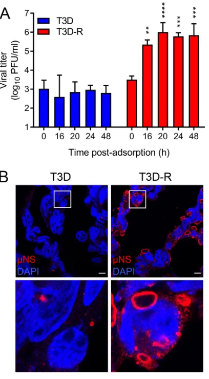
times postadsorption by plaque assay (Fig. 2A). T3D was incapable of forming new
progeny when infecting
NS-siRNA cells at all intervals tested. In contrast, T3D-R yields
increased throughout the time course of infection, reaching
⬃
70-fold by 16 h and
⬃
220-fold by 48 h, confirming that
NS is required for progeny particle production.
To determine whether
NS is required for inclusion formation, we allowed either
T3D or T3D-R to be adsorbed onto
NS-siRNA cells, stained the cells at 24 h with
polyclonal serum specific for the reovirus nonstructural protein
NS to visualize
inclusions, and imaged the cells by confocal microscopy (Fig. 2B). T3D-infected
NS-FIG 2Reovirus T3D does not replicate inNS-siRNA cells. T3D or T3D-R virus was allowed to adsorb ontoNS-siRNA cells at an MOI of 1 (A) or 100 (B) PFU/cell. (A) Viral titers at 0, 16, 20, 24, and 48 h postadsorption were determined by plaque assay. The results are presented as mean viral titers and SD for three independent experiments. Values that differ significantly from the value at 0 h by one-way analysis of variance (ANOVA) with Tukey’s multiple-comparison test are indicated (**,P⬍0.01;***,P⬍0.001;****,P⬍0.0001). (B) Cells were fixed at 24 h postinfection (hpi), stained usingNS-specific antiserum (red) and DAPI (blue) to visualize nuclei, and imaged using confocal microscopy. The boxed regions are shown enlarged below. Scale bars, 10m.
Zamora et al. Journal of Virology
on November 6, 2019 by guest
http://jvi.asm.org/
[image:4.585.103.307.70.439.2]siRNA cells contained few small inclusions (
⬍
1
m), similar to those detected early in
infection (22). In contrast, T3D-R-infected
NS-siRNA cells were filled with globular
inclusions, some of which were more than 10
m in diameter. These results suggest
that
NS is required for replication steps leading to the maturation of reovirus
inclu-sions.
Viral RNA levels are diminished in T3D-infected
NS-siRNA cells.
To estimate
the timing at which primary and secondary rounds of transcription occur in HEK293T
cells, we allowed either T3D or T3D-R to be adsorbed onto
NS-siRNA cells and
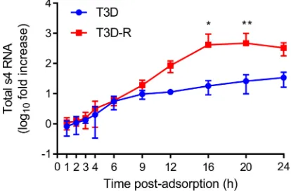
quantified s4 RNA by reverse transcription-quantitative PCR (RT-qPCR) (Fig. 3). (A
small-case letter preceding a number indicates an ssRNA species by convention.) Levels
of s4 RNA in T3D- and T3D-R-infected
NS-siRNA cells increased at similar rates until 6
h postadsorption. At later times, s4 RNA levels increased at higher rates in
T3D-R-infected
NS-siRNA cells than in T3D-infected cells. These results suggest that
second-ary rounds of transcription do not begin until after 6 h postadsorption, as previously
observed (50), and that late rounds of gene expression are impaired in T3D-infected
NS-siRNA cells.
To test whether steady-state levels of viral RNAs are altered by
NS disruption, we
quantified RNA levels for the L (large), M (medium), and S (small) gene segments by
NanoString (NanoString Technologies, Inc.) analysis during a time course of infection in
T3D- and T3D-R-infected
NS-siRNA cells. NanoString technology is based on
fluores-cent probes that, following hybridization with their target RNAs, allow direct
quantifi-cation of RNAs in a sample without the requirement for reverse transcription or
amplification. We allowed either T3D or T3D-R to be adsorbed onto
NS-siRNA cells and
quantified RNA levels corresponding to the 10 reovirus gene segments at various
intervals postadsorption using unique probes targeting each reovirus positive-sense
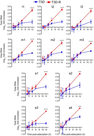
RNA (Table 1 and Fig. 4). The levels of all viral RNAs increased comparably in cells
infected with T3D or T3D-R until 4 to 6 h postadsorption, after which time RNA levels
in T3D-infected cells increased modestly. In contrast, levels of viral RNAs in cells
infected with T3D-R increased exponentially, with levels at 12 h postadsorption
ele-vated
⬃
250-fold for the L-class RNAs,
⬃
340-fold for the M-class RNAs, and
⬃
550-fold
for the S-class RNAs relative to the respective levels of these RNAs at the 0-h time point.
Because secondary transcription does not occur until after 6 h postadsorption (Fig. 3)
(50), these results indicate that
NS is dispensable for primary, but not secondary,
rounds of transcription.
Reovirus T3D protein synthesis is diminished in infected
NS-siRNA cells.
We
hypothesized that the reduction in abundance of viral RNAs observed in T3D-infected
NS-siRNA cells would diminish viral protein levels. To test this hypothesis, either T3D
FIG 3Total s4 RNA levels are diminished in T3D-infectedNS-siRNA cells. T3D or T3D-R virus was allowed to adsorb ontoNS-siRNA cells at an MOI of 1 PFU/cell. Cells were lysed at the intervals shown, and s4 RNA was quantified by RT-qPCR. The results are presented as mean RNA levels normalized to the RNA levels at 0 h for at least three independent experiments. The error bars indicate SD. Values that differ significantly between T3D- and T3D-R-infectedNS-siRNA cells at each time point by two-way ANOVA followed by Sidak’s multiple-comparison test are indicated (*,P⬍0.05;**,P⬍0.01).on November 6, 2019 by guest
http://jvi.asm.org/
[image:5.585.102.309.74.211.2]TABLE
1
Custom-designed
probes
used
for
NanoString
a
Gene
Protein
Target
sequence
(5
=
¡
3
=
)
L1
3
GAGCTCAGTCGTCGGTGAGCTTCGTAAACGGACAAAGACGTATGTTAAACATGACTTTGCTTCAGTGAGGTACATTCGTGACGCTATGGCATGTACTAGC
L2
2
TGGACGAAGGCGATCTGATGGTTAGTCGGCTTACGCAACTCCCGTTACGTCCTGATTATGGTAATATCTGGGTCGGCGATGCGCTATCCTATTATGTGGA
L3
1
CCAACGCGCATGGGAACGCCGAATGTATCCAAAATATGTAATTTCGTCGCCTCTTGTGTGCGAAATCGGGTTGGACGGTTTGATCGAGCACAGATGATGA
M1
2
ACGTTTGAGCAGGCGGTTATGGAGATATACAAAGGGATTGCTGGCGTTGACTCGCTGGATGATCTCATCAAGTGGGTGCTGAACTCGGATCTCATTCCGC
M2
1
GCTGGGATCCAAACGGCAAGAAGGTCGGATTCATCGTTTTTCAATCGAAGATACCATTCGAACTTTGGACTGCTGCTTCACAGATCGGTCAAGCCACGGT
M3
NS
GTGTCGTATGACGTTACGCTCACTCATGAAGACCGGACGCGACGTTGATGCACACAGAGCTTTTCAGCGAGTCCTCTCTCAAGGATACACATCGCTAATG
S1
1
ACTTGCAGAGCTACGCGTTGATCACGACAATCTCGTTGCGAGAGTGGATACTGCAGAACGTAACATTGGATCATTGACCACCGAGCTATCAACTCTGACG
S2
2
GCTTTGCAAGCACAGGCAGATCGAGTGTACGACTGCGATGATTATCCATTTCTAGCGCGTGATCCAAGATTCAAACATCGGGTGTATCAGCAATTGAGTG
S3
NS
TGGCCACGTCATCTTTAAGTATTTCCCTGGACCGGGGTCGATGGGTGGCGGCTGACGCCAGTGATGCTAGACTGCTGGTTTTTCCGATTCGCGTGTAATG
S4
3
CTGCTCACTGGAAGCGGGGTATGCTGTCCTTCGTTGCGCAGATGCACGAGATGATGAATGACGTGTCGCCAGATGACCTGGATCGTGTGCGTACTGAGGG
aThe
table
shows
the
target
sequence
of
each
reovirus
gene
segment
and
protein.
Zamora et al. Journal of Virology
on November 6, 2019 by guest
http://jvi.asm.org/
or T3D-R was adsorbed onto
NS-siRNA cells, and expression of the RNA-dependent
RNA polymerase
3 and nonstructural proteins
NS and
NS was quantified by
SDS-PAGE, followed by immunoblotting at various intervals postadsorption (Fig. 5).
Newly synthesized viral proteins were detected at 12 h postadsorption in
T3D-R-infected
NS-siRNA cells, and levels of the three immunoblotted viral proteins
in-creased over time. In contrast, viral protein synthesis was impaired in T3D-infected
NS-siRNA cells. These results suggest that
NS is required for replication steps leading
to viral protein synthesis.
FIG 4Levels of all reovirus RNAs are reduced in T3D-infectedNS-siRNA cells. T3D or T3D-R virus was allowed to adsorb ontoNS-siRNA cells at an MOI of 1 PFU/cell. Cells were lysed at the intervals shown, and RNAs were quantified by NanoString technology using custom-designed probes (Table 1) specific for each reovirus gene segment. The results are presented as mean RNA levels normalized to the RNA levels at 0 h for three independent experiments. The error bars indicate SD. Values that differ significantly between T3D- and T3D-R-infectedNS-siRNA cells at each time point by two-way ANOVA followed by Sidak’s multiple-comparison test are indicated (**,P⬍0.01;***,P⬍0.001;****,P⬍0.0001).
on November 6, 2019 by guest
http://jvi.asm.org/
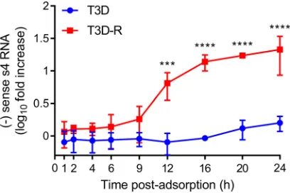
[image:7.585.42.370.71.543.2]Reovirus
NS is required for genome replication.
To elucidate whether
NS is
required for replication of the reovirus genome, we used a modified qPCR assay to
quantify s4 negative-sense ssRNA (50). In this assay, s4 RNA is reverse transcribed using
a primer that binds to the negative-sense strand of the S4 dsRNA gene segment. Once
the s4 negative-sense cDNA is synthesized, a complementary primer is added, and
quantification by qPCR proceeds. As the genome is the only source of negative-sense
viral RNA, the assay allows genome copies to be quantified. We allowed either T3D or
T3D-R to be adsorbed onto
NS-siRNA cells and quantified s4 negative-sense RNA at
various intervals postadsorption (Fig. 6). By this analysis, genome replication appeared
to initiate at 9 h postadsorption in T3D-R-infected
NS-siRNA cells, and by 12 h, T3D-R
S4 dsRNA levels were
⬃
10-fold higher than those at the initiation of infection. T3D-R S4
dsRNA levels continued to increase over time, and by 24 h, these levels were
⬃
20-fold
higher than those at 0 h. In contrast, T3D S4 dsRNA was undetectable throughout most
of the time course of infection, and by 24 h, T3D S4 dsRNA levels had increased only
⬃
1.5-fold in
NS-siRNA cells compared with the initiation of infection. These results
indicate that
NS functions at a step leading to the replication of the reovirus genome.
Thus, by examining the kinetics of individual replication steps, we conclude that
NS
mediates a process immediately prior to reovirus genome replication, following primary
but before secondary rounds of transcription.
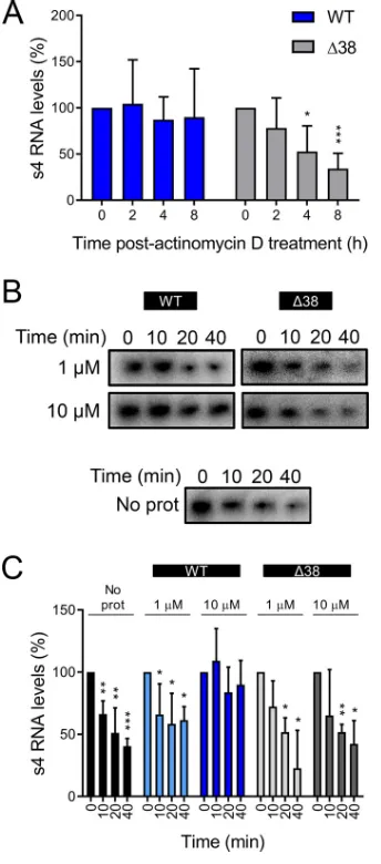
The
NS protein protects viral RNA from degradation.
Based on the known
affinity of
NS for ssRNA (42, 51) and the requirement for
NS in a step leading to
FIG 5T3D protein synthesis is diminished in T3D-infectedNS-siRNA cells. T3D or T3D-R virus was allowed to adsorb onto NS-siRNA cells at an MOI of 1 PFU/cell. (A) Cell lysates were collected at the intervals shown and analyzed by immunoblotting using antibodies specific for the3,NS, andNS proteins. (B) Pixel intensity analysis of the ratios of3,NS, andNS proteins to␣-tubulin. The results are presented as mean pixel intensity and SD for three independent experiments. Values that differ significantly between T3D- and T3D-R-infected NS-siRNA cells at each time point by two-way ANOVA followed by Sidak’s multiple-comparison test are indicated (*,P⬍0.05;***,P⬍0.001;****,P⬍0.0001).
Zamora et al. Journal of Virology
on November 6, 2019 by guest
http://jvi.asm.org/
[image:8.585.48.467.72.403.2]genome replication (Fig. 6), we hypothesized that
NS functions as an RNA stability
factor increasing the half-life of viral RNAs. To test this hypothesis, we used two
NS
constructs: full-length
NS (wild type [WT]) and a deletion mutant that lacks the first 38
amino acids (Δ38
NS). Δ38
NS was identified in a limited proteolysis assay in which
purified WT
NS was incubated with trypsin, followed by identification of the digestion
products using mass spectrometry (data not shown). The deletion mutant was
antici-pated to be impaired in RNA binding because it lacks a series of positively charged
residues predicted to be required for that activity (51, 52). We first tested whether
NS
expression increases RNA half-life in cells. HEK293T cells were transfected with plasmids
encoding either WT or Δ38
NS. At 20 h posttransfection, the cells were transfected
again with a plasmid encoding
3, incubated for 4 h, and treated with actinomycin D
to inhibit further transcription. Transcription derived from the
3-encoding plasmid
produces RNAs that are capped and polyadenylated (53). At various times following
actinomycin D treatment, we quantified total
3-encoding s4 RNA by RT-qPCR (Fig. 7A).
As actinomycin D inhibits transcription, this approach allowed us to determine the RNA
half-life. Transfection of cells with WT
NS led to the maintenance of s4 RNA levels
throughout the time course of actinomycin D treatment. At 8 h after treatment
initiation, we detected only a 10% reduction in s4 RNA levels. In contrast, transfection
of cells with Δ38
NS failed to protect s4 RNA. At 8 h after treatment initiation, s4 RNA
levels had decreased 66% relative to the initiation of actinomycin D treatment. These
results suggest that expression of WT
NS increases the half-life of a viral RNA and that
this activity requires the amino-terminal 38 amino acids of the protein.
To more directly test whether
NS protects RNA from degradation, we developed an
in vitro
cell-free RNA protection assay. We incubated purified, recombinant WT or Δ38
NS with internally radiolabeled uncapped and nonpolyadenylated s4 RNA.
NS-RNA
complexes were incubated with HeLa S100 cytoplasmic lysates for various intervals.
RNA was resolved by electrophoresis and quantified by phosphorimaging (Fig. 7B and
C). Incubation with WT
NS protected the RNA from degradation in a
concentration-dependent manner. At a concentration of 1
M,
NS did not protect s4 RNA from
degradation in HeLa S100 lysates, and similar results were obtained in the presence of
Δ38
NS or in the absence of any additional protein. However, 10
M
NS protected
the s4 RNA, so that by 40 min of incubation, only 10% of the input s4 RNA was lost.
Thus,
NS protects viral RNA from degradation.
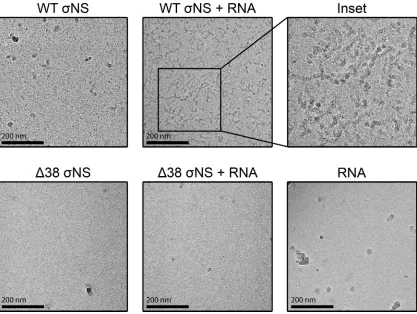
NS and RNAs organize in filamentous structures.
To better understand how
NS interacts with RNA, we used cryo-electron microscopy (cryo-EM) to visualize
NS-RNA complexes. We incubated purified, recombinant WT or Δ38
NS with
un-capped and nonpolyadenylated s4 RNA at a molar ratio of 50:1 on ice for 1 h and
FIG 6Reovirus T3D does not synthesize dsRNA inNS-siRNA cells. T3D or T3D-R virus was allowed to adsorb ontoNS-siRNA cells at an MOI of 1 PFU/cell. Cells were lysed at the intervals shown, and negative (⫺)-sense s4 RNA was quantified by single-strand RT-qPCR. The results are presented as mean RNA levels at each time point normalized to the RNA levels at 0 h for at least three independent experiments. The error bars indicate SD. Values that differ significantly between T3D- and T3D-R-infected NS-siRNA cells at each time point by two-way ANOVA followed by Sidak’s multiple-comparison test are indicated (***,P⬍0.001;****,P⬍0.0001).on November 6, 2019 by guest
http://jvi.asm.org/
[image:9.585.104.308.72.208.2]imaged
NS-RNA complexes by cryo-EM (Fig. 8). In the absence of RNA, WT
NS
appeared to form higher-order structures, as demonstrated by large electron-dense
configurations. In contrast, Δ38
NS appeared smaller and scattered throughout the EM
grids. Following incubation with s4 RNA, WT
NS organized into long filamentous
structures approaching 200 nm in length. As expected, we did not observe interactions
of Δ38
NS with s4 RNA. These data suggest that WT
NS coats RNA and that these
complexes arrange into long filamentous structures.
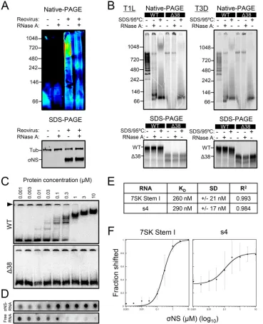
The
NS protein forms higher-order complexes with viral and nonviral ssRNAs.
To examine
NS-RNA complexes in detail, we sought to determine whether
NS exists
in an RNA-bound complex in infected cells. We allowed reovirus T3D to be adsorbed
onto HEK293T cells and, at 24 h postadsorption, we lysed the cells under mild
FIG 7TheNS protein protects viral RNA from degradation. (A) HEK293T cells were transfected with plasmids encoding either WT or Δ38NS and incubated for 20 h, followed by a second transfection with a 3-encoding plasmid and incubation for 4 h. The cells were treated with 10 g/ml of actinomycin D and lysed at the intervals shown, and3-encoding s4 RNA was quantified by RT-qPCR. The results are presented as mean RNA levels normalized to the RNA levels at 0 h for at least three independent experiments. The error bars indicate SD. (B and C) Radiolabeled uncapped and nonpolyadenylated s4 RNA was incubated without protein (No prot) or with 1 or 10M purified recombinant WT or Δ38NS at room temperature for 10 min, followed by addition of HeLa S100 lysates. (B) RNA was purified at the intervals shown, resolved by electrophoresis, and visualized by phosphorimaging. (C) Pixel intensity analysis of the s4 RNA bands. The results are presented as mean RNA levels normalized to the RNA levels at 0 min for three independent experiments. The error bars indicate SD. Values that differ significantly from the values at the start of the time course by one-samplettest for each time point are indicated (*,P⬍0.05;**,P⬍0.01;***,P⬍0.001).Zamora et al. Journal of Virology
on November 6, 2019 by guest
http://jvi.asm.org/
[image:10.585.119.291.72.462.2]conditions to preserve protein-RNA interactions. Samples were treated with RNase A or
left untreated and resolved by native PAGE and SDS-PAGE, followed by
immunoblot-ting using
NS-specific polyclonal serum (Fig. 9A). In the absence of RNase A treatment,
NS migrated predominantly as a high-molecular-mass complex of
⬃
500 kDa to 1 MDa.
Following RNase A treatment, the levels of these high-molecular-mass complexes
diminished. Thus,
NS appears to exist as a complex with RNA in infected cells.
We next tested the effect of RNase A treatment on the electrophoretic mobility of
WT and Δ38
NS from reovirus strains type 1 Lang (T1L) and T3D produced by coupled
in vitro
transcription and translation using rabbit reticulocyte lysates in the presence of
[
35S]methionine. Transcription derived from these
NS-encoding plasmids yielded
RNAs that were uncapped and nonpolyadenylated. Following translation, samples were
treated with RNase A, denatured with SDS and heat, or left untreated; resolved by
native PAGE and SDS-PAGE; and imaged by phosphorimaging (Fig. 9B). In the absence
of RNase A, WT
NS migrated as higher-order structures ranging from
⬃
250 kDa to
⬃
900 kDa, suggesting that several distinct oligomeric forms of
NS bind RNA. The
migration pattern appeared to be regularly spaced, suggesting a specific stoichiometry
of
NS-RNA binding. Treatment with RNase A disassembled
NS-RNA complexes.
Intermediate-molecular-mass bands became undetectable, and bands with estimated
sizes of
⬃
900 kDa and
⬃
80 kDa predominated. SDS and heat also disrupted the
higher-order structures. As expected, Δ38
NS did not form higher-order species with
RNA. Lastly, we did not observe differences in the migration patterns of T1L and T3D
NS, suggesting that the
NS proteins of both strains interact similarly with RNA. Thus,
multiple subunits of
NS bind RNA with an apparently defined stoichiometry.
FIG 8TheNS protein forms filamentous structures in the presence of RNA. Uncapped and nonpolyadenylated s4 RNA was incubated with 50⫻molar excess of purified recombinant WT or Δ38NS on ice for 1 h, followed by plunge freezing. Frozen specimens were imaged at⫻40,000 magnification with defocus levels ranging from⫺2.0m to⫺3.5m. Representative images for WTNS, WTNS complexed with s4 RNA, an enlargement of WTNS and s4 RNA filaments, Δ38NS, Δ38NS incubated with s4 RNA, and s4 RNA alone are shown.
on November 6, 2019 by guest
http://jvi.asm.org/
[image:11.585.44.461.70.384.2]FIG 9TheNS protein binds viral and nonviral ssRNAs. (A) Reovirus T3D was allowed to adsorb onto HEK293T cells at an MOI of 100 PFU/cell and was incubated for 24 h. The cells were lysed and treated with 0.25g/ml of RNase A at 30°C for 1 h. Proteins were resolved by native PAGE or SDS-PAGE, followed by immunoblotting using antibodies specific forNS and␣-tubulin (SDS-PAGE only). Representative immunoblots from triplicate experi-ments are shown. Pixel intensity is depicted in a rainbow scale (native PAGE only). (B) T1L and T3D WT and Δ38 NS proteins were translatedin vitroin the presence of [35S]methionine and incubated with 0.25g/ml of RNase A at 30°C for 1 h after translation, heated at 95°C for 10 min in SDS-PAGE sample buffer, or left untreated. Samples were resolved by native PAGE or SDS-PAGE and visualized by phosphorimaging. (C) Increasing concentrations of purified recombinant WT or Δ38NS were incubated with radiolabeled uncapped and nonpolyadenylated 7SK stem I RNA at room temperature for 10 min, followed by native electrophoresis and visualization by phosphorim-aging. The arrowhead indicates RNA that failed to enter the gel. (D) Increasing concentrations of purified recombinant WTNS protein were incubated with radiolabeled uncapped and nonpolyadenylated s4 RNA at room temperature for 10 min.NS-RNA complexes were spotted onto nitrocellulose membranes, and unbound free RNA was collected on nylon membranes. The membranes were visualized by phosphorimaging. (E)KD, SD, andR2values for WTNS and 7SK stem I (panel C, top) or s4 RNA (panel D) were determined using KaleidaGraph. (F) Langmuir isotherm curve fitting for WTNS and 7SK stem I or s4 RNA. The results are presented as the mean percent shift at each concentration ofNS for at least three independent experiments (7SK stem I RNA,n⫽5; s4 RNA,n⫽3). The error bars indicate SD.
Zamora et al. Journal of Virology
on November 6, 2019 by guest
http://jvi.asm.org/
[image:12.585.40.409.72.526.2]distinct
NS-RNA complexes following incubation of RNA with WT
NS, suggesting that
each
NS unit binds
⬃
27 nucleotides at saturation. Langmuir isotherm curve-fitting
yielded an estimated
K
D(equilibrium dissociation constant) value of 260 nM for the
interaction of
NS and 7SK stem I RNA (Fig. 9E and F). Increasing concentrations of WT
NS decreased the amount of free RNA, and at a concentration of 3
M, no free RNA
was detected, suggesting that at this concentration, the 7SK stem I RNA is saturated
with
NS. Increasing concentrations of
NS also decreased the amount of aggregated
RNA incapable of entering a polyacrylamide gel (Fig. 9C, arrowhead), suggesting that
NS has a function in unwinding RNAs.
To determine the affinity of
NS for viral RNA, we conducted a filter-binding assay
using purified recombinant WT
NS and terminally radiolabeled uncapped and
non-polyadenylated s4 RNA. Increasing concentrations of WT
NS were incubated with s4
RNA.
NS-RNA complexes were spotted on a nitrocellulose membrane, and the
un-bound RNA was collected on a nylon membrane. The radiolabeled RNA on each
membrane was quantified by phosphorimaging (Fig. 9D). Increasing concentrations of
NS increased the levels of RNA retained on the nitrocellulose membrane and
de-creased the free RNA collected on the nylon membrane. Langmuir isotherm curve
fitting yielded a
K
Dvalue of 290 nM for the
NS-s4 RNA interaction, which is
compa-rable to the estimated
K
Dvalue for the
NS-stem I 7SK RNA interaction (Fig. 9E and F).
Collectively, these data indicate that
NS binds a viral RNA with affinity similar to that
for a nonviral RNA, suggesting that
NS does not recognize a specific RNA sequence
under these conditions.
The
NS protein impairs translation of viral and nonviral RNAs.
As
NS is an
ssRNA-binding protein, we hypothesized that it might alter the translation of RNAs. To
test whether
NS affects translation, we cotransfected HEK293T cells with increasing
concentrations of plasmids encoding either WT or Δ38
NS and a fixed concentration
of a luciferase-encoding plasmid (Fig. 10A). Transcription derived from these plasmids
yields RNAs that are capped and polyadenylated (53). Increasing concentrations of WT
NS plasmid decreased luciferase activity, a marker for luciferase translation. In
con-trast, transfection of Δ38
NS did not decrease luciferase activity. These data indicate
that
NS impairs translation of a nonviral capped and polyadenylated RNA and that this
inhibition depends on the amino-terminal 38 amino acids of
NS.
To test whether
NS displays a broad-spectrum inhibitory effect on translation, we
conducted
in vitro
translation assays using an uncapped and polyadenylated RNA in the
presence or absence of
NS. Increasing concentrations of purified recombinant WT or
Δ38
NS or bovine serum albumin (BSA) as a control were incubated with uncapped
and polyadenylated luciferase RNA prior to incubation with wheat germ extracts to
initiate translation. Wheat germ extracts were chosen for these experiments instead of
rabbit reticulocyte lysates because of their higher stringency and dependency on the
type of RNA and the reaction conditions for efficient translation (56). After incubation,
we quantified luciferase activity (Fig. 10B). WT
NS at a concentration of 10
M (
⬃
100:1
molar excess) decreased translation of luciferase RNA by 67%, corroborating the
observations made in experiments using HEK293T cells. In contrast, Δ38
NS and the
BSA control did not diminish translation efficiency.
on November 6, 2019 by guest
http://jvi.asm.org/
Lastly, we examined the effect of
NS on translation of a viral RNA. We incubated
increasing concentrations of purified, recombinant WT or Δ38
NS with capped,
2
=
-O-methylated and nonpolyadenylated s4 RNA prior to incubation with wheat germ
extracts to initiate translation in the presence of [
35S]methionine. The protein products
were resolved by SDS-PAGE and quantified by phosphorimaging (Fig. 10C and D).
Similar to findings made in the luciferase experiments, 10
M WT
NS (
⬃
80:1 molar
excess) significantly impaired translation of the s4 RNA. Thus, when present at high
concentrations,
NS inhibits translation of different types of RNAs.
DISCUSSION
In this study, we found that the mammalian reovirus
NS protein, a nonstructural
protein expressed early in reovirus infection (9, 22), is required for a process leading to
viral genome replication (Fig. 11). In the absence of
NS, reovirus infection progressed
through primary transcription. However, subsequent replication steps, including
pro-tein synthesis, genome replication, secondary transcription, formation of reovirus
in-clusions, and generation of progeny, were impaired. Considering the kinetics of the
individual replication steps, we found that the first function of
NS must occur between
6 and 9 h postadsorption (Fig. 3, 4, and 6). Using
in vitro
and cell-based approaches, we
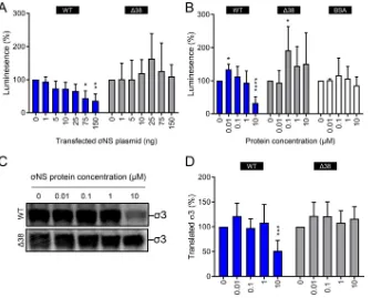
FIG 10The NS protein diminishes translation of viral and nonviral RNAs. (A) HEK293T cells were transfected with increasing concentrations of plasmids encoding either WT or Δ38 NS and a fixed concentration of renilla luciferase-encoding plasmid and incubated for 24 h. The cells were lysed, and luciferase levels were quantified. The results are presented as mean luminescence percentages normalized to luciferase levels in the absence ofNS plasmid for at least three independent experiments. The error bars indicate SD. (B) Firefly luciferase uncapped and polyadenylated RNA was incubated with increasing concentrations of purified recombinant WT or Δ38NS or BSA at room temperature for 10 min. Protein-RNA complexes were added to wheat germ extracts and incubated at 25°C for 1 h. Luciferase synthesis was quantified by luciferase assay. The results are presented as mean luminescence percentages normalized to luciferase levels in the absence of protein for at least three independent experiments. The error bars indicate SD. (C) Capped, 2=-O-methylated, and nonpolyadenylated s4 RNA was incubated with increasing concentrations of purified recombinant WT or Δ38NS at room temperature for 10 min. Protein-RNA complexes were added to wheat germ extracts and incubated at 25°C for 1 h in the presence of [35S]methionine. Samples were resolved by SDS-PAGE and visualized by phosphorimaging. (D) Pixel intensity analysis of the3 protein band for at least three independent experiments. The error bars indicate SD. Values that differ significantly from the no-protein condition values by one-samplettest for each time point are indicated (*,P⬍0.05;**,P⬍0.01;***,P⬍0.001;****,P⬍0.0001).Zamora et al. Journal of Virology
on November 6, 2019 by guest
http://jvi.asm.org/
[image:14.585.43.378.70.339.2]discovered that
NS forms complexes with RNA that organize into long filamentous
structures. In addition, we observed that
NS increases the RNA half-life and protects
RNA from degradation. These results suggest a function for
NS as an RNA-binding
protein that prepares RNA transcripts for replication.
The
NS protein, originally called essential noncapsid protein
2A (57), was first
identified as a nucleic acid-binding protein in cellular extracts derived from
reovirus-infected cells (40).
NS binds nucleic acids with positive cooperativity and covers a
length of
⬃
25 to 27 nucleotides at saturation (42). In experiments using purified
NS
and stem I 7SK RNA, a nucleic acid that is 108 nucleotides in length, we observed that
at least four units of
NS bound stem I 7SK RNA at saturation (Fig. 9C). These data
suggest that
NS covers a length of
⬃
27 nucleotides, consistent with previous results
(42). Residues 2 to 11 of
NS contribute to RNA binding, although Δ2-11
NS mutants
retain the capacity to bind RNA, albeit poorly (51). Using a
NS mutant lacking the
amino-terminal 38 amino acids, we found that these residues are absolutely required
for RNA binding (Fig. 9C).
By conducting coupled
in vitro
transcription and translation of WT and Δ38
NS in
the presence of [
35S]methionine and resolving the radiolabeled translation products by
native PAGE (Fig. 9B), we observed that WT
NS forms higher-order complexes when
RNA is present, whereas Δ38 does not. When we treated WT
NS samples with RNase
A, most of the high-molecular-mass species disappeared, except one of
⬃
900 kDa. This
⬃
900-kDa band was also observed following transcription and translation of Δ38
NS,
further indicating that the existence of the complex does not depend on the presence
of RNA. There are at least two possibilities that could account for this
⬃
900-kDa
complex. First,
NS may have the capacity to form large oligomers in the absence of
RNA and independently of its amino-terminal 38 amino acids. Second, both WT and
Δ38
NS may interact with a component of rabbit reticulocyte lysates in an
RNA-independent manner, forming an
⬃
900-kDa complex.
Previous competition assays indicated that
NS does not preferentially bind viral
over nonviral RNA (42). Our findings corroborate these results, as we calculated similar
K
Dvalues for
NS binding to viral s4 RNA and nonviral stem I 7SK RNA (290 nM and 260
nM, respectively) (Fig. 9E and F). Studies to quantify the affinity of a protein for RNA are
FIG 11Model ofNS function during reovirus replication. Reovirus enters cells by receptor-mediated endocytosis, and following acid-dependent proteolytic disassembly, the inner capsid or core is released into the cytoplasm (15, 16). Cores transcribe viral mRNAs corresponding to each gene segment (1° txn), which are subsequently translated to yield each viral protein. We hypothesize thatNS mediates at least two functions. (i) At low concentrations,NS enhances translation. (ii) At high concentrations,NS (either alone or with other viral proteins) binds and folds viral mRNAs into a conformation optimal for assortment and genome replication. After synthesis of the negative-sense strand and formation of new cores, secondary rounds of transcription occur (2° txn). The outer capsid assembles, silencing secondary transcription, and particles are released from infected cells by an unknown mechanism.on November 6, 2019 by guest
http://jvi.asm.org/
[image:15.585.84.324.74.256.2]usually conducted
in vitro
, and it is possible that during infection, other factors
contribute to the affinity and specificity of
NS-RNA interactions. For example,
seques-tration of
NS in discrete cellular environments (22) might account for increased
specificity for viral RNAs. In addition,
NS and
2, each of which interacts with
NS in
reovirus inclusions (23, 24, 43, 45), also display affinity for RNA (58, 59). A complex of
these proteins and
NS might be responsible for binding viral RNAs. It is also possible
that
NS binds cellular RNAs to promote the translation of cellular proteins within these
structures (47), as several host proteins are found within inclusions, for example, Hsc70
(60) and the TRiC chaperonin (61).
In the EMSAs conducted in our study, we observed an interesting feature of
NS-RNA interactions (Fig. 9C, arrowhead). Increasing concentrations of
NS resulted in
a reduction of aggregated RNA that did not enter the gel. These results suggest that
NS binding to RNA leads to RNA rearrangements allowing the RNA to migrate through
the gel matrix. RNA-remodeling proteins that resolve RNA structures are called RNA
chaperones (62), which are encoded by some RNA viruses (63). There are three lines of
evidence from our study and others suggesting that
NS is an RNA chaperone. First,
RNA chaperones differ from other RNA-folding proteins by binding nucleic acids
nonspecifically (Fig. 9) and acting in an ATP-independent manner, which has been
demonstrated for
NS in strand displacement experiments (42). Second, the avian
reovirus
NS protein, which is functionally homologous to mammalian reovirus
NS
(64, 65), acts as an RNA chaperone
in vitro
(27), as does the rotavirus NSP2 nonstructural
protein (28, 63). Third, our cryo-EM analysis demonstrated that
NS coats RNAs and
forms filamentous structures (Fig. 8). Coating of RNAs by viral proteins has been
suggested to mediate the activity of some viral RNA chaperones, and this activity
ensures protection and folding of their target RNAs. This is the case for HIV-1 NCp7 (66,
67), poliovirus 3AB (68), and tomato bushy stunt virus p33 (69). Therefore, it is likely that
viruses of the family
Reoviridae
encode RNA chaperones to resolve kinetically trapped
RNA conformations and facilitate replication.
Coating of RNAs also might account for the impairment in translation observed
when
NS is incubated with RNAs at molar excess, as it might sterically hinder effective
ribosome scanning (Fig. 10) (70). In this regard, we envision a bimodal function for
NS
in the regulation of translation. At low concentrations of
NS before RNAs become
saturated, translation is enhanced, as our
in vitro
luciferase translation experiments
suggested (Fig. 10B). It is possible that
NS directly stimulates translation of viral
mRNAs via interactions with a domain that differs from that responsible for RNA
binding, as our experiments also showed a tendency for Δ38
NS to increase
transla-tion (Fig. 10). This
NS-mediated translation enhancement effect might be partially
responsible for the impairment in protein synthesis observed in T3D-infected
NS-siRNA cells (Fig. 5). At high concentrations of
NS, translation is impaired via a
mechanism dependent on the amino-terminal 38 amino acids of the protein (Fig. 10).
Concentration-dependent inhibition of translation is a property of other viral proteins,
like HIV Gag, which inhibits translation of its own RNA at high concentrations (71), and
the coat protein from potato virus A (72). Considering that the same viral RNA is used
for translation and replication, a concentration-dependent bimodal function for
NS
might be necessary to transition between these replication cycle steps.
There is compelling evidence indicating that
NS is required for genome replication
(Fig. 6) (37, 73, 74), and we propose the following model for
NS function during
reovirus replication (Fig. 11). Early in infection,
NS concentrations are low, and viral
ssRNAs are in excess. Therefore, viral ssRNAs are minimally occupied by
NS. Perhaps
at this point,
NS recruits the host translational apparatus to enhance viral translation.
Interactions of
NS with the translation initiation machinery have been demonstrated,
as
NS colocalizes and coimmunoprecipitates with the ribosomal subunit pS6R and the
translation initiation factor eIF3A during infection (47). In addition, stress granules form
following infection and disassemble as infection proceeds, and their disruption is
required for efficient viral translation (75, 76).
NS is required to disrupt stress granules
during infection, as it associates with the stress granule effector protein, G3BP1 (77).
Zamora et al. Journal of Virology
on November 6, 2019 by guest
http://jvi.asm.org/
RNA decay machinery (80).
NS also might localize viral RNAs to sites required for
interaction with the RNA-dependent RNA polymerase
3. The
NS-interacting protein
NS has a defined
3-interacting domain (43) and might be responsible for bridging
NS to the genome replication machinery.
NS also could directly interact with
3
before the replication of the genome. Finally, an effect of
NS on genome replication
could be a consequence of its function to promote translation of
3 and other viral
proteins (Fig. 5 and 11).
We still do not understand the mechanism by which
NS dissociates from viral RNAs
before the formation of viral cores. Posttranslational modifications of RNA-binding
proteins can neutralize the basic residues responsible for interaction with RNAs. This
mechanism has been suggested for reovirus
NS (52) and shown for rubella virus
capsid protein (81), tomato bushy stunt virus p33 (82), and hepatitis B virus core protein
(83). Rotavirus NSP2 is phosphorylated (84), and similar to the case of
NS, NSP2
temperature-sensitive mutants fail to synthesize dsRNA (85). Whether
NS directly
participates in the replication of the dsRNA genome remains a key unanswered
question.
Collectively, data gathered in our study provide evidence of a function for
NS in
promoting the replication of the reovirus genome and establish a foundation for future
studies to determine the mechanism by which
NS acts prior to dsRNA synthesis. This
work sets the stage to better understand reovirus RNA assortment and replication.
MATERIALS AND METHODS
Cells and viruses.HEK293T cells engineered to express an siRNA against theNS-encoding S3 gene of reovirus strain T3D (48) were single-cell sorted at the Vanderbilt University Medical Center Flow Cytometry Shared Resource. Single-cell sorting was conducted by resuspending⬃2.5⫻106cells in phosphate-buffered saline (PBS) supplemented to contain 2% fetal bovine serum (FBS) (Gibco) in a volume of 500l. Propidium iodide (Sigma) was added at a concentration of 1g/ml, and single cells were sorted into wells of a 96-well plate and maintained in 100l of Dulbecco’s modified Eagle medium (DMEM) (Life Technologies) supplemented to contain 5% FBS, 2 mML-glutamine (Life Technologies), 100 U/ml of penicillin, 100g/ml of streptomycin (Life Technologies), 0.25 mg/ml of amphotericin B (Sigma), and 5g/ml of puromycin (Sigma). Cell clones were propagated for approximately 2 weeks, after which time the cells were divided into two plates: one was maintained in supplemented DMEM, and the other was infected with reovirus T3D for 18 h. Infection was scored by fluorescent focus assay using guinea pig NS polyclonal antiserum (44) as described previously (86). Clones with high knockdown efficiency were selected and passaged. HEK293T cells (ATCC CRL-3216), HEK293T cells expressing an S3-specific siRNA (NS-siRNA cells), and HEK293T cells expressing a GFP-specific siRNA (GFP-siRNA cells) (48) were maintained in DMEM supplemented to contain 5% FBS, 2 mML-glutamine, 100 U/ml of penicillin, 100 g/ml of streptomycin, 0.25 mg/ml of amphotericin B, and 5g/ml of puromycin (forNS-siRNA and GFP-siRNA cells). Murine L929 fibroblasts adapted to spinner bottles were maintained in Joklik’s modified Eagle’s minimal essential medium (JMEM) (Lonza) as described previously (11).
Reoviruses were recovered using plasmid-based reverse genetics (49, 87) and purified as described previously (88, 89). For generation of T3D-R virus, plasmid pT7-S3T3D (Addgene; 33284) was altered by QuikChange (Stratagene) site-directed mutagenesis to contain the synonymous mutations C632T, A635C, A642G, and A649G (Fig. 1B) using T3D_R_QC primers (Table 2). To confirm mutations, viral RNA was extracted from purified virions and subjected to OneStep RT-PCR (Qiagen) using T3D_S3 primers (Table 2). Purified PCR products were analyzed by Sanger sequencing. Viral titers were determined by plaque assay using L929 cells (89).
Plasmid cloning and DNA transfections.Plasmids forin vitrotranslation of the T3D s4 mRNA were engineered by introducing an EagI restriction site at the 3=end of the S4 cDNA in pT7-S4T3D (Addgene; 33285) using QuikChange site-directed mutagenesis and T3D_S4_EagI primers (Table 2). Plasmids for coupledin vitrotranscription and translation of strain T1LNS were generated by subcloning the open
on November 6, 2019 by guest
http://jvi.asm.org/
TABLE
2
PCR
primers
Primer
name
Sequence
(5
=
¡
3
=
)
Forward
(F)
Reverse
(R)
T3D_R_QC
AGATATCCATGCATGCCAATCCCTTGAGGCCATCGTCCAGAAGCTGTTCCG
CGGAACAGCTTCTGGACGATGGCCTCAAGGGATTGGCATGCATGGATATCT
T3D_T7S4
GCGGGTTAATACGACTCACTATAGCTATT
GATGAATGAAGCCTGTCCCACGT
T3D_S3
CCTGATGCGCCAATGTCTAA
CTGTCTCCTCGCAATACAACTC
T3D_S4
GAAGCATTTGCCTCACCATAG
GATCTGTCCAACTTGAGTGTATTG
human_GADPH
CCCATCACCATCTTCCAG
ATGACCTTGCCCACAGCC
T3D_S4_EagI
AGATGCCATGCCGACGGCCGATGAATGAAGCC
GGCTTCATTCATCGGCCGTCGGCATGGCATCT
T1L_S3_RS
CGACGGATCCATGGCTTCCTCACTCAGGGC
ATCACAGGCGGCCGCTTACACGCGAATTGGAAACACCAGC
T1L_S3del38
AACGTTGTTGAGTATCAAATCCGTACAGG
CATGGATCCGAGCTCGGTACCAA
T3D_S3_GA
TTGGTACCGAGCTCGGATCCATGGCTTCCTCACTCAGAG
TCTAGACTCGAGCGGCCGCCTTACACGCGAATCGGAAAAAC
pcDNA3.1_GA
GGCGGCCGCTCGAGTCTA
GGATCCGAGCTCGGTACC
T3D_S3del38_GA
CATGGATCCGAGCTCGGTACC
TTGGTACCGAGCTCGGATCCATGAATGTGGTTGAGTATCAAATTCG
T3D_S4_qPCR
CGCTTTTGAAGGTCGTGTATCA
CTGGCTGTGCTGAGATTGTTTT
Zamora et al. Journal of Virology
on November 6, 2019 by guest
http://jvi.asm.org/
[image:18.585.134.284.104.739.2]Laboratory at Baylor College of Medicine and purified as described previously (90). A rabbit was immunized with 100g of3 protein in complete Freund’s adjuvant at Cocalico Biologicals. Antigen boosts containing 50g of3 protein in incomplete Freund’s adjuvant were administered at days 14, 21, 49, and 134 following the initial inoculation. Antigen boosts containing 100g of3 protein in incomplete Freund’s adjuvant were administered at days 120, 183, and 211 following the initial inoculation. Serum samples were obtained at days 35, 56, 148, and 197 following the initial inoculation and tested for reactivity against purified T3D virions by immunoblotting. Final exsanguination was conducted 225 days after the initial inoculation.
Native PAGE, SDS-PAGE, immunoblotting, and phosphorimaging.Samples for native PAGE were diluted in 4⫻native PAGE sample buffer (ThermoFisher) and resolved in 4 to 16% native PAGE Bis-Tris acrylamide gels (ThermoFisher) using the Blue Native PAGE Novex Bis-Tris gel system (ThermoFisher) as described previously (61).
Samples for denaturing SDS-PAGE were diluted in 5⫻SDS-PAGE sample buffer, incubated at 95°C for 5 min, and resolved in 10% Mini-Protean TGX gels (Bio-Rad).
Native PAGE and SDS-PAGE gels were transferred to polyvinylidene difluoride (PVDF) and nitrocel-lulose membranes, respectively (61). The following antibodies were used for immunoblotting: guinea pig NS polyclonal antiserum (44), chickenNS polyclonal antiserum (47), rabbit3 polyclonal antiserum, and mouse␣-tubulin monoclonal antibody (Cell Signaling Technology). The following antibodies were used as detection reagents: IRDye800CW donkey anti-guinea pig, IRDye680RD donkey anti-chicken, IRDye800CW goat anti-rabbit, and IRDye680LT goat anti-mouse (Li-Cor).
The membranes were scanned using an Odyssey CLx imaging system (Li-Cor). The pixel intensities of the bands for all gels and membranes were quantified using Image Studio Software (Li-Cor).
Native PAGE and SDS-PAGE gels containing radiolabeled products were incubated in 40% methanol, 10% acetic acid at room temperature for 1 h, washed, and dried. The dried gels were exposed on a phosphorimaging screen and imaged using a PerkinElmer Cyclone phosphor system scanner.
Purification of recombinantNS protein.Escherichia coliBL21(DE3)pLysS (Promega) cultures were transformed with T3D WT or Δ38NS-encoding plasmids and induced with 0.5 mM isopropyl-D -1-thiogalactopyranoside (IPTG) (Sigma) after reaching an optical density (600 nm) of 0.6 to 0.7. The cells were resuspended in 50 mM Tris-HCl (pH 8), 500 mM NaCl, 10 mM imidazole, and 1 mM dithiothreitol (DTT) and supplemented with protease inhibitor cocktail (Roche). The cells were lysed using a micro-fluidizer, followed by removal of cell debris by centrifugation. The clarified cell lysates were loaded onto Ni-nitrilotriacetic acid (NTA) columns (Qiagen), andNS was eluted using a gradient of 50 mM Tris-HCl (pH 8), 0.5 M NaCl, 0.5 M imidazole, and 1 mM DTT. Elution fractions were dialyzed against 20 mM Tris-HCl (pH 8), 100 mM NaCl, 10 mM imidazole, 1 mM EDTA, and 1 mM DTT. The eluted protein was incubated with thrombin (1 U per 100 g of recombinant protein) at 4°C for 12 h to remove the His tag. Protein samples were reloaded onto a Ni-NTA column to remove uncleaved His-taggedNS. The flowthrough was loaded onto a Superose 6 10/300 GL column (GE Healthcare) in 10 mM Tris-HCl (pH 7.4), 100 mM NaCl, and 1 mM DTT. The eluted fractions were concentrated using a 30-kDa centrifugal filter unit (Millipore).
Immunofluorescence microscopy of reovirus-infected cells.Reovirus was allowed to adsorb onto cells at a multiplicity of infection (MOI) of 100 PFU per cell and incubated at 37°C for 24 h. The cells were fixed in 4% paraformaldehyde (PFA) (Electron Microscopy Sciences) in PBS at room temperature for 20 min; permeabilized with 1% Triton X-100 in PBS at room temperature for 5 min; blocked with 0.5% BSA, 0.1% glycine, and 0.05% Tween 20; and stained with chickenNS polyclonal antiserum, Alexa-Fluor secondary antibody (Invitrogen), and 4=,6-diamidino-2-phenylindole (DAPI) (Invitrogen) to visualize nuclei. Images were captured using a Zeiss LSM 710 laser scanning confocal microscope and analyzed using a Zeiss LSM5 series image browser.
RNA extraction and purification.To purify cellular RNA, cells were lysed using TRIzol reagent (ThermoFisher), and RNA was extracted with chloroform. To purify RNA fromin vitro transcription reactions, samples were combined with an equal volume of lysis buffer (ThermoFisher) supplemented to contain 1%-mercaptoethanol (BME). An equal volume of 70% ethanol was added, and the RNA was purified using a PureLink RNA minikit (ThermoFisher) according to the manufacturer’s instructions.
S4 quantitative RT-PCR.Total and single-strand negative-sense s4 RNAs were quantified using qScript XLT one-step RT-qPCR ToughMix, Low ROX (Quanta Bioscience), and T3D_S4_qPCR primers (Table 2) according to the manufacturer’s instructions. The following RT-qPCR cycling protocol was used: cDNA synthesis (50°C for 10 min), initial denaturation (95°C for 1 min), and 40 PCR cycles (95°C for 10 s
on November 6, 2019 by guest
http://jvi.asm.org/
followed by a data collection step at 60°C for 1 min). S4 cDNA was detected using a fluorogenic probe (5=-FAM [fluorescent fluorescein]-AGCGCGCAAGAGGGATGGGA-BHQ [black hole quencher]-1-3=; Bio-search Technologies). For the single-strand RT-qPCR, the following modifications were included: RNA was incubated at 95°C for 3 min and immediately placed on ice; reverse transcription was conducted using only the forward primer T3D_S4_qPCR; and for the quantitative-PCR step, the reverse T3D_S4_qPCR primer was included (50).
NanoString RNA quantification and analysis. Probes specific for each reovirus gene segment positive-sense RNA were designed by NanoString Technologies using proprietary software (Table 1). RNA was purified from infected cells and incubated with probes in hybridization buffer according to the manufacturer’s instructions. Following hybridization, excess probe was removed using an nCounter Prep Station automated liquid handler. Probe-target complexes were transferred to an nCounter cartridge for immobilization and loaded onto an nCounter digital analyzer for imaging and quantification of each target RNA. The quantified expression data were analyzed using nSolver analysis software, including image quality controls and background subtraction. Counts for each probe were normalized using the geometric mean of four cellular transcripts (GUSB, HPRT, SARM1, and RSP6).
In vitrotranscription reactions.Templates forin vitrotranscription of the T3D S4 gene segment were generated from pT7-S4T3D by PCR amplification using T3D_T7S4 primers (Table 2). Templates were transcribedin vitrousing a HiScribe T7 high-yield RNA synthesis kit (New England BioLabs) according to the manufacturer’s instructions with the following modifications. For synthesis of radiolabeled RNA, 125 ng of PCR product was used in the presence of radiolabeled [␣-32P]UTP (PerkinElmer) in 20-l reaction mixtures at room temperature for 2 h. For synthesis of nonradiolabeled RNA, 2g of plasmid DNA template was used in 40-l reaction mixtures at 37°C for 2 h. DNA templates were degraded by on-column incubation with PureLink DNase (ThermoFisher). For some experiments, RNA was capped using vaccinia capping enzyme (New England BioLabs) and 2=-O-methyltransferase (New England BioLabs) in one-step reaction mixtures according to the manufacturer’s instructions.
Stem I of 7SK RNA (5=-GGAUGUGAGGGCGAUCUGGCUGCGACAUCUGUCACCCCAUUGAUCGCCAGGG UUGAUUCGGCUGAUCUGGCUGGCUAGGCGGGUGUCCCCUUCCUCCCUCACCGCUCC-3=) was synthesized using T7 RNA polymerase from PCR-generated DNA templates. The RNA was purified from a gel slice following electrophoresis in a 7 M urea–1⫻Tris-borate-EDTA (TBE)– 6% 29:1 polyacrylamide gel.
In vitrocell-free RNA degradation assay and electrophoresis.s4 RNA transcripts (⬍10 pM; 5⫻105 cpm) were incubated with various concentrations ofNS in a volume of 10l at room temperature for 10 min.NS-RNA complexes were incubated with 26l of a 6-mg/ml stock of HeLa S100 cytoplasmic extracts (Speed Biosystems). At various intervals, RNA was purified from 8-l aliquots of the treatment mixtures. The RNA was diluted in 2⫻Novex TBE-urea sample buffer (ThermoFisher), resolved in 6% TBE-urea gels (ThermoFisher), exposed on a phosphorimaging screen, and visualized using a PerkinElmer Cyclone phosphor system scanner.
RNA degradation assay in cells.HEK293T cells seeded in 12-well plates were transfected using FuGene 6 with 500 ng of plasmid encoding either WT or Δ38NS and incubated for 20 h. The cells were transfected a second time using FuGene 6 with 50 ng of pCAG_S4T3D plasmid (49) and incubated for 4 h. The cells were treated with 10g/ml of actinomycin D (Sigma) and lysed at various intervals prior to RNA extraction. Expression of s3, s4, and human GADPH RNAs was confirmed by RT-PCR amplification using primers T3D_S3, T3D_S4, and human_GADPH.
In vitrotranslation reactions.RNAs were translatedin vitrousing wheat germ extracts (Promega) according to the manufacturer’s instructions with the following modifications. RNAs at a concentration of 10g/ml were incubated at 65°C for 3 min, placed immediately on ice, and incubated with various concentrations of WT or Δ38 NS at room temperature for 10 min. Protein-RNA complexes were incubated with wheat germ extracts supplemented with 80M amino acid mixture minus methionine (Promega) and 0.8 U/l of RNasin (Promega). To synthesize nonradioactive and radioactive proteins, 80 M amino acid mixture minus leucine (Promega) or 0.7Ci/l of [35S]methionine (PerkinElmer) was added, respectively. Translation reaction mixtures were incubated at 25°C for 1 h, followed by freezing at⫺20°C.
Coupled in vitro transcription and translation reactions.Coupled in vitro transcription and translation reactions were conducted using the TNT-coupled rabbit reticulocyte lysate system (Promega) according to the manufacturer’s instructions. Reaction mixtures were incubated at 30°C for 1 h and terminated by 4-fold dilution in stop buffer (20 mM HEPES-KOH [pH 7.4], 100 mM potassium acetate, 5 mM magnesium acetate, 5 mM EDTA, 2 mM methionine) supplemented to contain a final concentration of 1 mM DTT and 2 mM puromycin. Protein samples were incubated with RNase A (Qiagen) under variable treatment conditions, depending on the experiment.
5=labeling of RNA.RNAs were dephosphorylated using calf intestinal alkaline phosphatase (CIP) (New England BioLabs) according to the manufacturer’s instructions with the following modifications. CIP was used at a concentration of 0.5 U per 1g of RNA in 20-l reaction mixtures. Following incubation at 37°C for 30 min, RNA was precipitated using phenol-chloroform (s4 RNAs) or gel purified (7SK stem I RNA) and labeled using T4 polynucleotide kinase (New England BioLabs) and [␥32P]ATP (PerkinElmer). The RNAs were purified using a G25 desalting column (GE Healthcare).
Luciferase assays.To quantify RLuc activity in transfected cells, HEK293T cells seeded in 96-well plates were transfected with increasing concentrations of WT or Δ38NS plasmids (0 to 150 ng), decreasing concentrations of a noncoding plasmid (pT7-S2T3D; Addgene; 33283; 150 to 0 ng), and 50 ng of simian virus 40 (SV40) renilla luciferase plasmid, achieving a final DNA concentration of 200 ng/well, and incubated for 24 h. The cells were lysed with 20l of renilla luciferase lysis buffer (Promega) at room
Zamora et al. Journal of Virology
on November 6, 2019 by guest
http://jvi.asm.org/
Filter-binding assay.s4 RNA (⬍10 pM; 500 cpm) was incubated with increasing concentrations of WTNS in 100 mM NaCl, 50 mM HEPES (pH 7.2), 10% glycerol, and 1g BSA at room temperature for 10 min. Presoaked nitrocellulose (top) and nylon (bottom) membranes (GE) were used in a 96-well hybrid dot manifold assembly as described previously (93). After filtration, the membranes were air dried, exposed to phosphorimaging screens, and visualized using ImageQuant TL (GE). Data were plotted using KaleidaGraph (Synergy Software).
Electron microscopy ofNS-RNA complexes.s4 RNA was incubated with either WT or Δ38NS at a molar ratio of 50:1 on ice for 1 h before plunge freezing using a Vitribot (MIV). The frozen specimens were imaged at⫻40,000 magnification with defocus levels ranging from⫺2.0 to⫺3.5m using a JEM2010 (200-kV) cryo-electron microscope equipped with a charge-coupled-device (CCD) camera at the CryoEM Center at Baylor College of Medicine.
Statistical methods.All experiments were conducted independently at least three times. Data are presented as means and standard devi