Structural Definition of Duck Major
Histocompatibility Complex Class I
Molecules That Might Explain Efficient
Cytotoxic T Lymphocyte Immunity to
Influenza A Virus
Yanan Wu,
aJunya Wang,
aShuhua Fan,
aRong Chen,
aYanjie Liu,
a,bJianhua Zhang,
aHongyu Yuan,
aRuiying Liang,
aNianzhi Zhang,
aChun Xia
a,c Department of Microbiology and Immunology, College of Veterinary Medicine, China Agricultural University, Beijing, Chinaa; Key Laboratory for Insect-Pollinator Biology of the Ministry of Agriculture, Institute of Apiculture, Chinese Academy of Agricultural Sciences, Beijing, Chinab; Key Laboratory of Animal Epidemiology and Zoonosis, Ministry of Agriculture, China Agricultural University, Beijing, ChinacABSTRACT
A single dominantly expressed allele of major histocompatibility
com-plex class I (MHC I) may be responsible for the duck’s high tolerance to highly
pathogenic influenza A virus (HP-IAV) compared to the chicken’s lower tolerance. In
this study, the crystal structures of duck MHC I (
Anpl
-UAA
*
01) and duck

2-microglobulin (

2m) with two peptides from the H5N1 strains were determined.
Two remarkable features were found to distinguish the
Anpl
-UAA
*
01 complex from
other known MHC I structures. A disulfide bond formed by Cys
95and Cys
112and
connecting the

5 and

6 sheets at the bottom of peptide binding groove (PBG) in
Anpl
-UAA
*
01 complex, which can enhance IAV peptide binding, was identified.
Moreover, the interface area between duck MHC I and

2m was found to be larger
than in other species. In addition, the two IAV peptides that display distinctive
con-formations in the PBG, B, and F pockets act as the primary anchor sites. Thirty-one
IAV peptides were used to verify the peptide binding motif of
Anpl
-UAA
*
01, and the
results confirmed that the peptide binding motif is similar to that of HLA-A
*
0201.
Based on this motif, approximately 600 peptides from the IAV strains were partially
veri-fied as the candidate epitope peptides for
Anpl
-UAA
*
01, which is a far greater number
than those for chicken BF2
*
2101 and BF2
*
0401 molecules. Extensive IAV peptide
bind-ing should allow for ducks with this
Anpl
-UAA
*
01 haplotype to resist IAV infection.
IMPORTANCE
Ducks are natural reservoirs of influenza A virus (IAV) and are more
resis-tant to the IAV than chickens. Both ducks and chickens express only one dominant MHC
I locus providing resistance to the virus. To investigate how MHC I provides IAV
resis-tance, crystal structures of the dominantly expressed duck MHC class I (p
Anpl
-UAA
*
01)
with two IAV peptides were determined. A disulfide bond was identified in the peptide
binding groove that can facilitate
Anpl
-UAA
*
01 binding to IAV peptides.
Anpl
-UAA
*
01
has a much wider recognition spectrum of IAV epitope peptides than do chickens. The
IAV peptides bound by
Anpl
-UAA
*
01 display distinctive conformations that can help
in-duce an extensive cytotoxic T lymphocyte (CTL) response. In addition, the interface area
between the duck MHC I and

2m is larger than in other species. These results indicate
that HP-IAV resistance in ducks is due to extensive CTL responses induced by MHC I.
KEYWORDS
crystal structure, duck, MHC class I, influenza A virus, disulfide bond
I
nfluenza A virus (IAV) poses a large threat for both animal and human health and is
a growing problem (1–4). Ducks play a pivotal role in its epidemiology because they
are natural reservoirs of IAV (5). Although all subtypes of IAV are perpetuated in ducks
Received6 January 2017Accepted26 April
2017
Accepted manuscript posted online10 May
2017
CitationWu Y, Wang J, Fan S, Chen R, Liu Y,
Zhang J, Yuan H, Liang R, Zhang N, Xia C. 2017. Structural definition of duck major
histocompatibility complex class I molecules that might explain efficient cytotoxic T lymphocyte immunity to influenza A virus. J Virol 91:e02511-16.https://doi.org/10.1128/JVI .02511-16.
EditorAdolfo García-Sastre, Icahn School of
Medicine at Mount Sinai
Copyright© 2017 Wu et al. This is an
open-access article distributed under the terms of theCreative Commons Attribution 4.0 International license.
Address correspondence to Nianzhi Zhang, [email protected], or Chun Xia, [email protected].
STRUCTURE AND ASSEMBLY
crossm
on November 7, 2019 by guest
http://jvi.asm.org/
(6, 7), they typically do not show serious signs of the disease, even that caused by
highly pathogenic IAVs (HP-IAVs), such as the H5N1 strain, which is lethal to chickens
(8, 9). Since 2002, many Asian lineage H5N1 HP-IAVs have been shown to produce
similar symptoms and mortality, although clinical outcomes are also affected by the age
and species of the ducks and the strains of IAVs (10, 11). Moreover, ducks have more
active and robust cellular immune responses than chickens (12). These findings suggest
that at least some species of duck show efficient immune responses against IAV
infection.
Studies have indicated that major histocompatibility complex class I (MHC I) plays a
role in the anti-IAV response (13–15). MHC I can present viral epitope peptides to
specific T cell receptors (TCRs), resulting in the proliferation of cytotoxic T lymphocytes
(CTLs) and eventual clearance of the virus from the host (16–18). CTL responses
mediated by MHC I play a significant role in primary IAV infection and provide
cross-protection against different IAV strains in chickens, mice, and humans (19–23).
Suppressive subtractive hybridization libraries have been constructed to enrich
differ-entially expressed genes from the lungs of ducks infected with IAVs of high or low
pathogenicity compared to mock-infected controls (13). These data showed that the
MHC I gene was upregulated in ducks under both conditions, especially in the highly
pathogenic group, in which the MHC I gene was increased by more than 1,000-fold (13).
Another study showed that there was an increase in MHC I gene expression in two
different duck species after injection with a commercial inactivated vaccine (24). These
results suggest that duck MHC I may play a role similar to that of the human HLA
against infection.
[image:2.585.41.372.84.370.2]MHC I molecules are encoded by polymorphic alleles from several loci. These loci
make up the peptide binding groove (PBG) and contain epitope peptides against
TABLE 1Data collection and refinement statistics for pAnpl-UAA*01–MVM9/RLI9
Parameter
Valueafor:
pAnpl-UAA*01–MVM9 pAnpl-UAA*01–RLI9 Data collection
Space group P1211 P1211
Cell dimensions
a,b,c(Å) 46.27, 64.39, 77.92 46.49, 63.54, 79.95
␣,,␥(°) 90.00, 105.74, 90,00 90.00, 106.10, 90.00
Resolution (Å) 50.00–1.80 (1.86–1.80) 50.00–2.05 (2.09–2.05)
No. of reflections
Total 112,298 180,543
Unique 41,752 27,433
RsymorRmergeb 0.063 (0.542) 0.092 (0.568)
I/I 20.759 (2.667) 24.100 (3.691)
Completeness (%) 96.2 (98.2) 99.8 (99.9)
Redundancy 4.5 (4.7) 4.1 (4.1)
Refinement
Resolution (Å) 50.00–1.71 50.00–2.06
No. of reflections 40612 26122
Rwork/Rfree(%)c 18.27/21.06 20.80/25.33
Root mean square deviation
Bond length (Å) 0.019 0.006
Bond angle (°) 1.931 1.057
Avg B factor 32.794 33.678
Ramachandran plot quality (%)
Most favored region 98.33 98.92
Allowed region 1.67 1.08
Disallowed 0.00 0.00
aValues in parentheses are for the highest-resolution shell. bR
merge⫽ ⌺hkl⌺i|Ii(hkl)⫺ 具I(hkl)典|/⌺hkl⌺iIi(hkl), whereIi(hkl) is the observed intensity and具I(hkl)典is the
average intensity from multiple measurements.
cR⫽ ⌺
hkl||Fobs|⫺k|Fcalc | |⌺hkl|Fobs|, whereRfreeis calculated for a randomly chosen 5% of reflections
andRworkis calculated for the remaining 95% of reflections used for structure refinement.
Wu et al. Journal of Virology
on November 7, 2019 by guest
http://jvi.asm.org/
multiple pathogens. In humans and mice, there are three functional MHC I loci that
provide polymorphic alleles for peptides (25, 26); however, many birds predominantly
express only one MHC class I gene, such as in chickens and ducks (27). Chickens express
only one dominant MHC I locus, referred to as BF2, which is adjacent to the
TAP
gene
(28). Although there are five different loci in the duck MHC I genome region (named the
UAA to UEA loci), only UAA, which lies adjacent to the polymorphic
TAP2
gene, is
predominantly expressed for the CTL immune response (29, 30). To date, more than 400
structures of the peptide-MHC I-

2-microglobulin

2m) protein complexes (here called
pMHC I) of different species have been determined, and most of them are from human
and mouse. Structural studies revealed that the epitope peptides are fixed in the PBG
of the MHC I heavy chain by six pockets (A to F) (31). Some pockets, typically the B and
F pockets, play a critical role to bind the peptides and determine the peptide binding
motif of a certain MHC I molecule. In recent years, pMHC I structures of other species
have been solved, while in avian species, only chicken MHC I (BF2) was resolved
(32–34). These structures have greatly facilitated the identification of MHC I-restricted
CTL epitopes.
Several studies have indicated that “minimal MHC” is related to resistance and the
susceptibility of chickens to viruses, such as the Rous sarcoma virus and Marek’s disease
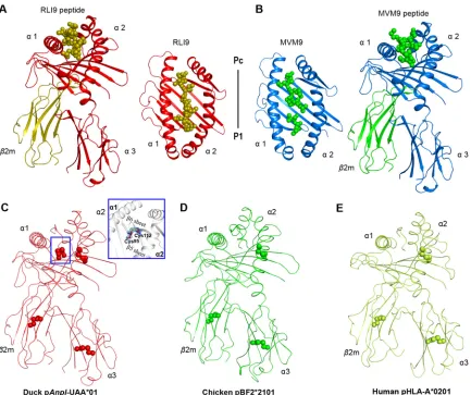
FIG 1Structural overview and additional disulfide bond of the pAnpl-UAA*01 complex. (A and B) The overall structures of pAnpl-UAA*01– RLI9 (A) and pAnpl-UAA*01–MVM9 (B). pAnpl-UAA*01 chains are shown with distinct colors: for pAnpl-UAA*01–RLI9, H in red and L in olive, and for pAnpl-UAA*01–MVM9, H in marine and L in green). Peptides are shown as spheres using the same colors with their light chains. (C to E) The additional disulfide bond of pAnpl-UAA*01 compared with chicken and human pMHC I complexes. Disulfide bonds are marked with spheres. The additional disulfide bond formed by Cys95-Cys112of pAnpl-UAA*01 is labeled in a box and amplified to show its position
and electronic density map. Both chicken pBF2*2101 (green, Protein Data Bank code 3BEV) and human pHLA-A*0201 (lemon, Protein Data Bank code 3PWN) have just three disulfide bonds distributed in the␣2 and␣3 domains and2m chains.
Structures of Duck MHC Class I Journal of Virology
on November 7, 2019 by guest
http://jvi.asm.org/
[image:3.585.43.476.69.432.2]virus (MDV) (32, 35, 36). Chickens expressing BF2
*
0401 (B4 haplotype) are susceptible
to MDV, and chickens expressing BF2
*
2101 (B21 haplotype) are resistant to MDV.
Crystal studies of BF2 structures have illustrated its resistance or susceptibility to MDV
(32). The MHC I complex presents viral peptides to CTLs, depending on the six (A to F)
pockets in its peptide binding groove (PBG). The MHC I polymorphisms determine the
distinct three-dimensional (3D) structure of the MHC I PBG, and each classical MHC I
molecule has a specific peptide binding motif. Crystal studies of B21 have shown that
it can remodel its pockets to accommodate different peptides based on its wide
binding groove and interplay of two charged residues (Arg
9and Asp
24); B21 could bind
multiple peptides from MDV and activate extensive T cell repertories to clear MDV (32).
In contrast, B4 has a narrow peptide binding groove and strong polar pockets that
restrict its peptide binding motif and the presented MDV peptides (33). Therefore, the
CTL response to MDV induced by B4 is not strong enough to prevent infection and
suggests that resistance is controlled by the type of MHC I and its 3D structure.
MHC I could also play a role in the susceptibility of viruses. For IAV, the higher
resistances of some species of duck indicate that their MHC I alleles would present more
IAV peptide epitopes than those of chickens (11, 14). Our previous data, as well as a
recently published study, confirmed the presence of two or more cysteines, which have
never been found in humans or other species, in the PBGs of many duck MHC I alleles
(30, 37). To date, little is known about the 3D structure and peptide presentation of
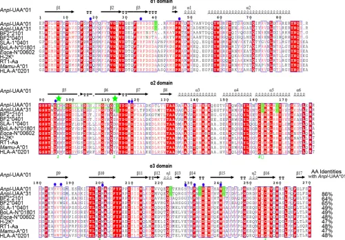
FIG 2Structure-based amino acid sequence alignment ofAnpl-UAA*01 and representatives of other crystallized MHC I molecules, with the secondary structure elements indicated. Black arrows above the alignment indicate-strands; cylinders denote␣-helices. Green numbers denote residues that form disulfide bonds. Cysteines at positions 95 and 112 are marked by green stars. Conserved residues are highlighted in red. Residues highlighted in green are species-specific amino acids that differ between duck and other animals. Solid blue dots indicate that the residues interact withAnpl-2m. The total amino acid (AA) identities between Anpl-UAA*01 and the listed MHC I molecules are given at end of each sequence.
Wu et al. Journal of Virology
on November 7, 2019 by guest
http://jvi.asm.org/
[image:4.585.42.543.75.424.2]duck MHC I. In this study, we determined two crystal structures of the dominantly
expressed duck MHC I (
Anpl
-UAA
*
01) molecules with two IAV peptides. The crystal
structures showed that
Anpl
-UAA
*
01 exhibits characteristics consistent with the duck
MHC I-peptide-

2m complex (pMHC I) architecture; however, we also identified a
disulfide bond in its PBG. Unlike the chicken MHC I, only B and F pockets of
Anpl
-UAA
*
01 are the primary anchor sites for peptide binding. The two pockets, whose
compositions are similar to those of the B and F pockets of HLA-A2, could
accommo-date diverse residues and make
Anpl
-UAA
*
01 an extensive peptide binding motif. By
screening the entire sequences of IAV strains, approximately 600 nonapeptides fit the
identified peptide binding motif of
Anpl
-UAA
*
01. Our study demonstrates that
Anpl
-UAA
*
01 could present more IAV epitope peptides, suggesting that ducks with this MHC
I haplotype may be protected from infection.
RESULTS
The
Anpl
-UAA
*
01 structure shows an unexposed disulfide bond in the PBG and
a large interface between heavy and light (H and L) chains.
Two trimer complexes
formed by
Anpl
-UAA
*
01, duck

2m (
Anpl
-

2m), and the MVM9 and RLI9 peptides were
crystallized in the P1211 space group at resolutions of 1.71 Å and 2.06 Å, respectively
(Table 1). Both of the complexes displayed a canonical pMHC I structure, in which the
peptide was located in the platform formed by the
␣
1 and
␣
2 domains of
Anpl
-UAA
*
01
above the
␣
3 domain and light-chain
Anpl
-

2m (Fig. 1A and B). The most remarkable
features of the p
Anpl
-UAA
*
01 complexes are the two disulfide bonds formed by 4
cysteines in the PBG (Fig. 1C). In other known pMHC I structures, there is only one
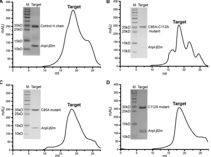
FIG 3Gel filtration and SDS-PAGE results of no-peptide refolding.Anpl-UAA*01 andAnpl-2m can co-refold in the absence of participation of the peptide, either the wild-typeAnpl-UAA*01 heavy chain or the C-A mutant. Different heavy chains andAnpl-2m are marked in the corresponding positions. Target, co-refolding products eluted via gel filtration. M, protein ladder. (A) Co-refolding results for the wild-type Anpl-UAA*01 andAnpl-2m. (B) Co-refolding results for the C95A-C112A double mutant heavy chain andAnpl-2m. (C) Co-refolding results for the C95A mutant heavy chain andAnpl-2m. (D) Co-refolding results for the C112A mutant heavy chain andAnpl-2m.
Structures of Duck MHC Class I Journal of Virology
on November 7, 2019 by guest
http://jvi.asm.org/
[image:5.585.43.478.74.397.2]disulfide bond formed by a pair of cysteines in their PBGs, such as in chicken pBF2
*
2101
(32) and human pHLA-A
*
0201 (38) (Fig. 1D and E). These two cysteines have been
found only in ducks (Fig. 2). This novel disulfide formed by Cys
95and Cys
112of
p
Anpl
-UAA
*
01 complexes connects the

5 and

6 sheets at the bottom of the PBG.
The no-peptide refolding product (only
Anpl
-UAA
*
01 and
Anpl
-

2m) was used as a
negative control to judge the refolding efficiencies of different peptides; however, a
peak (approximately 300 milli-absorbance units [mAU]) eluted at the position
repre-senting the p
Anpl
-UAA
*
01. The gel filtration and SDS-PAGE results showed that the
peak represented the complex of
Anpl
-UAA
*
01 and
Anpl
-

2m (Fig. 3), which is different
from results of studies on cattle, pig, chicken, and horse pMHC I refolding (33, 39–41).
This result could be due to the Cys
95-Cys
112disulfide bond of p
Anpl
-UAA
*
01. In order
to clarify the impact of this disulfide bond, Cys
95and Cys
112were mutated to alanine
(A), alone and together. However, the results for refolding of the mutated
Anpl
-UAA
*
01s
were similar to those for the wild type (Fig. 3), indicating that the additional disulfide
bond is not directly related to the stability of
Anpl
-UAA
*
01 without peptide binding.
The formation of a complex without the peptide may be due to the strong binding
affinity between H and L chains. There are a total of 18 hydrogen bonds and salt
bridges between
Anpl
-UAA
*
01 and
Anpl
-

2m (Fig. 4A), which is greater than the
numbers found in the pMHC I structures of chickens (13) (pBF2
*
0401, PDB code 4E0R)
and pigs (16) (pSLA-1
*
0401, PDB code 3QQ3). In addition, the size of the interface
between
Anpl
-UAA
*
01 and
Anpl
-

2m is 1,554.2 Å
2, which is the largest among the
known pMHC I structures of different species (Fig. 4B), including human (PDB code
3PWN), monkey (PDB code 1ZVS), mouse (PDB code 3TID), rat (PDB code 1ED3), swine
(PDB code 3QQ3), cattle (PDB code 3PWU), horse (PDB code 4ZUV), and chicken (PDB
code 3BEV) (33, 38–44). The interactions and interface area between
Anpl
-UAA
*
01 and
Anpl
-

2m likely contribute to the stability of the complex without binding peptides.
The distinct peptide conformations presented by the
Anpl
-UAA
*
01 molecule.
The conformations of IAV-MVM9 and IAV-RLI9 presented by
Anpl
-UAA
*
01 are distinct,
FIG 4Strong binding between duck H and L chains. (A) Interactions between H and L chains of pAnpl-UAA*01. pAnpl-UAA*01 is shown in red, with the interactive residues shown as sticks and colored according to the atom type (blue, N; red, O). The interactive residues on the L chain are colored white. The hydrogen bonds and salt bridges are shown as green dashed lines. All of the interactions and interactive residues are magnified within a blue dotted box and labeled. (B) Interface areas of the L chains of pAnpl-UAA*01 and pMHC I structures from the other known species. All the L chains are surface representations with different colors. The white areas indicate the interactive regions contacting H chains. Detailed interface areas are shown under each structure. The structures are as follows: chicken (BF2*2101, PDB code 3BEV), human (HLA-A2, PDB code 3PWN), bovine (BoLA-N*01801, PDB code 3PWU), swine (SLA-1*0401, PDB code 3QQ3), horse (Eqca-N*00602, PDB code 4ZUV), rat (RT1-Aa, PDB code 1ED3), macaque (Mamu-A*01, PDB code 1ZVS), and mouse (H-2 Kb, PDB code 3TID).
Wu et al. Journal of Virology
on November 7, 2019 by guest
http://jvi.asm.org/
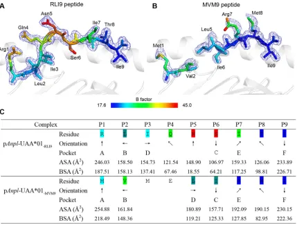
[image:6.585.45.485.68.307.2]especially in the middle region from the residue at position 3 (P3) to that at P6 (Fig. 5).
The IAV-RLI9 peptide is clear on the electronic density map and adopts an “M” overall
conformation, which is common to other pMHC I structures. The B factors of the
IAV-RLI9 middle part are relatively higher than at the N and C termini, indicating that
P4 to P6 are more flexible than other residues (Fig. 5A). In IAV-MVM9, the electronic
density map is missing at P3 and P4, which means that these two residues are quite
flexible (Fig. 5B). The side chain orientations of P5 are obviously different between the
IAV-MVM9 and IAV-RLI9 peptides. In IAV-MVM9, the side chain of P5 stretches to the
␣
2
helix but in RLI9, its side chain stretches upward and most parts are exposed from the
PBG (Fig. 5C).
The conformations of the MVM9 and RLI9 peptides were significantly distinct when
the two p
Anpl
-UAA
*
01 structures were superimposed (Fig. 6A and B); their deviation is
focused mainly at the center, where the distance between the C
␣
atoms of MVM9 and
RLI9 P5 residues can reach 4.4 Å. The N and C termini of the two peptides matched well.
Structural analysis showed that the N termini of the MVM9 and RLI9 peptides (P1 and
P2 residues) form the same hydrogen bonds with the PBG (Fig. 6C). From the P3 to P7
residues, there are no hydrogen bonds between RLI9 and the PBG. Although the P3 and
P4 residues are missing in MVM9, three hydrogen bonds were found between P5, P7,
and the PBG (Fig. 6D). These distinct peptide conformations of this part should be
related to the differences in hydrogen bonds found here. In the C termini, the hydrogen
bonds between the main chain of peptides and the PBG are the same; the only
FIG 5Divergent presentations of RLI9/MVM9 peptides presented byAnpl-UAA*01. (A and B) Electron densities and overall conformations of the structurally defined peptides RLI9 and MVM9. Simulated annealing omit maps (CNS) calculated for the two peptides are shown in blue at a contour of 1.5, and the coloration of the two peptides is according to the isotropic B factors. (C) General side chain orientations and the different interfacing areas of RLI9/MVM9 presented byAnpl-UAA*01 PBG, as viewed in profile from the peptide N terminus toward the C terminus. Black arrows indicate the directions in which the residues point: up is toward the T cell receptor, down is toward the floor of the peptide binding groove, left is toward the␣1 helix domain, and right is toward the␣2 helix domain. Pockets accommodating each residue are listed under the corresponding anchors within the peptide binding groove. ASA, accessible surface area of each residue; BSA, buried surface areas of the residues.
Structures of Duck MHC Class I Journal of Virology
on November 7, 2019 by guest
http://jvi.asm.org/
[image:7.585.46.475.70.395.2]difference is that the Lys
142of p
Anpl
-UAA
*
01 forms 3 hydrogen bonds with the side
chains of the P8 and P9 residues in the RLI9 peptide but does not form a bond with the
MVM9 peptide (Fig. 6E).
These data suggest that the conformation of the middle section of the peptide
presented by
Anpl
-UAA
*
01 is flexible and is determined by the peptide’s specific amino
acid sequence.
Anpl
-UAA
*
01 selects peptides by relying on the B and F pockets.
The
compo-sitions of the six pockets of
Anpl
-UAA
*
01 are shown in Fig. 7, and the interactions
between peptides and these pockets are listed in Table 2. The RLI9 peptide was
selected to illustrate the interactions with the six pockets in Fig. 6 because of its
completeness.
The A pocket of
Anpl
-UAA
*
01, composed of Leu
5, Tyr
7, Tyr
58, Glu
62, Tyr
156, Thr
160,
Cys
161, Trp
164, and Tyr
168, fixes P1-Arg by hydrogen bonds and strong van der Waals
forces (VDWs) (Fig. 7A; Table 2). As in the known pMHC I structures, P1-Arg forms the
hydrogen bonds with the A pocket by its main-chain atoms, and its side chain stretches
upward out of the A pocket (45, 46). Therefore, although the binding between the A
pocket and P1 residue is strong, the A pocket dose not play a restrictive role in the PBG
of
Anpl
-UAA
*
01.
The B pocket is a primary anchor site and plays a restrictive role in peptide binding.
The B pocket of
Anpl
-UAA
*
01 accommodates P2-Leu (Fig. 7B) The charged Glu
62, which
is on the top of the B pocket, can form a hydrogen bond with the main chain of P2-Leu.
FIG 6Comparisons of dissimilar conformations and interactions of RLI9/MVM9 peptides. (A and B) Superposition of RLI9 (olive) and MVM9 (green) presented byAnpl-UAA*01. To conveniently observe the different conformations of the two peptides, the side chains of residues are presented. Both the side (A) and top (B) views of the peptide alignments demonstrate that most conformational distinctions are located in the central region of the peptides, with the farthest distance between the backbone carbon atoms on P4 of the two peptides marked using red dashes. (C to E) Hydrogen bond comparisons of RLI9 and MVM9 peptides interacting with their PBGs. The interacting residues on PBG are shown (pAnpl-UAA*01– RLI9, red; pAnpl-UAA*01–MVM9, blue), and the hydrogen bonds are represented as dashed lines with the corresponding colors. (C) Similar interactions at the P1 and P2 positions of RLI9/MVM9 peptides with the residues in the A pocket ofAnpl-UAA*01. (D) Different conformations and hydrogen bonds between the central region of peptides andAnpl-UAA*01. Three hydrogen bonds are formed in pAnpl-UAA*01–MVM9, and no hydrogen bonds are found in pAnpl-UAA*01–RLI9. (E) Interactions between the C-terminal residues of the RLI9/MVM9 peptides and their PBGs. Three more hydrogen bonds of RLI9 with the Lys142residues ofAnpl-UAA*01 are observed.
Wu et al. Journal of Virology
on November 7, 2019 by guest
http://jvi.asm.org/
[image:8.585.47.497.69.368.2]The side chain of the P2 residue inserts into the B pocket and is fixed by the VDWs
provided by the surrounding residues (Table 2).
The C, D, and E pockets usually connect with residues in the middle part of the
binding peptides. The amino acid compositions of these three pockets of
Anpl
-UAA
*
01
are shown in Fig. 7C to E. The C and D pockets can interact with the side chains of the
P3 and P6 residues, respectively, but the E pocket can only interact with the main chain
of the P7 residue, as the orientation of the P7 side chain faces upward (Table 2).
The additional disulfide bond formed by Cys
95and Cys
112is in the C pocket of
Anpl
-UAA
*
01. The impact of this disulfide bond on peptide binding was checked by the
refolding of MVM9 and RLI9 peptides with the C95A, C112A, and C95A-C112A double
mutant heavy chains. We found that that the mutant H chains can still form a complex
with peptides, but the refolding is worse than that of the wild-type H chain, especially
for the C112A mutant H chain (Fig. 8A and B). The reduced refolding efficiencies of the
mutants indicated that the disulfide bound in the C pocket helps with the peptide
binding of
Anpl
-UAA
*
01; however, the distances between the peptide residues and
disulfide bond are over 5.0 Å, which means that their direct interactions are negligible
(Fig. 6C). Previous studies have suggested that Cys
95and Cys
112do not meet the
preferred geometry to form the disulfide bond (30), and our p
Anpl
-UAA
*
01 structures
show that the distance between Cys
95and Cys
112is less than that for the corresponding
positions in HLA-A2 and B21 (Fig. 8C). To some extent, the additional disulfide bond of
Anpl
-UAA
*
01 alters the bottom of the C pocket and strengthens the stability. Improved
peptide binding efficiency may strengthen the stability of the C pocket with the
additional disulfide bound.
The F pocket is the most important anchor site at the C terminus of PBG. The F
pocket of
Anpl
-UAA
*
01 is composed of Asn
76, Thr
79, Ala
80, Arg
83, Tyr
84, Trp
93, Phe
119,
and Ala
135, and it has a strong binding affinity with P9-I (Fig. 7F). The main chain of P9-I
can form 2 salt bridges with Arg
83and Lys
142and 2 hydrogen bonds with Asn
76and
FIG 7Composition of the pockets ofAnpl-UAA*01. Pockets are shown as surface representations in light pink. Residues comprising these pockets (light pink) and the bound peptide RLI9 (C, olive; N, blue; O, red) are labeled. The hydrogen bonds between RLI9 and pockets are shown as red dashed lines. (A) Pocket A with the P1-Arg. (B) Pocket B with the P2-Leu. (C) Pocket C with the P6-Ser. The additional disulfide bond are shown as thick sticks and colored by atom type (C, red; N, blue; O, red; S, yellow). (D) Pocket D with the P3-Ile. (E) Pocket E with the P7-Ile. (F) Pocket F with the PC-Ile.
Structures of Duck MHC Class I Journal of Virology
on November 7, 2019 by guest
http://jvi.asm.org/
[image:9.585.43.477.73.347.2]Thr
139in the F pocket. The side chain of P9-I inserts into the F pocket and is fixed by
the strong hydrophobic forces (Table 2).
In order to determine the primary anchor residues of
Anpl
-UAA
*
01-presenting
peptides and the vital restriction pockets for peptide binding, the RLI9 peptide was
mutated by alanine scanning and circular dichroism (CD) spectral analysis was used to
test the stabilities of
Anpl
-UAA
*
01 complexes with these mutant peptides (Fig. 9). The
wild-type RLI9 peptide was used as a control, and its midpoint transition temperature
(
T
m) value was 43.3°C. Among all of the alanine mutant peptides, only the
T
mvalues of
the P2-Ala and P9-Ala mutant peptides were significantly lower than that of the
wild-type RLI9 peptide, indicating that the side chains of P2 and P9 play key roles in
RLI9 peptide binding and that these two residues are the primary anchor residues. The
pockets (B and F) at the two termini of the
Anpl
-UAA
*
01 PBG anchor the peptides and
determine the peptide binding motif of
Anpl
-UAA
*
01.
[image:10.585.41.543.81.434.2]The peptide binding motif of
Anpl
-UAA
*
01 is most similar to that of HLA-A2.
The peptide binding manner of p
Anpl
-UAA
*
01 is similar to those of some mammalian
pMHCs but not similar to that of chicken pMHC I; only the B and F pockets are the key
anchor caves, similar to what is observed in human MHC I (also named human
lymphocyte antigen [HLA]). By comparison with the solved structures in the Protein
Data Bank (PDB), the peptide conformation and binding manner of p
Anpl
-UAA
*
01 were
found to be most similar to those of HLA-A2 (Fig. 10). In the B pockets of
Anpl
-UAA
*
01
and HLA-A2, the side chains of P2-Leu residues have similar orientations and are fixed
in a similar way by the residues at the same positions in the two pockets (Fig. 10A). A
similar situation was also found in the two F pockets, where the hydrogen bonds fixing
TABLE 2Interactions between the RLI9/MVM9 peptide andAnpl-UAA*01
Complex
Hydrogen bonds and salt bridges
van der Waals contact residuesa
Peptide Heavy chain
Residue Atom Residue Atom
pAnpl-UAA*01–RLI9 P1-Arg N Tyr168 OH Leu5, Tyr7, Tyr58, Glu62, Tyr156, Thr160, Trp164,
Tyr168 (59)
N Tyr7 OH
O Tyr156 OH
P2-Leu N Glu62 OE1 Tyr7, Tyr9, Thr24, Met43, Glu62, Thr65, Ser66,
Tyr156 (46)
P3-Ile Thr65, His97, Trp153, Tyr156 (42)
P4-Gln Arg61, Thr65 (18)
P5-Asn (0)
P6-Ser Asn69, Ile72 (5)
P7-Ile Trp143, Lys142, Glu146, Val149, Ile72 (21)
P8-Thr OG1 Lys142 NZ Ile72, Lys142, Asn76, Val75, Trp143 (31)
O Lys142 NZ
O Trp143 NE1
P9-Ile N Asn76 OD1 Lys142, Trp143, Asn76, Thr139, Phe119, Thr79,
Ala80, Arg83, Trp93 (69)
OXT Arg83 NH2 (S)
OXT Thr139 OG1
O Lys142 NZ (S)
Total 11 291
pAnpl-UAA*01–MVM9 P1-Met N Tyr7 OH Leu5, Tyr7, Tyr58, Arg61, Glu62, Tyr156, Trp164,
Tyr168 (56)
O Tyr156 OH
N Tyr168 OH
P2-Val N Glu62 OE1 Tyr7, Tyr9, Glu62, Thr65, Asn69, His97, Tyr156 (37)
P5-Leu O Trp153 NE1 Gln152, Trp153, Tyr156 (33)
P6-Ile Tyr9, Asn69, Ile72, Phe73, Trp153 (26)
P7-Arg O Asn76 ND2 Ile72, Asn76, Trp143, Glu146, Val149, Gln152 (31)
NH2 Glu146 OE2 (S)
P8-Met O Trp143 NE1 Ile72, Val75, Asn76, Lys142, Trp143 (22)
P9-Ile N Asn76 OD1 Asn76, Thr79, Ala80, Arg83, Trp93, Phe119,
Thr139, Lys142, Trp143 (70)
OXT Arg83 NH2 (S)
OXT Thr139 OG1
Total 11 275
aNumbers in parentheses are the amounts of van der Waals force.
Wu et al. Journal of Virology
on November 7, 2019 by guest
http://jvi.asm.org/
P9-I are almost the same (Fig. 10B). The peptide presentations and pocket compositions
make the peptide conformations of p
Anpl
-UAA
*
01 and pHLA-A2 almost identical (Fig.
10C and D).
Previous studies have shown that the B pocket of HLA-A2 prefers hydrophobic
anchor residues, such as I, L, V, A, and F, and can also accommodate the same polar
residues, such as T. The most frequently occurring anchor residues in the F pocket of
HLA-A2 are I, V, and L. The two peptides presented by
Anpl
-UAA
*
01, MVM9 and RLI9,
perfectly fit the peptide binding motif of HLA-A2. Eighteen additional peptides from
IAV and 11 mutant peptides were used to verify the peptide binding motif of
Anpl
-UAA
*
01 (Table 3). The results confirmed that the peptide binding motifs of these two
MHC I molecules from different species are mostly overlapping (Fig. 10E and F). In brief,
the nonapeptide binding motif of
Anpl
-UAA
*
01 is x-(A, V, I, L, M, T, S, or F)-x-x-x-x-x-x-(A,
V, I, L, M, or F).
Many IAV peptides match the motif of p
Anpl
-UAA
*
01.
Using the motif of
Anpl
-UAA
*
01 that we identified, the proteomes of several IAV strains were screened to
identify the epitope peptides that could be presented by
Anpl
-UAA
*
01, including the
H1N1, H3N2, H3N8, H4N6, H5N1, H7N9, and H9N2 subtypes (47–53). There were
FIG 8Mutants ofAnpl-UAA*01 influence thein vitrorefolding efficiency with L chain and RLI9/MVM9 peptides. The refolding products were analyzed by chromatography on a Superdex 200 10/300 GL column (GE Healthcare). Curves in different colors indicate different H chains. (A) Refolding results for the wild-typeAnpl-UAA*01 and three mutants with the MVM9 peptide. (B) Refolding results with the RLI9 peptide. (C) The bottomsheet ofAnpl-UAA*01 is more compact than those in B21 and HLA-A2. All the structures are in ribbon models. The distances between C95 and C112 inAnpl-UAA*01 and their corresponding positions in B21 and HLA-A2 are labeled by dashed lines.
Structures of Duck MHC Class I Journal of Virology
on November 7, 2019 by guest
http://jvi.asm.org/
[image:11.585.43.498.70.460.2]approximately 600 candidate nonapeptides from different IAV strains matching the
peptide binding motif of
Anpl
-UAA
*
01 (Fig. 11). Most selected peptides also fit the
motif of HLA-A2, and 11 nonapeptides were proven to activate HLA-A2-restricted CTL
responses efficiently according to IEDB data (
http://www.iedb.org/
) (54). The
in vitro
refolding results for 31 peptides confirmed that the predicted IAV epitopes could be
bound by
Anpl
-UAA
*
01 (Table 3).
Based on the motifs of chicken B4 and B21, the longer peptides from IAV strains
were also screened. There are fewer than 20 octapeptides for B4 and 24 to 27
decapeptides for B21 (Fig. 11), which are much less than the numbers of peptides
found in
Anpl
-UAA
*
01.
DISCUSSION
The most distinguishable feature of
Anpl
-UAA
*
01 was the additional disulfide bond
in the PBG. The mutant experiments showed that this disulfide bond located in the C
pocket can increase the efficiency of peptide binding to p
Anpl
-UAA
*
01. Although all of
the mutants could still form complexes with the RLI9/MVM9 peptide and

2m, their
refolding efficiencies were significantly lower than that of the wild type (Fig. 8A and B).
Without the peptide, this disulfide bond did not increase the refolding production of
FIG 9P2 and P9 of the peptide are critical for binding toAnpl-UAA*01. The denaturation and elution curves of the complexes with the different peptides are indicated in different colors. (A) Thermostabilities ofAnpl-UAA*01 with the RLI9 peptide and its substitutions (residues at each position in RLI9 are replaced with alanine). Circular dichroism spectropolarimetry was utilized to assess the thermosta-bilities of purified pAnpl-UAA*01 complexes. Denaturation was monitored at 218 nm as the temperature was ramped up from 25 to 75°C at 1°C/min. Shown here are the data for fitting to the denaturation curves using the Origin 9.1 program (OriginLab).TheTms of different
peptides are indicated by the gray line at 50% fraction unfolded. (B and C) Peptide-induced assembly and stabilization assay of Anpl-UAA*01 andAnpl-2m with RLI9 and its mutants byin vitrorefolding. (B) Gel filtration chromatograms of the refolded products obtained using a Superdex 200 10/300 GL column (GE Healthcare). The aggregated H chain, the correctly refolded pAnpl-UAA*01 complex (⬃45 kDa), and the extra2m are indicated. The refolding efficiencies are represented by the heights of pAnpl-UAA*01 complex peaks. A higher peak indicates a better efficiency of the peptide to help the MHC renature. (C) Results of further stabilization assays of the refolded products tested by anion exchange. A higher peak here also indicates better stability. With peptides P2-A and P9-A, the refolded complex proteins dissociated at the corresponding eluting NaCl concentration (9% to 11%), implying poor stabilities.
Wu et al. Journal of Virology
on November 7, 2019 by guest
http://jvi.asm.org/
[image:12.585.46.471.69.394.2]the complex of
Anpl
-UAA
*
01 and

2m (Fig. 3). These data indicate that this additional
disulfide bond can improve peptide binding to
Anpl
-UAA
*
01 and lead to the increased
formation of a trimer complex; however, no significant contacts between the presented
peptide and disulfide bond were identified. We also discovered that the additional
disulfide bond makes the bottom

sheet of the C pocket of
Anpl
-UAA
*
01 more
compact than in other MHC I molecules. The refolding effect of the C112A mutant
indicated that the free side chain of Cys
95could disturb peptide binding in the C pocket
region (Fig. 8A and B). Therefore, we hypothesize that this additional disulfide bond
provides a more stable C pocket, which helps with peptide presentation of
Anpl
-UAA
*
01.
Another characteristic of the
Anpl
-UAA
*
01 structure is the intensive interaction
FIG 10Alignments of B pocket, F pocket, and binding peptides indicate similar peptide binding motifs of Anpl-UAA*01 with HLA-A*0201 (PDB code 3PWN). Residues making up these pockets and binding peptides are as follows: for pAnpl-UAA*01, C, red; N, blue; O, red; and for pHLA-A*0201, C, lemon; N, blue; O, red). (A and B) B and F pocket alignments ofAnpl-UAA*01 with HLA-A*0201. The same compositions of P2-Leu and P9-Ile in both RLI9/HuDG2Lpeptides are shown using the same color as their H chains. Hydrogen bonds formed by P2/P9 and
their H chains are represented as dashed lines with each corresponding color. (C and D) Side and top views of the RLI9 peptide alignment with HuDG2Lpeptide presented by HLA-A*02. Both of the peptides’ main chains are shown
as bold sticks. Shared P2-Leu and P9-Ile in RLI9/HuDG2L peptides are colored black in their sequences in the upper left corner. (E and F) Residues fitting the B and F pockets ofAnpl-UAA*01 and HLA-A*0201 can mostly overlap. Residues within the solid red circle are motifs for Anpl-UAA*01, and those within the circle are motifs for HLA-A*0201.
Structures of Duck MHC Class I Journal of Virology
on November 7, 2019 by guest
http://jvi.asm.org/
[image:13.585.43.405.68.497.2]between its H and L chains. In comparison with other known pMHC I structures,
p
Anpl
-UAA
*
01 has the greatest interface area and many interchain bonds (Fig. 5). The
interface area in ducks and chickens is significantly larger than that in mammals.
Compared with BF2
*
2101 (32), p
Anpl
-UAA
*
01 had a 100-Å
2-larger interface area and 4
more hydrogen bonds; this relatively stronger interaction could lead to the formation
of complexes containing only
Anpl
-UAA
*
01 and

2m through
in vitro
refolding, which
has never been identified in pMHC I structures of other species (33, 39–41).
Anpl
-UAA
*
01 is similar to human HLA in peptide presentation (but not to chicken
BF2), although the amino acid identities among
Anpl
-UAA
*
01 and BF2 molecules are
much higher than those between
Anpl
-UAA
*
01 and HLA (
⬎
60% versus
⬍
50%,
respec-tively). There are only two primary anchor pockets in p
Anpl
-UAA
*
01, the B and F
pockets, which is common in HLA molecules (Fig. 10A and B). Using structural analysis
and mutant peptide refolding tests, we found that the peptide binding motif of
p
Anpl
-UAA
*
01 overlaps with HLA-A
*
0201 to a large degree. The B pocket of
Anpl
-UAA
*
01 can accommodate extensive uncharged residues, and the F pocket can
ac-commodate multiple hydrophobic residues (Fig. 10E). However, BF2 exhibits
peptide-presenting strategies that differ from those of HLA. B4 has a narrow and highly charged
PBG, which limits the binding peptides that must fit its B, C, and F pockets together (33).
B21 has a large central cavity and flexible Arg9, which make its binding motif
promis-cuous with three anchor residues (32). The moderate limits of the peptide binding motif
suggest that ducks can accommodate more peptides from pathogens than chickens.
[image:14.585.46.541.83.404.2]The role of MHC I mediating the CTL response in clearance of IAV has been
confirmed in human and mouse. Although substantial data on anti-IAV CTL responses
in duck and chicken are scarce, some evidence indicates that duck MHC genes respond
TABLE 3Predicted peptides used in this study and their binding toAnpl-MHC I, evaluated byin vitrorefolding
Name Derived protein Position Sequence % randoma
Stability with Anpl-MHC Ib
IAY9 Nucleocapsid protein (IAV) 45–53 IAYERMCNI 0.185 ⫹⫹
RTS9 Nucleocapsid protein (IAV) 22–30 RTSDMRTEI 0.168 ⫹⫹
MVM9 Nucleocapsid protein (IAV) 17–25 MVMELIRMI 0.172 ⫹⫹
RLI9 Nucleocapsid protein (IAV) 55–63 RLIQNSITI 0.245 ⫹⫹
WMA9 Nucleocapsid protein (IAV) 68–76 WMACHSAAF 0.213 ⫹⫹
IFL9 Nucleocapsid protein (IAV) 18–26 IFLARSALI 0.125 ⫹⫹
VSG9 Nucleocapsid protein (IAV) 28–36 VSGIGRFYI 0.134 ⫹⫹
ATA9 Nucleocapsid protein (IAV) 53–61 ATAGLTHLM 0.200 ⫹/⫺
SLAP5 Hemagglutinin (IAV) 25–33 TSADQQSLY 0.100 ⫹/⫺
SLAP9 Polymerase PA (IAV) 6–14 GTFDLGGLY 0.041 ⫹/⫺
AI-4 Hemagglutinin (IAV) 442–450 VAMENQHTI 0.387 ⫹⫹
EN5 Gag protein (IAV) 226–234 MTARFIRGL 0.130 ⫹⫹
KMN-P9I Hemagglutinin (IAV) 402–410 KMNTQFIAI 0.207 ⫹⫹
NS1-VKN Nonstructural protein 1 (IAV) 174–182 VKNAVGVLI 0.054 ⫹/⫺
PA-ENK Polymerase PA (IAV) 613–621 ENKSETWPI 0.035 ⫹/⫺
PA-KTN Polymerase PA (IAV) 497–505 KTNLYGFII 0.150 ⫹⫹
HA-ITN Hemagglutinin (IAV) 393–401 ITNKVNSVI 0.162 ⫹⫹
HA-YIN Hemagglutinin (IAV) 182–190 YINDKGKEV 0.073 ⫹/⫺
PA-RRN Polymerase PA (IAV) 442–450 RRNYFTAEV 0.059 ⫹/⫺
PB1-KM Polymerase PB1 (IAV) 531–539 KNNMINNDL 0.046 ⫹/⫺
RLI9-P1A Mutant RLI9 ALIQNSITI ⫹⫹
RLI9-P2A Mutant RLI9 RAIQNSITI ⫹/⫺
RLI9-P3A Mutant RLI9 RLAQNSITI ⫹⫹
RLI9-P4A Mutant RLI9 RLIANSITI ⫹⫹
RLI9-P5A Mutant RLI9 RLIQASITI ⫹⫹
RLI9-P6A Mutant RLI9 RLIQNAITI ⫹⫹
RLI9-P7A Mutant RLI9 RLIQNSATI ⫹⫹
RLI9-P8A Mutant RLI9 RLIQNSIAI ⫹⫹
RLI9-P9A Mutant RLI9 RLIQNSITA ⫹/⫺
RLI9-P9V Mutant RLI9 RLIQNSITV ⫹/⫺
RLI9-P9L Mutant RLI9 RLIQNSITL ⫹⫹
aBase value for estimating the binding affinities of peptides with the NetMHCpan 3.0 server (http://www.cbs.dtu.dk/services/NetMHCpan/): rank threshold for strongly
binding peptides, 0.100; rank threshold for weakly binding peptides, 1.000.
b⫹⫹, peptide binds strongly and can tolerate anion-exchange chromatography;⫹/⫺, peptide bindsAnpl-MHC I but cannot tolerate anion-exchange chromatography.
Wu et al. Journal of Virology
on November 7, 2019 by guest
http://jvi.asm.org/
FIG 11Peptide predictions from different influenza virus subtypes according to the distinct binding motifs ofAnpl-UAA*01, BF2*0401, and BF2*2101. Genome-wide scanning results of peptides matching the motifs ofAnpl-UAA*01 (A), BF2*0401 (B), and BF2*2101 (C) are as follows:Anpl-UAA*01, x-(A, V, I, L,
(Continued on next page)
Structures of Duck MHC Class I Journal of Virology
on November 7, 2019 by guest
http://jvi.asm.org/
[image:15.585.73.344.66.695.2]to infection with IAV (13, 55). The studies showed that duck MHC locus genes (MHC I/II
and TAP) are overexpressed after infection with IAV, especially the duck MHC I gene,
and under the condition of HP-IAV infection its expression could be increased by about
1,000 times in the lung (13). Unlike mammals, both ducks and chickens have a “minimal
MHC” region with a limited set of genes and express only one dominant MHC I allele.
The “minimal MHC” in chickens is critical to defense against a particular pathogen
because it is completely dependent on whether or not it can load peptides from that
pathogen. The best illustrated example is that chickens of the BF2
*
2101 genotype
could defend against MDV, while chickens of the BF2
*
0401 genotype could not (32, 33,
56). The difference in duck versus chicken responses to IAV might also relate to the
different peptide loading abilities of duck and chicken MHC I. Approximately 600
nonapeptides from different IAV strains matching the motif of p
Anpl
-UAA
*
01 were
identified in this study, which is much higher than the numbers of BF2
*
2101 and
BF2
*
0401 from chicken (Fig. 11; see Data Set S1 in the supplemental material).
There-fore, the duck
Anpl
-UAA
*
01 should exhibit stronger resistance to IAV strains than
chicken BF2
*
2101 and BF2
*
0401 (57, 58). In addition, the
Anpl
-UAA
*
01 structure
showed that it can present IAV peptides in distinct conformations, which is believed to
be useful for activating the T cell repertoire and inducing stronger CTL responses. Our
study illustrates the structural basis of duck MHC I and provides a novel approach for
explaining why ducks are more resistant to HP-IAV.
MATERIALS AND METHODS
Prediction and synthesis of IAV-derived peptides.All nonamer peptides that potentially bound to Anpl-UAA*01 were predicted using the NetMHCpan 3.0 server (http://www.cbs.dtu.dk/services/ NetMHCpan/) (59, 60). The 31 peptides used in this study (Table 3) were synthesized and purified to 90% by reverse-phase high-performance liquid chromatography (HPLC) and mass spectrometry (SciLight Biotechnology). These peptides were stored in lyophilized aliquots at⫺80°C after synthesis and dissolved in dimethyl sulfoxide (DMSO) before use.
Protein preparation. DNA fragments encoding extracellular domains ofAnpl-UAA*01 (GenBank accession no.AB115245, residues 1 to 270 of the mature protein with EcoRI and HindIII restriction sites) (37) andAnpl-2-microglobulin (Anpl-2m) (GenBank accession no.AB246408, residues 1 to 101 of the mature protein) were synthesized by Shanghai Invitrogen Life Technologies and then cloned into pET21a(⫹) vectors (Novagen) and expressed inEscherichia coliBL21(DE3). The recombinantAnpl-UAA*01 and2m were expressed in inclusion bodies and purified as previously described (61, 62). Finally, the Anpl-UAA*01 heavy chain andAnpl-2m bodies were separately dissolved in 6 M guanidinium chloride buffer to a protein concentration of 30 mg/ml.
Assembly of the pAnpl-UAA*01 complex.To assemble the pAnpl-UAA*01 complex, the peptide, Anpl-UAA*01, andAnpl-2m inclusion bodies were refolded (in a 1:1:1 molar ratio) according to the gradual dilution method that we described previously (39, 63). After a 24-h refolding step at 277 K, the remaining soluble portion of the complex was concentrated and purified using a Superdex 200 16/60 column (GE Healthcare), followed by Resource Q anion-exchange chromatography (GE Healthcare). Purified proteins were buffer exchanged with 10 mM Tris-HCl and 50 mM NaCl at a pH of 8.0.
Crystallization and data collection.The purified pAnpl-UAA*01 complex was ultimately concen-trated to 10 mg/ml. After mixing with reservoir buffer at a 1:1 ratio, the purified protein was crystallized using the sitting-drop vapor diffusion technique at 277 K. Index, Crystal Screen I/II, and Crystal Screen Cryo I/II kits (Hampton Research, Riverside, CA) were used to screen for optimal crystal growth conditions. After several days, crystals (pAnpl-UAA*01–MVM9 and pAnpl-UAA*01–RLI9) were observed with solutions NO.7 from the Crystal Screen Cryo II kit (8% polyethylene glycol 1000, 8% [wt/vol] polyethylene glycol 8000, and 20% [vol/vol] glycerol) and NO.43 from the Crystal Screen Cryo I kit (24% [wt/vol] polyethylene glycol 1500 and 20% [vol/vol] glycerol), respectively. Diffraction data for pAnpl-UAA*01 crystals were collected to resolutions of 1.71 Å (pAnpl-UAA*01–MVM9) and 2.06 Å (pAnpl-UAA*01–RLI9) at the Shanghai Synchrotron Radiation Facility (SSRF) using beamline BL17U at a wavelength of 1.5418 Å (Shanghai, China) (64).The crystals were first soaked in reservoir solution containing 25% glycerol as a cryoprotectant and were then flash-cooled in a stream of gaseous nitrogen at 100 K (65). The collected
FIG 11Legend (Continued)
M, T, S, or F)-x-x-x-x-x-x-(A, V, I, L, M, or F); BF2*0401: x-(D, E)-x-x-(D, E)-x-x-E; BF2*2101, x-(H, K, or R)-x-x-x-x-x-(E or D)-x-(A V, L, I, F, or W), x-(E or D)-x-x-x-x-x-L-x-(A, V, L, I, F, or W). The numbers of peptides derived from the PB1, PB2, PA, HA, NA, M, NP, and NS proteins of each IAV subtype are marked, as well as the total numbers. Vacuum electrostatic surface potential presentations (red, negative; blue, positive; gray, neutral) and predominant lengths of binding peptides of PBGs inAnpl-UAA*01, BF2*0401, and BF2*2101 are shown below. The peptides are shown as ribbons (olive) with their lengths labeled: nonamers forAnpl-UAA*01, octamers for B4, and 10-mers for B21.
Wu et al. Journal of Virology
on November 7, 2019 by guest
http://jvi.asm.org/
intensities were indexed, integrated, corrected for absorption, scaled, and merged using the HKL2000 package (66).
Structure determination and refinement.The crystals of pAnpl-UAA*01–MVM9/RLI9 all belong to the P1211 space group, and their structures were solved by molecular replacement using Molrep and Phaser in the CCP4 package, with the chicken BF2*0401 structure (PDB code 4E0R) as the search model (67–69). Extensive model building was performed by hand with COOT (70), and restrained refinement was performed using REFMAC5. Additional rounds of refinement were performed using the Phenix refine program implemented in the PHENIX package (71) together with isotropic atomic displace-ment parameter (ADP) refinedisplace-ment and bulk solvent modeling. The stereochemical quality of the final model was assessed with the PROCHECK program (72). Detailed information about collection and refinement is shown in Table 1.
Structural analysis and generation of illustrations.Peptide-contacting residues were identified using the program CONTACT and were defined as residues containing an atom within 3.3 Å of the target partner (69). Structural illustrations and electron density-related figures were generated using the PyMOL molecular graphics system (http://www.pymol.org/). Solvent-accessible surface areas and the B factor were calculated with CCP4.
Preparation of Cys-to-Ala mutants of pAnplMHC I.To investigate the function of the additional disulfide bond inAnpl-UAA*01, Cys95and Cys112were mutated to Ala by overlap PCR (the primers used
for Cys95-to-Ala mutation were 5=-GGCAGGCGATGCATGGCTGTG-3= and 5=-GCCATGCATCGCCTGCC
ATG-3=and those for Cys112-to-Ala mutation were 5=-CAACAAGCGGGCTATGATGGG-3=and 5=-CCCATCA
TAGCCCGCTTGTTG-3=, where the underlined sequences mutated the codon encoding Ala). These mutants were inserted into the pET21a vector and expressed in BL21(DE3) cells. The mutants were termedAnpl-UAA*01-C95A,Anpl-UAA*01-C112A, andAnpl-UAA*01-C95A-C112A, respectively. Recombi-nant mutants were expressed as inclusion bodies and further purified as described above. The mutant inclusion bodies were refolded withAnpl-2m using thein vitrogradual dilution method as described above (61, 62). In addition, all of the mutant pAnpl-UAA*01 complexes formed by refolding were further purified by gel filtration and anion-exchange chromatography as described above (39, 63).
CD spectra and thermal unfolding.Circular dichroism (CD) experiments for the pAnpl-UAA*01 with parental RLI9 or mutant peptides were performed on a Jasco J-810 spectropolarimeter equipped with a water-circulating cell holder. The CD spectra were collected at a protein concentration of 8 m⌴in pH 8.0 Tris buffer (20 mM Tris and 50 mM NaCl), using a 1-mm-optical-path-length cuvette with ellipticity at 218 nm. Thermal denaturation curves were determined as the temperature was raised from 25 to 80°C at a linear rate of 1°C/min. The temperature of the sample solution was directly measured with a thermistor. The fraction of unfolded protein was calculated from the mean residue ellipticity () using the standard method. The unfolded fraction (%) was expressed as (⫺N)/(U⫺N), whereN andU are the mean residue ellipticity values in the fully folded and fully unfolded states, respectively. The midpoint transition temperature (Tm) was determined by fitting data to the denaturation curves using the Origin 9.1 program
(OriginLab) (73).
Accession number(s). The coordinates and structure factors for pAnpl-UAA*01–RLI9 and pAnpl -UAA*01–MVM9 have been deposited in the Protein Data Bank (http://www.rcsb.org/pdb/home/home .do) under accession numbers5GJXand5GJY, respectively.
SUPPLEMENTAL MATERIAL
Supplemental material for this article may be found at
https://doi.org/10.1128/JVI
.02511-16
.
SUPPLEMENTAL FILE 1,
XLSX file, 0.1 MB.
ACKNOWLEDGMENTS
This work was supported financially by grants from the National Basic Research
Program of China (973 Program) (2013CB835302,
http://program.most.gov.cn
), the 13th
Five-Year National Key Research project (2016YFD0500106,
http://program.most.gov
.cn/
), and the National Natural Science Foundation of China (NSFC) (31572493,
http://
www.nsfc.gov.cn
).
The funders had no role in study design, data collection and interpretation, or the
decision to submit the work for publication.
We thank the staff at the Shanghai Synchrotron Radiation Facility of China (SSRF) for
their technical assistance during data collection.
We declare no conflict of interest.
REFERENCES
1. Kong W, Wang F, Dong B, Ou C, Meng D, Liu J, Fan ZC. 2015. Novel reassortant influenza viruses between pandemic (H1N1) 2009 and other influenza viruses pose a risk to public health. Microb Pathog 89:62–72. https://doi.org/10.1016/j.micpath.2015.09.002.
2. Del Rio C, Franco-Paredes C. 2009. The perennial threat of influenza
pandemics. Arch Med Res 40:641– 642.https://doi.org/10.1016/j.arcmed .2009.09.006.
3. Yen HL, Webster RG. 2009. Pandemic influenza as a current threat. Curr Top Microbiol Immunol 333:3–24. https://doi.org/10.1007/978-3-540 -92165-3_1.
Structures of Duck MHC Class I Journal of Virology
on November 7, 2019 by guest
http://jvi.asm.org/
4. Capua I, Alexander DJ. 2007. Animal and human health implications of avian influenza infections. Biosci Rep 27:359 –372.https://doi.org/10 .1007/s10540-007-9057-9.
5. Huang YH, Li YR, Burt DW, Chen HL, Zhang Y, Qian WB, Kim H, Gan SQ, Zhao YQ, Li JW, Yi K, Feng HP, Zhu PY, Li B, Liu QY, Fairley S, Magor KE, Du ZL, Hu XX, Goodman L, Tafer H, Vignal A, Lee T, Kim KW, Sheng ZY, An Y, Searle S, Herrero J, Groenen MAM, Crooijmans RPMA, Faraut T, Cai QL, Webster RG, Aldridge JR, Warren WC, Bartschat S, Kehr S, Marz M, Stadler PF, Smith J, Kraus RHS, Zhao YF, Ren LM, Fei J, Morisson M, Kaiser P, Griffin DK, Rao M, Pitel F, Wang J, Li N. 2013. The duck genome and transcriptome provide insight into an avian influenza virus reservoir species. Nat Genet 45:776.https://doi.org/10.1038/ng.2657.
6. Hinshaw VS, Webster RG, Turner B. 1980. The perpetuation of ortho-myxoviruses and paraortho-myxoviruses in Canadian waterfowl. Can J Micro-biol 26:622– 629.https://doi.org/10.1139/m80-108.
7. Webster RG, Bean WJ, Gorman OT, Chambers TM, Kawaoka Y. 1992. Evolution and ecology of influenza A viruses. Microbiol Rev 56:152–179. 8. Kim JK, Negovetich NJ, Forrest HL, Webster RG. 2009. Ducks: the “Trojan horses” of H5N1 influenza. Influenza Other Respir Viruses 3:121–128. https://doi.org/10.1111/j.1750-2659.2009.00084.x.
9. Guan XT, Li YB, Liu JL, Chai HL, Wang YL, Hua YP. 2014. Evaluation of specific pathogen-free ducks infected with the highly pathogenic avian influenza virus H5N1 subtype derived from wild birds. Pakistan J Zool 46:625– 631.
10. Pantin-Jackwood MJ, Suarez DL. 2013. Vaccination of domestic ducks against H5N1 HPAI: a review. Virus Res 178:21–34.https://doi.org/10 .1016/j.virusres.2013.07.012.
11. Bingham J, Green DJ, Lowther S, Klippel J, Burggraaf S, Anderson DE, Wibawa H, Hoa DM, Long NT, Vu PP, Middleton DJ, Daniels PW. 2009. Infection studies with two highly pathogenic avian influenza strains (Vietnamese and Indonesian) in Pekin ducks (Anas platyrhynchos), with particular reference to clinical disease, tissue tropism and viral shedding. Avian Pathol 38:267–278.https://doi.org/10.1080/03079450903055371. 12. Huang Z, Fang D, Lv P, Bian X, Ruan X, Yan Y, Zhou J. 2012. Differential
cellular immune responses between chickens and ducks to H9N2 avian influenza virus infection. Vet Immunol Immunopathol 150:169 –180. https://doi.org/10.1016/j.vetimm.2012.09.010.
13. Vanderven HA, Petkau K, Ryan-Jean KE, Aldridge JR, Jr, Webster RG, Magor KE. 2012. Avian influenza rapidly induces antiviral genes in duck lung and intestine. Mol Immunol 51:316 –324.https://doi.org/10.1016/j .molimm.2012.03.034.
14. Liang QL, Luo J, Zhou K, Dong JX, He HX. 2011. Immune-related gene expression in response to H5N1 avian influenza virus infection in chicken and duck embryonic fibroblasts. Mol Immunol 48:924 –930.https://doi .org/10.1016/j.molimm.2010.12.011.
15. Adams SC, Xing Z, Li J, Cardona CJ. 2009. Immune-related gene expres-sion in response to H11N9 low pathogenic avian influenza virus infec-tion in chicken and Pekin duck peripheral blood mononuclear cells. Mol Immunol 46:1744 –1749.https://doi.org/10.1016/j.molimm.2009.01.025. 16. McMichael AJ, Gotch FM, Noble GR, Beare PA. 1983. Cytotoxic T-cell immunity to influenza. N Engl J Med 309:13–17.https://doi.org/10.1056/ NEJM198307073090103.
17. Kane K, Clark WR. 1984. The role of class I MHC products in polyclonal activation of CTL function. J Immunol 133:2857–2863.
18. Jondal M, Schirmbeck R, Reimann J. 1996. MHC class I-restricted CTL responses to exogenous antigens. Immunity 5:295–302.https://doi.org/ 10.1016/S1074-7613(00)80255-1.
19. Bender BS, Croghan T, Zhang L, Small PA, Jr. 1992. Transgenic mice lacking class I major histocompatibility complex-restricted T cells have delayed viral clearance and increased mortality after influenza virus challenge. J Exp Med 175:1143–1145.https://doi.org/10.1084/jem.175.4 .1143.
20. Langley WA, Bradley KC, Li ZN, Talekar GR, Galloway SE, Steinhauer DA. 2010. The effects of preexisting immunity to influenza on responses to influenza vectors in mice. Vaccine 28:6305– 6313.https://doi.org/10 .1016/j.vaccine.2010.06.112.
21. Liu J, Wu B, Zhang S, Tan S, Sun Y, Chen Z, Qin Y, Sun M, Shi G, Wu Y, Sun M, Liu N, Ning K, Ma Y, Gao B, Yan J, Zhu F, Wang H, Gao GF. 2013. Conserved epitopes dominate cross-CD8⫹T-cell responses against in-fluenza A H1N1 virus among Asian populations. Eur J Immunol 43: 2055–2069.https://doi.org/10.1002/eji.201343417.
22. Sridhar S, Begom S, Bermingham A, Ziegler T, Roberts KL, Barclay WS, Openshaw P, Lalvani A. 2012. Predominance of heterosubtypic IFN-gamma-only-secreting effector memory T cells in pandemic H1N1 naive
adults. Eur J Immunol 42:2913–2924. https://doi.org/10.1002/eji .201242504.
23. Yap KL, Ada GL. 1978. Cytotoxic T cells in the lungs of mice infected with an influenza A virus. Scand J Immunol 7:73– 80.https://doi.org/10.1111/ j.1365-3083.1978.tb00428.x.
24. Cagle C, To TL, Nguyen T, Wasilenko J, Adams SC, Cardona CJ, Spackman E, Suarez DL, Pantin-Jackwood MJ. 2011. Pekin and Muscovy ducks respond differently to vaccination with a H5N1 highly pathogenic avian influenza (HPAI) commercial inactivated vaccine. Vaccine 29:6549 – 6557. https://doi.org/10.1016/j.vaccine.2011.07.004.
25. McKenzie LM, Pecon-Slattery J, Carrington M, O’Brien SJ. 1999. Taxo-nomic hierarchy of HLA class I allele sequences. Genes Immun 1:120 –129.https://doi.org/10.1038/sj.gene.6363648.
26. Rawle FC, Knowles BB, Ricciardi RP, Brahmacheri V, Duerksen-Hughes P, Wold WS, Gooding LR. 1991. Specificity of the mouse cytotoxic T lym-phocyte response to adenovirus 5. E1A is immunodominant in H-2b, but not in H-2d or H-2k mice. J Immunol 146:3977–3984.
27. Kaufman J. 1999. Co-evolving genes in MHC haplotypes: the “rule” for nonmammalian vertebrates? Immunogenetics 50:228 –236.https://doi .org/10.1007/s002510050597.
28. Kaufman J, Milne S, Gobel TW, Walker BA, Jacob JP, Auffray C, Zoorob R, Beck S. 1999. The chicken B locus is a minimal essential major histo-compatibility complex. Nature 401:923–925. https://doi.org/10.1038/ 44856.
29. Moon DA, Veniamin SM, Parks-Dely JA, Magor KE. 2005. The MHC of the duck (Anas platyrhynchos) contains five differentially expressed class I genes. J Immunol 175:6702– 6712.https://doi.org/10.4049/jimmunol.175 .10.6702.
30. Fleming-Canepa X, Jensen SM, Mesa CM, Diaz-Satizabal L, Roth AJ, Parks-Dely JA, Moon DA, Wong JP, Evseev D, Gossen DA, Tetrault DG, Magor KE. 2016. Extensive allelic diversity of MHC class I in wild mallard ducks. J Immunolhttps://doi.org/10.4049/jimmunol.1502450. 31. Saper MA, Bjorkman PJ, Wiley DC. 1991. Refined structure of the human
histocompatibility antigen HLA-A2 at 2.6 A resolution. J Mol Biol 219: 277–319.https://doi.org/10.1016/0022-2836(91)90567-P.
32. Koch M, Camp S, Collen T, Avila D, Salomonsen J, Wallny HJ, van Hateren A, Hunt L, Jacob JP, Johnston F, Marston DA, Shaw I, Dunbar PR, Cerundolo V, Jones EY, Kaufman J. 2007. Structures of an MHC class I molecule from B21 chickens illustrate promiscuous peptide binding. Immunity 27:885– 899.https://doi.org/10.1016/j.immuni.2007.11.007. 33. Zhang J, Chen Y, Qi J, Gao F, Liu Y, Liu J, Zhou X, Kaufman J, Xia C, Gao
GF. 2012. Narrow groove and restricted anchors of MHC class I molecule BF2*0401 plus peptide transporter restriction can explain disease sus-ceptibility of B4 chickens. J Immunol 189:4478 – 4487.https://doi.org/10 .4049/jimmunol.1200885.
34. Chappell P, Meziane el K, Harrison M, Magiera L, Hermann C, Mears L, Wrobel AG, Durant C, Nielsen LL, Buus S, Ternette N, Mwangi W, Butter C, Nair V, Ahyee T, Duggleby R, Madrigal A, Roversi P, Lea SM, Kaufman J. 2015. Expression levels of MHC class I molecules are inversely corre-lated with promiscuity of peptide binding. eLife 4:e05345.
35. Kaufman J, Volk H, Wallny HJ. 1995. A “minimal essential Mhc” and an “unrecognized Mhc”: two extremes in selection for polymorphism. Im-munol Rev 143:63– 88. https://doi.org/10.1111/j.1600-065X.1995 .tb00670.x.
36. Wallny HJ, Avila D, Hunt LG, Powell TJ, Riegert P, Salomonsen J, Skjodt K, Vainio O, Vilbois F, Wiles MV, Kaufman J. 2006. Peptide motifs of the single dominantly expressed class I molecule explain the striking MHC-determined response to Rous sarcoma virus in chickens. Proc Natl Acad Sci U S A 103:1434 –1439.https://doi.org/10.1073/pnas.0507386103. 37. Xia C, Lin CY, Xu GX, Hu TJ, Yang TY. 2004. cDNA cloning and genomic
structure of the duck (Anas platyrhynchos) MHC class I gene. Immuno-genetics 56:304 –309.https://doi.org/10.1007/s00251-004-0685-y. 38. Borbulevych OY, Piepenbrink KH, Baker BM. 2011. Conformational
meld-ing permits a conserved bindmeld-ing geometry in TCR recognition of foreign and self molecular mimics. J Immunol 186:2950 –2958.https://doi.org/ 10.4049/jimmunol.1003150.
39. Li X, Liu J, Qi J, Gao F, Li Q, Li X, Zhang N, Xia C, Gao GF. 2011. Two distinct conformations of a rinderpest virus epitope presented by bovine major histocompatibility complex class I N*01801: a host strategy to present featured peptides. J Virol 85:6038 – 6048. https://doi.org/10 .1128/JVI.00030-11.
40. Zhang N, Qi J, Feng S, Gao F, Liu J, Pan X, Chen R, Li Q, Chen Z, Li X, Xia C, Gao GF. 2011. Crystal structure of swine major histocompatibility complex class I SLA-1 0401 and identification of 2009 pandemic
swine-Wu et al. Journal of Virology
on November 7, 2019 by guest
http://jvi.asm.org/
origin influenza A H1N1 virus cytotoxic T lymphocyte epitope peptides. J Virol 85:11709 –11724.https://doi.org/10.1128/JVI.05040-11. 41. Yao S, Liu J, Qi J, Chen R, Zhang N, Liu Y, Wang J, Wu Y, Gao GF, Xia C.
2016. Structural illumination of equine MHC class I molecules highlights unconventional epitope presentation manner that is evolved in equine leukocyte antigen alleles. J Immunolhttps://doi.org/10.4049/jimmunol .1501352.
42. Chu F, Lou Z, Chen YW, Liu Y, Gao B, Zong L, Khan AH, Bell JI, Rao Z, Gao GF. 2007. First glimpse of the peptide presentation by rhesus macaque MHC class I: crystal structures of Mamu-A*01 complexed with two immunogenic SIV epitopes and insights into CTL escape. J Immunol 178:944 –952.https://doi.org/10.4049/jimmunol.178.2.944.
43. Shen ZT, Nguyen TT, Daniels KA, Welsh RM, Stern LJ. 2013. Disparate epitopes mediating protective heterologous immunity to unrelated vi-ruses share peptide-MHC structural features recognized by cross-reactive T cells. J Immunol 191:5139 –5152. https://doi.org/10.4049/ jimmunol.1300852.
44. Speir JA, Stevens J, Joly E, Butcher GW, Wilson IA. 2001. Two different, highly exposed, bulged structures for an unusually long peptide bound to rat MHC class I RT1-Aa. Immunity 14:81–92.https://doi.org/10.1016/ S1074-7613(01)00091-7.
45. Mohammed F, Cobbold M, Zarling AL, Salim M, Barrett-Wilt GA, Sha-banowitz J, Hunt DF, Engelhard VH, Willcox BE. 2008. Phosphorylation-dependent interaction between antigenic peptides and MHC class I: a molecular basis for the presentation of transformed self. Nat Immunol 9:1236 –1243.https://doi.org/10.1038/ni.1660.
46. Petersen J, Wurzbacher SJ, Williamson NA, Ramarathinam SH, Reid HH, Nair AKN, Zhao AY, Nastovska R, Rudge G, Rossjohn J, Purcell AW. 2009. Phosphorylated self-peptides alter human leukocyte antigen class I-restricted antigen presentation and generate tumor-specific epitopes. Proc Natl Acad Sci U S A 106:2776 –2781.https://doi.org/10.1073/pnas .0812901106.
47. Neumann G, Noda T, Kawaoka Y. 2009. Emergence and pandemic potential of swine-origin H1N1 influenza virus. Nature 459:931–939. https://doi.org/10.1038/nature08157.
48. Russell CA, Jones TC, Barr IG, Cox NJ, Garten RJ, Gregory V, Gust ID, Hampson AW, Hay AJ, Hurt AC, de Jong JC, Kelso A, Klimov AI, Kageyama T, Komadina N, Lapedes AS, Lin YP, Mosterin A, Obuchi M, Odagiri T, Osterhaus AD, Rimmelzwaan GF, Shaw MW, Skepner E, Stohr K, Tashiro M, Fouchier RA, Smith DJ. 2008. The global circulation of seasonal influenza A (H3N2) viruses. Science 320:340 –346. https://doi.org/10 .1126/science.1154137.
49. Subbarao K, Klimov A, Katz J, Regnery H, Lim W, Hall H, Perdue M, Swayne D, Bender C, Huang J, Hemphill M, Rowe T, Shaw M, Xu X, Fukuda K, Cox N. 1998. Characterization of an avian influenza A (H5N1) virus isolated from a child with a fatal respiratory illness. Science 279: 393–396.https://doi.org/10.1126/science.279.5349.393.
50. Chen Y, Liang W, Yang S, Wu N, Gao H, Sheng J, Yao H, Wo J, Fang Q, Cui D, Li Y, Yao X, Zhang Y, Wu H, Zheng S, Diao H, Xia S, Zhang Y, Chan KH, Tsoi HW, Teng JL, Song W, Wang P, Lau SY, Zheng M, Chan JF, To KK, Chen H, Li L, Yuen KY. 2013. Human infections with the emerging avian influenza A H7N9 virus from wet market poultry: clinical analysis and characterisation of viral genome. Lancet 381:1916 –1925.https://doi.org/ 10.1016/S0140-6736(13)60903-4.
51. Guan Y, Shortridge KF, Krauss S, Webster RG. 1999. Molecular character-ization of H9N2 influenza viruses: were they the donors of the “internal” genes of H5N1 viruses in Hong Kong? Proc Natl Acad Sci U S A 96: 9363–9367.https://doi.org/10.1073/pnas.96.16.9363.
52. Ferreira HL, Vangeluwe D, Van Borm S, Poncin O, Dumont N, Ozhelvaci O, Munir M, van den Berg T, Lambrecht B. 2015. Differential viral fitness between H1N1 and H3N8 avian influenza viruses isolated from mallards (Anas platyrhynchos). Avian Dis 59:498 –507.https://doi.org/10.1637/ 11074-033015-Reg.
53. VanDalen KK, Franklin AB, Mooers NL, Sullivan HJ, Shriner SA. 2010. Shedding light on avian influenza H4N6 infectio