(ms. received 4.6.1982) THREE NEW SPECIES AND A NEW RECORD OF mCROPHALLlD TREMATODES FROM TASMANIA,
WITH OBSERVATIONS ON THEIR IN VITRO DEVELOPMENT. by S.J. Smith
The Friends' School, Hobart (with nine tables and six text-figures)
SMITH, S.J., 1983 (31 viii): todes from Tasmania, with Soc. Tasm., 117: 105-123. Australia.
ABSTRACT
Three new species and a new record of microphallid trema-observations on their in vitro development. Pap. Proc. R.
ISSN 0080-4703. The Friends' School, Hobart, Tasmania,
Four species of microphallid trematodes occurring as metacercariae in the estuarine crab Paragpapsus gaimardii (M. Edw.) at Great Bay, Bruny Island, Tasmania, are described from specimens excysted and cultured to maturity in vitro. Gynaecotyla hickmani n.sp., and G. macpocotylata n.sp., occur in the green gland and body cavity, Maritrema eroliae Yamaguti,1939 occurs in the body cavity and Microphallis paragrapsi n.Eip. inhabits the nervous system of the crab.
INTRODUCTION
In 1968 Dr P.S. Lake, who at that time was a lecturer in the Department of Zoology, University of Tasmania, was engaged in neuro-endocrinological studies on the crab Paragrapsus gaimardii (M. Edw.) when he found in histological sections what he concluded were cysts of a parasite. He submitted the material to his colleague Dr J.L. Hickman who identified them as trematode cysts. On subsequent investigation P. gaimardii from Great Bay, Bruny Island, Tasmania, were found to be infected with metacercarial cysts of four microphallid species: Gynaecotyla hickmani n.sp., G. macrocotylata n.sp., Maritrema eroliae Yamaguti, 1939 and Microphallus paragrapsi n.sp.
The Tasmanian vertebrate and molluscan hosts of these trematodes have not yet been discovered. The following descriptions of the four species are based mainly on adults obtained in vitro by culturing excysted metacercariae to maturity.
MATERIALS AND METHODS
The estuarine crab Paragrapsus gaimardii was collected at low tide, from under rocks at Great Bay, Bruny Island, and maintained in running seawater in a marine aquarium for up to two weeks prior to dissection. Crabs were examined for trematodes in seawater, in plastic petri dishes. They were prepared for dissection by pithing, and then removal of limbs at the basi-ischium/merus joint, using strong scissors. The carapace was cut around the margins of the dorsal surface, and removed. Internal organs were first examined in situ under a dissecting microscope and then removed for further examination. The loca-tion of any trematodes present was recorded.
Metacercarial cysts were dissected free of host tissue, washed twice in isotonic saline and then transferred for excystment to test-tubes containing either 0.5% pancreatin or 0.5% pancreatin and 0.2% sodium taurocholate, dissolved in Hank's Balanced Salt
Solution (Hank's BSS) , gassed with C02 to pH 7.4 and pre-heated to 41 ± 1°C. The test-tubes were shaken for 30 seconds overy 15 minutes for a period of 60 minutes in an inter-mittently shaking water-bath kept at 41 ± 1°C. The trematodes were then rinsed free of the enzyme with Hank's BSS and maintained in Hank's BSS in the water-bath for a further 60 minutes. Excystment occurred iri the enzyme solution and in the saline.
Observations on live excysted metacercariae were facilitated by slightly compressing specimens under coverslips supported by a little vaseline. For measurement, some excysted
106
Microphallid Trematodes from Tasmania
metacercariae were fixed without flattening in boiling 10% phosphate buffered formol saline. Such worms were generally stained in alum carmine or Gower's carmine. Some ex-cysteJ metacercariae and adults, flattened and fixed in 70% ethanol, were treated with Fast Red Salt B to stain phenolic egg-shell precursors in the vitellaria and vitelline ducts, and usually counter-stained in Gower's carmine. Permanent whole amounts of stained worms were prepared by clearing in clove oil, and mounting in Canada balsam, via standard procedures.
Some excysted metacercariae were cultured in vitro. Routine in vitro culture pro-cedures were carried out under sterile conditions inside a laminar flow cabinet, using sterile culture media (Hank's BSS, Eagle's MEM and foetal calf serum), supplied by the Commonwealth Serum Laboratories. Serum was inactivated before use, by heating for one hour in a water-bath at 56°C. Excysted metacercariae were transferred to a sterile Universal vial at 41°C, and given four sterile washes of 10 minutes each in Hank's BSS before being transferred to the culture medium (Eagle's MEM, or Eagle's MEM pI us 20% or 40% foetal calf serum) in a sterile Leighton tube. Cultures were maintained in an inter-mittently shaking (30 seconds every 13 minutes) water-bath at 41 ± 1°C. The pH of cul-tures was held at 7.4 by periodic gassing with 5% C02 in air.
Some sules and Linnaeus. dissected
metacercarial cysts of each microphallid species were placed in gelatine cap-force fed to approximately one-week old uninfected ducklings, Anas platyrhynahos
The ducklings were killed with chloroform after periods of up to 5 weeks, and examined for trematodes.
In the following descriptions, unless otherwise stated, all measurements are given in microns (the mean first, followed by the size range in parenthesis) and are based, except in the case of metacercarial cysts, on specimens fixed without compression. Cysts were measured live and uncompressed. Drawings were made with the aid of a camera lucida.
GYNAECOTYLA HICKfuWNI n.sp.
1. Adult
Dimensions of relatively mature, non-ovigerous, unflattened excysted metacercariae are shown in table 1. The adult is illustrated in fig. 1 and is described below from ovi-gerous adults obtained by in vitro culture of excysted metacercariae. Dimensions of un-flattened ovigerous adults are not available.
Description
S.J. Smith
50 IJm
avs
u
t
FIG. 1 _ Gynaecotyla hickmani n.sp. A: ho1otype, mature excysted metacercariae after 12 hours at 41°C, dorsal view; B: slightly flattened gravid adult, ventral view; C: detail from B, of cornucoty1e region. (avs: antipora1 ventral sucker, c: caecum, cc: cornucotyle, e: egg, pvs: pora1 ventral sucker,
[image:3.471.82.401.112.660.2]108
fvlicrophall id Trematodes from Tasmania
Crustacean intermediate host
Paragrapsus gaimardii (M. Edw.).
Geographical location
Great Bay, Bruny Island, Tasmania.
Site of encystment
Green gland, general body cavity.
Type material
Tasmanian Museum and Art Gallery - K895 holotype, mature excysted metacercaria (ringed); K896 paratypes, gravid adults (flattened); K897 and K898 paratypes, mature excysted metacercariae.
TABLE 1
EXCYSTED METACERCARIAE OF GYNAECOTYLA HICKMANI n. sp. Dimensions 'of metacercariae excysted in vitro:
(a) after about 3 hours at 41°C, and (b) the holotype, after 12 hours at 41°C. All specimens relatively mature
with active vitallaria, but no eggs.
(a) (b)
n
=
20 n=
1Body:length (BL) 735 (582-839) 703
width 220 (190-255) 217
Oral sucker:1ength (OS) 54 (48-61) 54
width 54 (49-61) 48
Prepharynx:length 43 (23-57) 38
Oesophagus:length 156 ( 118-190) 129
Pharynx: length 38 (34-42) 34
width 29 (25-32) 27
Left caecum:length 264 (220-304) 232
Right caecum:length 264 (224-304) 234
Antiporal ventral sucker:length (AVS) 69 (61-84) 68
width 65 (53-80) 65
Poral ventral sucker:length (PVS) 43 (38-48) 42
width 36 (32-40) 40
Cornucotyle:length (C) 44 (38-49) 42
width 46 (42-49)
Vesiculoprostatic pouch: 1 ength (VPL) 122 (106-148) 137
width 35 (30-40) 34
Ovary: length 60 (49-67) 72
width 53 (46-61) 63
Left testis:length 85 (63-99) 89
width 66 (53-84) 74
Right testis:length 81 (68-91) 91
width 63 (42-80) 68
VPL:BL ratio 0.17 0.19
Relative size (1+w) of organs AVS > OS > C > PVS 2. Metacercarial cyst (fig. 2)
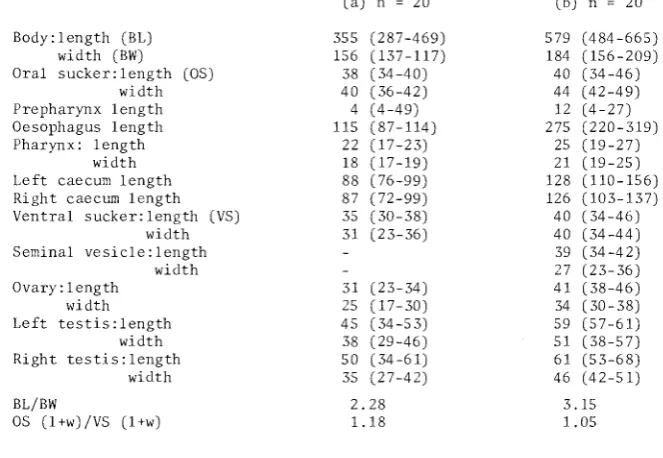
The large round cyst occurs mainly in the green gland, but also in the body cavity of
Paragrapsus gaimardii, either free or loosely bound by connective tissue. The cyst wall, about 28 W thick, is composed of a wide translucent outer layer, often yellowish, and a narrow, clear inner layer. Dimensions of 20 cysts, their identity confirmed by in vitro
[image:4.471.101.398.325.629.2]FIG. 2 - Trematode cysts infect-ing the crab Paragrapsus gaimardii. The cysts of Gynaecotyla hickmani n.sp. and Maritrema eroliae are shown free in the body cavity. Cysts of
Microphallus paragrapsi n.sp. are embedded in a large nerve from the coxa of a peraeopod.
~}'naecot}'la
hickmani n.sp.
200
J,lmMaritrema eroliae
Microphallus paragrapsi
n.sp.t>letacercariae excyst at varying stages of maturity from those in which sex organs are not well developed, to those in which gametogenesis and vitellogenesis are advanced. Eggs are produced by more advanced specimens after one day in Eagle's MEM plus 20% foetal calf serum at 41°C.
TABLE 2
CYSTS OF GYNAECOTYLA HICKMWNI n.sp. Dimensions of live metacercarial cysts
dissected from naturally infected Paragrapsus gaimardii (n=20).
External dimensions Internal dimensions
length width length width
[image:5.471.187.433.138.490.2]110
Microphallid Trematodes from Tasmania
3. Relationships
This new species of Gynaecotyla is named after Dr J.L. Hickman, in recognition of his initial investigations of the microphallids infecting the crab P. gaimardii at Great Bay. Using the key to species of the genus Gynaecotyla, proposed by Deblock (1974), G. hickmani
n.sp. keys out with G. brisbanensis Deblock
&
Pearson, 1968a, and G. bridgmani Deblock, 1974. It is distinctly larger than both of those species, and although similar in shape and general anatomy, differs from them in several respects. The sclerotized pieces in the cornucotyle of G. bridgmani, arranged like a Y, are quite distinct. The form and orn:ament-ation of the cornucotyle of G. brisbanensis are similar to those of G. hickmani n.sp. The arc of the vesiculoprostatic pouch of G. brisbanensis is not sub tended by bundles of muscle fibres, and the diameter of the oral sucker is greater than or equal to the diameter of the antiporal ventral sucker; however, the vesiculoprostatic pouch of G. hickmani n.sp. is subtended by conspicuous muscle fibres, extending from the pouch to the cornucotyle, and the antiporal ventral sucker is much larger than the oral sucker.GYNAECOTYLA MACROCOTYLATA n.sp.
1. Adult
A minority of Gynaecotyla metacercariae that excysted in vitro from large, round cysts taken from the body cavity of Paragrapsus gaimardii, were obviously different from
Gynaecotyla hickmani n.sp., and were found to belong to another new species, Gynaecotyla macrocotylata n.sp.
Dimensions of relatively mature, non-ovigerous, unflattened excysted metacercariae of this species are presented in table 3. The adult; illustrated in figure 3, is described from ovigerous specimens cultured in vitro. Dimensions of the unflattened ovigerous holotype are included in table 3.
Description
Body elongate spatulate. Tegumental spines diminish in size posteriorly, extending to level of testes. Tegumental gland cells distributed in anterior of body. Oral sucker round, mouth subterminal ventral. Prepharynx about one-quarter length of oesophagus, pharynx oval; oesophagus bifurcates in anterior half of body. Caeca long, convoluted, terminating posterior to testes. Two ventral suckers well developed, antiporal ventral sucker slightly larger than poral ventral sucker. Testes posterolateral, oval, symmetric-al. Vesiculoprostatic pouch arcuate, partly dorsal to ventral suckers, between caeca. Seminal vesicle occupies proximal two-thirds; pars prostatica opens through ejaculatory orifice, into genital atrium. Cornucotyle larger than oral and ventral suckers. External lobe largely ventral, enveloping smaller internal lobe, measures about 91 x 48~. Internal lobe measures about 47 x 42~. Surfaces of lobes smooth except for scattered small pointed projec+ions. Three sclerotized pieces on, partly embedded in, anterior part of cornucotyle: according to scheme of Deblock (1974, p.323), no.2 elongate bent obliquely in middle, about 32 x 10 ~; no.S rod shaped, about 20 x 7 ~; no.6 forked, about 17 x 8~. Two bundles of well developed muscles extend from piece no.6 to posterior wall of vesiculoprostatic pouch. Ovary round, sinistral, situated between sinistral testis and proximal end of vesiculo-prostatic pouch, partly dorsal to antiporal ventral sucker. Oviduct passes posteromedially to median ootype. Uterus forms loops posterior to testes, then passes anteriad to genital atrium. Metraterm enters atrium dorsally, opening near ejaculatory orifice. Vitelline glands clustered in two distinct post-testicular bunches. Vitelline ducts pass anteriad, joining to form small round median vitelline reservoir. Eggs numerous, measure 17(16-19) x 12(11-13) ~ (fixed, flattened). Flame-cell formula not determined. Excretory vesicle typical of genus.
Crustacean intermediate host
Paragrapsus gaimardii (M. Edw.).
Geographical location
112
Microphallid Trematodes from Tasmania
Site of encystment
Green gland, general body cavity.
Type material
Tasmanian Museum and Art Gallery - K899 holotype, gravid adult (ringed); K900 para-types, gravid adults (flattened); K90l and K902 parapara-types, mature excysted metacercariae.
TABLE 3
EXCYSTED METACERCARIAE OF GYNAECOTYLA MACROCOTYLATA n.sp. Dimensions of; (a) mature, non-ovigerous specimens after about 3 hours
in vitro at 41°C, and (b) the holotype, a gravid specimen, after about 12 hours in vitro at 41°C.
(a) (b)
n
=
12 n=
1Body:length (BL) 779 (665-937) 703
width 238 (224-258) 227
Oral sucker:length (OS) 55 (49-61) 48
width 56 (53-61) 53
Prepharynx length 44 (34-57) 38
Oesophagus length 201 (160- 228) 160
Pharynx: length 40 (36-44) 36
width 30 (27-32) 27
Left caecum length 323 (289-372) 285
Right caecum length 329 (296-380) 308
Antiporal ventral sucker:length (AVS) 68 (61-74) 68
width 59 (53-68) 57
Poral ventral sucker:length (PVS) 63 (57-68) 61
width 46 (42-51) 48
Cornucotyle:length (C) 78 (65-95) 84
width 71 (68-72) 72
Vesiculoprostatic pouch: 1 ength (VPL) 166 (156-182) 160
width 45 (42-49) 46
Ovary:1ength (0) 65 (57-72) 70
width 57 (53-61) 61
Left testis:length 70 (65-80) 68
width 57 (51-65) 53
Right testis:length 66 (61-72) 65
width 58 (49-65) 53
VPL:BL ratio 0.21 0.23
Relative size (l+w) of organs C > AVS > PVS OS
2. Metacercarial cyst
The cysts are round and large, and occur mainly in the green gland, but also in the general body cavity of Paragrapsus gaimardii. To date they have not been distinguished from those of G. hiekmani n.sp. In January 1980, about 50 large, round metacercarial cysts from the body cavity of P. gaimardii were measured, and then exposed to digestive enzymes in vitro, however all of the metacercariae that excysted were G. hiekmani n.sp. In December 1979, similar, unmeasured cysts were exposed to digestive enzymes in vitro,
and the ratio of the two Gynaeeotyla species in 100 excysted metacercariae was
85 G. hiekmani n.sp. to 15 G. maeroeotylata n.sp.
[image:8.471.96.410.265.545.2]3. Relationships
The mOTe distinct differences between G. macrocotylata n.sp. and G. hickmani n.sp.
are that in the former the cornucotyle is larger than the oral and ventral suckers, the two ventral suckers are similar in muscular development, the poral ventral sucker is similar in size to the oral sucker, the cornucotyle contains three large sclerotized pieces, and the caeca are relatively long, terminating posterior to the testes; whereas in
G. hickmani n.sp. the cornucotyle is smaller than the oral sucker and antiporal ventral
sucker, the antiporal ventral sucker is more powerfully developed than the poral ventral sucker, the cornucotyle contains only a single thin sclerotized layer, and the caeca do not extend posterior to the testes.
According to the key to the species of Gynaecotyla presented by Deblock (1974), G. macrocotylata n.sp. most closely resembles the large form of G. longiintestinata
Leonov, 1958 (syn. G. gallica Rebecq,~ 1961). In the latter species, however, the oral sucker is larger than the poral ventral sucker, and larger than or equal to the antiporal ventral sucker. The form and arrangement of sclerotized pieces in the cornucotyle, which are consistent within Gynaecotyla species, differ markedly between G. macrocotylata n.sp.
and G. longiintestinata, e.g. in the latter species no.6 is not branched, and no.2 is
absent, whereas in the former, no.6 is forked, and no.2 is well developed.
The name G. macrocotylata n.sp. is proposed for this new species as the cornucotyle
is massive, and both ventral suckers are large and muscular.
MARITREMA EROLIAE Y~~GUTI, 1939
Syn. (according to Deblock, 1975): M. urayensis Ogata, 1951; M. magnicirrus Belopolskaia, 1952; M. kitanensis Shibue, 1953.
1. Adult
The adult, illustrated in figure 4, is described from ovigerous specimens cultured in vitro, and from one gravid fluke recovered from a laboratory duckling. Dimensions of mature excysted metacercariae, some of which have commenced egg production, are shown in table 4. Ovigerous and non-ovigerous specimens are similar in size.
Dimensions
Body elongate pyriform to triangular. Tegumental spines small, cover entire body, except at posterior extremity. Tegumental gland cells.anterior to cirrus pouch. Oral sucker round. Prepharynx about half length of oesophagus; pharynx oval; oesophagal bifur-cation about one-third body length from anterior end. Caeca diverge acutely, extend to midlevel of metraterm. Ventral sucker round, larger than oral sucker, located in posterior half of body. Oval testes symmetrical, situated posterolaterally. J-shaped cirrus pouch massive, about 2.5 ~ thick. Flexure of pouch antero-dorsal to ventral sucker. Seminal vesicle large, clavate, in proximal half of dextral limb of cirrus pouch. Distal end of seminal vesicle tapers to short, narrow, folded seminal canal, which widens to voluminous, convoluted pars prostatica. Invaginated spiny cirrus leads from level of anterior seminal vesicle to sinistral genital atrium, proximal part of cirrus densely lined by simple sharp spines, increasing in size distally from 5 x 2 ~ to 10 x 3 ~; distal part lined by large, flattened, thorn-like spines, variable in size, 12 (6-19) x 7 (5-10)~. Evaginated cirrus, directed anteriorly, about 150 ~ long, (fixed flattened), with small simple spines covering distal part, increasing in size to several rows of thorn-like spines near base of cirrus. 'Aspinous ;heel' at base of evaginated cirrus. Ovary triangular, multilobed (usually three
longitud-114
Microphallid Trematodes from Tasmania
A
200
!Jm
u ____
~~~~~~~~~~~~~~~~AJ
..~ .0 . .
50
!Jm
[image:10.471.81.425.127.668.2]inal vitelline reservoir. Flame-cell formula not determined. Excretory vesicle Y-shaped, arms extending to anterior margins of testes.
Experimental definitive host
Anas pZatyrhynchoB Linn~eus.
Crustacean intermediate host
PapagrapBuB gaimardii (M. Edw.).
Geographical location
Great Bay, Bruny Island, Tasmania.
Site of encystment Body cavity.
Material
Tasmanian Museum and Art Gallery - K892 gravid adult (flattened, cirrus everted); K893 gravid adults; K894 non-ovigerous, excysted metacercariae.
TABLE 4
EXCYSTED METACERCARIAE OF MdRITREMA EROLIAE.
Dimensions of metacercariae excysted in vitro,
after about 3 hours at 41°C. All metacercariae relatively mature, with active vitel1aria (n
=
15).Body: length (BL) width
Oral sucker:length (OS) width Prepharynx length Oesophagus length Pharynx: length
width Left caecum length Right caecum length Ventral sucker:length (VS)
width Cirrus pouch:length (CPL)
width Seminal vesicle:1ength
width Metraterm: length
width Ovary: length
width Left testis: length
width Right testis:length
width Roundness (BL/BW) OS (l+w)/ VS (l+w) CPL/BL
683 (620-794) 258 (217-293)
47 (40-53) 51 (46-57) 40 (30-53) 86 (65-133) 28 (23-30) 24 (21-27) 292 (251-342) 307 (274-357)
64 (61-70) 61 (57-67) 350 (323-380)
82 (68-91) 141 (114-156)
59 (49-68) 117 (103-137)
62 (46-80) 108 (87-125)
59 (46-76) 71 (65-76) 52 (42-57) 76 (65-87) 53 (46-61)
2.65 0.78 0.51
2. Metacercarial cyst (fig. 2)
[image:11.471.110.343.376.642.2]116
Microphallid Trematodes from Tasmania
Encysted metacercariae reach an advanced state of sexual development. Although they excyst at varying degrees af maturity, more advanced specimens have phenolic egg-shell precursors in the vitellaria, and commence egg production within a few hours at 41°C, in Hank's BSS and in Eagle's MEM.
TABLE 5
CYSTS OF MARITREMA EROLIAE.
Dimensions of live metacercarial cysts, dissected from Paragrapsus gaimardii, (n=20).
External diameter
Internal diameter
length width
length width
3. Relationships
458 (423-484) 304 (289-331)
402 (370-423) 254 (239-274)
The anatomy of this microphallid, with very large cirrus pouch, spiny cirrus, vast metraterm and median ovary, is characteristic of a distinctive and homogeneous group of
Maritrema species: M. eroliae Yamaguti, 1939, M. echinocirrata Leonov, 1953, M. patulus
Coil, 1955, and M. misenensis (Palombi, 1940) Prevot, Bartoli and Deblock, 1976. The evaginated cirrus of the latter species is free of spines, except for a zone around the base, mainly proximal to a distinct "heel". M. patulus is morphologically identical to
M. e~oliae, and Prevot et al., 1976, consider that when its life-cycle is discovered it may fall into synonymy with M. eroliae. M. echinocirrata differs from M. eroliae only in the absence of the large thorn-like spines at the base of the evaginated cirrus. The Tasmanian flukes fall within the range of variation described for M. eroliae by Deblock (1975) .
The metacercariae of M. eroliae has previously been found encysting in various deca-pod crustaceans in Japan (Ogata 1951, Shibue 1953, and Bridgman et al. 1972). The oval cyst varies greatly in size and thickness, possibly in relation to the identity of the second intermediate host. The cysts found in Tasmanian crabs are intermediate in size between those infecting Macrophthalmus dilatatus , 520-690 x 360-450 ~ (Ogata 1951; and Bridgman et al. 1972), and those infecting Neocaridina denticulata, 280-320 x 240-270 ~
(Shibue 19}3), and Scopimer spp., 310-360 x 210-240 ~ (Ogata 1951). The adult of
M. eroliae has been found only in birds inhabiting the eastern border of the Pacific Ocean (Deblock 1975); and in Australia, has been recorded in the Mongolian dotterel,
Charadrius mongolus Pallas, in Queensland (Deblock
&
Pearson 1968b).MICROPHALLUS PARAGRAPSI N. SP .
1. Adult
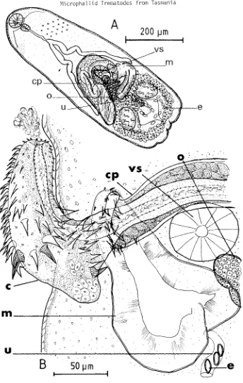
The adult, illustrated in fig. 5, is described below from ovigerous specimens cul-tured in vitro and non-ovigerous specimens taken from a laboratory duckling. Dimensions of unflattened, ovigerous specimens are not available, however gravid adults appear to be the same size as relatively mature excysted metacercariae (with phenolic egg-shell precursors in the vitellaria), dimensions of which are included in table 6b. Dimensions of very immature excysted metacercariae are shown in the same table. Non-ovigerous adults (some with phenolic egg-shell precursors), recovered from an experimentally infect-ed duckling, were interminfect-ediate in size between immature and mature excystinfect-ed metacercariae
(table 7).
Description
A
200
J.lm
50
IJm
200
IJm
[image:13.471.38.423.153.634.2]118
Microphallid Trematodes from Tasmania
arranged, diminishing in size posteriorly, extending to waist level. Tegumental gland cells distributed over anterior body. Oral sucker transversely oval to round, mouth sub-terminal ventral. Prepharynx short, about 1/20th length of oesophagus. Oesophageal bi-furcation in posterior half of body. Caeca relatively short, terminating at midlevel of ventral sucker. Ventral sucker round, smaller than oral sucker. Testes oval, equal, posterolateral. No cirrus pouch; seminal vesicle oval, partly dorsal to ventral sucker. Seminal canal short, leading posteriorly from seminal vesicle to expanded pars prostatica, about 6 ~ diameter, at base of male papilla. Prostate gland cells located between
seminal vesicle and male papilla, clustered around seminal canal; secreting through undet-ermined number of small ducts, into pars prostatica. Retracted male papilla coiled or folded within genital atrium, measures 34(30-38) x 29(27-30)~. Everted male papilla, tubular, not lobed, directed posteromedially; length 66(53-76) ~, width at base 26(23-30)
~, width at tip 22(19-25)~. Ejaculatory canal, about 4 ~ diameter, mainly axial within male papilla, but opens eccentrically. Ovary oval, contiguous to dextral caecum and testis, dextral and partly dorsal to ventral sucker. Oviduct passes posteromedially to bulbous seminal receptacle, from which Laurer's duct leads to dorsal surface. Ootype located between testes, posterior to ventral sucker. Uterus forms post-testicular loops, overlapping each testis. Metraterm enters left side of genital atrium. Vitelline gland cells clustered in compact bunches posterior to each testis. Vitelline ducts pass antero-mediad, joining to form longitudinal vitelline reservoir, posterior to ventral sucker. Oval eggs, numerous, 19(17-21) x 10(8-13) ~, (fixed, flattened). Flame-cell formula not determined. Excretory vesicle V-shaped, limbs extending to testes.
TABLE 6
EXCYSTED METACERCARIAE OF MICROPHALLUS PARAGRAPSI N.SP.
Dimensions of metacercariae excysted in vitro, after about 3 hours at 41°C:
(a) smaller, less mature specimens; (b) larger, more mature specimens, some with active vite1laria.
(a) n
=
20 (b) n=
20Body:length (BL) 355 (287-469) 579 (484-665)
width (BW) 156 (137-117) 184 (156-209)
Oral sucker:length (OS) 38 (34-40) 40 (34-46)
width 40 (36-42) 44 (42-49)
Prepharynx length 4 (4 -49) 12 (4-27)
Oesophagus length 115 (87-114) 275 (220-319)
Pharynx: length 22 (17-23) 25 ( 19-27)
width 18 (17-19) 21 (19-25)
Left caecum length 88 (76-99) 128 (110-156) Right caecum length 87 (72-99) 126 (103-137) Ventral sucker:length (VS) 35 (30-38) 40 (34-46)
width 31 (23-36) 40 (34-44)
Seminal vesicle: length 39 (34-42)
width 27 (23-36)
Ovary: 1 ength 31 (23-34) 41 (38-46)
width 25 (17-30) 34 ( 30-38)
Left testis:length 45 (34-53) 59 (57-61)
width 38 (29-46) 51 (38-57)
Right testis:length 50 (34-61) 61 (53-68)
width 35 (27-42) 46 (42~51)
BL/BW 2.28 3.15
[image:14.471.85.417.418.647.2]Experimental definitive host Anas platyphynchos Linnaeus
CrustaceaD intermediate host
PaTagpapsus gaimapdii (M.Edw.)
Geographical location
Great Bay, Bruny Island, Tasmania Si te of eDcystment
Nerves of legs and claws Type material
Tasmanian Museum and Art Gallery - K903 holotype, non-ovigerous adult (ringed); K903 paratypes, non-ovigerous adults (not ringed); K904 paratypes, ovigerous adults (ringed, flattened); K905 and K906 paratypes, excysted metacercariae.
TABLE 7
NON-OVIGEROUS ADULTS OF MICROPHALLUS PARAGRAPSI N.SP. Dimensions of the holotype (a), and other non-ovigerous
adults (b), from an experimentally infected duckling 17 hours after infection.
(a) n=l (b) n=9
Body:length (BL) 544 460 (325-544)
width (BW) 171 159 (137-179)
Oral sucker:length (OS) 42 38 ( 34-40)
width 44 41 (36-44)
Prepharynx length 11 14 (8-23)
Oesophagus length 243 199 (110-258)
Pharynx: length 27 22 (19-25)
width 19 19 (17-23)
Left caecum length 114 95 (72-110) Right caecum length 114 98 (80-110) Ventral sucker:length (VS) 40 39 (34-42)
width 34 34 (30-38)
Ovary: length 38 40 (36-46)
width 34 34 (27-38)
Left testis:length 57 55 (46-65)
width 38 40 (30-46)
Right testis:length 55 54 (46-61)
width 46 46 (38-49)
BL/BW 3.18 2.89
OS (l+w)/VS (l+w) 1.16 1.08
2. Metacercarial cyst (fig. 2)
The cyst is round, colourless, and quite thick-walled. Its dimensions are shown in table 8. The uniformly thick cyst wall, about 36(27-44) ~wide, is composed of two layers: a clear, homogeneous inner layer, 14(10-19) ~, and a darker, outer layer with fine radial striae, 22(17-27)~. The cyst occurs within the large nerves innervating the legs and claw muscles of the crab host, particularly near the coxae. No behavioural changes related to this infection have been noticed in the crab host.
[image:15.471.121.361.364.594.2]120
Microphallid Trematodes from Tasmania
egg-shell precursors in the vitellaria, and produce eggs in vitro after 2 days at 41°C, in Eagle's MEM plus 20% foetal calf serum.
TABLE 8
CYSTS OF MICROPHALLUS PARAGRAPSI N.SP. Dimensions of live metacercarial cysts, dissected from Paragrapsus gaimardii, (n=20). External diameter
Internal diameter
length width length width
TABLE 9
319 (300-353) 310 (285-331) 246 (2:<8-266) 239 (224-251)
SIZE VARIATION IN EXCYSTED METACERCARIAE OF MICROPHALLUS PARAGRAPSI N.SP. Size of metacercariae excysted in vitro at 41°C: (a) after 1~ hours; and
(b) from 1~ to 12 hours.
length width
(a) n=20 351 (249-507) 154 (125-175)
(b) n=20 479 (333-665) 185 (144-213)
3. Relationships
The species of Microphallus encysting in the nerves of the crab Paragrapsus gaimardii in Tasmania is considered to be new, and is named after its second intermediate host. Only one other microphallid species is known to encyst in the nerves of the limbs of its
crustacean host: Microphallus pachygrapsi Deblock
&
Prevot, 1968, encysts in the crab Pachygrapsus marmoratus on the Mediterranean coast of France. Microphallus paragrapsi n.sp. and M. pachygrapsi adults are similar in size, but differ in the following respects: the former has OS:VS greater than 1, and male papilla length greater than VS diameter; the latter has OS:VS less than 1, and male papilla length less than VS diameter. Metacercar-ial cysts of M. paragrapsi n.sp. are round whereas those of M. pachygrapsi are oval. According to the key to species of Microphallus presented by Deblock (1971), the,species most similar to M. paragrapsi n.sp. is M. minutus, a tiny fluke discovered in water rats captured on the banks of the Murray River at Tailem Bend, South Australia (Johnston 1948) and rediscovered and redescribed by Deblock ( Pearson (1969), in the same host, captured near Brisbane, Queensland. The adults of M. paragrapsi n.sp. and M. minutus are very 'similar, however the average size of gravid M. minutus adults, 290 x 160 \1 (Deblock &Pearson 1969), is slightly less than the average size of the more immature excysted meta-cercariae of M. paragrapsi n.sp., 355 x 156 \1, and markedly less than the average size of mature excysted metacercariae of M. paragrapsi n.sp., 579 x 184 \1. The male papilla of M. minutus. 53 x 27 \1, is less elongate than that of M. paragrapsi n.sp., 66 x 26 \1. In relation to body size, the oral and ventral suckers of M. minutus are much larger than those of M. paragrapsi n.sp. On the basis of these differences, and in the absence of further life-history information,
M.
paragrapsi n.sp. and M. minutus are considered to be distinct, but closely related species. Four other species of Microphallus has been recorded in Australia (Deblock&
Pearson 1969), however, all are readily distinguished from M. paragrapsi n.sp. by details of the reproductive system: M. minutus has a large, robust male papilla of the "papillorobustus" type; M. papillornatus has an ornamented male papilla; M. vaginosus has an unusual, large metraterm, with thick folded walls; andA
200
IJm
v
50
IJ m
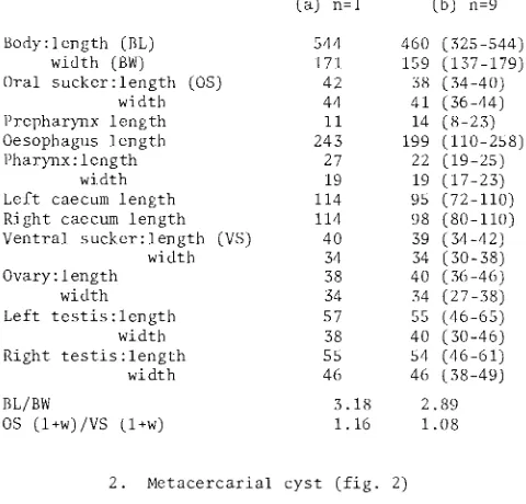
pp
sv
FIG. 6 - MiorophaZZus paragrapsi n.sp. A: excysted metacercaria, after 12 hours at 41°C, lateral view; B: detail of everted male papilla of metacercaria shown in A; C: variation in form and size of excysted metacercariae, after 12 hours at 4loC. (c: caecum,
[image:17.471.38.429.119.648.2]122
Microphallid Trematodes from Tasmania
DISCUSSION
The incidence of infection of Paragrapsus gaimardii with microphallid trematodes has
not been determined, however the impression gained from dissection of many crabs from Great Bay, at irregular intervals over several years, is that almost all adult crabs, of both sexes, are infected by Gynaecotyla spp., Maritrema eroliae, and Microphallus paragrapsi n.sp., throughout the year. Two large male crabs dissected in July 1977 were infected by an average of 197(118-275) metacercarial cysts of Gynaecotyla spp.,
195(90-300) of M. paragrapsi n.sp., and 14(2-26) of Maritrema eroliae. These crabs also
contained an average of 19(15-23) large, unencysted immature metacercariae of Gynaecotyla
spp. These specimens had an oral sucker, a large excretory bladder, and some individuals also haq ventral suckers and rudimentary digestive and reproductive systems. Delayed encystment is known in the life-cycles of a number of Gynaecotyla species, and in G. adunca
and G. longiintestinata, a small percentage of metacercariae mature and produce eggs,
with-out encysting (Deblock 1977). The cysts of the two Gynaecotyla species infecting P. gaimardii have not been distinguished, however, the ratio of the two species in 100
metacercariae, excysted in vitro from cysts dissected in December 1979, was about 6 G. hickmani n.sp. to 1 G. macrocotylata n.sp. The molluscan hosts of these four microphallid
species are as yet unknown. The Mongolian dotterel, which harbours Maritrema eroliae in
Queensland, 1S an infrequent visitor to Tasmanian shores, and might have introduced this trematode to the fauna of Great Bay. An estuarine fish, the leatherjacket, caught in the Derwent River, was found by Last (1975) to harbour a single microphallid in its intestine, which appears to be an immature adult of Maritrema eroliae. As experimental evidence indicates that a temperature of about 40°C is required for oviproduction in M. eroliae,
eggs are unlikely to be produced in this host. Attempts to experimentally infect eight domestic ducklings with hundreds of cysts of these four Tasmanian microphallids were not very successful. No ducklings were infected by the two Gynaecotyla species; three
duck-lings, dissected 17, 18 and 26 hours after being fed with cysts, were infected with 16, 1 and 1 immature adults of Microphallus paragrapsi n.sp. respectively; and one duckling
dissected after 17 hours was infected by one gravid adult of Maritrema eroliae that
con-tained 52 uterine eggs.
The discovery and identification of four microphallid species concurrently infecting a single species of crab in Tasmania presents many challenging problems. The site of en-cystment of Microphallus paragrapsi n.sp. is unusual, and presumably of selective value
to the parasite. This trematode may increase its chances of transmission by making its intermediate host more vulnerable to predation. An investigation of the effect of M. paragrapsi n.sp. cysts on the function of the nerves and the behaviour of Paragrapsus gaimardii, may help to explain why two microphallids, M. paragrapsi n.sp. in Tasmania and M. pachygrapsi in France, invade the nerves of their crustacean hosts. The life history
of none of the four microphallids infecting P. gaimardii is known. Elucidation of their life-cycles at Great Bay will facilitate discovery of their life-cycles at other locations in Australia, and the life-cycle of Maritrema eroliae in Japan.
ACKNOWLEDGEMENTS
This work was conducted as part of a Ph.D. project at the University of Tasmania, while I was the holder of a Commonwealth Postgraduate Research Award. I am indebted to Dr J.L. Hickman (Zoology Department, University of Tasmania) for his advice and encourage-ment and for critically reading this manuscript.
REFERENCES
Be1opo1skaia, M.M., 1952: Trematode family Micropha11idae Travassos, 1920. In Skrjabin, K.I. (Ed.): Trematodes of animals and man, 6: 619-756. (In Russian, cited by Deblock 1971).
Bridgman, J.F., Otagaki, K., Shitanda, I. and Tada, K., 1972: Nine metacercariae
Coil, W.H., 1955: Notes on the genus Maritrema Nicoll, 1907 (Trematoda, Microphallinae) with the description of two new species. J. Parasitol., 41: 533-537.
Deblock, S., 1971: Contribution
a
l'etude des Microphallidae Travassos, 1920. XXIV. Tentative de phylogenie et de taxonomie. Bull. Mus. Rist. Nat., Paris, 3e. serie, Zool., 7: 353-468.____________ ' 1974: Contribution
a
l'etude des Microphallidae Travassos, 1920 (Trematoda). XXIX. A propos d'especes decrites au Japon par s. Yamaguti B. Le genreYamaguti, 1939. Ann. Parasit. hum. comp., 49: 319-335.
---X~X-X--.--~ ~~:~~s ~~:~;~~~~i~~C!i~~:t~~eJ~:~n~·~:~p~~lt:~:u~~~va~~o~~r~;~~m~T~~~~~~a).
et le genre Pseudospelotren~. Ann. Parasit. hum. comp., 50: 45-54.
______ ~~~' 1977: De l'abregement du cycle evolutif chez les trematodes digenes micro-phallides. Sobretiro de Excerta Parasitologica en memoria del doctor Eduardo Caballero y Caballero. Instituto de Biologia Publicaciones Especiales (Universidad Nacional, Mexico), 4: 151-160.
______ ~----and Pearson, J.C., 1968a: Contribution
a
l'etude des Microphallidae Travassos, 1920 (Trematoda). XIV. Trois Gynaecotylinae nouveaux d'Australie. Considerations systematiques. Ann. Parasit. hum. comp., 43: 131-148.,1968b: Contribution
a
l'etude des Microphallides Travassos, 1920 (Trematoda). XV. De quelques especes d'Australie dont PseudospeZotrema anenteron n.sp. Ann. Parasit. hum. comp.,43: 457-465., 1969: Contribution
a
l'etude des Microphallidae Travassos, 1920 (Trematoda). XVIII. De cinq Microphallus d'Australie dont deux nouveaux.Essai de cle diagnostique des especes du genre. Ann. Parasit. hum. comp., 44:391-414. and Prevot, G., 1968: Contribution
a
l'etude des Microphallidae Travassos,----~1~9~2~0~(Trematoda). XVII. Microphallus pachygrapsi n.sp., adulte experimental d'une metacercaire de Pachygrapsus marmoratus Stemp. (Crustace Brachyoure). Bull. Soc.
zoo~. France, 93: 603-610.
Johnston, T.H., 1948: Microphallus minutus, a new trematode from the Australian water rat. Rec. S. Aust. MUs., 9: 93-100.
Last, P., 1975: ASPECTS OF THE TAXONOMY AND ECOLOGY OF TASMANIAN LEATHERJACKETS (FAMILY MONACANTHIDAE, PISCES). B.Sc. Honours thesis, unpub. University of Tasmania. Leonov, V.A., 1958: Faune helminthologique des mouettes des reserves de la Mer Noire et
des regions limitrophes du territoire de Kherson. C. R. de l'Inst. pedagogique d'etat de Gorkovsk, 20: 266-296. (In Russian, cited by Deblock, 1971.)
Nicoll, W., 1907: Observations on the trematode parasites of British birds. Ann. Mag. Nat Rist., 20: 254-271.
Ogata, T., 1951: Studies on the life histories of certain trematodes, the intermediate hosts of which are brackish water crustaceans, with the discussions on the systematic position of the species. Japan. J. Parasit., 1: 17-35.
Palombi, A., 1940: Gli stadi larvali dei trematodi del Golfo di Napoli. Rivista di Parassitol., 4: 7-30. (Cited by Prevot, Bartoli and Deblock 1976.)
Prevot, G., Bartoli, P. and Deblock, S., 1976: Cycle biologique de Maritrema misenensis (A. Palombi, 1940) n.comb. (Trematoda: Microphallidae Travassos, 1920) du Midi de la France. Ann. Parasit. hum. comp., 51: 433-446.
Rebecq, J., 1961: Sur un trematode nouveau de Larus argentatus michaellis Naumann, appartenant
a
la sous-famil1e cynaecotylinae Guschanskaia, 1952. C. R. Congres Soc. Sav., 86: 669-678.Shibue, H., 1953: A study of a new metacercaria encysted in the freshwater shrimp Neocaridina denticulata (de Haan) believed to belong to the genus Maritrema Nicoll, 1907. Japan. J. Med. Sci.
&
BioI., 6: 389-394. (Cited by Deblock 1971.)Ward, H.B., 1901: Notes on the parasites of the lake fish. III. On the structure of the copulatory organs in Microphallus nov. gen. Trans. Amer. Microscop. Soc., 22: 175-187.