Copyright © 2002, American Society for Microbiology. All Rights Reserved.
Antiviral Effects of Pyrrolidine Dithiocarbamate on
Human Rhinoviruses
Elisabeth Gaudernak,
1Joachim Seipelt,
1Andrea Triendl,
1Andreas Grassauer,
2and Ernst Kuechler
1*
Institute of Medical Biochemistry, University of Vienna, Vienna Biocenter, A-1030 Vienna,1and Department of
General Dermatology, University of Vienna Medical School, A-1090 Vienna,2Austria
Received 25 October 2001/Accepted 22 March 2002
Human rhinoviruses (HRVs) are the predominant cause of the common cold. The frequency of HRV infections in industrial countries and the lack of effective therapeutical treatment underline the importance of research for new antiviral substances. As viral infections are often accompanied by the generation of oxidative stress inside the infected cells, several redox-active substances were tested as potential antivirals. In the course of these studies it was discovered that pyrrolidine dithiocarbamate (PDTC) is an extremely potent compound against HRV and poliovirus infection in cell culture. Besides the ability to dramatically reduce HRV production by interfering with viral protein expression, PDTC promotes cell survival and abolishes cytopathic effects in infected cells. PDTC also protects cells against poliovirus infection. These effects were highly specific, as several other antioxidants (vitamin C, Trolox, 2-mercaptoethanol, andN-acetyl-L-cysteine) are inactive against HRV
infection. Synthesis of HRV proteins and cleavage of eucaryotic initiation factor 4G responsible for host cell shutoff of cellular protein synthesis are severely inhibited in the presence of PDTC.
Human rhinoviruses (HRVs), the main causative agents of the common cold occurring worldwide (20), constitute the most extensive genus of thePicornaviridae. The frequent ap-pearance of HRV infections and their economic importance in terms of employee absenteeism, physician visits, and medica-tion costs make it a subject of primary importance (17). De-spite the frequency of the disease, no cure for the common cold is presently available apart from symptomatic treatment. Infections of patients with HRVs elicit typical protory responses accompanied by massive release of inflamma-tory mediators (53). In particular, cytokines, including inter-leukin-1 beta (IL-1), tumor necrosis factor alpha, IL-8, IL-6, and IL-11 (12, 46, 77), and the vasoactive peptides bradykinin and lysyl-bradykinin (45, 54, 64) were found in nasal secretions of patients with colds. There is strong evidence that the acti-vation of these proinflammatory molecules is at least in part mediated by the transcription factor NF-B (77).
To successfully infect a host, viruses have to both exploit the cellular resources and at the same time avoid defense reactions of the host organism. In tissue culture, morphological changes observed in cells infected with picornaviruses, including cell rounding and detachment from the substrate, are generally termed cytopathic effects (CPE). During picornaviral infection, several essential cellular processes are modified by the action of viral proteins at the levels of both transcription (57, 75) and translation (14, 30). Furthermore, profound changes in cy-toskeletal architecture are found to occur (2, 32, 62). A hall-mark of infections with rhino- and enteroviruses is the cleavage of the eucaryotic translation initiation factors (eIF) eIF4GI and eIF4GII by viral proteinase 2A, resulting in the shutoff of cap-dependent host cell translation.
There is increasing evidence supporting the view that oxida-tive stress might play an important role in virus infection. Mechanistically, oxidative stress is characterized by increased levels of reactive oxygen intermediates (ROIs), which act as second messengers for the activation of transactivators such as NF-B (58, 59), AP-1 (41), Egr-1 (22), p53 (70) and c-fos (37). It has been shown that interference with the generation of ROIs by use of antioxidants can drastically reduce replication of various seemingly unrelated viruses, e.g., bovine diarrhea virus (61), Sindbis virus (36), hepatitis B virus (71), influenza A virus (16), and retroviruses such as the human and feline im-munodeficiency viruses (27, 39, 44). It is hypothesized that these viruses induce apoptosis via a pathway involving oxida-tive stress and the transcription factor NF-B. Interference with the generation of oxidative stress with antioxidants is believed to inhibit virus-induced apoptosis and thus virus rep-lication.
Although oxidative stress is also known to occur during infec-tion with picornaviruses, little informainfec-tion is available on its role in virus multiplication (28). Therefore, we investigated the effects of several antioxidants including pyrrolidine dithiocarbamate (PDTC), vitamin C, the vitamin E derivative Trolox, 2-mercap-toethanol, andN-acetyl-L-cysteine (NAC) on infections of
epithe-lial cells with several serotypes of HRVs.
In this report, we provide evidence that PDTC has a drastic inhibitory effect on the multiplication of several HRV sero-types in different cell sero-types. In the presence of PDTC, virus growth is greatly reduced and the CPE is absent. Interestingly, these effects are specific for PDTC, as other redox-active com-pounds do not interfere with virus multiplication. Similarly, PDTC protects cells against the CPE induced by poliovirus infection.
MATERIALS AND METHODS
Media, reagents, and chemicals.HeLa cells (strain Ohio; European Collection of Cell Cultures, Salisbury, United Kingdom) and A549 alveolar carcinoma cells
* Corresponding author. Mailing address: Ernst Kuechler Institute of Medical Biochemistry, Bohrgasse 9/3, A-1030 Vienna, Austria. Phone: 43-1-4277-61610. Fax: 43-1-4277-9616. E-mail: kuechler @bch.univie.ac.at.
6004
on November 8, 2019 by guest
http://jvi.asm.org/
(American Type Culture Collection) were cultured in Dulbecco modified Eagle’s medium (Gibco) supplemented with 10% heat-inactivated fetal calf serum (FCS)
(Gibco), 2 mML-glutamine (Dipro), 100 U of penicillin/ml (Dipro), and 100g
of streptomycin/ml (Dipro). HeLa S3 cells were grown in RPMI medium (Gibco)
for experiments with poliovirus. 16HBE14o⫺cells (obtained from D. Gruenert,
San Francisco, Calif.) were grown in minimal essential medium (Gibco)
supple-mented with 10% FCS, 2 mML-glutamine, 100 U of penicillin/ml, and 100g of
streptomycin/ml. Dishes were coated with 10g of bovine serum albumin/ml
(Sigma), 30g of bovine collagen type I/ml (Promocell) and 10g of human
fibronectin/ml (Becton Dickinson) in Ham’s F-12 medium (HyClone). Human
recombinant IL-1␣was obtained from Genzyme Diagnostics. PDTC was
pur-chased from Alexis Biochemicals, Lausen, Switzerland. A 0.6 M stock solution in
water was stored at⫺20°C. The cell-permeative vitamin E derivative Trolox
(6-hydroxy-2,5,7,8-tetramethylchroman-2-carboxylic acid) (Alexis) was stored at 4°C at a concentration of 10 mM. NAC (Sigma) was dissolved in water to a concentration of 1 M and diluted subsequently. 2-Mercaptoethanol was obtained from Calbiochem. Ascorbic acid (vitamin C) (Sigma) was dissolved in water before use. Guanidine HCl (Sigma) was dissolved in water at a concentration of 100 mM.
Virus preparation.HRV1A, HRV2, HRV14, and HRV16 (American Type Culture Collection) were grown in suspension cultures of HeLa cells (strain Ohio) and purified as described previously (65). Virus titers in 50% tissue culture
infectious doses (TCID50)/ml were determined according to Reed and Muench
(55). Poliovirus strain Mahoney was cultivated in HeLa S3 cells. Cell supernatant was used for infection without further purification.
Infection of cells with HRV serotypes.HeLa, 16HBE14o⫺, and A549 cells
seeded into 6-well plates were infected at 70% confluence with HRV serotypes HRV1A, HRV2, HRV14, or HRV16 in infection medium (minimal essential
medium [MIM] [Gibco] containing 2% FCS, 2 mML-glutamine, 30 mM MgCl2,
100 U of penicillin/ml, 100g of streptomycin/ml). If not otherwise indicated,
input virus was used at 100 TCID50per cell. PDTC was added simultaneously
with HRV preparations. Cellular toxicity was tested by visual inspection and by the proliferation assay (see below) after 24 h with different concentrations of PDTC. The optimal concentrations of PDTC treatment as determined by the
toxicity tests differed among various cell types. Routinely, 125M PDTC was
used for HeLa cells, 60M was used for A549 cells, and 6M was used for
16HBE14o⫺cells. Cells were incubated at 37°C and 5% CO
2in a humidified
incubator. At 4 h after infection, unbound viral particles were removed by washing three times with 10 mM HEPES (pH 5.3) and 140 mM KCl and once with phosphate-buffered saline (PBS). Cells were then incubated for 2, 4, 6, 8, and 24 h in MIM supplemented with or without PDTC. Cells were investigated by light microscopy, by staining with 0.2% crystal violet in 20% methanol, or by immunofluorescence. At 2, 4, 6, 8, and 24 h after infection, supernatants were
collected for the determination of TCID50(55) or cells were lysed to obtain
extracts for Western blotting.
Proliferation assay.To determine cell viability, the Cell Titer 96 AQueous
Non-radioactive Cell Proliferation Assay (Promega) was performed as instructed
by the manufacturer. Briefly, 106cells were seeded into a 96-well plate 1 day
prior to infection. Cells were infected using HRV serotypes HRV1A, HRV2,
HRV14, and HRV16 at 20 TCID50/cell for HeLa cells. PDTC, guanidine HCl,
2-mercaptoethanol, vitamin C, and Trolox were added as indicated in the figure legends. At 24 h after infection, cell viability was determined by adding the tetrazolium compound followed by incubation for 2 h at 37°C and subsequent measurement of absorbance at 492 nm in a Labsystems Multiscan RC plate reader.
Western blot analysis.Cells were infected in 6-well plates as described for virus infection. At 0, 2, 4, 6, 8, and 24 h after infection, the medium was removed
and the cells were lysed by the addition of 100l of protein sample buffer (8%
sodium dodecyl sulfate [SDS], 20%-mercaptoethanol, 20% glycerol, 0.04%
bromophenol blue). Protein extract (20l per lane) was subjected to
SDS-polyacrylamide gel electrophoresis and electroblotted onto polyvinylidene diflu-oride membranes. Blocking and incubation with antibodies was done using 5% nonfat dry milk and 0.1% Tween 20 in Tris-buffered saline (50 mM Tris–HCl [pH 7.4], 150 mM NaCl). For immunodetection, a rabbit polyclonal antibody against eIF4GI (76) and an antiserum against HRV2 prepared by conventional methods were used. Secondary antibodies were alkaline phosphatase-conjugated goat anti-rabbit immunoglobulin (Sigma). Staining using an alkaline phosphatase reaction was performed as described previously (35). Molecular sizes were de-termined using prestained molecular weight marker SDS-7B (Sigma).
Immunofluorescence.Immunofluorescence was essentially performed as de-scribed elsewhere (3). Briefly, cells were grown on sterile coverslips in 6-well tissue culture dishes. Fixation was done with 4% paraformaldehyde–PBS for 10 min at room temperature. Cells were permeabilized by incubation in 0.5% Triton
X-100–PBS for 5 min. Blocking was done at 4°C using 1% goat serum–PBS. The primary antibody was a rabbit polyclonal antiserum raised against human p65 (Santa Cruz) diluted 1:500 in PBS–1% bovine serum albumin. As secondary antibody, Alexa 488-labeled goat anti-rabbit immunoglobulin G conjugates were used at 1:800 dilution (Molecular Probes). Finally, slides were mounted with DAKO fluorescent mounting medium and examined with a Leica TCS-NT con-focal microscope using fluorescein isothiocyanate settings.
RESULTS
PDTC drastically reduces production of infectious HRV particles.Viral infections frequently result in the generation of oxidative stress in the infected cells (16, 23, 28, 61). To study the influence of antioxidants on HRV infections, the effect of PDTC on the replication was investigated in epithelial cells by using several serotypes of HRV. HeLa cells, A549 alveolar carcinoma cells, and 16HBE14o⫺human bronchial epithelial
cells were infected with HRV2 or HRV14 by using 100 TCID50
per cell. At the same time, PDTC was added at concentrations of 125M for HeLa cells, 60M for A549 cells, and 6M for 16HBE14o⫺cells. At 4 h after infection, input virus was
re-moved and the MIM was replaced by growth medium contain-ing the same concentrations of PDTC. Supernatants were col-lected 24 h postinfection (p.i.), and the amount of progeny virus was determined by titration using TCID50assays on HeLa
cells (Fig. 1).
Under these conditions, HRV replication in untreated cells resulted in the generation of a viral titer between 107and 109
TCID50/ml after 24 h p.i. (Fig. 1A). Presence of PDTC during
infection dramatically changes the outcome of viral infection: depending on the cell type, viral titers decrease between 2 orders of magnitude in the case of HRV14-infected 16HBE14o⫺cells
and nearly 6 orders of magnitude in the case of HRV14-infected HeLa cells (Fig. 1A). Although there are differences in the amount of virus reduction, the inhibitory effect of PDTC does not appear to be restricted to a particular cell type.
To investigate whether this effect was serotype specific, both minor group viruses HRV1A and HRV2 and major group viruses HRV14 and HRV16 were used to infect HeLa cells. In this experiment, the amount of input virus was reduced to 20 TCID50/cell. Concurrent treatment of cells with PDTC
re-duced viral titers after 24 h by 3 orders of magnitude for all HRV serotypes tested (Fig. 1B). Viral titers in the superna-tants harvested from 24 to 48 h p.i. were also significantly reduced by PDTC treatment (Fig. 1C), implying that the effect is due to a reduction in virus growth rather than to a delay in virus release. As a lower amount of input virus (20 TCID50/
cell) was used in this experiment, the absolute number of virus particles produced was reduced compared to that for the re-sults presented in Fig. 1A. Nevertheless, the antiviral effect of PDTC was in a similar range to that observed in the experi-ments using a higher amount of input virus.
These experiments demonstrate that PDTC has strong an-tiviral properties against several serotypes of HRV indepen-dent of cell types. There is also no dependence on the viral receptor, as major group HRVs bind to intercellular adhesion molecule 1 whereas minor group HRVs use the low-density lipoprotein receptor and/or related proteins for viral entry. Different amounts of input virus change the absolute amount of progeny virus but do not affect significantly the percentage
VOL. 76, 2002 ANTIVIRAL EFFECTS OF PDTC ON HUMAN RHINOVIRUS 6005
on November 8, 2019 by guest
http://jvi.asm.org/
of reduction of viral titers, indicating that the antiviral prop-erties of PDTC are independent of the multiplicity of infection. Search for PDTC-resistant HRV variants.Following a pro-tocol similar to that described for the generation of guanidine-resistant mutants of poliovirus (68), HRV14 was propagated through five passages in HeLa cells in the presence of increas-ing concentrations of 15 to 30M PDTC. This resulted in a complete loss of viral titer (data not shown), whereas under
similar conditions guanidine-resistant poliovirus variants had been isolated previously (68). In another experiment, HRV14 was carried through nine passages at increasing concentrations from 4 to 12 M PDTC. Again no PDTC-resistant HRV14 variants were obtained (data not shown). Under similar con-ditions, HRV14 variants resistant to the inhibitor enviroxime have been produced (21). From these experiments it can be concluded that mutations in HRV14 conferring resistance against PDTC do not occur with high frequency.
PDTC inhibits HRV-induced CPE.Many viruses are capable of inducing cell death, leading to lysis of infected cells (1, 10, 33). In late stages of HRV infections, morphological changes, commonly known as CPE, can be microscopically observed. HRV-induced CPE is characterized by cell rounding, shrink-age, deformation of nuclei, and chromatin condensation.
During infections of HeLa cells with HRV1A, HRV2, HRV14, and HRV16 using 100 TCID50/cell, a clear CPE was
visible within 8 h p.i. Figure 2 shows the morphology of HeLa cells infected with HRV14. Mock-infected cells (Fig. 2A) or cells treated with 125 M PDTC (Fig. 2B) showed typical spread-out shapes and normal morphology. At this concentra-tion, no signs of cytotoxicity of PDTC were seen. Infection with HRV14 in the absence of PDTC resulted in a severe CPE (Fig. 2C). Addition of PDTC upon infection inhibited the formation of a visible CPE. As shown in Fig. 2D, the morphology of cells 8 h after infection with HRV14 in the presence of PDTC was virtually undistinguishable from that of mock-infected cells (Fig. 2A). A similar protective effect was shown in 16HBE14o⫺
cells infected with HRV14 and in A549 cells infected with FIG. 1. PDTC inhibits HRV replication in cell culture. (A) HeLa
cells, 16HBE14o⫺cells, and A549 cells were infected with HRV2 or
HRV14 using 100 TCID50/cell as described in Materials and Methods.
PDTC was added at the time of infection. Viral titers in the superna-tants of infected cells were determined after 24 h by TCID50assay.
Representative experiments are shown. (B and C) HeLa cells were infected with HRV serotypes HRV1A, HRV2, HRV14, and HRV16 using 20 TCID50/cell. At the same time, 125M PDTC was added.
Virus titers of supernatants collected from 0 to 24 h p.i. (B) and from 24 to 48 h p.i. (C) as determined by TCID50assay are shown.
FIG. 2. PDTC inhibits HRV-induced CPE. HeLa cells were in-fected with HRV14 in the absence or presence of 125M PDTC. Cells were examined by light microscopy 8 h p.i. (A) Noninfected cells; (B) noninfected cells with PDTC; (C) HRV14-infected cells without PDTC; (D) HRV14-infected cells with PDTC.
on November 8, 2019 by guest
http://jvi.asm.org/
HRV2 (data not shown). Thus, the CPE of the virus infection is prevented by the presence of PDTC.
PDTC promotes viability of infected cells. To quantify the effect of PDTC on cells during viral infection, a proliferation assay was employed. Conversion of the tetrazolium substrate into a formazan dye is accomplished by dehydrogenase en-zymes. The observed optical density therefore reflected the metabolic activity of cells. HeLa cells infected with HRV2 (Fig. 3A) and with HRV14 (Fig. 3B) showed a complete loss of metabolic activity after 24 h of infection. However, PDTC concentrations as low as 31 M protected cells efficiently against virus-induced cell death. Even at lower concentrations of PDTC, cellular survival was significantly improved. PDTC did not show cytotoxic effects on uninfected cells in the range of concentrations used. In fact, a weak stimulatory effect upon PDTC treatment was seen in the proliferation assay in com-parison to that for untreated cells (Fig. 3A and B). It is also noteworthy that under certain conditions of cellular stress, PDTC can exert cytotoxic effects (data not shown; see refer-ence 13). Similar experiments have been carried out using guanidine HCl, a well-known inhibitor of enterovirus multipli-cation (56). As seen in Fig. 3C, guanidine had essentially no effect on HRV2 infection even at high concentrations. In con-trast, HeLa cells were protected significantly against HRV14 infection at concentrations of guanidine between 250M and 4 mM (Fig. 3D). However, the fact that PDTC inhibited all HRV serotypes tested whereas guanidine did not prevent HRV2 infection indicates that the mechanisms of action of these two inhibitors are different.
PDTC inhibits poliovirus-induced CPE.In terms of genomic structure and sequence identity, HRV and enteroviruses are closely related. It was therefore of interest to investigate whether PDTC is specific for HRV or whether the drug exhib-its a more general antiviral action. Figure 4 presents the results of experiments in which PDTC was tested for its inhibitory activity against poliovirus. A clear reduction of the poliovirus-induced CPE can be seen. Interestingly, the HeLa S3 subtype used for infection experiments with poliovirus had a much higher tolerance for PDTC. The poliovirus-inhibiting effect was found to occur at a 5- to 20-fold higher concentration of PDTC compared to that of HRV2 (Fig. 4; see Fig. 9).
Early steps of viral life cycle are unaffected by PDTC treat-ment.These data raised the question of whether PDTC exerts its antiviral activity by a direct mode of action or whether cells are converted into an antiviral state, e.g., by formation of stable protective proteins. However, preincubation of the cells with PDTC for 1 h followed by removal of the drug did not protect the cells against subsequent HRV infection (data not shown). This suggests that the PDTC pretreatment did not induce a long-lasting antiviral state.
To clarify at which step of the viral life cycle PDTC inter-feres, proliferation assays were performed by adding PDTC at different time points after virus infection (Fig. 5). Under the conditions used, a complete life cycle lasts approximately 10 h. Addition of PDTC within the first 6 h p.i. resulted in the best protection from virus-induced loss of proliferation. Again, this effect is not specific for a single serotype, as HRV serotypes HRV1A, HRV2, HRV14, and HRV16 show similar results (Fig. 5). If PDTC treatment started as late as 8 h p.i., there was a decrease of inhibition but still significant protection,
[image:4.587.304.543.75.564.2]partic-ularly for HRV2 and HRV14. Similar results were obtained by determining viral titers in the supernatants of infected and PDTC-treated cells (Fig. 6). Presence of PDTC from 4 h to 24 h p.i. reduced the titers of newly produced HRV2 by 3 orders of magnitude. Even if PDTC treatment started as late as FIG. 3. PDTC promotes cell viability and survival of HRV-infected cells. Proliferation assays of cells 24 h after mock infection or infection of cells with HRV2 (A) and HRV14 (B). No PDTC (–) or a PDTC concentration of between 250M and 4M was added. Results using guanidine HCl as inhibitor in the concentration range of 4,000M to 62M are presented for HRV2 (C) and for HRV14 (D). Absorption values at 492 nm are shown, corresponding to relative mitochondrial activities. The values represent the means (⫾ standard deviations [SD]) for at least four samples.
VOL. 76, 2002 ANTIVIRAL EFFECTS OF PDTC ON HUMAN RHINOVIRUS 6007
on November 8, 2019 by guest
http://jvi.asm.org/
6 h p.i., the viral titers were significantly decreased. Both in proliferation assays and in titers of progeny virus, a striking antiviral effect of PDTC can be seen. We therefore conclude that early events such as receptor binding and internalization of the virus are not affected by the drug but that PDTC acts at a later stage of infection. This is in line with experiments employing freezing and thawing of infected cells, which show a drastic reduction in the number of infectious particles in PDTC-treated cells compared to that in untreated controls. This supports the notion that PDTC inhibits virus multiplica-tion directly rather than indirectly by blocking the CPE and/or by preventing the release of infectious viral particles from the cells (data not shown).
PDTC treatment greatly reduces cleavage of cellular sub-strates during HRV infection.To follow the progress of rhi-noviral infection in the presence and absence of PDTC in more detail, we analyzed the proteolytic cleavage events mediated by the viral 2A proteinase. A distinctive feature of infections with rhino- and enteroviruses is the early and efficient cleavage of the cellular translation initiation factors eIF4GI and eIF4GII by the viral 2A proteinase (18, 19, 30, 31) leading to the shutoff of host cell protein synthesis (15). Another recently described cleavage event during rhinoviral infection is the cleavage of the intermediate filament protein cytokeratin 8. This cleavage is also mediated by the viral 2A proteinase and occurs late in the infection (62). To study the influence of PDTC treatment on the progress of the HRV infection, Western blotting was per-formed to analyze the cleavage of these cellular 2A proteinase substrates (Fig. 7).
Under the conditions used, cleavage of eIF4GI in the ab-sence of PDTC was seen at 4 h p.i. in HRV2-infected HeLa cells and complete cleavage was accomplished at 8 h p.i. (Fig. 7A). At these time points, cells infected in the presence of PDTC did not show any detectable cleavage of eIF4GI. Only at
a very late stage (24 h p.i.) was a partial eIF4GI cleavage detected in PDTC treated cells. Similarly, cleavage of eIF4GII was greatly reduced (data not shown). In untreated cells, cleav-age of cytokeratin 8 was visible at 6 h p.i. In infected cells treated with PDTC, cytokeratin cleavage was completely ab-sent, indicating either that 2A proteinase function was blocked or that the expression level of the viral 2A proteinase was greatly diminished (data not shown). However, there was also no effect of PDTC on cleavage of synthetic peptide substrates by highly purified HRV2 2A proteinase (66) or on eIF4GI cleavage activity of in vitro synthesized 2A proteinase (data not shown) in a coupled transcription-translation system (TnT; Promega) (18, 52).
PDTC treatment reduces expression of viral coat proteins. To study the interference of PDTC with the expression of viral proteins, capsid proteins of HRV2 were analyzed in protein extracts of HRV2-infected HeLa cells by Western blotting (Fig. 7B). Significant amounts of the rhinoviral proteins VP1, VP2, and VP3 were detectable starting at 6 h p.i. Treatment with PDTC strongly reduces the expression of the capsid pro-teins within the first 8 h of infection. Low expression of VP1, VP2, and VP3 was found at the very late time point of 24 h p.i. Other antioxidants fail to inhibit HRV replication.From the preceding data, the mechanism of the antirhinoviral action of PDTC cannot be explained. One widely accepted property of PDTC is its function as an antioxidant (41). In order to deter-mine whether the observed antiviral activity is a specific effect of PDTC or a general feature of antioxidants, other commonly used antioxidative substances were tested during infection of HeLa cells with HRV2 (Fig. 8). Vitamin C (Fig. 8A), the vitamin E derivate Trolox (Fig. 8B), and 2-mercaptoethanol (Fig. 8C) did not show any antiviral effect in proliferation assays. The range of concentrations used was based on previ-ously published data about the biological effects of the corre-sponding substance. In the absence of virus, toxic effects of the applied substances were seen only in the case of vitamin C at concentrations of 50g/ml or higher (Fig. 8A). In addition, the redox-active compound NAC was tested. As NAC reacts with the formazan dye of the proliferation assay, its potential anti-viral property was tested in a different assay (Fig. 9). Cells were infected with HRV2 in the presence or absence of NAC or PDTC. After 24 h, the monolayer was stained with crystal violet. Figure 9 shows that untreated infected cells were lysed completely. PDTC reduced the CPE, whereas NAC had no effect. Based on these results, it can be concluded that the inhibition of HRV production by PDTC is not simply due to a general action as an antioxidant.
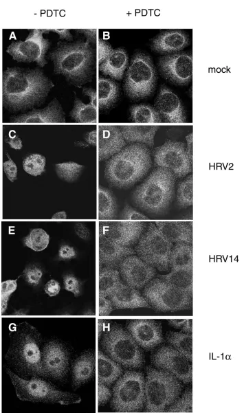
PDTC inhibits HRV-mediated NF-B activation in infected cells.A well-defined function of PDTC is the suppression of activation of the ubiquitous transcription factor NF-B under various conditions of induction (58, 59). As some reports em-ploying transgenic mice show that activation of NF-B is es-sential for picornavirus virulence (60, 63), we analyzed the cellular distribution of the p65 subunit of NF-B during HRV infection by using immunofluorescence (Fig. 10).
In uninfected HeLa cells, the p65 subunit is localized exclu-sively in the cytoplasm (Fig. 10A). During infection with HRV2 or HRV14, activation and nuclear translocation of NF-B were observed. At 8 h p.i., the p65 subunit of NF-B was detected exclusively in the nucleus (Fig. 10C and E). Com-FIG. 4. PDTC promotes cell survival following poliovirus infection.
HeLa S3 cells grown in RPMI medium were infected with poliovirus strain Mahoney at a multiplicity of infection of 1. After 3 h, the virus inoculum was removed and replaced by RPMI medium containing the indicated amounts of PDTC. At 24 h after infection, cells were stained with crystal violet.
on November 8, 2019 by guest
http://jvi.asm.org/
[image:5.587.55.266.74.277.2]FIG.
5.
Early
steps
in
the
viral
life
cycle
are
unaf
fected
by
PDTC
treatment.
HeLa
cells
were
infected
with
HRV1A
(A),
HRV2
(B),
HRV14
(C),
or
HRV16
(D).
PDTC
(125
M)
was
added
at
dif
ferent
time
points
between
1
an
d
8
h
after
infection
(w/o
PDTC,
no
PDTC
added).
Proliferation
assays
were
performed
24
h
after
infection.
All
values
represent
the
means
(
⫾
SD)
for
at
least
four
samples.
VOL. 76, 2002 ANTIVIRAL EFFECTS OF PDTC ON HUMAN RHINOVIRUS 6009
on November 8, 2019 by guest
http://jvi.asm.org/
pared to direct induction of NF-B by IL-1␣, the activation of NF-B during rhinovirus infection was a late event. IL-1␣
stimulation resulted in massive translocation of p65 to the nucleus after 1.5 h (Fig. 10G). IL-1␣-mediated translocation was a transient event. At 8 h after induction, NF-B was again found exclusively in the cytoplasm (data not shown).
Incubation of cells with PDTC completely inhibited NF-B translocation independent of whether NF-B was induced by HRV (Fig. 10B, D, and F) or by IL-1␣(Fig. 10H). This can be seen both in HeLa cells infected with HRV2 or with HRV14 (Fig. 10) and in 16HBE14o⫺cells infected with HRV14 (data
not shown). Although signaling leading to NF-B activation is supposed to occur via the generation of ROIs, it is interesting
that other antioxidants such as NAC or Trolox failed to pre-vent HRV-induced NF-B activation (data not shown).
DISCUSSION
Most of the inhibitors of picornavirus infection act at an early stage of infection, i.e., during virus binding, internaliza-tion, and/or uncoating. Only very few of the known inhibitors are effective late in the viral life cycle (8). In this study, we demonstrated novel functions of PDTC during infection with HRV. Treatment of infected cells with PDTC resulted in dras-tic reduction of rhinoviral replication. Even if the substance was applied several hours after HRV infection, the survival of the infected cells was greatly improved. HRV infection in the presence of PDTC was characterized by a strong reduction in the processing of cellular substrates by the viral 2A proteinase; at the same time, the expression of viral capsid proteins was reduced. The activation of the transcription factor NF-B usu-ally observed in HRV infections was absent in the PDTC-treated cells.
The events leading to the generation of ROIs during viral infections are still unclear. It was reported previously that the administration of antioxidative compounds during infection can lead to the inhibition of viral replication in a variety of viral systems, e.g., Sindbis virus (36), hepatitis B virus (71), bovine viral diarrhea virus (61), and retroviruses (27). As these viruses are unrelated to each other, it is postulated that these com-pounds affect a central process required for viral replication.
Mechanistically, it is believed that these viruses induce ap-optosis by oxidative stress mediated via ROIs. In several cases a pathway is triggered, leading to activation of the transcription factor NF-B. Interference with this pathway by antioxidants is believed to inhibit virus-induced apoptosis and thus inhibit efficient virus multiplication. In contrast, there are also reports indicating that under certain conditions PDTC acts as a pro-FIG. 6. Addition of PDTC at later stages still reduces viral
multi-plication. HeLa cells were infected with HRV2, and PDTC was added for the indicated intervals. Viral titers in the supernatants of cells after 24 h are shown for a representative experiment (in TCID50per
milli-liter).
FIG. 7. PDTC inhibits HRV-mediated cleavage of eIF4GI and reduces the expression of rhinoviral proteins. Protein extracts from HRV2-infected HeLa cells in the absence or presence of 125M PDTC were collected at the indicated time points after infection. (A) Western blot analyses were performed to detect cleavage of eIF4GI. c.p., cleavage products. (B) Western blot showing the expression of the rhinoviral proteins VP1, VP2, and VP3. Mock-infected cells served as controls.
on November 8, 2019 by guest
http://jvi.asm.org/
[image:7.587.59.267.71.237.2]FIG. 8. Other antioxidative compounds fail to show an antiviral effect. HeLa cells were infected with HRV2 (⫹HRV2) at 20 TCID50/cell and
treated with a wide range of concentrations of vitamin C (A), the vitamin E derivate Trolox (B), and 2-mercaptoethanol (2-ME) (C). Mock-infected cells (⫺HRV2) served as controls. All values represent the means (⫾SD) of at least four samples.
6011
on November 8, 2019 by guest
http://jvi.asm.org/
apoptotic drug (11, 34, 51). Depending on the viral system analyzed, antioxidative compounds differ in the ability to duce virus growth. For example, NAC and several other re-ducing substances like 2-mercaptoethanol and dithiothreitol completely inhibited Sindbis virus-induced cell death whereas other antioxidants such as Trolox and catalase were ineffective (36). NAC was also shown to inhibit hepatitis B virus replica-tion (71). In contrast, NAC and PDTC failed to prevent apo-ptosis in the case of a cytopathic strain of bovine viral diarrhea virus where butylated hydroxyanisole was effective (61). In this report we present evidence that PDTC, but not other com-monly used antioxidants, is able to protect cells from HRV-induced cell death. Similarly, PDTC inhibited cell death fol-lowing poliovirus infection.
Dithiocarbamates are potent agents and have been shown to be involved in a number of processes, suggesting that the antiviral effect of PDTC might not be due to its antioxidant function alone. Although dithiocarbamates are widely used as antioxidants, they can exert both pro- and antioxidative func-tions in the cell. Their antioxidant funcfunc-tions include eliminat-ing hydrogen peroxide, scavengeliminat-ing superoxide radicals, per-oxynitrite, hydroxyl radicals, and lipid peroxidation products (reference 72 and references therein). These reactions ulti-mately generate thiuram disulfide, the oxidized form of dithio-carbamates (48). In some cases, the formation of thiurams appears to be metal dependent (6, 47). Thiuram disulfides are responsible for the prooxidant effects of dithiocarbamates characterized by their potent oxidation of glutathione and pro-tein thiols (48, 50). There is evidence that this prooxidative effect inactivates caspases by thiol oxidation and thereby blocks apoptosis (49). As the picornaviral proteinases 2A and 3C are cysteine proteinases, they are highly sensitive to oxidation and are strictly dependent on the reducing environment of the cell. Changes in the reduction-oxidation balance would have detri-mental effects on proteinase function and hence on viral rep-lication. However, our preliminary experiments employing peptide cleavage assays by highly purified 2A proteinase as well
as eIF4G cleavage by nascent 2A proteinase do not support a mechanism of direct action of PDTC on 2A enzymatic activity. A well-described function of PDTC is the inhibition of ac-tivation of transcription factor NF-B (24, 58, 59). On the one hand, the inhibition of NF-B activation by PDTC is attributed to its radical scavenging properties (41, 59); on the other hand, it has been suggested that PDTC might exert its inhibitory effect via oxidation of critical thiols in NF-B (4, 5). This has also been shown for the tumor suppressor and transcription factor p53 (73, 74). Several investigators have demonstrated that PDTC exhibits anti-inflammatory effects through inhibit-ing NF-B activation in response to a variety of stimuli includ-ing IL-1, tumor necrosis factor alpha, LPS, and H2O2in several
cell types (59).
In this report we provide evidence that during HRV infec-tion NF-B is translocated to the nucleus at a late time point. Under the conditions used, NF-B activation was observed at 6 to 8 h p.i. At this stage, host cell protein synthesis is to a large extent shut off due to the cleavage of eIF4GI and eIF4GII. Under these conditions, mRNAs can be translated only by a cap-independent mechanism. However, recently several cellu-lar genes translated under cap-independent conditions were identified (25, 26, 38). Whether NF-B is involved in activation of these genes is not clear at present.
How can inactivation of NF-B affect viral multiplication? NF-B activation is a frequent process during viral infections, e.g., influenza A virus (7), alphavirus (36), dengue virus (40), Newcastle disease virus (69), and human T-cell leukemia virus type 1 (67). In some cases NF-B has been shown to be re-quired for maximal viral replication. NF-B activation is thought to provide a cellular environment favorable for effi-cient viral nucleic acid or protein synthesis, intracellular trans-port of viral proteins, or assembly and release of progeny virions. Thus, viral replication may be impaired by PDTC in-hibition of NF-B activation.
In the case of encephalomyocarditis virus, it was shown recently that NF-B knockout mice (p50⫺/⫺) survive infections
that readily kill normal mice (63). However, NF-B activation is not essential for virus multiplication in cell culture but is required for the inhibition of apoptosis and for prevention of premature cell death, thus affecting virulence in mice (60). In contrast, no significant apoptosis and/or cell death was de-tected when NF-B activation by HRV was inhibited by PDTC treatment (Fig. 10D and F). PDTC was also shown to be involved in activation of pleiotropic transcription factors, e.g., AP-1 (41), transactivator C/EBP(9), and heat shock factor 1 (29). Activation of genes regulated by these factors may ac-count for the stimulatory effect of PDTC on uninfected cells as observed in our experiments. Another biological property of PDTC is its activity as a chelator of Zn and Cu (42, 43). As many enzymatic functions are dependent on essential ions, chelation of these ions could lead to inhibition of enzyme function.
[image:9.587.62.265.75.262.2]To clarify the mechanism of PDTC action, it would be ex-tremely useful to obtain HRV variants resistant to PDTC. However, despite many attempts we have so far been unable to produce any PDTC-resistant HRV. Further long-term experi-ments are in progress to select for PDTC-resistant variants. Nevertheless, the fact that PDTC equally inhibits all tested HRV serotypes, whereas guanidine inhibits HRV14 but does FIG. 9. NAC does not show an antiviral effect. Cell viability of
NAC-treated HeLa cells was determined by crystal violet staining and compared to that of PDTC-treated cells.
on November 8, 2019 by guest
http://jvi.asm.org/
FIG. 10. PDTC inhibits HRV- and IL-1␣-induced NF-B activation. Immunofluorescence experiments located the NF-B subunit p65 in HeLa cells. Left column, cells without PDTC; right column, PDTC-treated cells. (A and B) Mock-infected cells. (C and D) Cells infected with HRV2 at 8 h p.i. (E and F) Cells infected with HRV14 at 8 h p.i. (G and H) Cells incubated with IL-1␣at 1.5 h after stimulation.
VOL. 76, 2002 ANTIVIRAL EFFECTS OF PDTC ON HUMAN RHINOVIRUS 6013
on November 8, 2019 by guest
http://jvi.asm.org/
not affect HRV2, can be taken to indicate that the mechanisms of action of these two inhibitors are different.
In conclusion, PDTC is an extremely potent antirhinoviral substance which reduces HRV growth and inhibits the CPE of infected cells in culture. PDTC also promotes cell viability following poliovirus infection. Elucidation of the antiviral mechanism of PDTC will be the subject of further research and should facilitate understanding of the complex interactions of viruses and cells which may eventually lead to novel ap-proaches for the treatment of picornavirus infections such as the common cold, the most frequent infection of humans.
ACKNOWLEDGMENTS
This work was supported by grant F508-MED from the Austrian Science Foundation to E.K. and by EMBO fellowship ASTF 9688 to J.S.
We thank D. Gruenert, San Francisco, Calif., for supplying 16HBE14o⫺cells, D. Blaas for providing HRV2 antibody, R.
Perez-Bercoff for his hospitality, and Z. Rattler for stimulating discussions.
REFERENCES
1.Agol, V. I., G. A. Belov, K. Bienz, D. Egger, M. S. Kolesnikova, N. T. Raikhlin, L. I. Romanova, E. A. Smirnova, and E. A. Tolskaya.1998. Two types of death of poliovirus-infected cells: caspase involvement in the
apo-ptosis but not cytopathic effect. Virology252:343–353.
2.Badorff, C., G. H. Lee, B. Lamphear, M. E. Martone, K. P. Campbell, R. E. Rhoads, and K. U. Knowlton.1999. Enteroviral protease 2A cleaves dystro-phin: evidence of cytoskeletal disruption in an acquired cardiomyopathy.
Nat. Med.5:320–326.
3.Bonifacino, J. S. (ed.).1998. Current protocols in cell biology, p. 4.3.1–4.3.4. John Wiley & Sons, Inc., New York, N.Y.
4.Brennan, P., and L. A. O’Neill.1996. 2-Mercaptoethanol restores the ability of nuclear factor kappa B (NF kappa B) to bind DNA in nuclear extracts from interleukin 1-treated cells incubated with pyrrolidine dithiocarbamate (PDTC). Evidence for oxidation of glutathione in the mechanism of
inhibi-tion of NF kappa B by PDTC. Biochem. J.320:975–981.
5.Brennan, P., and L. A. O’Neill.1995. Effects of oxidants and antioxidants on nuclear factor kappa B activation in three different cell lines: evidence against a universal hypothesis involving oxygen radicals. Biochim. Biophys. Acta1260:167–175.
6.Burkitt, M. J., H. S. Bishop, L. Milne, S. Y. Tsang, G. J. Provan, C. S. Nobel, S. Orrenius, and A. F. Slater.1998. Dithiocarbamate toxicity toward thymo-cytes involves their copper-catalyzed conversion to thiuram disulfides, which oxidize glutathione in a redox cycle without the release of reactive oxygen
species. Arch. Biochem. Biophys.353:73–84.
7.Bussfeld, D., M. Bacher, A. Moritz, D. Gemsa, and H. Sprenger.1997. Expression of transcription factor genes after influenza A virus infection.
Immunobiology198:291–298.
8.Carrasco, L.1994. Picornavirus inhibitors. Pharmacol. Ther.64:215–290. 9.Chinery, R., J. A. Brockman, D. T. Dransfield, and R. J. Coffey.1997.
Antioxidant-induced nuclear translocation of CCAAT/enhancer-binding protein beta. A critical role for protein kinase A-mediated phosphorylation
of Ser299. J. Biol. Chem.272:30356–30361.
10.Connolly, J. L., S. E. Rodgers, P. Clarke, D. W. Ballard, L. D. Kerr, K. L. Tyler, and T. S. Dermody.2000. Reovirus-induced apoptosis requires
acti-vation of transcription factor NF-B. J. Virol.74:2981–2989.
11.Della Ragione, F., V. Cucciolla, A. Borriello, V. Della Pietra, C. Manna, P. Galletti, and V. Zappia.2000. Pyrrolidine dithiocarbamate induces apoptosis
by a cytochromec-dependent mechanism. Biochem. Biophys. Res. Commun.
268:942–946.
12.Einarsson, O., G. P. Geba, Z. Zhu, M. Landry, and J. A. Elias.1996. Interleukin-11: stimulation in vivo and in vitro by respiratory viruses and
induction of airways hyperresponsiveness. J. Clin. Investig.97:915–924.
13.Erl, W., C. Weber, and G. K. Hansson.2000. Pyrrolidine
dithiocarbamate-induced apoptosis depends on cell type, density, and the presence of Cu2⫹
and Zn2⫹. Am. J. Physiol. Cell Physiol.278:1116–1125.
14.Etchison, D., and S. Fout.1985. Human rhinovirus 14 infection of HeLa cells result in the proteolytic cleavage of the p220 cap-binding complex subunit
and inactivates globin mRNA translation in vitro. J. Virol.54:634–638.
15.Etchison, D., S. C. Milburn, I. Edery, N. Sonenberg, and J. W. Hershey.
1982. Inhibition of HeLa cell protein synthesis following poliovirus infection correlates with the proteolysis of a 220,000-dalton polypeptide associated with eucaryotic initiation factor 3 and a cap binding protein complex. J. Biol.
Chem.257:14806–14810.
16.Flory, E., M. Kunz, C. Scheller, C. Jassoy, R. Stauber, U. R. Rapp, and S.
Ludwig.2000. Influenza virus-induced NF-B-dependent gene expression is mediated by overexpression of viral proteins and involves oxidative radicals
and activation of IB kinase. J. Biol. Chem.275:8307–8314.
17.Garibaldi, R. A. 1985. Epidemiology of community-acquired respiratory tract infections in adults. Incidence, etiology, and impact. Am. J. Med.
78:32–37.
18.Glaser, W., and T. Skern.2000. Extremely efficient cleavage of eIF4G by
picornaviral proteinases L and 2A in vitro. FEBS Lett.480:151–155.
19.Gradi, A., Y. V. Svitkin, H. Imataka, and N. Sonenberg.1998. Proteolysis of human eukaryotic translation initiation factor eIF4GII, but not eIF4GI, coincides with the shutoff of host protein synthesis after poliovirus infection.
Proc. Natl. Acad. Sci. USA95:11089–11094.
20.Gwaltney, J. M.1975. Rhinoviruses. Yale J. Biol. Med.48:17–45. 21.Heinz, B. A., and L. M. Vance.1995. The antiviral compound enviroxime
targets the 3A coding region of rhinovirus and poliovirus. J. Virol.69:4189–
4197.
22.Huang, R. P., J. X. Wu, Y. Fan, and E. D. Adamson.1996. UV activates
growth factor receptors via reactive oxygen intermediates. J. Cell Biol.133:
211–220.
23.Israel, N., and M. A. Gougerot-Pocidalo.1997. Oxidative stress in human
immunodeficiency virus infection. Cell. Mol. Life Sci.53:864–870.
24.Janssen, Y. M., and C. K. Sen.1999. Nuclear factor kappa B activity in
response to oxidants and antioxidants. Methods Enzymol.300:363–374.
25.Johannes, G., M. S. Carter, M. B. Eisen, P. O. Brown, and P. Sarnow.1999. Identification of eukaryotic mRNAs that are translated at reduced cap bind-ing complex eIF4F concentrations usbind-ing a cDNA microarray. Proc. Natl.
Acad. Sci. USA96:13118–13123.
26.Johannes, G., and P. Sarnow.1998. Cap-independent polysomal association of natural mRNAs encoding c-myc, BiP, and eIF4G conferred by internal
ribosome entry sites. RNA4:1500–1513.
27.Kalebic, T., A. Kinter, G. Poli, M. E. Anderson, A. Meister, and A. S. Fauci.
1991. Suppression of human immunodeficiency virus expression in
chroni-cally infected monocytic cells by glutathione, glutathione ester, andN
-ace-tylcysteine. Proc. Natl. Acad. Sci. USA88:986–990.
28.Kaul, P., M. C. Biagioli, I. Singh, and R. B. Turner.2000. Rhinovirus-induced oxidative stress and interleukin-8 elaboration involves p47-phox but is independent of attachment to intercellular adhesion molecule-1 and viral
replication. J. Infect. Dis.181:1885–1890.
29.Kim, S. H., S. I. Han, S. Y. Oh, H. Y. Chung, H. D. Kim, and H. S. Kang.
2001. Activation of heat shock factor 1 by pyrrolidine dithiocarbamate is mediated by its activities as pro-oxidant and thiol modulator. Biochem.
Biophys. Res. Commun.281:367–372.
30.Kräusslich, H. G., M. J. Nicklin, H. Toyoda, D. Etchison, and E. Wimmer.
1987. Poliovirus proteinase 2A induces cleavage of eucaryotic initiation
fac-tor 4F polypeptide p220. J. Virol.61:2711–2718.
31.Lamphear, B. J., R. Q. Yan, F. Yang, D. Waters, H. D. Liebig, H. Klump, E. Kuechler, T. Skern, and R. E. Rhoads.1993. Mapping the cleavage site in protein synthesis initiation factor–eIF-4-gamma of the 2A proteases from
human coxsackievirus and rhinovirus. J. Biol. Chem.268:19200–19203.
32.Lenk, R., and S. Penman.1979. The cytoskeletal framework and poliovirus
metabolism. Cell16:289–301.
33.Levine, B., Q. Huang, J. T. Isaacs, J. C. Reed, D. E. Griffin, and J. M. Hardwick.1993. Conversion of lytic to persistent alphavirus infection by the
bcl-2 cellular oncogene. Nature361:739–742.
34.Li, W. G., L. Coppey, R. M. Weiss, and H. J. Oskarsson.2001. Antioxidant therapy attenuates JNK activation and apoptosis in the remote noninfarcted myocardium after large myocardial infarction. Biochem. Biophys. Res.
Com-mun.280:353–357.
35.Liebig, H. D., E. Ziegler, R. Yan, K. Hartmuth, H. Klump, H. Kowalski, D. Blaas, W. Sommergruber, L. Frasel, B. Lamphear, R. Rhoads, E. Kuechler, and T. Skern.1993. Purification of two picornaviral 2A proteinases: inter-action with eIF-4 gamma and influence on in vitro translation. Biochemistry
32:7581–7588.
36.Lin, K. I., S. H. Lee, R. Narayanan, J. M. Baraban, J. M. Hardwick, and R. R. Ratan.1995. Thiol agents and Bcl-2 identify an alphavirus-induced apoptotic pathway that requires activation of the transcription factor
NF-kappa B. J. Cell Biol.131:1149–11461.
37.Lo, Y. Y., and T. F. Cruz.1995. Involvement of reactive oxygen species in cytokine and growth factor induction of c-fos expression in chondrocytes.
J. Biol. Chem.270:11727–11730.
38.Macejak, D. G., and P. Sarnow.1991. Internal initiation of transcription
mediated by the 5⬘leader of a cellular mRNA. Nature353:90–94.
39.Malorni, W., R. Rivabene, M. T. Santini, and G. Donelli.1993.N -Acetyl-cysteine inhibits apoptosis and decreases viral particles in HIV-chronically
infected U937 cells. FEBS Lett.327:75–78.
40.Marianneau, P., A. Cardona, L. Edelman, V. Deubel, and P. Despres.1997.
Dengue virus replication in human hepatoma cells activates NF-B which in
turn induces apoptotic cell death. J. Virol.71:3244–3249.
41.Meyer, M., R. Schreck, and P. A. Baeuerle.1993. H2O2and antioxidants
have opposite effects on activation of NF-kappa B and AP-1 in intact cells:
AP-1 as secondary antioxidant-responsive factor. EMBO J.12:2005–2015.
42.Morpurgo, L., E. Agostinelli, O. Befani, and B. Mondovi.1987. Reactions of
on November 8, 2019 by guest
http://jvi.asm.org/
bovine serum amine oxidase withN,N-diethyldithiocarbamate. Selective
re-moval of one copper ion. Biochem. J.248:865–870.
43.Morpurgo, L., A. Desideri, A. Rigo, P. Viglino, and G. Rotilio.1983. Reaction
ofN,N-diethyldithiocarbamate and other bidentate ligands with Zn, Co and
Cu bovine carbonic anhydrases. Inhibition of the enzyme activity and evi-dence for stable ternary enzyme-metal-ligand complexes. Biochim. Biophys.
Acta746:168–175.
44.Mortola, E., M. Okuda, K. Ohno, T. Watari, H. Tsujimoto, and A. Hasegawa.
1998. Inhibition of apoptosis and virus replication in feline
immunodefi-ciency virus-infected cells byN-acetylcysteine and ascorbic acid. J. Vet. Med.
Sci.60:1187–1193.
45.Naclerio, R. M., D. Proud, L. M. Lichtenstein, A. Kagey-Sobotka, J. O. Hendley, J. Sorrentino, and J. M. Gwaltney.1988. Kinins are generated
during experimental rhinovirus colds. J. Infect. Dis.157:133–142.
46.Noah, T. L., F. W. Henderson, I. A. Wortman, R. B. Devlin, J. Handy, H. S. Koren, and S. Becker.1995. Nasal cytokine production in viral acute upper
respiratory infection of childhood. J. Infect. Dis.171:584–592.
47.Nobel, C. I., M. Kimland, B. Lind, S. Orrenius, and A. F. Slater.1995. Dithiocarbamates induce apoptosis in thymocytes by raising the intracellular
level of redox-active copper. J. Biol. Chem.270:26202–26208.
48.Nobel, C. S., D. H. Burgess, B. Zhivotovsky, M. J. Burkitt, S. Orrenius, and A. F. Slater.1997. Mechanism of dithiocarbamate inhibition of apoptosis: thiol oxidation by dithiocarbamate disulfides directly inhibits processing of
the caspase-3 proenzyme. Chem. Res. Toxicol.10:636–643.
49.Nobel, C. S., M. Kimland, D. W. Nicholson, S. Orrenius, and A. F. Slater.
1997. Disulfiram is a potent inhibitor of proteases of the caspase family.
Chem. Res. Toxicol.10:1319–1324.
50.Orrenius, S., C. S. Nobel, D. J. van den Dobbelsteen, M. J. Burkitt, and A. F. Slater.1996. Dithiocarbamates and the redox regulation of cell death.
Bio-chem. Soc. Trans.24:1032–1038.
51.Ozaki, K., H. Takeda, H. Iwahashi, S. Kitano, and S. Hanazawa.1997.
NF-B inhibitors stimulate apoptosis of rabbit mature osteoclasts and inhibit
bone resorption by these cells. FEBS Lett.410:297–300.
52.Pelham, H. R., and R. J. Jackson.1976. An efficient mRNA-dependent
translation system from reticulocyte lysates. Eur. J. Biochem.67:247–256.
53.Pitkaranta, A., and F. G. Hayden.1998. What⬘s new with common colds?
Pathogenesis and diagnosis. Infect. Med.15:50–59.
54.Proud, D., C. R. Baumgarten, R. M. Naclerio, and P. E. Ward.1987. Kinin metabolism in human nasal secretions during experimentally induced
aller-gic rhinitis. J. Immunol.138:428–434.
55.Reed, L. J., and H. Muench.1938. A simple method of estimating fifty per
cent endpoints. Am. J. Hyg.27:493–497.
56.Rightsel, W. A., J. R. Dice, R. J. McAlpine, E. A. Timm, I. W. McLean, G. J. Dixon, and F. M. Schabel.1961. Antiviral effect of guanidine. Science134:
558–559.
57.Rubinstein, S. J., T. Hammerle, E. Wimmer, and A. Dasgupta.1992. Infec-tion of HeLa cells with poliovirus results in modificaInfec-tion of a complex that
binds to the rRNA promoter. J. Virol.66:3062–3068.
58.Schreck, R., K. Albermann, and P. A. Baeuerle.1992. Nuclear factor kappa B: an oxidative stress-responsive transcription factor of eukaryotic cells. Free
Radic. Res. Commun.17:221–237.
59.Schreck, R., B. Meier, D. N. Mannel, W. Droge, and P. A. Baeuerle.1992. Dithiocarbamates as potent inhibitors of nuclear factor kappa B activation in
intact cells. J. Exp. Med.175:1181–1194.
60.Schwarz, E. M., C. Badorff, T. S. Hiura, R. Wessely, A. Badorff, I. M. Verma, and K. U. Knowlton.1998. NF-B-mediated inhibition of apoptosis is
re-quired for encephalomyocarditis virus virulence: a mechanism of resistance
in p50 knockout mice. J. Virol.72:5654–5660.
61.Schweizer, M., and E. Peterhans.1999. Oxidative stress in cells infected with bovine viral diarrhea virus: a crucial step in the induction of apoptosis.
J. Gen. Virol.80:1147–1155.
62.Seipelt, J., H. D. Liebig, W. Sommergruber, C. Gerner, and E. Kuechler.
2000. 2A proteinase of human rhinovirus cleaves cytokeratin 8 in infected
HeLa cells. J. Biol. Chem.275:20084–20089.
63.Sha, W. C., H. C. Liou, E. I. Tuomanen, and D. Baltimore.1995. Targeted disruption of the p50 subunit of NF-kappa B leads to multifocal defects in
immune responses. Cell80:321–330.
64.Shibayama, Y., D. Skoner, S. Suehiro, J. E. Konishi, P. Fireman, and A. P. Kaplan.1996. Bradykinin levels during experimental nasal infection with
rhinovirus and attenuated influenza virus. Immunopharmacology33:311–
313.
65.Skern, T., W. Sommergruber, D. Blaas, C. Pieler, and E. Kuechler.1984. Relationship of human rhinovirus strain 2 and poliovirus as indicated by
comparison of the polymerase gene regions. Virology136:125–132.
66.Sommergruber, W., H. Ahorn, A. Zöphel, I. Maurer-Fogy, F. Fessl, G. Schnorrenberg, H. D. Liebig, D. Blaas, E. Kuechler, and T. Skern.1992. Cleavage specificity on synthetic peptide substrates of human rhinovirus 2
proteinase 2A. J. Biol. Chem.267:22639–22644.
67.Sun, S. C., and D. W. Ballard.1999. Persistent activation of NF-B by the tax
transforming protein of HTLV-1: hijacking cellular IB kinases. Oncogene
18:6948–6958.
68.Tershak, D. R.1982. Guanidine-resistant defective interfering particles of
poliovirus. J. Virol.41:615–625.
69.Umansky, V., V. A. Shatrov, V. Lehmann, and V. Schirrmacher.1996. In-duction of NO synthesis in macrophages by Newcastle disease virus is
asso-ciated with activation of nuclear factor-kappa B. Int. Immunol.8:491–498.
70.Vile, G. F.1997. Active oxygen species mediate the solar ultraviolet radia-tion-dependent increase in the tumour suppressor protein p53 in human skin
fibroblasts. FEBS Lett.412:70–74.
71.Weiss, L., E. Hildt, and P. H. Hofschneider.1996. Anti-hepatitis B virus
activity ofN-acetyl-L-cysteine (NAC): new aspects of a well-established drug.
Antivir. Res.32:43–53.
72.Wild, A. C., and R. T. Mulcahy.1999. Pyrrolidine dithiocarbamate up-regulates the expression of the genes encoding the catalytic and regulatory subunits of gamma-glutamylcysteine synthetase and increases intracellular
glutathione levels. Biochem. J.338:659–665.
73.Wu, H. H., and J. Momand.1998. Pyrrolidine dithiocarbamate prevents p53
activation and promotes p53 cysteine residue oxidation. J. Biol. Chem.273:
18898–18905.
74.Wu, H. H., J. A. Thomas, and J. Momand.2000. p53 protein oxidation in cultured cells in response to pyrrolidine dithiocarbamate: a novel method for relating the amount of p53 oxidation in vivo to the regulation of
p53-responsive genes. Biochem. J.351:87–93.
75.Yalamanchili, P., K. Harris, E. Wimmer, and A. Dasgupta.1996. Inhibition of basal transcription by poliovirus: a virus-encoded protease (3Cpro)
inhib-its formation of TBP-TATA box complex in vitro. J. Virol.70:2922–2929.
76.Yan, R., W. Rychlik, D. Etchison, and R. E. Rhoads.1992. Amino acid sequence of the human protein synthesis initiation factor eIF-4g. J. Biol.
Chem.267:23226–23231.
77.Zhu, Z., W. Tang, A. Ray, Y. Wu, O. Einarsson, M. L. Landry, J. Gwaltney, and J. A. Elias.1996. Rhinovirus stimulation of interleukin-6 in vivo and in vitro. Evidence for nuclear factor kappa B-dependent transcriptional
activa-tion. J. Clin. Investig.97:421–430.
VOL. 76, 2002 ANTIVIRAL EFFECTS OF PDTC ON HUMAN RHINOVIRUS 6015