A STUDY ON PREVALENCE OF HEARING LOSS AS A
COMPLICATION OF DIABETES
DISSERTATION
SUBMITTED FOR
M.S IN OTO RHINO LARYNGOLOGY
THE TAMILNADU Dr. M.G.R MEDICAL UNIVERSITY
DEPARTMENT OF E.N.T
PSG INSTITUTE OF MEDICAL SCIENCES & RESEARCH PEELAMEDU, COIMBATORE- 641 004
TAMILNADU, INDIA
CERTIFICATE
This is to certify that the thesis entitled ‘A study on prevalence of
hearing loss as a complication of diabetes’ is a bonafide work of
Dr. Prathula Sivakumar done under the guidance and supervision of
Dr. S. Palaninathan, MS in the Department of E.N.T, PSG Institute of Medical Sciences and Research, Coimbatore in fulfillment of the
regulations of Dr. MGR Medical University for the award of M.S. Degree
in Oto-Rhino-Laryngology.
DR. S. PALANINATHAN Dr. George Zacharias
Professor Professor & HOD
Dept. Of E.N.T Dept. Of E.N.T
DECLARATION
I hereby decalre that this study dissertation entitled “A study on prevalence of hearing loss as a complication of diabetes” was prepared by me under the direct guidance and supervision of Professor of E.N.T,
Dr.S. Palaninathan, MS, PSG Institute of Medical Sciences & Research,
Coimbatore.
This dissertation is submitted to the Tamil Nadu Dr. MGR Medical
University in fulfillment of the University regulations for the award of
M.S. degree in Oto-Rhino-Laryngology. This dissertation has not been
submitted for the award of any other Degree or Diploma.
CERTIFICATE BY THE GUIDE
This is to certify that the thesis entitled “A study on prevalence of hearing loss as a complication of diabetes” is a bonafide work of Dr. Prathula Sivakumar done under my direct guidance and supervision in the
Department of E.N.T, PSG Institute of Medical Sciences and Research,
Coimbatore in fulfillment of the regulations of DR. MGR Medical
University for the award of M.S. degree in Oto-Rhino-Laryngology.
Dr. S. PALANINATHAN Professor
ACKNOWLEDGEMENTS
It is my greatest pleasures to recall the people who have helped me
in the completion of my dissertation.
It gives me immense pleasure to express my heartfelt gratitude and
sincere thanks to my beloved teacher, Professor and HOD Dr. George Zacharias, Dept. of E.N.T, PSG IMS & R, Coimbatore for being an inspiration and for providing all support during this study, without whose
towering presence this study would not have been possible.
I thank my teacher, Dr. S. Palaninathan, Professor of E.N.T for his staunch support during the period of this study. His suggestions have
helped me in improving the quality of this work.
I would like to express my sincere gratitude to Dr. A. Dayanand, Associate Professor who provided valuable tips from time to time which
helped me complete my work. My associate Professor, Dr. V. Ravishankar has guided me in all stages of study.
I take this opportunity to thank Dr. Anil Mathew, Professor in Biostatistics whose inputs were very valuable in completion of this study.
I take this opportunity to recognize the efforts by E.N.T department
staff, Mrs. Chitrapriya, Mrs. Gomathi , Mrs. Anandhi and Ms. Nithya for helping me collect all necessary patient details. I also want to mention a special thanks to the audiology technicians Mrs. Prema and Mr. keerthiraj for helping me in the audiology session.
I am thankful to my family especially my father and my husband
who were the real source of encouragement for me during my dissertation
work. I thank my post Graduate colleagues for their whole-hearted
support.
At the outset I would like to sincerely thank Dr. S. Ramalingam, Principal and the Ethics Committee, PSG IMS & R for having consented
to this study.
Finally my heartfelt appreciation & greatest thanks to all the
CONTENTS
SL.No CONTENTS PAGE NO
1 INTRODUCTION 1
2 AIMS OF STUDY 3
3 REVIEW OF LITERATURE 4
4 RATIONALE FOR STUDY 71
5 MATERIALS AND METHODS 72
6 RESULTS 78
7 DISCUSSION 94
8 LIMITATIONS OF STUDY 103
9 CONCLUSIONS 104
10 BIBLIOGRAPHY
INTRODUCTION
Hearing impairment is the most common sensory deficit in human beings.
It affects more than 250 million people in the world. The consequences of
hearing impairment include difficulty in interpret of speech, often
producing a decreased ability to communicate, delay in language
acquisition, educational and economic drawbacks, social isolation and
stigmatization. It may turn bad along with by medical conditions such as
hypothyroidism, diabetes, and hyperlipidemia, among others.
Hearing loss can be classified into 3 types:
A. Conductive hearing loss
B. Sensorineural hearing loss
C. Mixed hearing loss
Hearing of an individual could by assessed by clinical and audiometric
tests. Pure tone audiometery is a simple audiometric test which is being
used to measure the degree of hearing impairment.
Diabetes mellitus is an increasing health problem worldwide and the
prevalence is steadily increasing. And it is more pronounced in India (an
Diabetes Mellitus is a metabolic disorder. It presents because of partial or
complete reduction of insulin levels which results in increased levels of blood
glucose in association with long term complications which may be vascular or
neurological. Among the metabolism disorders of glucose, diabetes is found
commonly related to auditory disorders. It also affects metabolism of lipids
and proteins. Though Diabetes Mellitus has various etiologies, the most
common presentation is hyperglycemia. The physiological basis of type 2
diabetes mellitus is a combined form of both impairment of beta cell function,
with significant raise in peripheral levels of insulin resistance near the levels
of receptor and post receptor. The complex arrangement of inner ear makes it
potential target of hyperglycaemic damage.
Diabetic patients are more prone to complications of hyperglycemia, as all
body cells are exposed to high levels of plasma glucose. The organ of corti
cells are important structures for hearing mechanism and turn out to be the
potential target for damage, due to high glycemic levels, micro vascular
compromise, their complex structure and arrangement. Thus screening of
these patients at a higher risk of developing sensory neural hearing loss, will
AIM OF STUDY
1. To Study the prevalence of Hearing loss and its association with
Diabetes.
2. To assess the hearing loss in diabetics and correlate it with age and
REVIEW OF LITERATURE
The pure tone hearing average in frequencies 250, 500, 1000, 2000,
4000, 6000, 8000 Hz heard in more than 25 decibel in the ear is called as
hearing loss. Hearing loss is a dysfunction of hearing. Its severity varies
from mild; moderate; severe or profound. In general hearing loss can be
divided into conductive type, sensorineural type or mixed type.[1]
Hearing impairment is the most common sensory deficit in human
populations, affecting more than 250 million people in the world. The
consequences of hearing impairment include inability to interpret speech
sounds, often producing a reduced ability to communicate, delay in
language acquisition, economic and educational disadvantage, social
isolation and stigmatization. It may be worsened by some medical
conditions such as diabetes, hypothyroidism and possibly hyperlipidemia,
among others.[2]
Global Burden of Disease 2000 study which was published in the World
Health Report 2001 the adult-onset hearing loss is the 2nd most leading
cause of Years lead with diseases in global level, which accounts for
ANATOMICAL CONSIDERATIONS:
The human ear has three parts the outer, the middle and the inner
ear. During the 6th week of embryonic life, about 6 tubercles appear near
the 1st branchial cleft. These tuberceles gradually coincide to become
auricle. By 20th week, pinna achieves adult shape.
The external auditory canal forms from the 1st branchial cleft. In
the 16th embryonic week, the cells multiply from below ectodermal cleft
and forms the meatal plug. Recanalisation from this forms the epithelial
lining in the bony meatus. Recanalisation starts deep near the tympanic
membrane and grows outwards. External ear canal is completely formed
in the 28th week.
The development of tympanic membrane is from the three
germinal layers. The outer epithelial layer develops from the ectoderm,
the inner mucosal layer is from the endoderm and the middle fibrous
layer is from the mesoderm.
The Eustachian tube, tympanic cavity, attic, antrum and mastoid air
cells forms from the endoderm in the tubotympanic recess. This is found
arising from the first and second pharyngeal pouch partially. The malleus
develops from the 2nd arch except the footplate and the annular ligament
which is formed from the Otic capsule.
The inner ear is the first organ of special senses to be formed in
man. The evolution of the inner ear starts during the 3rd week of
embryonic life and is completely from the sixteenth week. Ectoderm in
this region of the hind brain becomes thick to form the auditory placode
which invaginates to become the Auditory vesicle or the Otocyst. This
then differentiates to form the endolymphatic duct and sac, the
semicircular ducts, saccule, the utricle and the cochlea. The cochlea is
fully developed by 20th week of gestation.
The external ear consists of the auricle or pinna, external acoustic
canal and tympanic membrane. The entire pinna, except the lobule, and
the outer part of external acoustic meatus are derived from a single
framework of yellow elastic cartilage which is covered by skin. This is
attached tightly to the perichondrium over the lateral surface and it is
loosely attached over the medial surface. The external acoustic canal
starts below the concha till the tympanic membrane. It measures around
24mm in its posterior wall. There are two parts of the canal namely the
cartilaginous and the bony part. The cartilaginous part measures about
8mm and is the outer part of the canal. The skin, covering this region is
hair follicles are limited only in the outer part of canal. The bony part
forms inner two- thirds and measures 16mm. Skin lining this region is
thiner and it continuity with the tympanic membrane. It is free of hair
follicles and cerumin containing gland.
The tympanic membrane forms the division from the external canal
and the middle ear. They are placed obliquity, measures about 9- 10mm
in height. It is divided into two parts,the pars tensa and the pars flaccida.
Pars tensa forms most of the tympanic memebrane . The central part is
withdrawn inwards near the tip of the malleus and this region is called
Umbo. Bright cone of light is usually found radiating from the tip of
malleus extending till the periphery of the anteroinferior quadrant. Pars
flaccid also known as Sharpnel’s membrane is situated above the lateral
process of malleus between the notch of Rivinus and the anterior and
posterior malleal folds. It appears pink and not so taut. The tympanic
membrane is divided into 3 layers (i) The epithelial layer is outer most,
found in continuity with the meatus skin lining (ii) The mucosal layer is
innermost, continous from the mucosa in the middle ear. (iii) Fibrous
layer is in middle, encloses the handle of malleus. This layer has 3 types
The middle ear along with eustachian tube, aditus, antrum and mastoid air
cells is called the middle ear cleft. It is covered by mucosa and it is filled
with air. Middle ear is compared to a box with six sides with a roof, a
floor, lateral, medial, anterior and posterior walls. The mastoid antrum is
the largest cell and it contains air and communicates with the attic
through the aditus in the upper part. The tegmen antri forms the roof and
this separates it from the middle cranial fossa. Aditus is an opening
through which the attic communicates with the antrum. The facial nerve
is found just below the aditus
Three ossicles are found in the middle ear. They are the malleus,
the incus and the stapes. Malleus has a head, handle, neck, a lateral
process and anterior process. The head and neck of malleus lie in the
attic. The lateral process forms a knob- like projection and gives
attachment to anterior and posterior malleal folds. The incus consists of
the body and short process both of which lie in the attic. The long process
is attached to the head of stapes. The stapes is divided into the head, foot
plate, neck, anterior and posterior crura. It is attaches to the oval window
by the annular ligament. The ossicles direct the sound energy from the
The internal ear also called the labyrinth is an organ of importance for
balance and hearing. It contains the bony and membranous labyrinth.
Membranous labyrinth contains fluid which is clear called endolymph.
Perilymph is seen in the space seperating the membranous and bony
labyrinth . The bony labyrinth has three parts: the vestibule, the
semicircular canals and the cochlea. The membranous labyrinth consists
of the cochlear duct, the utricle, saccule, the endolymphatic duct and sac
and three semicircular ducts,
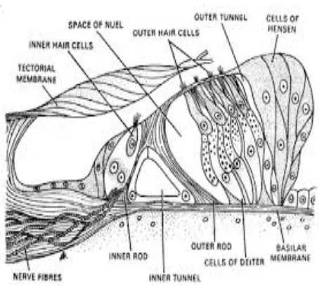
The organ of Corti is situated on the basilar membrane. It is the
sense organ of hearing and balance. The organ of Corti contains the
tunnel of Corti , hair cells , supporting cells , tectorial membrane . The
inner and outer rods form the tunnel of corti. The fluid inside is called the
cortilymph. The hair cells act as a significant receptor cells for hearing. It
transforms sound to electrical energy. Inner hair cells are arranged in a
single row while the outer hair cells are placed in 3 to 4 rows. The inner
hair cells are richly supplied by afferent cochlear fibres and it is probably
more important in transmission of auditory impulses. The outer hair cells
mostly receive efferent innervations from the Olivary complex. It is
concerned with modulating the function of the inner hair cells. There are
a total of 3500 inner hair cells and they are flask shaped . The outer hair
Acoustics:
Sound is a form of energy produced by a vibrating object. A sound wave
consists of compression and rarefaction of molecules of the medium in
which it travels. The sound velocity differs from media to media. Sound
travels fastest in solid medium.
Frequency is the number of cycles per second. The unit of frequency is
Hertz (Hz) named after Heinrich Rudolf Hertz who was a German
scientist. A sound of 1000 Hz means 1000 cycles per second.
Pure tone is a single frequency of sound. In pure-tone audiometry the
threshold of hearing is measured in decibles for varied pure tones ranging
from 125 to 8000.
Complex sound is a sound which contains many frequency. Human voice
is an example of complex sound.
Pitch is a subjective sensation which is produced by frequency of sound.
More the frequency, more is the pitch.
A complex sound consists of basic frequency, i.e. the lowest frequency in
which a source is set into vibrations. Frequencies above this level are
termed as overtones. The latter determines the quality or the timber of
Intensity is the sound strength which determines its loud nature. It is
usually calculated in decibles. At an approximate distance of 1 metre, the
intensity of
Whisper = 30 dB
Normal conversation = 60 dB
Shout = 90 dB
Discomfort of the ear = 120 dB
Pain in the ear = 130 dB
Loudness is a sensation that is subjective, which is produced by intensity.
More the intensity of sound, more is the loudness.
Decibel(dB) is denoted as 1/10th of a bel and it is named in remembrance
of Sir Alexander Graham Bell. It is the logarithmic ratio among two
sounds which is the sound being detailed and sound of reference. Sound
can be measured as power. Level of sound pressure is the measure of
sound in audiology. SPL is compared in reference of sound which
containd an SPL of 0.0002 dynes/cm2 or 20µ Pa (micropascals) this
approximately corresponds to the threshold in normal subjects with
hearing under normal limits at 1000 Hz. Decibel is used to avoid large
figures of sound pressure level.
Noise is descirbed as an aperiodic complex of sound. The three varities of
noise are:
(a)White noise: this consists of all the frequencies in the entire audible
spectrum. It can be compared to white light which contains all the
colours in the visible spectrum. This is a broad-band noise
generally used for purpose of masking.
(b)Narrow band noise: This is white noise that contains specific
frequencies which are above and below the given noise which is
filtered out. Thus its frequency range is smaller than that of
broad-band white noise. This is also used in masking specific test
frequency during pure tone audiometry.
(c)Speech noise: All noises in speech range frequencies are called
speech noise (300-3000 Hz). All the other frequencies are generally
filtered out.
Masking is a mechanism to produce inaudibility of one sound by the
producing another. In clinical audiometry, one ear is occupied by a sound
while the other ear is being tested. Masking of ear which is not tested is
very essential in all forms of bone conduction tests, while in case of air
conduction tests, it is required only when the difference of hearing
In clinical audiometry, one ear is occupied by a sound while the other ear
is being tested. Masking of ear which is not tested is very essential in all
forms of bone conduction tests, while in case of air conduction tests, it is
required only when the difference of hearing between two ears exceeds
more than 40 dB.
Wide range of human voices falls in this range. PTA is the average
threshold measure of all three frequencies of hearing. This roughly equal
to the speech reception threshold.
Hearing level is the sound pressure level which is delivered by an
audiometer at a respective frequency. It is measured in decibels. The
reference is maintained at audiometric zero. If an audiometer produces a
sound at 70dB, it is represented as 70dB HL.
Physiology of hearing:
Sound waves travel through external auditory meatus and produce
vibrations in the tympanic membrane. Vibrations from tympanic
membrane travel through malleus and incus and reach the stapes resulting
in the movement of stapes. Movements of stapes produce vibrations in
the fluid of cochlea. These vibrations stimulate the hair cells in the organ
of Corti. This in turn, causes generation of action potential in the auditory
perception of hearing occurs. Thus, during the process of hearing, ear
converts enery of sound waves into action potentials in the auditory nerve
fibers. This process is called sound transduction.
Role of inner ear:
Traveling wave: Movement of foot plate of stapes against oval
window causes movement of perilymph in scala vestibuli. This fluid does
not move all the way from oval window to round window through
helicotrema. It immediately hits the vestibular membrane near oval
window. This causes movement of fluid in scala media, since the
vestibular membrane is flexible. This causes bulging of the basilar
membrane towards scala tympani. This increases the elastic tension in
basilar fibres in the portion of the basilar membrane. This tension initiates
a wave,which travels along basilar membrane towards the helicotrema.
Resonance point: It is a part of basilar membrane, which is
activated by traveling wave. Initially each wave is weak. When it travels
through the basilar membrane from base towards apex, the wave becomes
stronger and at one point it becomes very strong and activates the basilar
membrane. This resonance point of the basilar membrane immediately
vibrates back and forth. The traveling wave stops here. Distance between
stapes and resonance point is inversely propotional to frequency of sound
disappears near the base of the chochlea, medium-pitched sound reaches
half of the way and wave generated by low pitched sounds travel the
entire distance of the basilar membrane.
Excitation of hair cells:
Stereocilia of hair cells in organ of cortii are embedded in tectorial
membrane. Haircells are tightly fixed by cuticular lamina reticularis and
the pillar cells. When travelling wave causes vibration of basilar
membrane at the resonance point, the basilar fiber, pillar cells, hair cells
and lamina reticularis move as a single unit. It causes movements of
stereocilia leading to excitement of hair cells and generation of receptor
potential.
Sound Transduction:
It is type of sensory transduction in the hair cells in the organ of
Corti by which sound energy is converted into action potentials in the
auditory nerve fiber. Three types of electrical events that occur during
sound transduction are :
(1)Receptor potential or chochlear microphonic potential
(2)Endochochlear potential or endolymphatic potential
Role of hair cells:
Inner hair cells and outer hair cells have different roles during sound
transduction. The inner haircells are responsible for sound transduction,
i.e. these receptor cells are the primary sensory cells, which causes the
generation of action potential in auditory nerve fibers. Outer hair cells
have a different action. These hair cells are shortened during
depolarization and lengthened during hyperpolarisation. This process is
called electromotility. This action of outer hair cells facilitates the
movement of basilar membrane and increases the amplitude and the
sharpness of sound. Hence, the outer hair cells are collectively called
chochlear amplifier.
Role of efferent nerve fibers of hair cells:
They play an important role during sound transduction by releasing acetyl
choline. Efferent nerve fiber to inner hair cell terminates on the
auditory(afferent) nerve fiber where it leaves the inner hair cell. It
controls the generation of action potential in auditory nerve fibre by
inhibiting the release of glutamate from inner hair cells.
Theories of hearing:
(1)Telephone theory: It was postulated by Sir Rutherford in 1880. It is also called frequency theory. According to this theory, the
telephone, sound vibrations are converted into electrical impulses,
which are transmitted by cables to the receiving end. Where
electrical impulses are converted to sound waves. Similarly,
chochlea converts sound waves into electrical impulses of same
frequency. Impulses are transmitted by auditory nerve fibres to
cerebral cortex, where perception and analysis of sound are done. It
is approximated that, the nerve fibres can transmit maximum of
thousand impulses per second. Thus,the telephone theory fails to
explain the transmission of sound waves with frequency above
1000 cycles per second.
(2)Volley theory: Wever postulated this theory in 1949. According to this theory, the impulses of sound waves with frequency above
1000 cycles per second are transmitted by different group of nerve
fibers. However there was no evidence to prove it. Thus not
accepted by many.
(3)Resonance theory of Helmholtz: This theory was proposed by Helmholtz in 1863. According to this theory analysis of sound
frequency is the function of chochlea. Helmholtz named the basilar
fibers as resonators and compared them with resonators of piano.
When a string in piano is struck, sound with a particular note is
applied , the basilar fibes in a particular portion of the basilar
membrane are stimulated.
(4)Place theory: According to this theory, nerve fibers from different portions of organ of cortii on basilar membrane give response to
sounds of different frequency. Accordingly, corresponding nerve
fiber from organ of corti send information to the brain regarding
the portion of organ of corti that is stimulated. Many evidences are
present to support the place theory. E.g. If a person is exposed to
loud noise of a particular frequency for a long period, he becomes
deaf for that frequency. It is found that the specific portion of organ
of corti is destroyed.
(5)Traveling wave theory: This theory was derived from place theory. It explains the travelling wave generation in basilar
membrane.
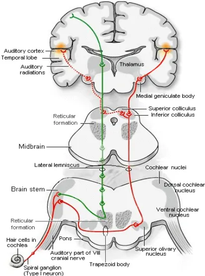
Auditory Pathway:
Hair cells in the organ of corti are the receptors of auditory sensation. All
hair cells are innervated by afferent and efferent nerve fibers. The afferent
cells forms the auditory pathway. First order neurons of auditory pathway
as cochlear nerve fibers and enter medulla oblongata, where they divide
into dorsal and ventral cochlear nucleus on same side of medulla.
These act as second order neurons, where they run as different
groups to cross the superior olivary nucleus, lateral lemniscus and
recticular formation. The third order neurons are in the superior olivary
nuclei and nucleus of lateral lemniscus. Fibers from medial geniculate
body go the tremporal cortex, via the internal capsule as auditory
radiation. Some fibers run to inferior colliculus which are responsible for
reflex movements of head in response to auditory stimuli. The cortical
auditory centers in the temporal lobe of cerebral cortex lie are the primary
auditory area 41; 42 and Wernicke area. The secondary auditory
Hearing loss:
Hearing loss can be of three types (i) Conductive hearing loss (ii)
Sensorineural hearing loss (iii) Mixed hearing loss. While auditory
function is being assessed it is important to find out the type of hearing
loss; degree of hearing loss; site of lesion and cause of hearing loss.
Hearing of an individual can be tested by clinical and audiometric tests.
Sensorineural hearing loss: This results from cochlear lesions, VIII th nerve or central auditory pathways. It may be congenital or acquired. The
characteristics of sensorineural hearing loss are:
- A positive Rinne test
- Weber lateralized to better ear
- Bone conduction reduced on Schwabach and absolute bone
conduction test.
- Mostly high frequency loss
- No gap between air and bone conduction curve on audiometry
- Loss may exceed 60dB
- Speech discrimination is poor
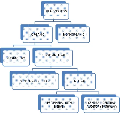
Table: 1:
CONDUCTIVE
SENSORY(COCHLEAR)
Table: 1: Classification of hearing loss
HEARING LOSS
ORGANIC
SENSORINEURAL
SENSORY(COCHLEAR) NEURAL
PERIPHERAL (8TH NERVE)
CENTRAL (CENTRAL AUDITORY PATHWAY) NON-ORGANIC
Classification of hearing loss
Congenital sensorineural hearing loss is present from birth and is the
result of anomalies of the inner ear or damage to the hearing apparatus by
prenatal or perinatal factors.
Acquired sensorineural hearing loss appears later in life. The cause may
be genetic or non- genetic. The genetic cause of hearing loss may
presents late and damages only the hearing. It can even be a component
of a larger syndrome with other the body systems being involved.
Common causes of acquired sensorineural hearing loss are:
- Infections of labyrinth
- Trauma to labyrinth
- Noise-induced hearing loss
- Ototoxic drugs
- Presbycusis
- Menier’s disease
- Acoustic neuroma
- Sudden hearing loss
- Systemic disorders like diabetes, hypothyroidsm, kidney disease
Specific forms of hearing loss:
A. Inflammation of the labrynth
1. Viral labrynthitis : viruses usually reach the inner ear by blood
stream affecting stria vascularis , endolymph and organ of corti.
Measels,mumps and cytomegalovirus are known to cause
labrynthitis.
2. Bacterial : these infections reach the labrynth through the
middle year or through CSF.sensory neural deafness following
meningitis is a known complication .
3. Bacteria can invade the labrynth along nerves , vessels, coclear
aqueduct or endo lymphatic sac and this causes complete
distruction of the membraneous labrynth.
4. Syphilitic : sensory neural hearing loss is caused both by
congenital and acquired syphilis . syphilitic involvement of the
inner ear can cause sudden sensorineural hearing loss which
may be unilateral or bilateral. Menier’s syndrome with episodic
hearing loss, tinnitus and aural fullness. Tullio phenomenon
B. Familial Progressive Sensorineural hearing loss: It is genetic
disorder characterized by progressive degeneration of cochlea. It
may start late in childhood or in early adult life.
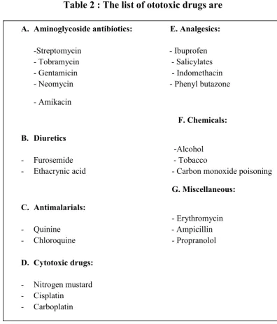
C. Ototoxicity: Various chemicals and drugs affect the inner ear and
produces sensorineural hearing loss and tinnitus. Symptoms of
ototoxicity-hearing loss, tinnitus or giddiness may be noted during
[image:37.595.111.508.299.764.2]treatment or even after completion of entire treatment.
Table 2 : The list of ototoxic drugs are OTOTOXIC DRUGS
A. Aminoglycoside antibiotics: E. Analgesics:
-Streptomycin - Ibuprofen - Tobramycin - Salicylates - Gentamicin - Indomethacin - Neomycin - Phenyl butazone
- Amikacin F. Chemicals:
B. Diuretics
-Alcohol - Furosemide - Tobacco
- Ethacrynic acid - Carbon monoxide poisoning
G. Miscellaneous: C. Antimalarials:
- Erythromycin - Quinine - Ampicillin - Chloroquine - Propranolol
D. Cytotoxic drugs:
- Nitrogen mustard - Cisplatin
D. Noise trauma:
Hearing loss due to exposure to noise exposure is known in boiler
makers, iron smiths, copper smith and artillery men. Now a day’s
noise trauma has become very significant as it has become an
occupational hazard. Hearing loss caused by excessive noise can be
divided into two groups:
1. Acoustic trauma: Permanent damage to hearing can occur even on single brief exposure to very intense sound. E.g.:
Gunfire; explosion. Sudden loud sounds damage cause damage
to the outer hair cells disrupting the organ of corti and will
result in rupture of the Reissner’s membrane. In some cases
rupture of tympanic membrane and disruption of ossicular chain
can happen in severe blast.
2. Noise-induced hearing loss: Here hearing loss occurs following chronic exposure to less intense sounds. This is
mostly noted as occupational hazard in people working in noisy
The damage caused by noise trauma depends on many factors like:
a. Frequency of noise
b. Intensity and duration of noise
c. Continuous or interrupted noise
d. Susceptibility of the individual
e. Any pre-existing ear disease
E. Sudden Hearing loss: It is a sensorineural hearing loss, that is developed over a period of hours or few days. Hearing loss may be
partial or complete. It is mostly unilateral and may be accompanied
by tinnitus or spells of vertigo. Mostly the cause of this sudden deafness remains unclear and termed as idiopathic. The three main
aetiological factors generally considered are: viral, vascular or
rupture of cochlear membrane. The other factors causing sudden
hearing loss are:
- Infections
- Trauma
- Vascular
- Ear pathology like Meniere’s disease.
- Toxic
- Neoplastic
F. Presbycusis: Sensorineural haering loss associated with aging physiology in the ear is termed as presbycusis. Generally it
manifests at 65 years or even earlier. The four pathological types
are:
- Sensory
- Neural
- Strial or metabolic
- Cochlear conductive
The tests for hearing are:
A] Clinical tests of hearing are (i) Finger Friction Test (ii) Watch Test (iii) Speech Tests (iv) Tuning Fork Tests which include (a) Rinne test (b)
Weber test (c) Absolute bone conduction test (d) Schwabach’s test (e)
Bing test (f) Gelle’s test.
Rinne tuning fork test: is formulated to find the difference between air
conduction with that of bone conduction. In normal conditions, air
conduction is more than bone conduction and the tuning fork will be
heard loud in the opposite ear canal than when it is placed over the
mastoid bone behind.
The alternate method for doing this test is by threshold comparison
external canal till it is no longer heard. It is then placed over the mastoid
process. If sound is heard once more, then it is considered that bone
conduction is better than air conduction and the test is considered
negative. The test when performed with 256 Hz tuning fork has greater
sensitivity and specificity. The specificity of this test increases above
30dB conductive deafness, decreases when narrow air-bone gap is found.
False positivity of this test is about 20%.
Weber tuning fork test: This test is used only if asymmetrical or
unilateral hearing is seen in patients. The basis of this is by placing a
tuning fork in the centre of the skull being heard louder in the ear with a
conductive impairment in case of sensorineural loss it is louder in the
better ear.
This difference can be distinctive only, if the examiner has done a clinical
hearing test previously and if he knows which ear has better hearing. The
test is done by keeping an activated tuning fork on the forehead or over
the bridge of nose or on the incisor teeth. The tuning fork can also be
placed over the vertex of the skull in the midline. Then the patient is
asked to identify the ear in which the sound is heard or otherwise in
which ear the sound is louder. If the sound is heard in the better hearing
it is heard in the affected ear, then it is considered as conductive
impairment. This test has a low sensitivity and specificity.
Absolute bone conduction test: Bone conduction is measurement of the
function of cochlea. Here bone conduction of the patient is compared
with the examiner, assuming the examiners have hearing within normal
limits. The external auditory canal of both the examiner and the patient
are closed, so that ambient noise entering through air conduction route is
prevented. In case of conductive hearing loss, the subject and the
examiner hears the tuning fork for equal time period. Whereas in case of
sensorineural loss, the subject perceives the tuning fork for a brief period
of time.
Schwabach’s test: Here again the bone conduction of the patient is
compared with the examiner, assuming that he has hearing within normal
limits. But in this test, the meatus is not occluded. Schwabach’s test is
reduced in case of sensorineural deafness and lengthened in case of
conductive deafness.
The Bing test: it is done on the same basis of Weber test in which closing
of the external auditory canal increases the tuning fork sound in the ear
vibrating tuning fork over the mastoid process and the external auditory
meatus is also occluded.
If increase in sound is present then there is less likelihood that there is
conductive hearing loss. However, if it remains the same, then it is more
likely to be a conductive deafness. The specificity and sensitivity of this
test is also very low. Most of the situations normal individuals are
identified as conductive deafness. This test is not used widely.
Tuning fork tests should generally be reserved for situations where
audiometry is not satisfactory. The results must be interpreted keeping in
mind the low sensitivity and specificity.
B] Audiometric tests are (i) Pure tone audiometry (ii) Speech audiometry (iii) Bekesy audiometry (iv) Impedance audiometry.
C] Special tests of hearing are (i) Recruitment (ii) Short increment sensitivity index (SISI) (iii) Threshold tone decay test (iv) Evoked
response audiometry (v) Otoacoustic emissions (vi) Central auditory tests
(vii) Hearing assessment in children and infants.
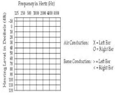
Pure tone audiometry : Pure tones are produced by an electronic device
called an audiometer. The intensity of these tones can be increased or
calculated for tones of 125, 250, 500, 1000, 2000, 4000 and 8000 Hz and
for thresholds of bone conduction, it is done at 250, 500, 1000, 2000 and
4000 Hz.
The measure of intensity of tones that has to be increased more than the
level of normal is considered as the degree of hearing impairment in that
particular frequency. Audiogram is a graphical representation of the
charted values. The bone conduction threshold is a measurement of
function of cochlea. The variations in the air conduction and bone
conduction thresholds (A-B gap) are a measurement of the degree of
conductive hearing loss. It is observed that the calibration of audiometer
is done in such a way that the perception in a normal person, both the air
and bone conduction remains at zero dB and no A-B gap is noted. The
turning fork test generally shows AC>BC.
When the difference of hearing in the 2 ears is 40 dB or more in
thresholds of air conduction then the ear with better hearing is masked so
as to not to get a shadow curve in the better ear that is not being tested. In
the same way masking is important in all studies of bone conduction.
Masking is carried out by delivering a narrow-band noise to the ear that is
not being tested. The benefits of pure tone audiogram are (i) It is a
measurement threshold for hearing in both air and bone conduction, and
record. (iii) Hearing aids can be prescribed only after an audiometery (iv)
Speech reception thresholds can be predicted with its help (v) The degree
[image:45.595.114.485.185.425.2]of handicap can be assessed for medicolegal issuses.
Figure. 4. Audiogram
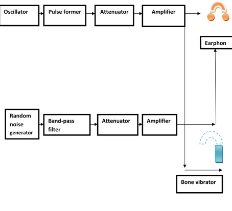
Equipment:
A pure tone audiometer is designed with a set of basic functions. The
technical requirements for the instrument are specified by international
standard, in which four types are identified based on complexity of
Pulse former Attenuator Amplifier
Random noise
generator
Band-pass filter
Attenuator Amplifier
Bone vibrator Oscillator
[image:47.595.109.564.136.549.2]Earphon
speech audiometry is a test in which the patient’s capacity to hear and to
understand speech is calculated. 2 parameters are taken into
consideration: (i) Speech reception threshold (ii) The discrimination
score. SRT is the intensity at which minimum words of 50% are
reciprocated righttly by the patient. The headphones of an audiometer
delivers a set of spondee words to both nears.
An SRT more than pure tone average of 10dB suggests that hearing
loss is of functional type. Discrimination score is also termed as speech
recognition score. It is a measure of patient’s capacity to understand
speech. Here phonetically balanced words are delivered to both ears at an
intensity 30-40 dB above the SRT.
Thus, speech audiometry helps (a) to differentiate an organic
hearing from the functional one. (b) to indicate at which intensity the
discrimination score is best, and this is useful for fitting apt hearing aid
and setting its volume for optimal hearing and (c) to differentiate a
MANAGEMENT OF HEARING IMPAIRMENT:
Management of individuals with hearing impairment can be determined
by the degree of hearing impairment, whether the impairment is
sensorineural, conductive or mixed. Ears with mild, moderate or severe
impairment are given the option of hearing aids. Those with profound or
total impairment, cochlear implants may be suggested. Middle ear
implants were developed for those with conductive hearing loss but in
long term it could be used for sensorineural hearing loss.
All individuals with hearing impairment need some rehabilitation for
communication. The various means of rehabilitation are:
1. INSTRUMENTAL DEVICES
HEARING AIDS COCHLEAR IMPLANTS ASSISTIVE DEVICES
2. TRAINING
Hearing aids:
Hearing aids partially overcome the deficits associated with hearing loss.
For a sensorineural hearing loss, there are several deficits to overcome.
Some sounds are inaudible and some are heard because partial spectra are
audible.
Hearing aids are devices that amplify sound by electrical or chemical
means. The basic components of a hearing aid are:
- Microphone: They collect sounds and transform it into electrical
energy. The microphones are generally magnetic in type. It consists
of diaphragm that converts sound energy into mechanical energy.
- Amplifier: The function is to increase the electrical voltage which
is received from the microphone. An implanted transmitter in the
amplifier increases the voltage.
- Receiver: It transfers electrical energy back into sound waves with
a much greater amplitude than those which fell upon the
microphone. The receivers are both air conduction type as well as
bone conduction type. These magnetic receivers have their
diaphragm connected to a vibrator which is placed over the mastoid
process. The vibrations are transmitted to the bony labyrinth and
fluid in the cochlea.
Diabetes mellitus is characterized by chronic hyperglycemia with
disturbances of carbohydrate, protein and fat metabolism resulting from
defects in insulin secretion, insulin action, or both. The effects of diabetes
mellitus include long term damage, dysfunction and failure of organs like
eye, ears, heart, kidneys and blood vessels. It may present with symptoms
like thirst, polyuria, polyphagia, blurring of vision and weight loss. And
in more severe forms it may present as ketoacidosis or nonketotic
hyperosmolarity, which in the absence of treatment leads to stupor, coma
and death.
A number of specific causes of diabetes mellitus have been
identified, the etiology and pathogenesis is not clear. The majority of
cases fall under two broad etiopathological categories, now called as type
1 and type 2 diabetes. Recently the WHO along with expert committee of
Table 4: Etiological classification of disorders of glycemia
This classification differs considerably from the previous
recommendation which classified using terms such as insulin-dependent
diabetes and non-insulin dependent diabetes [4]. These terms were
misused and classified patients based on treatment needs rather than
etiologic characteristics. So the terms type 1 and type 2 diabetes have
been adopted for the most common forms of diabetes mellitus.
TYPE 1
Beta cell destruction usually leading to absolute insulin deficiency
A) Autoimmune B) Idiopathic
TYPE 2
A) Predominantly insulin resistance
B) Predominantly insulin secretory defects
OTHER SPECIFIC TYPES OF DIABETES
A) Genetic defects of beta cell dysfunction
B) Genetic defects in insulin action e.g: Type A insulin resistance C) Diseases of exocrine pancreas e.g: Fibro calculus pancreatopathy D) Endocrinopathies e.g: Acromegaly, cushings etc
E) Drugs or chemical – induced e.g: Glucocorticoids F) Infections e.g: congenital rubella
G) Uncommon forms of immune-mediated diabetes e.g: Stiff Man Syndrome H) Other genetic syndromes
Clinical Stages:
Individuals who develop diabetes pass through several clinical stages
during the development. Initially, glucose regulation is normal and no
abnormality of glycemia can be identified even if the individual
undergoes oral glucose tolerance test. This stage is followed by a period
of variable duration where in glucose regulation is impaired. Some
amount of fasting glucose concentration may be noted or oral glucose test
may show impairment.
Once diabetes develops, glycemia may be controlled by lifestyle changes
like diet and increased physical activity in some patients, whereas in
others oral hypoglycemic agents or insulin is needed for control or
prevention against ketosis and ketoacidosis.
If insulin is required to prevent ketosis such patients are labeled as
“insulin requiring for survival”. In all forms remission may be present.
Patients may revert to having impaired glucose regulation or even normal
glycemia, particularly if diabetes is of recent onset. This is seen most
frequently in patients with recent-onset type 2 DM in whom lifestyle
intervention or early aggressive treatment may result in apparent reversal
of the abnormality with reversion to impaired or normal glucose
This may be seen in type 1 diabetes also, where in a short period of
insulin treatment may result in a variable period when insulin is no longer
required for survival and glucose tolerance may improve, the so called
honeymoon period. Eventually these patients need insulin treatment for
survival [6].
All subjects with diabetes can be classified according to clinical stages
regardless of underlying etiology of diabetes. The stage of glycemia may
change over time depending on the extent of underlying disease process.
Impaired glucose regulation refers to the metabolic state intermediate
between normal glucose homeostasis and diabetes that can be identified
by impaired glucose tolerance or impaired fasting glucose [7]. They are
not synonymous and represent different abnormalities of glucose
regulation, even if they occur together. These patients are at a higher risk
of progressing to diabetes.
ETIOLOGIC TYPES
Type 1 Diabetes Mellitus: It is a form of diabetes primarily due to β- cell destruction. This usually leads to a type of diabetes in which insulin is
required for survival. These individuals are metabolically normal before
the disease is clinically manifested, but the process of β- cell destruction
can be detected earlier by the presence of certain antibodies like
more of these antibodies can be subclassified as type 1A, immune
mediated type 1 diabetes. Type 1 diabetes can also occur in the absence
of autoimmune antibodies and without evidence of any autoimmune
disorders. It is a progressive form of disease with marked hyperglycemia
resulting in insulin requirement for prevention of ketosis and survival.
These individuals are classified as type 1B or idiopathic diabetes[8].
The rate of β- cell destruction is variable, being rapid in some individuals
especially in infants and children, and slower in adults. Individuals
become dependent on insulin for survival only many years after the
detection of diabetes. Type 1 diabetes patients have low or undetectable
levels of insulin and plasma C- peptide. These patients are more prone for
ketoacidosis, although they have no clinical evidence of autoimmune
antibodies. They may suffer from episodic ketoacidosis, but pathogenetic
basis for this insulinopenia remains unclear.
Type 2 Diabetes Mellitus: It is the most common form of diabetes. It is characterized by disorders of insulin action and insulin secretion. usually
The specific etiology of this form of diabetes is yet unknown. Auto
immune destruction of β- cell does not occur in this form of diabetes.
Type 2 diabetes patients usually have insulin resistance rather than
absolute insulin deficiency. These patients do not need insulin treatment
for survival although many may require it for glycemic control. This form
of diabetes is associated with progressive β- cell failure with increasing
duration of diabetes [9]. Ketoacidosis rarely occurs by itself but
conditions associated with stress and other illnesses such as infection can
increase the risk
Type-2 diabetes patients are mostly obese when they develop diabetes
and obesity aggravates the insulin resistance.this form of diabetes goes
undiagnosed for many years because the hyperglycemia develops
gradually and in early stages it is not severe enough to produce the classic
symptoms of diabetes.however these patients are at increased risk of
developing macrovascular and microvascular complications.
The circulating levels of insulin may be normal or elevated yet
insufficient to control glucose levels in the blood within the normal range
because of insulin resistance. Thus they have relative insulinopenia.
Insulin resistance may improve with weight reduction or pharmacologic
history of gestational diabetes and individuals with characteristics of
insulin resistance syndrome like hypertension and dyslipidemia are at
higher risk of developing type 2 diabetes. The risk also increases with
age, obesity and physical inactivity. Lifestyle measures to reduce weight
such as dietary regulation and increased physical activity reduce or delays
the development of diabetes [10; 11].
Other Specific Types of Diabetes Mellitus
Maturity-onset diabetes of the young (MODY):
This subgroup includes a relatively rare monogenic disorder characterized
by non–insulin dependent diabetes with autosomal dominant inheritance
and the age of onset is 25 years or younger. Patients are not obese, and
their hyperglycemia is due to impaired glucose-induced secretion of
insulin. There are Six types of MODY which have been described.
Except that in MODY 2, a glucokinase gene is defective, all other types
involve mutations of a nuclear transcription factor that regulates islet
gene expression. The enzyme glucokinase is a rate-limiting step in
glycolysis and determines the rate of adenosine triphosphate (ATP)
MODY 2, due to glucokinase mutations, is usually mild, associated with
only slight fasting hyperglycemia and few diabetic complications. It
responds well to hygienic measures or low doses of oral hypoglycemic
agents.
MODY 3, is due to mutations in hepatic nuclear factor 1α and is the most
common form. It accounts for two thirds of all MODY cases. The clinical
course is of progressive beta cell failure and needs insulin for therapy.
Mutations in both alleles of glucokinase present with more severe
neonatal diabetes.
Diabetes due to mutant insulin receptors: This is a rare subtype of type 2 diabetes who are not obese. There are less than ten families being
described in the past. Since affected individuals were heterozygous,
diabetes presentation was mild, and appeared only in middle age. It also
showed genetic autosomal dominant transmission. There is no evidence
of clinical insulin resistance, and these patients respond well with
standard therapy
Defects in insulin receptor genes are found in more than 40 people with
diabetes. These individuals have the highest insulin resistance associated
genes are abnormal , newborns present with leprechaun-like phenotype
and do not survive through infancy.
Wolfram syndrome:
It is an autosomal recessive neurodegenerative disorder. It first becomes
evident in childhood. It consists of diabetes insipidus, diabetes mellitus,
optic atrophy, and deafness. This protein forms a part of the unfolding
protein response and this protects the beta cells from endoplasmic
reticulum stress and apoptosis during periods of high insulin demand.
Diabetes mellitus along with optic atrophy presents in the first decade of
life.
Sensorineural deafness and diabetes insipidus starts in the second decade
of life in 60–70% of individuals . Ureterohydronephrosis, neurogenic
bladder, peripheral neuropathy, cerebellar ataxia and psychiatric illness
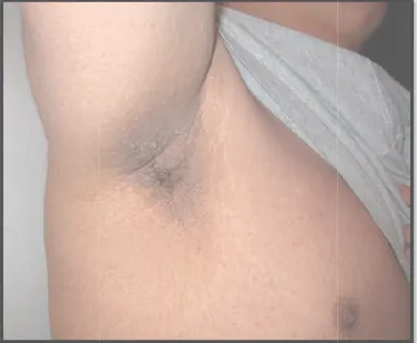
Fig 6: Acanthosis nigricans in the nape of the neck, with typical dark
Fig 7: Acanthosis nigricans of the
Acanthosis nigricans in the nape of the neck, with typical dark and velvety appearance
Acanthosis nigricans of the axilla, with typical dark coloration and velvety texture
Acanthosis nigricans in the nape of the neck, with typical dark
[image:61.595.126.502.403.712.2]Autosomal recessive syndromes:
In this form, there is homozygous mutations in a number of pancreatic
transcription factors like
NEUROG3, PTF1A, and GLI. It causes neonatal or childhood diabetes.
Homozygous PTF1A
mutations results in absent pancreas and cerebellar atrophy; NEUROG3
mutations causes severe malabsorption and results in diabetes before
puberty. Homozygous mutations in RFX6 causes the Mitchell-Riley
syndrome. It is characterized by the absence of all islet cell types apart
from pancreatic polypeptide cells, hypoplasia of the pancreas and
gallbladder, and intestinal
atresia. The gene EIF2AK3 helps in controlling the pathway for unfolding
protein response. Absence of this leads to inadequate response to stress
and accelerated beta cell apoptosis. Patients with mutation in this gene
leads to neonatal diabetes, epiphyseal dysplasia, developmental delay,
hepatic and renal dysfunction (Wolcott-Rallison syndrome).
Diabetes mellitus secondary to other causes:
Glucose intolerance can be caused by endocrinr tumors secreting growth
hormone like catecholamines, glucocorticoids, glucagon or somatostatins.
In these incidences there is impairment of peripheral responsiveness to
insulin. With increase in levels of hormones hepatic output of glucose
insulin release and becomes an added factor in producing carbohydrate
intolerance, somatostatin inhibits insulin secretion and plays as a major
factor.
Diabetes predominantly occurs in individuals with underlying defects in
insulin secretion and hyperglycemia spontaneously resolves when the
hormone excess is resolved. Extreme insulin resistance with acanthosis
nigricans is a rare syndrome which affects young woman with
androgenic features and also in older woman who have circulating
immunoglobulin binds to insulin receptors and this reduces the affinity to
insulin. Many medications like calcineurin inhibitors, thiazides,
β-blockers, corticosteroids, niacin, atypical antipsychotics are known to
produce carbohydrate intolerance or even frank diabetes. These
medications act by either decreasing insulin secretion or by increasing
insulin resistance or both. Corticosteroids are known to increase insulin
resistance Thiazide diuretics and β-blockers moderately increase the risk
for diabetes. Treating the hypokalemia produced by these thiazides may
reverse the hyperglycemia. Atypical antipsychotics, olanzapine and
clozapine, in 63articular have been associated with increased risk of
glucose intolerance. These medications also causes weight gain and
insulin resistance. An increase in rate of developing diabetic ketoacidosis
DIAGNOSTIC CRITERIA FOR DIABETES:
Patients having symptoms such as thirst, polyuria, unexplained weight
loss, drowsiness or coma with marked glucosuria, the diagnosis of
diabetes can be established by demonstrating fasting hyperglycemia. If
the fasting glucose concentration lies in the diagnostic range for diabetes,
an oral glucose tolerance test is not required for diagnosis.
A confirmatory test has to be performed because diagnosis of diabetes
carries a considerable consequence for the patient. Intraindividual
variation or incomplete fasting may result in wrong diagnosis. Whereas if
the patient is asymptomatic or has minimal symptoms with fasting or
plasma concentration values which are not diagnostic, an oral glucose
tolerance test is required to confirm or exclude the diagnosis of diabetes.
Normal glucose tolerance cannot be established on basis of fasting
glucose determination alone. In normal healthy individuals, fasting
glucose levels should be less than 100mg/dl in venous or capillary plasma
and 90mg dl or less in whole blood. Individuals with fasting glucose
levels above the characteristic value for normal healthy subjects but
below diagnostic value of diabetes have a high likelihood of having either
diabetes or impaired glucose tolerance. These levels represent a primary
indication for an oral glucose tolerance test to confirm or exclude the
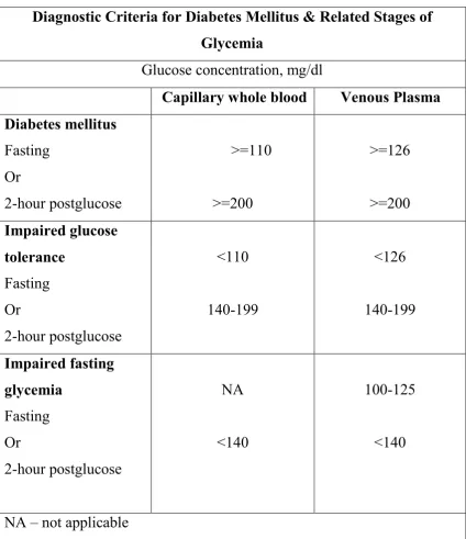
Table 5: Diagnostic Criteria
Diagnostic Criteria for Diabetes Mellitus & Related Stages of Glycemia
Glucose concentration, mg/dl
Capillary whole blood Venous Plasma Diabetes mellitus Fasting Or 2-hour postglucose >=110 >=200 >=126 >=200 Impaired glucose tolerance Fasting Or 2-hour postglucose <110 140-199 <126 140-199 Impaired fasting glycemia Fasting Or 2-hour postglucose NA <140 100-125 <140
NA – not applicable
The 2- hour postglucose values are measured after a 75 gm oral glucose
Chronic complications of diabetes mellitus:
Many organ in the body are affected as a chronic complications of
diabetes and it acts as the major cause for morbidity and mortality . There
are tow types of complications - vascular and nonvascular . The vascular
complications can be again subdivided into microvascular and
macrovascular complications.
Non vascular complications include conditions such as gastroparesis,
infections, and skin changes. Long standing diabetes may be associated
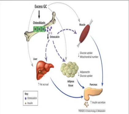
with hearing loss. Among metabolic disorders of glucose diabetis mellitus
is most commonly associated with auditory disfunction[12]
Damage to the nerves and vessels of the inner ear are noted in many
Histopathological studies in patients with diabetes. Neuronal degeneration in
the auditory system was linked to these vascular changes as an important
causative factor. The organ of corti cells which are important structures of
hearing are complex components and its arrangement makes it also a potential
target for hyperglycaemic damage. Such damage to any part of hearing
Chronic complications of Diabetes Mellitus
Microvascular complications
Eye disease
- Retinopathy
- Macular edema
Neuropathy
- Sensory and motor (mono- and polyneuropathy)
- Autonomic
Nephropathy
Macrovascular complications
Coronary artery disease Peripheral arterial disease
Cerebrovascular disease
Others
Gastrointestinal (gastroparesis, diarrhea)
Genitourinary ( uropathy/ sexual dysfunction)
Dermatologic
Infections Cataracts
Hearing loss
Glaucoma
The risk of chronic complications increases as duration and degree of
hyperglycemia increases. These complications do not become apparent
till the second decade of hyperglycemia. As the type 2 diabetic patients
have a long asymptomatic period of hyperglycemia, they may present
with complications when diagnosis is made. The microvascular changes
in both type 1 and type 2 diabetes are due to chronic hyperglycemia.
Clinical trials prove that reduction in chronic hyperglycemia prevents or
delays retinopathy, nephropathy and neuropathy.
Evidence to prove that chronic hyperglycemia is a cause for developing
macrovascular changes is less provided. However complications like
coronary heart disease and mortality rates are 2 to 4 folds greater in type
2 diabetes. These complications correlate with fasting and postprandial
plasma glucose levels asa well as with HbA1C values. Other factors like
dyslipidemia and hypertension play an important role in macrovascular
complications.
Mechanisms of complications:
Chronic hyperglycemia is a well known etiologic factor leading to
complications of diabetes, the actual mechanism by which it leads to such
diverse cellular and organ dysfunction is not clear. There are few
prominent theories that have been enumerated to explain how
One theory states that raise in levels of intracellular glucose leads to the
production of advanced glycosylation end products, which bind to a cell
surface receptor via the nonenzymatic glycosylation of intra and
extracellular proteins. The interaction of glucose with amino groups on
proteins results due to nonenzymatic glycosylation . The AGEs are
known to cross –link proteins, accelerate atherosclerosis, promotes
glomerular dysfunction, reduces nitric oxide synthesis, induces
endothelial dysfunction and leads to alteration of extracellular matrix
composition and structure. Serum levels of AGEs correlates with the
levels of glycemia, and these products accumulate as the glomerular
filtration rate declines.
Second theory is proposed on basis of observation that hyperglycemia
increases glucose metabolism by sorbitol pathway. Increased levels of
sorbitol concentration alters redox potential, increases cellular osmolality
leading to generation of oxygen species which is reactive and further
leads to other cellular dysfunction. Trails based on this theory have lead
into clinical clues on complications like retinopathy, neuropathy and
nephropathy.
Third hypothesis suggests that the formation of diacylglycerol leading to