COAGULATION PROFILE STUDY IN PREGNANCY INDUCED HYPERTENSION
Dissertation submitted in
Partial fulfilment of the regulations required for the award of M.D.DEGREE
IN
PATHOLOGY
THE TAMILNADU
DR.M.G.R. MEDICAL UNIVERSITY CHENNAI
DECLARATION
I hereby declare that the dissertation entitled “COAGULATION
PROFILE STUDY IN PREGNANCY INDUCED
HYPERTENSION” is a bonafide research work done by me in the Department of Pathology, Coimbatore Medical College during the period from July 2013 to July 2014 under the guidance and supervision of Dr.A.Arjunan, M.D., Professor, Department of Pathology, Coimbatore Medical College.
This dissertation is submitted to The TamilnaduDr.MGR Medical University, Chennai towards the partial fulfilment of the requirement for the award of M.D., Degree (Branch III) in Pathology. I have not submitted this dissertation on any previous occasion to any University for the award of any Degree.
Place: Coimbatore
CERTIFICATE
This is to certify that the dissertation entitled “COAGULATION
PROFILE STUDY IN PREGNANCY INDUCED
HYPERTENSION” is a record of bonafide work done by Dr. S.VIJAYALAKSHMI in the Departmentof Pathology, Coimbatore Medical College, Coimbatore under the guidance and supervision of Dr. A. ARJUNAN M.D., Professor, Department of Pathology, Coimbatore Medical College and submitted in partial fulfilment of the requirements for the award of M.D. Degree (Branch III) in Pathology by The TamilnaduDr. MGR University Chennai.
Guide HOD of the Department
Dr.A.ARJUNAN, M.D., Dr.C.Lalitha, M.D.,
Professor, Professor,
Department of Pathology, Department of Pathology, Coimbatore Medical College, Coimbatore Medical College,
Coimbatore. Coimbatore.
Dean
Dr.S.Revwathy,MD,D.G.O.,DNB, Dean,
ACKNOWLEDGEMENT
To begin with, I thank the Almighty in making this project a successful one.
I wish to record my sincere gratitude to our respectful Dean, Professor, Dr.S.Revwathy,MD,D.G.O.,DNB, Coimbatore Medical College, Coimbatore, for permitting me to carry out this study and to avail all the facilities in this institution.
I am deeply indebted to our Professor, Dr.C.Lalitha,M.D.,Professor and Head of the Department of Pathology, Coimbatore Medical College, Coimbatore, but whose guidance, this study would not have come through. It has been a great privilege to work under her and especially on this topic.
I am thankful to our Professor, Dr.Arjunan,M.D., for his timely help and guidance.
I wish to record my sincere thanks to Dr.P.Sundhari,D.G.O.,M.D., Professor and Head of the Department of Obstetrics and Gynaecology for allowing me to select cases.
I take this opportunity to thank my fellow Post Graduates and all the Technical Staffs for their contribution to this study.
ABBREVIATIONS
PIH - Pregnancy Induced Hypertension
PT - Prothrombin Time
APTT - Activated Partial Thromboplastin Time
BT - Bleeding Time
CT - Clotting Time
sFlt - Soluble fms-like tyrosine kinase.
INR - International Normalised Ratio
EDTA - Ethylene DiamineTetraacetic Acid
vWF - von Willebrand Factor
mpl receptor - Myeloproliferative leukemia virus receptor
nm - Nanometer
fl - femtolitre
CONTENTS
SI.NO PARTICULARS PAGE NO.
1. INTRODUCTION 1
2. AIM 3
3. OBJECTIVES 4
4. REVIEW OF LITERATURE 5
5. MATERIALS AND METHODS 67 6. OBSERVATION AND RESULTS 77
7. DISCUSSION 104
8. SUMMARY 108
9. CONCLUSION 110
10. BIBLIOGRAPHY APPENDICES
LIST OF TABLES
Sl.No TABLES Page.No
1. Distribution of patients according to bleeding time 77
2.
Mean Bleeding Time (in seconds) for Normal Pregnancy, Non Severe PIH and Severe PIH Patients.
79
3.
Analysis of variance of Bleeding Time among group
Normal Pregnancy, Non severe PIH and Severe PIH 81
4.
Multiple Comparisons of Bleeding time among group Normal Pregnancy, Non severe PIH and Severe PIH
82
5. Distribution of patients according to Clotting Time 83
6.
Mean Clottingtime (in seconds) for Normal Pregnancy, Non Severe PIH and Severe PIH Patients
85
7.
Analysis of variance of Clotting Time among group Normal Pregnancy, Non severe PIH and Severe PIH
87
8.
Multiple Comparisons of Clotting time among group Normal Pregnancy, Non severe PIH and Severe PIH
88
9. Distribution of patients according to Platelet Count 89
10.
Mean Platelet Count (in Lakhs) for Normal Pregnancy, Non Severe PIH and Severe PIH Patients.
90
11.
Analysis of variance of Platelet Count among group Normal Pregnancy, Non severe PIH and Severe PIH
12.
Multiple Comparisons of Platelet Count among group Normal Pregnancy, Non severe PIH and Severe PIH
93
13. Distribution of patients according to Prothrombin Time. 94
14.
Mean Prothrombintime (in seconds) for Normal Pregnancy, Non Severe PIH and Severe PIH Patients.
95
15.
Analysis of variance of Prothrombin Time among group Normal Pregnancy, Non severe PIH and Severe PIH.
97
16.
Multiple Comparisons of Prothrombin Time among group Normal Pregnancy, Non severe PIH and Severe PIH.
98
17. Distribution of patients according to APTT. 99
18.
Mean APTT (in seconds) for Normal Pregnancy, Non Severe PIH and Severe PIH Patients.
100
19.
Analysis of variance of APTT among group Normal Pregnancy, Non severe PIH and Severe PIH
102
20.
Multiple Comparisons of APTT among group Normal Pregnancy, Non severe PIH and Severe PIH
LIST OF CHARTS
Sl. No CHARTS Page.
No
1.
The mean Bleeding Time of Normal Pregnancy, Non severe PIH and severe PIH.
80
2.
The mean Clotting Time of Normal Pregnancy, Non severe PIH and severe PIH.
86
3.
The mean Platelet Count of Normal Pregnancy, Non severe PIH and severe PIH.
91
4.
The mean Prothrombin Time of Normal Pregnancy, Non severe PIH and severe PIH.
96
5.
The mean APTT of Normal Pregnancy, Non severe PIH and severe PIH.
LIST OF EQUIPMENTS AND REAGENTS USED
Sl.No Equipment and Reagents Page. No
1.
Vaccutinor used for the collection of blood for the estimation of platelet count.
71
2. Analyser used for estimation of platelet count 72
3.
Vaccutinor used for collection of blood for PT and APTT.
74
4.
Reagents used for estimation of prothrombin time and activated partial thromboplastin time.
75
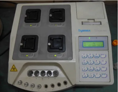
5.
Analyser used for estimation of prothromin time and activated partial thromboplastin time.
ABSTRACT
AIM:
To study coagulation profile changes in pregnancy induced hypertension. Methods and Materials: Control group consists of 60 normotensive pregnant women compared with 30 cases of non-severe PIH and 30 cases of severe PIH. Coagulation parameters BT, CT, Platelet Count, PT and APTT were done for all cases. Observations: Patients with severe PIH patients showed low Platelet Count, prolonged BT, PT and APTT compared to normotensive pregnant women and non-severe PIH. No changes observed in CT. Summary and Conclusion: The changes in Coagulation Profile in severe PIH is helpful in assessing the Coagulation abnormalities at earlier stage. Changes observed in the coagulation parameters as the severity of PIH increases.
KEYWORDS:
Pregnancy Induced Hypertension, Bleeding Time, Clotting Time, Platelet Count, Prothrombin Time, Activated Partial Thromboplastin Time.
INTRODUCTION
Pregnancy induced hypertension is an elevated blood pressure that appears first time after five months of pregnancy. It is one of the most common cause of increased mortality and morbidity both in the mother and in the foetus.
Preeclampsia is a combination of elevated blood pressure, proteinuria and edema. Eclampsia is the presence of convulsions along with above three features of preeclampsia.
Normal pregnancy is a hypercoagulable state due to elevation of the most of the coagulation factors and reduced anticoagulant activity. In pregnancy induced hypertension, there is an accentuation of hypercoagulable state as a result of injury to the endothelium.
AIM OF THE STUDY
OBJECTIVES
1. To study the changes in platelet count, bleeding time, clotting time, prothrombin time and activated partial thromboplastin time in pregnancy induced hypertension.
2. To study the above parameters in normal pregnant women.
3. To compare the above parameters between normotensive pregnant women and women with pregnancy induced hypertension.
REVIEW OF LITERATURE
COAGULATION PROFILE STUDY IN PREGNANCY INDUCED HYPERTENSION
Hypertensive disorders complicate 5 to 10% of all pregnancies and it is one of the major causes of maternal and foetal mortality and morbidity. As per WHO (Khan and colleagues, 2006) Hypertension and its complications contribute to 16% of maternal deaths in developed countries. Erg and colleagues study in United States (2003) reported that 16% of maternal deaths are related to pregnancy related hypertension. TERMINOLOGY AND CLASSIFICATION
Working Group NHBPEP – National High Blood Pressure Education Programme (2000) Classification of Hypertensive Disorders complicating Pregnancy.[1]
1. Gestational Hypertension or Pregnancy Induced Hypertension.[2],[3],[4]
2. Preeclampsia and eclampsia Syndrome.
3. Preeclampsia Syndrome superimposed on chronic hypertension. 4. Chronic hypertension.[5],[6]
PREGNANCY INDUCED HYPERTENSION
above 140 mm Hg systolic and 90 mm Hg diastolic and it is called as severe if the systolic blood pressure is above 160 mm Hg and the diastolic blood pressure is more than 110 mmHg. Severity is determined by the presence or absence of certain factors. Presence of symptoms like Headache, Visual Disturbances, Upper abdominal Pain, Oliguria, Convulsion, Low platelet Count, Elevated serum enzymes and creatinine indicate severe pregnancy induced Hypertension. In the absence of abovementioned features it is called as non-severe.
PATHOPHYSIOLOGY OF PREGNANCY INDUCED
HYPERTENSION
In 1939 Ernest introduced the concept that pathophysiology of pregnancy induced hypertension is impaired perfusion of the placenta. According to Oxford Group in 1991 and supported by Roberts there are two stages in the development of pregnancy induced hypertension. Stage one is reduced placental perfusion and the stage two is maternal endothelial cell activation.[7],[8]
In PIH the incomplete endovascular trophoblastic invasion occurs only in the decidual vessels but not in the myometrial vessels and these vascular changeslead toendothelial damage, proliferation of myointimal cells, medial necrosis and lipid accumulation in myointimal cells and macrophages. This was referred as atherosis by Hertig (1945).Superimposed thrombosis leads to hypoperfusion, hypoxia and release of placental debris.
PATHOPHYSIOLOGICAL CHANGES IN PREGNANCY INDUCED HYPERTENSION
Hyperdynamic circulation is caused by increase in maternal cardiac output .The systemic vascular resistance is altered compared to normotensive pregnant women and there is hyperdynamic left ventricular function.[10]
Intravascular volume during normal pregnancy is increased whereas in PIH there is minimal increase or no increase in intravascular volume. This is because of generalised constriction of the venules.[11]
The limited blood volume mainly affects the plasma volume so that there is development of hemoconcentration.
MORPHOLOGICAL CHANGES IN PREGNANCY INDUCED HYPERTENSION
KIDNEY:
The characteristic lesion is glomerular endotheliosis which is well demonstrated by electron microscopy. There is deposition of osmophilic material between the basal membrane and the endothelial cells which leads to the narrowing of the capillary lumen and there is increase in the cytoplasm of endothelial cells and mesangial cells. There is no change in the epithelial cells or foot processes, no proliferation ofmesangial cells, and no alteration in the architecture of the renal medulla. The nature of the osmophilic deposits on immunofluorescent technique shows material that reacts with antibodies against fibrinogen and fibrin.[12],[13],[14],[15]
LIVER:
The same kind of material present in the kidney is also found in the liver. There are many areas of subcapsular haemorrhage. In the periphery of the lobules there is presence of fibrin thrombi and surrounding this thrombi are areas of haemorrhage and necrosis called asperiportal haemorrhagicnecrosis.[16],[17]
FACTORS RELEASED FROM THE PLACENTA IN
PREGNANCY INDUCED HYPERTENSION
1.
Oxidative stress[18],[19],[20]
3. Insulin like growth factors
4. Heparin binding endothelial growth factor-like growth factor. 5. Endothelin
6. Arachidonic acid metabolites.
7.
Angiotensin type II – type1 receptor autoantibody and angiogenic factors.[21]
Study conducted by Reem Mustafa[22] et al in the year 2012 reveals that the imbalance of proangiogenic and antiangiogenic factors is an important factor in the pathogenesis of pregnancy induced hypertension. There is increased expression of soluble fms –like tyrosine kinase.It is an antiangiogenic factor.It antagonizes vascular endothelial growth factor and placental growth factor. Soluble endoglin is a placenta derived coreceptor for transforming growth factor beta. It antagonises the effect of transforming growth factor beta which is an angiogengic factor.
[23],[24],[25],[26],[27]
ENDOTHELIAL CELL ACTIVATION
As a result of damaged endothelial cells the amount of nitric oxide and prostacyclin production by the endothelium becomes less.[28],[29],[30],[31],[32],[33],[34],[35],[36]
ROLE OF ANGIOGENIC AND ANTIANGIOGENIC PROTEINS By the time of 21 days of pregnancy placental vasculogenesis is established.Two antiangiogenic peptides involved in PIH are [38],[39]
1. Soluble Fms- like tyrosine kinase 1
Increased levelof this cause reduction of placental growth factor and vascular endothelial growth factor leading to dysfunction of endothelium.[40],[41]
2. Soluble endoglin
It inhibits Transforming Growth factor-beta isotopes. COAGULATION CHANGES IN NORMAL PREGNANCY 1. Fibrinogen
Fibrinogenlevelin nonpregnant women ranges from 200 to 400 mg/dL. In pregnant women the level increases and can reach upto600mg/dL in the third trimester. [42],[43],[44]
2. Clotting factors VII, VIII, IX and X levels are increased and there is no change in factors II and XI.
3. Activated Partial Thromboplastin Time is shortened.
4. Protein C is unchanged and there is acquired activated protein C resistance.
5. Protein S levels are reduced.
8. Fibrinolysis
It is suppressed and there is elevation of plasminogen activator inhibitor. Placenta produces plasminogen Activator inhibitor-1 and it is the main source of plasminogen activator inhibitor-2 which plays a main role in reduction in tissue plasminogen activator activity. Compared to non-pregnant level at normal term pregnancy the level of plasminogen activator inhibitor-2 is 25 times high.[45],[46],[47]
9. Platelets
Hence pregnancy is a hypercoagulable state because of the changes in the coagulation and fibrinolysis. Thromboplastic substances released at the time of delivery from the placenta stimulate clot formation. Physiological adaptations occurring during pregnancy are to help the mother from untoward bleeding complications during labour.[52],[53],[54],[55],[56],[57],[58],[59],[60],[61],[62],[63],[64]
COAGULATON CHANGES IN PREGNANCY INDUCED
HYPERTENSION
Reduction in the formation of vasodilator and antiplatelet agents such as prostacyclin and nitric oxide in association with increased levels of endothelin the endothelium becomes antithrombotic to prothrombotic.Downregulation of fibrinolytic system occurs.
Thrombocytopenia is due to
1. Reduction of platelet lifespan 2. Increased platelet consumption 3. Decreased prostacyclin synthesis 4. Immunological mechanisms.
Rahim[65] et al study in the year 2010 revealed that low platelet count was present in disseminated intravascular coagulation.
the assessment of pregnancy induced hypertension the platelet estimation is a rapid and simple cost effective method to diagnose the haematological changes.
Fitz Gerald [67] et al study in 1966 said that the estimation of prothrombin time, activated partial thromboplastin time and fibrinogen level are important in patients with pregnancy induced hypertension. In their study they found out in the presence of normal platelet count, there were minor changes in the prothrombin time, activated partial thromboplastin time and fibrinogen level.
S.Kawaguchi[68] et al study in year 2013 showed that increased D-Dimer levels during third trimester of pregnancy in patients with pregnancy induced hypertension.
Srivatsava[69] et al study in the year1995 revealed thatseverity of pregnancy induced hypertension can be assessed by reduction in antithrombinIIIlevel.
NORMAL HAEMOSTASIS
Depends upon three important factors such as 1. Platelets
PLATELETS
In 1841 Addison described as extremely minute granules. Recognised in 1882 by Bizzozero as cell structure having adhesive properties termed platelets and does not come under white blood cells or red blood cells. Osler and schaeter, in 1970 described the role in haemostasis and thrombosis. The bone marrow cell was called as megakaryocyte by Howell in 1890.James Homer Wright identified that platelets are produced from bone marrow precursor cell megakaryocyte in 1906.
They are discoid shaped cells and products of fragments of megakaryocyte cytoplasm.Interaction of platelets with coagulation system and vascular endothelium plays a major role in normal haemostasis.
DEVELOPMENT
STAGES OF DEVELOPMENT
Pleuripotent hematopoietic stem cell ↓
Common myeloid progenitor ↓
Colony forming unit erythroid megakaryocyte ↓
Burst forming unit megakaryocyte ↓
Megakaryoblast(stage-1) ↓
Megakaryocyte (stage II/III/IV) ↓
Platelet
Cytoplasmic maturation begins during endomitosis and formation of specific protein organelles and membrane systems. The internal membrane system in the mature megakaryocyte is referred as demarcation membrane system.There is formation of platelet territories within the cytoplasm. Fragmentation of cytoplasm produces platelets along demarcation membrane fracture lines between platelet territories. Young platelets contain ribonucleic acid and are called reticulate platelets. Large and beaded platelets are produced under stress are called as stress platelets.The time taken for the production of 1000 to 5000 platelets from one megakaryocyte is about ten days.
THROMBOPOIETIN
It is a glycoprotein hormone produced mainly from the parenchymal cells and sinusoidal endothelial cells of liver and proximal convoluted tubule cells of the kidney. It is also produced in minimal amount by skeletal muscle and stromal cells of the bone marrow. It binds to the mpl receptor present in the megakaryocyte and platelet.
Functions of thrombopoietin are
1. Proliferation and maturation of megakaryoblast
2. Regulate the development of megakaryocyte in all stages from the stem cell.
Other factors involved in thrombopoiesis are Cytokine stem cell factor,Granulocyte macrophage colony stimulating factor and Interleukins-3,6and11.
Once released from the bone marrow majority will be present in the circulation and 20% present in the spleen.There is no reserve in the bone marrow. The normal platelet count is 250x109(Range150-400x109). Each platelet makes 14,000 drips through the blood stream in its life span of 7 to 10 days. In the circulation they exist in two forms,one is resting form and the other one is active form. Mean diameter of platelet is 1-2 µm and the mean cell volume is 5.8fl.[71],[72],[73]
STRUCTURE OF PLATELETS By ultrastructure three zones are studied
ZONE ONE – PERIPHERAL ZONE-It contains exterior coat,cell membrane and open canalicular system.
ZONE TWO – CYTOSKELETAN OR SOL-GEL ZONE - This consists of microfilaments, circumferential microtubules and dense tubular system.
Ultra structure of platelets
EXTERIOR COAT
It contains glycoproteins, glycolipids,mucopolysaccharides and absorbed plasma proteins.Thickness of this coat is 15 to 20 nm. It has close contact with cellular and fluid component of the blood.It covers the plasma membrane and also the surface connected canalicular system. The interior contents pass through this coat into blood by endocytosis.
THE PLASMA MEMBRANE
SURFACE CONNECTED SYSTEM
It is the modification of the plasma membrane over the surface. The functions of this system are storage for membrane glycoproteins such as GPIb-IX-V and GPIIb-IIIa, pathway for release of secretory granules when the platelets are activated, act as a barrier between blood and platelet and helpful for adhesion of platelets by spreading the membrane.[74],[75],[76],[77]
CYTOSKELETON(SOL-GEL ZONE) DENSE TUBULAR SYSTEM
It is one of the sites for prostaglandin production. It is nothing but smooth endoplasmic reticulum of the megakaryocyte and in electron microscopy it appears as an amorphous electron dense material. It contains peroxidase, glucose-6 phosphatase, adenylatecyclase and calcium and magnesium activated adenine triphosphatase. [78]
MICRO FILAMENTS
MICROTUBULES:
Disassembly and reassembly occur under platelet activation. It consists of two subunits which are not identical. Micro tubule coil lies just adjacent to the plasma membrane. It plays an important role in platelet formation from megakaryocytes and in maintaining the discoid shape of the platelet.
ORGANALLE ZONE:
Responsible for platelet storage and platelet release functions. ALPHA GRANULES:
They are the predominate granules present in the platelet. Each platelet contains about 50-80 alpha granules. The size is 200-300nm.It contains Thrombospondin, von Willebrand factor, Multimerin, Fibrinogen, Fibronectin, Vitronectin and P-selectin which are responsible for platelet adhesion. P-selectin is transferred to the plasma membrane under the effect of platelet activation. The growth modulators present are platelet derived growth factor and transforming growth factor-β. The platelet specific proteins present are platelet factor 4 and
β-thromboglobulin.
low electron dense zone. The platelet specific proteins are present in dense region. Adhesive proteins are present in low electron dense zone.
Megekrayocyte produces β-thromboglobulin, platelet factor 4,
thrombospondin and von Willebrand factor. DENSE GRANULES
They are few in number and opaque, normally 3-8 per platelet. The size of the granule is 20-30nm.The content of the dense granules are high concentrations of serotonin, calcium, adenosine diphosphate and adenosine triphosphate .Granule membrane contains P-selectin and granulophysin.From the plasma high concentrations of serotonin is transferred to the granules through the plasma membrane. Serotonin is responsible for platelet coagulant activity, liver regeneration and vasospasm. Adenosinediphosphate and adenosine triphosphate are synthesized by megakaryocytes. The high concentration of adenosine diphosphate is an important mediator of platelet aggregation.
LYSOSOMES:
They play a major role in lysis of thrombi. It contains
MITOCHONDRIA:
They are the site for respiratory chain and citric acid cycle. They are approximately 7 per platelet and smaller in size compared to other areas.
MICROPEROXISOMES:
They are the source of platelet activating factor and the size is about 90nm in diameter.
COATED VESICLES:
They transfer the contents of the plasma into the granules and their number is increased with the stimulation of adenosine diphosphate.Its size is about 70-90 nm in diameter.
PLATELET FUNCTION:
To maintain the normal haemostasis following damage to vascular system platelets play a main role in forming mechanical plugs. Both platelet adhesion and aggregation reactions are needed for the formation of haemostatic plug at the site of vascular injury.
PLATELET ADHESION:
Adhesive receptors present in the platelets are GPIb-V-IX,GPIIb-IIIa, GPIc-IIa,GPIa-IIa and vitronectin receptor. After vascular injury there is exposure of subendothelial matrix to the blood and blood components. The first reaction is formation ofcoating ofvWFmultimer at the exposed surface. After that the platelets with the help of GPIb-V-IX binds to vWF. Then rolling of platelet occurs.[80],[81],[82]
PLATELET ADHESION –FUNCTION OF GPIb-V-IX
RECEPTOR:
It acts a link between platelets and subendothelial matrix by binding to von Willebrandfactor(vWF). It is a receptor for vWF.vWFdependent adhesion of platelet depends on the shear rate of the blood flow. Because of the high shear rate conformational changes occur in platelet receptor GPIb or in the Collagen typeI and collage type III helps in binding of vWF and platelets. Interaction of vWF with receptor causes rolling of platelets along the surface of vascular injury.
There is another mechanism of platelet adhesion with endothelial cell p-selectin which is present in alpha granules and weibel –palade bodies of endothelial cells. Exposure of p-selectin on the surface of the endothelium may promote platelet adhesion by binding to GP-Ibcomplex on platelets.[83]
Various other substrates capable of binding platelets are laminin, thrombospondin, fibronectin, and vitronectin.
SUBSTANCES PREVENTING PLATELET ADHESION TO ENDOTHELIUM
1. Nitric oxide
It is potent vasodilator synthesised from the endothelium by the action of endothelial nitric oxidesynthase. Both cyclic GMP-dependent and independent mechanisms play a role in nitric oxide induced inhibition of platelet adhesion and activation. Nitric oxidemay also inhibit leukocyte-dependent platelet adhesion to endothelium.
2. Prostacyclin
It is a prostaglandin derivative. It increases the activation of adenylatecyclase, thereby increasing the level of cyclic adenosine monophosphate, causing smooth muscle relaxation. It is a potent inhibitor of platelet aggregation.
3. Ecto-adenosine diphosphatase(CD39or ADPase)
It is present in the endothelial cell surface membrane. Adenosine diphosphate is a most potent stimulator of platelets. It is metabolised by this enzyme ADPase
4. Endothelial surface layer
PLATELET SHAPE CHANGE:
In response to agonist’s action on platelets shape change occurs. Agonists are von Willibrand factor, thromboxaneA2, thrombin, adhesion, epinephrine, serotonin, Adenosinediphosphate, thrombospondin, fibrinogen, vasopressin and immunecomplexes. Upon activation a change in shape from disc to sphere is observed as first event. Intracellular calcium level in platelets is elevated and the protein gelsolin is activated.As a result number of actin filaments in the platelet is increased.Swelling of membrane cytoskeleton occurs. Then incorporation of plasma membrane with open canalicular system develops. Polimerisation of actin leads to formation of lamillopodia and filopodia. Two proteins present in the platelets involved in the polymerisation of the actin are profilin and thymosin-β4. Peripheral microtubule coil compressed into the center of the platelet and surrounds the granules and organelles.Myosin light chain kinase causes activation of myosin and then interaction of actin and myosin occurs.
PLATELET SPREADING AND SURFACE INDUCED
ACTIVATION
platelets developed by αIIbβ3receptors resulting in broad lamellopodia.Actin filament crosslinking protein filaminA form orgagonal network of actin filaments in lamellopodia.Calcium-dependent calpains also helps in spreading.
PLATELET CONTRACTION AND SECRETION
Contraction of actin and myosin occurs by changes in the cytoplasmic calcium concentration. Cacium-calmodulin complex formed by the rise in calcium concentration activate myosin light-chain kinase.The proteins involved in platelet granule plasma membrane fusion are core or integral proteins,they are known as soluble N-ethylmaleimide-sensitive factor attachment protein receptors along with accessory proteins.
Platelets Secretion (release reaction) of both granule types occurs soon after adhesion. Various agonists can bind platelet surface receptors and initiate an intracellular protein phosphorylation cascade ultimately leading to degranulation.
PLATELET AGGREGATION
Interaction of one platelet with another is a major function of platelets. Release of the contents of dense-bodies is especially important since calcium is required in the coagulation cascade and Adenosine diphosphate (ADP) is a potent activator of platelet aggregation. ADP also begets additional ADP release, amplifying the aggregation process. A configurational change in membrane GPIIb-IIIa and binding of fibrinogen molecules to GPIIb-IIIa receptors on adjacent platelets induces aggregation of platelets.[84],[85],[86],[87],[88],[89],[90]
COAGULATION SYSTEM HISTORY OF COAGULATION
400 BC Hippocrates, the father of medicine covered the wound of a soldier with skin and observed that the bleeding was stopped. Once the skin is removed the bleeding started again. The cooling theory or blood coagulation was introduced by Aristotle and he found that cooling of blood occurs when the blood was removed from the body. No clotting occurs if fibres were removed.
presence of calcium and thromboplastin, prothrombin was converted to thrombin which inturn converts fibrinogen into a fibrin clot.
In 1994 Paul Owren discovered the coagulation factor V and cofactor. Loeliger in 1952 called the cofactor as coagulation factor VII. Then Christmas factor or factor IX was identified. In the year 1957 factor X was introduced by Prower and Stuart.In 1953 factorXI was introduced into the coagulation system. In 1955 Ratnoff and Colopy discovered factor XII. In 1960 factorXII or fibrin stabilizing factor was demonstrated by Duckert. In 1965 prekallikrein or fletcher factor was discovered and in 1975 high molecular weight kininogen or fitzgerald factor was identified. By WilliamHewson the first whole blood clotting time was done in 1780 and he found that the blood of a healthy normal patient clotted in 7 minutes. In 1910 Koaguloviskosismeter was identified by Kottman.
COAGULATION SYSTEM
Plasma proteins in haemostasis can be divided into the following groups: 1. Coagulation system-
Factor I ,II ,III, IV, V, VII, VIII ,IX, X, XI, XII, XIII, prekallikrein andhigh molecular weight kininogen.
2. Fibrinolytic system-
Plasminogen,plasmin,tissue plasminogen
3. Inhibitor system-
Protein C,protein Sand antithrombin-III. COAGULATION SYSTEM
Coagulation is a host defence system that maintains the integrity of the high pressure closed circulatory system. It is one of the important components in the normal haemostasis. It is a cascade of events in which inactive substances are activated ending in thrombin formation which in turn converts fibrinogen into fibrin monomers. Subsequently these monomers are polymerised in which platelets and other cells are encased and finally secondary haemostatic plug is formed.
Every step of coagulation cascade consists of activated coagulation factor that is enzyme, proenzyme form of coagulation factor that is a substrate and a reaction accelerator that is called as cofactor. All the above are combined with calcium ions on the phospholipid surface. Usually the coagulation occurs over the surface of activated platelets or endothelium.
Proenzymes called zymogens grouped into phospholipid bound zymogens and surface bound zymogens. Phospolipid bound zymogens are proteins which are vitamin k dependant.[91]
VITAMIN K DEPENDANT COAGULATION FACTORS
Factor VII
Factor II
Factor IX
Protein C
These proteins require vitamin K for their synthesis. They have 10-12 residues of γ-carboxyglutamic acid. This carboxylic reaction occurs by the help of membrane bound γ –carboxylase,which is located on endoplasmic reticulum. By this enzyme there is addition of γ-carboxyl group to the sialic acid residue group which are present in these zymogens. This carboxylation reaction enable these zymogens to bind to cell membrane and phospholipids followed by their activation there by helps in the formation of clot.
SURFACE BOUND ZYMOGENS: They are-
Factor XII
Prekallikrein
Factor XI
COFACTORS:
They are also called as reaction accelerator. They are
High molecular weight kininogen
Factor VIII
Factor V
Fibrinogen
Protein S
They act as a receptor for coagulation proteins. For example factor VIIIa acts as a receptor for factor IXa and factor Va is a receptor for factor Xa and high molecular weight kininogen functions as receptor for prekallikrein. These cofactors help in the activation of coagulation cascade. They also function as substrates of enzymes in coagulation events.
COAGULATION SYSTEM COAGULATION:
BLOOD COAGULATION FACTORS:
FACTOR SYNONYM ACTIVE FORM
I Fibrinogen Fibrin subunit
II Prothrombin Serine protease
III Tissue factor, thromboplastin Receptor/cofactor
IV Calcium
V Labile factor,proaccelerin Cofactor
VI
F VI has been determined to be activated form of FV and the term FVI is no longer
used.
Serine protease
VII Stable factor
VIII Antihaemophilic factor or globulin Cofactor
IX
Christmas factor, Plasma thromboplastin components
Serine protease
X Stuart-Prower factor Serine protease XI Plasma thromboplastin antecedent Serine protease XII Hageman factor Serine protease XIII Fibrin stabilising factor, Lakilorand factor Transglutaminase Fletcher
factor
Prekallikrein Serine protease
Fitzgerald factor
FIBRINOGEN:
It is a soluble glycoprotein synthesised from liver hepatocytes. Its level is highest among the coagulation proteins, ranges from 200-400milligram/dL in which 75% are present in the plasma and 25% are distributed in the lymph and interstitium. Molecular weight is 340 kilodaltons.
It is made up of 6 polypeptide chains. Two Aα chains, two Bβ chains and two γ chains are linked by disulphide bonds. There are two
domains, domain E and domain D. Domain E is formed by amino terminal of all chains which participates in the formation of cross linking of chains. D domain is formed by carboxy terminal regions of Bβ and γ
chains which is meant for protein- protein interactions. In the α and β chains there is a small peptide sequence which prevents the formation of fibrin polymers. Normal half-life is 3-5 days.
In the coagulation process formation of fibrin from fibrinogen takes place in various stages. The N terminals of the fibrinogen α and β
procarboxypeptidase and binding to several adhesive proteins of various cells.
Both the activation of factor XIII by thrombin and plasminogen activator are catalysed by fibrin. Fibrin binds the activated coagulation factor Xa and thrombin and entraps them in the network of fibres. Thus functioning as a temporary inhibitor of these enzymes it can form bridges between platelets by binding to their GPIIb/IIIa surface membrane proteins.
PROTHROMBIN:
It is a single polypeptide glycoprotein produced from the liver having 579 amino acids with a molecular weight of 72,000daltons. The gene for prothrombin and thrombin is located on chromosome number 11. Normal concentration of prothrombin in the blood is approximately 100µg/ml. Half-life of prothrombin is 3 days.
After synthesis it undergoes post translational modification by vitamin K dependant reaction. In this reaction glutamic acid in the 10th position is replaced by γ carboxy glutamic acid, it is called as γ
carboxylation reaction.
α thrombin is a potent activator of platelet and induces the release
of contents of both α and dense granules. From the α granules many
adhesive and procoagulant factors are released and from the dense granules adenosine diphosphate is released. α thrombin also induces the exposure of anionic surface of platelets. It cleaves the fibirinogen into fibrinopeptide A and fibrinopeptide B. It activates factor XI to XIa, factor VII to VIIa, factor V to Va and factor XIII to XIIIa. By combining with thrombomodulin it activates protein C. The activated protein C inactivates factor Va and VIIIa. A serine protease inhibitor antithrombin III blocks the action of thrombin.
Meizothrombin is a potent vasoconstrictor and activates factor XI and factor V and also augments α thrombin formation by activating factor XII.
Thrombin activatable fibrinolysis inhibitor:
It is otherwise known as plasma carboxypeptidase B, synthesised by the liver. It circulates in the plasma as a plasminogen bound zymogen. It is activated bythrombin/thrombomodulin complex. It reduces the fibrinolytic activity by removing the binding site for plasminogen. [93] TISSUE FACTOR:
lungs and also present in leukocytes . It is a non-enzymatic cofactor for factor VIIa in the extrinsic tenase complex. It has got a high affinity receptor for factor VII. Gene for this is located on chromosome 1p21-22. It is a single chain glycoprotein.
Tissue factor can bind either factor VII or factor VIIa in the presence of calcium. This extrinsic tenase complex activates factor IX and X thereby extrinsic pathway of coagulation is initiated.
FACTOR V:
It is otherwise known as proaccelerin or labile factor. It is a coagulation protein synthesised from the liver and in the platelets as a large single chain glycoprotein. In the platelets it is presented in the α
granules that constitutes 25% of the total factor V. The average plasma concentration is 20 nmol/L. Its molecular weight is 330 kilodaltons. Gene for this factor is present on chromosome-1q23. In contrast to other factors it is not enzymatically active and it acts as a cofactor. Plasma half-life is 12 hours.
It binds to activated platelets. α thrombin cleaves the single chain
molecule into two chains, one is called light and the other one is called heavy. They are non-covalently bound to each other by calcium. Factor Xa cleaves factor V and produces active factor Va. Compared to α
Other factors like platelet calpain, cathepsin G, and human neutrophil elastase will also activate factor V partially.
It forms prothrombinase complex and stabilises the complex and helps in inactivation of prothrombin. On the cell membrane prothrombin is converted into thrombin with the help of activated factor X and activated factor V. In this reaction factor Va acts as a cofactor for factor Xa. It helps to anchor the factor Xa on the membrane surface of the platelets.
Thrombomodulin combines with thrombin and activates protein C. Protein C when it gets activated causes degradation of factor Va, thereby inhibiting the coagulation.
Lack of factor V in factor V Leiden mutation clotting will not occur. It is a very rare inherited coagulopathy.
FACTOR VII:
Activation of factor VII to factor VIIa is occurred by α thrombin factor IXa, factor Xa, autoactivation of factor VIIa and factor XIIa. After activation single chain is cleaved into light and heavy chain.
Tissue factor is found on the outside of blood vessels. It is normally not exposed to the bloodstream. Upon vessel injury, tissue factor is exposed to the blood and circulating factor VII. Factor VII combines with integral membrane protein tissue factor to form extrinsic tenase complex. The extrinsic tenase complex contains activated factor VIIa, tissue factor, membrane surface and calcium which is extrinsic to the plasma environment. This complex activates factor X into Xa and factor IX into IXa.
FACTOR VIII:
α thrombin activates factor VIII into factor VIIIa. After activation
it binds to factor IXa to form calcium and membrane dependant complex called as the intrinsic tenase complex. This complex activates factor Xa. VON WILLEBRAND FACTOR:
vWF is a large multimeric glycoprotein present in blood plasma and produced from the Weibel-Palade bodies of endothelium, α-granules of platelets, and subendothelial connective tissue. It is synthesized as prepro molecule which undergoes extensive post translational modification to form mature vWF protein. The vWF gene is located on chromosome 12. Average plasma concentration is 10 µg/ml. Its level is increased with pregnancy, surgical trauma and stress. Individuals with type O blood group have lower level of vWF than type A,B, or AB blood groups.
The vWF monomer contains 2050 amino acids.The domains which are present in the vWF factor are D,C1,A1,A3 and cysteine knot domain. D domain has got binding site for factor VIII whereas C1 binding domain binds to platelet receptor GPIIb-IIIa. The A1 domain binds to platelet GPIb-receptor, heparin and collagen. The A3 domain binds to collagen.
vWF binds to platelet GPIb when it forms a complex with GPIX and GPV, thereby promoting platelet aggregation and adhesion. vWF binds to other platelet receptors when they are activated and also binds to collagen. vWFmutimers produced by the endothelial cells are large and they bind to and agglutinate platelets in the presence of high shear stress. FACTOR IX:
It is also called as Christmas factor or hemophilia B factor or plasma thromboplastin component.
It is a glycoprotein consists of 415 aminoacids produced as zymogen in the liver. Factor IX gene is located on the chromosome X near factor VIII gene. It is a vitamin K dependent coagulation factor which undergoes gamma carboxylation in the presence of vitamin K.
It is cleaved by factor XIa or factor VIIa to produce two chains which are linked by a disulfide bridge. This cleavage occurs in two stages, the 1st stage forms factor IXα and then the 2nd stage forms IXa. It forms intrinsic tenase complex with its cofactor VIIIa, calcium and platelet membrane surface. This complex activates factor X to Xa.
DOMAINS:
Gla domain is for calcium binding, the EGF domain for tissue factor binding and platelet binding sites. The C-terminal trypsin like peptidase is for catalytic cleavage. The intrinsic tenase complex is more efficient in activation of factor X to factor Xa than extrinsic tenase complex. Factor IX is inhibited by antithrombin III.
FACTOR X (STUART- PROWER FACTOR):
It is also known as prothrombin, thrombokinase. It is a Vitamin K dependant glycoprotein produced from the liver. It is a serine endopeptidase. Its molecular weight is 59,000 Daltons. Plasma concentration is 170 nmol per litre.It contains two chains linked by disulfide bond. Its half-life is 40-45 hours.
` Activation of factor X is by extrinsic tenase complex and intrinsic tenase complex. Factor Xa cleaves prothrombin in two places, produces active thrombin. This occurs after the formation of prothrombinase complex.Factor Xa is activated by protein Z dependant protease inhibitor. FACTOR XI:
apple 4. In the circulation it forms complex with HMWK. Apple 1 domain binds with anionic surfaces and α thrombin and prothrombin. The
apple 2 and apple 3 both bind to factor IX.
Factor XI is activated into factor XIa by factor XIIa, thrombin and factor XIa itself. Factor XIa activates factor Xa by cleaving peptide bonds. Factor XIa is inhibited by protein Z dependant protease inhibitor. FACTOR XII
It is a plasma protein involved in blood coagulation, also named as Hageman factor. It is a zymogen precursor of factor XIIa. Average normal concentration in the plasma is 40 microgram per ml. In post-menopausal women with hormonal replacement therapy and in pregnant women the level of factor XII is increased. Structurally it consists of 596 amino acids in which a heavy chain and a light chain bound by disulfide bond. Its molecular weight is 18,000 Daltons. At the tip of long arm of chromosome 5 the gene for factor XII is present. It initiates coagulation on contact with thrombogenic surface and hence it is also called as contact factor.
negatively charged surfaces like glass, kaolin and interaction with hydrophobic surfaces.
In our body it is usually activated by a cell membrane associated proteinase. Factor XII, prekallikrein and high molecular weight kininogen form a complex on anionic phospholipids of the cell membrane.This is the starting point of intrinsic pathway of coagulation. The prekallikrein is divided to form the enzyme kallikrein. This in turn activates factor XII. In addition to this activation, plasmin also activates factor XII and the factor XII is cleaved to form alpha factor XIIa. This cleavage causes exposure of the active site of factor XIIa and factor XIIa can bind with negatively charged surfaces to activate factor XI and prekallikrein.
Factor XIIa activates factor XI and prekallikrein. It also activates the complement system and down regulates Fc receptor on the macrophages and monocytes to release interleukin 1 and interleukin 6. It stimulates neutrophils.Hence there is a link between inflammation and coagulation.
Factor XII deficiency is a very rare autosomal recessive condition in which there is no abnormality observed in the haemostatic system.
FACTOR XIII OR FIBRIN STABILIZING FACTOR:
of two A-chains and two B-chains. Gene for A-chains situated on chromosome 6 and for B-chains on chromosome 1.
Bone marrow is the main site of synthesis for the plasma factor XIII-A chain. Intracellular factor XIII is present in platelets, megakaryocytes and monocytes. It is a heterodimer which has two catalyticsubunits,A subunithas enzymatic activity and B subunit act as a carrier.
Factor XIII is converted to factor XIIIa in the presence of cofactor calcium. Factor XIII uses cysteine for catalysis. Fibrinogen is converted into fibrin by thrombin, then activated factor XIIIa acts on fibrin to form an insoluble clot.
Substrates for factor XIII include fibrin, fibrinectin, α2 plasmin
inhibitor, collagen, vitronectin, vWF, actin, myosin, factor V and thrombospondin.
PROTEIN C:
Light and heavy chains are linked by disulfide link. Protein C is activated when it binds to thrombin in the presence of thrombomodulin and endothelial protein C receptors.
PREKALLIKREIN (FLETCHER FACTOR):
In 1965 Hathaway described this factor and in the year 1973 this factor is named as “Fletcher factor’’. It is a serine protease precursor of kallikrein. In combination with high molecular weight kininogen Kallikrein activates kinins. Activated factor XII is needed for production of kallikrein from prekallikrein.
Plasma prekallikrein is a glycoprotein, synthesised in the liver and secreted into the blood as a single polypeptide chain.Gene is located on chromosome 4 close to factor IX gene. Normal concentration in the plasma is 486 nmol/L. 3/4 of the total concentration forms complex with high molecular weight kininogen and 1/4 of it is free in the plasma. It participates in the surface dependant activation of coagulation, fibrinolysis, kinine generation and inflammation.
Prekallikrein consists of a heavy chain which is for surface dependant procoagulant activity and the light chain containing active site or catalytic domain. Plasma prekallikrein deficiency causes prolonged activated partial thromboplastin time in patients. It acts as a link between coagulation and fibrinolytic system.
KALLIKREINS:
Plasma kallikrein produces kinins from kininogens for regulation of blood pressure and activation of inflammation. It helps in the formation of plasmin from plasminogen. Kallikrein with factor high molecular weight kininogen activates factor XII to factor XIIa and proteolysis of factor XIIa to β- factor XII and this reaction occurs with anionic surface.
Bradykinin is produced from high molecular weight kininogen by the action of kallikrein. Bradykinin induces the endothelial cell for the formation of prostacyclin from arachidonic acid metabolites which causes dilatation of vascular system and reduction of blood pressure. Kallikrein activates neutrophils and stimulates elastase release. C1 inhibitor, α
macrogloblin and antithrombin III regulates the activity of kallikrein. HIGH MOLECULAR WEIGHT KININOGEN:
the light chain contains 5 and 6 domains whereas the domain 4 links the light and heavy chains.
FUNCTIONS OF DOMAINS:
Domain 1 It has low affinity calcium binding capacity. Domain 2 Inhibits cysteine protease such as cathepsin.
Domain 3 Inhibits cysteine protease and it will have platelet and endothelial cell binding.
Domain 4 It is for bradykinin production and it also inhibits α- thrombin.
Domain 5 It binds to heparin and cell binding and also binds to negatively charged surfaces.
Domain 6 It has prekallikrein and factor XI binding capacity. MECHANISM OF BLOOD COAGULATION:
INITIATION OF COAGULATION:
As a result of vascular injury, membrane bound tissue factor is exposed. Tissue factor interacts with active form of factor VIIa. After that activation of both factor IX and factor X occurs by the activation of complex which consists of factor VIIa and tissue factor. The activated Xa forms small amount of thrombin from prothrombin. This reaction initiates coagulation.
AMPLIFICATION:
The small amount of thrombin formed in the initial stage of coagulation leads to amplification of coagulation system. In this phase intrinsic tenase complex is formed by factor IXa and VIIIa in the presence of calcium over the phospolipid surface. This process activates factor Xa which then in combination with Va, phospolipid and calcium forms the prothrombinase complex and results in the explosive generation of thrombin which acts on fibrinogen to form the fibrin clot.
COAGULATION PATHWAYS:
It is divided into extrinsic, intrinsic and common pathways. INTRINSIC PATHWAY:
This active form of XIIa converts prekallikrein to kallikrein and inactive factor IX to factor IXa. In the presence of high molecular weight kininogen, Factor XIa cleaves factor IX to give factor IXa. For this reaction calcium and phospholipid are needed. Factor X is converted into Xa by means of factor IXa complexes with factor VIII, phospholipid and calcium. Factor VIII is activated by thrombin and also by factor Xa. Factor VIII acts as a cofactor and accelerates the reaction.
EXTRINSIC PATHWAY:
In this pathway as a result of tissue injury tissue factor is released. This tissue factor complex with factor VII and in the presence of calcium leads to activation of factor X and factor IX. Factor Xa and thrombin converts the single chain form of factor VII to two chain form which has got enzymatic activity. This reciprocal activation of factor VII leads to auto amplification of the reaction.
COMMON PATHWAY:
This pathway starts with activation of factor X. Both intrinsic and extrinsic pathway generates factor Xa. This activated form of factor X complexes with factor V, phospholipid and calcium. This complex converts prothrombin to thrombin. Either thrombin or factor Xa activates factor V which functions as a cofactor.
Polymerisation of fibrin monomers occurs spontaneously by the formation of non-covalent bonds which are end-to-end and side-to-side. In this way the fibrin polymer is formed. Factor XIIIa which is formed from inactive factor XIII by the action of thrombin. It mediates the formation of covalent bonds between adjacent polypeptide chains in the presence of calcium. This cross linking of fibrin monomers forms stable clot.
FIBRINOLYTIC SYSTEM: PLASMINOGEN:
It is synthesised in the liver and has a molecular weight of 92 Kilo daltons. It is composed of a single chain and there are two forms Glu-plasminogen which is larger having N-terminal glutamic acid and other form is Lys-plasminogen. Lys-plasminogen is formed in the circulation by plasmin cleavage and it rapidly binds to fibrin via lysin binding sites. When plasminogen is activated it is converted into active two chain plasmin with a serine active site on heavy chain that is connected to light chain by disulphide bonds.
fragment X. Fragment X gives rise to fragments Y and D. Fragment Y is further proteolysed to give rise to a second fragment D and fragment E. TISSUE PLASMINOGEN ACTIVATOR:
It is a physiological activator of plasminogen synthesised in endothelial cells which has got high affinity for plasminogen. Its molecular weight is 68,000 Daltons. In the circulation it usually complexes with its inhibitor plasminogen activator inhibitor-1. During the process of coagulation this complex is separated and free tissue plasminogen activator combines with fibrin so that its activity is enhanced. Compared to single chain form, two chain form has got three fold higher activity.
UROKINASE PLASMINOGEN ACTIVATOR:
It is found in the kidney, urine and fibroblast-like cells and it is a single chain zymogen. It also activates plasminogen.
PLASMINOGEN ACTIVATOR INHIBITOR-1:
Synthesised by the endothelial cells and acts as a physiological inhibitor of tissue plasminogen activator. Plasminogen activator inhibitor-2 which is found in plasma and plasminogen activator inhibitor-3 also inhibit tissue plasminogen activator.
ANTIPLASMIN:
to fibrin and inhibits the proteolytic activity of plasmin. By the action of factor XIIIa it binds to fibrin in a covalent manner. By binding to fibrin antiplasmin competitively inhibits the binding of plasminogen to fibrin. ENDOTHELIUM:
Normal vascular endothelium is one cell thick, separating the blood and vascular smooth muscle. It lines the interior surface of the blood vessels forming an interface between circulating blood and the rest of the vessel wall.
Endothelial cells are exposed to shear stress of the blood and the interaction between the cells and the vascular wall. It regulates the fluidity of blood through thrombo resistance and profibrinolytic potential. It regulates vascular permeability and fragility. It plays a major role in maintaining the lumen patency in the vascular system. Thrombo regulatory compounds are produced from endothelial cells.
In thrombus formation these compounds control platelet and vascular reactivity. They are eicosanoids, nitric oxide,endothelin and CD39/ENTPDI (ecto-nucleoside triphosphate diphosphohydrolase). EICOSANOIDS
intracellular calcium brings the phospholipase A into the endoplasmic reticulum. It produces vascular relaxation, cytokine production and it blocks platelet reactivity.
Seventransmembrane G-protein coupled receptor that couples with adenyl cyclase which is present in platelet is type I prostaglandin receptor .This receptor activates protein kinaseA which inturn inhibit platelet activation and platelet recruitment.[94],[95]
NITRIC OXIDE:
It is a gas produced from vascular endothelial cells when vasodilators binds to the receptors over the endothelial cell membrane. It is short bind substance. It blocks the agonists induced platelet reactivity. It increases the cyclic guanosine monophosphate by acting on guanylatecyclase and inturn produces vascular relaxation and inhibition of platelet function. It reduces the expression of P-selectin on the platelets and inhibit the conformational changes in β2 integrin protein IIb-IIIa
which is required for fibrinogen cross binding.[96],[97],[98],[99] ENDOTHELIN:
ENDOTHELIAL CELL CD39/ENTPDI (ecto-nucleoside triphosphate diphosphohydrolase)
It is present in endothelial cells and leukocytes .It prevents further platelet activation and recruitment by metabolising adenosine diphosphate in the platelets. It has a good therapeutic application.
LATE THROMBO REGULATING SUBSTANCES:
They prevent excessive thrombin production. They also promote lysis of thrombi in the blood vessels. Anti-thrombin inhibits thrombin and factor Xa in the circulation.
It acts as natural anticoagulant. Heparin proteoglycans from endothelium play as cofactors for antithrombin. Tissue factor pathway inhibitor inhibits tissue factor and factor VIIIa complex. Direct anticoagulant effect on thrombin is mediated by thrombomodulin and protein C system. Endothelial cells control the formation of plasmin from plasminogen and also synthesise and secrete components of fibrinolytic system.Thromboregulatory system maintains the fluidity of the blood by interaction of blood cells and cells in the endothelium.
ROLE OF ENDOTHELIUM IN REGULATING CELL
CIRCULATING FUNCTION
endotheleial cells they are expressed on the cell surface and binds to the counter receptors on circulating platelets and neutrophils.
Cytokines such as tumor necrosis factor α and γ, Interleukin-I and Interleukin-4 act on endothelial cells and make the cells proadhesive. Thus the endothelium regulates the migration of platelets, leukocytes and monocytes.
INHIBITORS OF COAGULATION: Tissue factor pathway inhibitor (TFPI):
It is synthesized by micro vascular endothelial cells, monocytes and macrophages. Mostly they are bound to lipoproteins in the circulation. The free form is most active compared to bound form.
Tissue factor pathway inhibitorcontains three kunitz type protease inhibitor domains. The second kunitz domain inhibits the factor Xa. This action is needed for the action of first kunitz domain to inhibit factor VIIa/ tissue factor complex. The third kunitz domain binds to endothelial cell glycosaminoglycans and released by heparin.
ANTITHROMBIN:
thrombin. Heparin like substances enhances the anti-thrombin activity. It is a serine protease cofactor III.
ACTIVATED PROTEIN C:
It causes inactivation of factor Va and VIIIa by irreversible proteolysis. Protein C also enhances the fibrinolysis.
COFACTORS FOR ACTIVATED PROTEIN C: Protein S:
It acts as a cofactor for activated protein C. Factor V:
It enhances the action of activated protein C against factor S. It acts synergistically with factor V for the above reaction. High density lipoproteins and glycosphingolipids also act as cofactors for activated protein C.
TISSUE FACTOR PATHWAY INHIBITOR
It forms quaternary complex with factor VIIa, Tisssue factor and Factor Xa and causes neutralisation. It is a powerful inhibitor of FVIIa-tissue factor complex and it is serine protease inhibitor of Kunitz-type. SCREENING TESTS FOR HEMOSTASIS
TESTS TO ASSESS THE PLATELET FUNCTION
Bleeding time
Platelet count
BLEEDIND TIME:
By doing bleeding time,the function of the platelets are assessed and also the vascular components.So it is a measure of primary haemostasis.It is time taken for bleeding to stop after the puncture is produced on the skin surface allowing the free flow of blood
Increased bleeding time indicates impairment of platelet function or impaired contracture of the vasculature or both.
Methods for testing the bleeding time are 1. IVY’S METHOD
2. DUKE’S METHOD 3. TEMPLATE METHOD IVY’S METHOD
Principle: under fixed pressure three standard punctures are produced on the volar aspect of the forearm by using microlancet and the average time taken for the bleeding to stop from the puncture sites is measured.
INSTRUMENTS USED: 1. Sphygmomanometer
2. Sterile disposable microlancet –having cutting depth of 2.5 mm and width of 1.0mm
PROCEDURE:
A Sphygmomanometer cuff is put around the patient’s upper arm and pressure is fixed to 40 mm Hg by inflating the cuff.
With antiseptic 70% ethanol ,the dorsal surface of the forearm is cleaned
Three punctures are done by using microlancet at 5-10 cm apart.
One stopwatch is used for each puncture.As soon as the puncture is made the stopwatch is started.
By using the filter paper bloodoozingfrom the wound is blotted. Direct touching of the edges of the wound should be avoided.
The timer is stopped when there is no stains in the filter paper.
Time taken for bleeding to stop from all three puncture wound is noted. The average time is recorded as the bleeding time
Sterile adhesive strip is placed over the sites of puncture.
Normal bleeding time :2-7 minutes.
Conditions producing prolonged bleeding time are
Low platelet count
Platelet function disorders
Von Willebrand disease
DUKE’S METHOD
It is done by puncturing of ear lobe. Since it produces large haematoma it is not in use.
TEMPLATE METHOD
In this method larger cut is made with the help of surgical blade and the wound is 5mm long and 1mm deep. It produces a large scar. PLATELET COUNT
Platelet count can be done by manual method or by using of automated haematology analyser.
MANUAL COUNT:
Blood is collected from the vene puncture and the anticoagulant used is dipotassium salt of ethylene diaminetetraacetic acid (EDTA). EDTA is a choice of anticoagulant because it chelates the calcium ions and minimize platelet adherence and clumping.
By doing platelet count estimation the factors to be considered are 1. They should be separated from the dust particles or fragments of
red blood cells.
2. They are very small in size.
3. They will have a tendency to aggregate together or adhere to the glass ware.
METHODS:
Anticoagulated venous blood is thoroughly mixed with diluent in the ratio of 20:1.
Charge the improved Neubauer counting chamber.
Place the mounted counting chamber inside a moist chamber and leave it undisturbed for 20 minutes. This is for settling of platelets and for the prevention of drying of fluid.
Platelet is counted by using microscope with 40x objectives in the central large square. Platelets are round or oval, small bluish brightly refractile fragments.
Problems encountered in the peripheral smear examination of platelets are platelet clumps, platelet satellitism on white blood cells and poor smearing.
AUTOMATED METHOD:
Principle used by this method is principle of aperture impedance. This method was first introduced by Coulter Electronics and so it is called as coulter principle.
TESTS TO ASSESS THE COAGULATION SYSTEM 1. Clotting Time
2. Prothrombin Time
CLOTTING TIME: Methods:
Wright’s capillary tube method
Under sterile precautions a deep prick is made in the finger tip. Time is noted when bleeding starts by starting the stop watch.
The blood drop at the fingertip is touched with one end of the capillary tube which is kept tilted downwards.
The tube is filled by capillary action.
After about two minutes small lengths of the tube are snapped off at intervals of 15 seconds and each time it is noted whether the fibrin thread is formed between the ends of snapped tube.
When the fibrin thread is first seen time is noted Lee and White method
Two unsiliconised glass tubes with 10mm external bore should be prewarmed by keeping at 37° C bath.