Copyright © 2003, American Society for Microbiology. All Rights Reserved.
Immunization of Newborn Rhesus Macaques with Simian
Immunodeficiency Virus (SIV) Vaccines Prolongs Survival
after Oral Challenge with Virulent SIVmac251
Koen K. A. Van Rompay,
1Jennifer L. Greenier,
1Kelly Stefano Cole,
2Patricia Earl,
3Bernard Moss,
3Jonathan D. Steckbeck,
2Bapi Pahar,
1Tracy Rourke,
1Ronald C. Montelaro,
2Don R. Canfield,
1Ross P. Tarara,
1Christopher Miller,
1,4Michael B. McChesney,
1and Marta L. Marthas
1,4*
California National Primate Research Center1and Department of Pathology, Microbiology, and Immunology,4School of Veterinary
Medicine, University of California, Davis, California; Laboratory of Viral Diseases, National Institutes of Health, Bethesda, Maryland3; and Department of Molecular Genetics and Biochemistry, University of Pittsburgh
School of Medicine, Pittsburgh, Pennsylvania2
Received 22 July 2002/Accepted 30 September 2002
There is an urgent need for active immunization strategies that, if administered shortly after birth, could protect infants in developing countries from acquiring human immunodeficiency virus (HIV) infection through breast-feeding. Better knowledge of the immunogenic properties of vaccine candidates in infants and of the effect of maternal antibodies on vaccine efficacy will aid in the development of such a neonatal HIV vaccine. Simian immunodeficiency virus (SIV) infection of infant macaques is a useful animal model of pediatric HIV infection with which to address these questions. Groups of infant macaques were immunized at birth and 3 weeks of age with either modified vaccinia virus Ankara (MVA) expressing SIV Gag, Pol, and Env (MVA-SIVgpe) or live-attenuated SIVmac1A11. One MVA-SIVgpe-immunized group had maternally derived anti-SIV antibodies prior to immunization. Animals were challenged orally at 4 weeks of age with a genetically heterogeneous stock of virulent SIVmac251. Although all animals became infected, the immunized animals mounted better antiviral antibody responses, controlled virus levels more effectively, and had a longer disease-free survival than the unvaccinated infected monkeys. Maternal antibodies did not significantly reduce the efficacy of the MVA-SIVgpe vaccine. In conclusion, although the tested vaccines delayed the onset of AIDS, further studies are warranted to determine whether a vaccine that elicits stronger early immune responses at the time of virus exposure may be able to prevent viral infection or AIDS in infants.
The recent discovery of simplified zidovudine and nevirap-ine regimens to reduce peripartum human immunodeficiency virus type 1 (HIV-1) transmission is very promising (9, 16, 38, 50). However, the need for breast-feeding in developing coun-tries continues to be a considerable risk for postnatal mother-to-child transmission of HIV, since breast-feeding is estimated to account for 33 to 50% of new infant HIV infections world-wide (2, 11, 20, 21, 31, 41). The efficacy and safety of prolonged administration of zidovudine or nevirapine to nursing infants to reduce infection through breast-feeding has not been deter-mined. In addition, the high cost and complexity of such drug regimens would preclude their implementation in most of the developing world. These problems underscore the need for a vaccine that, when administered to the infant shortly after birth, could protect against HIV transmission via breast-feed-ing. However, there are several challenges to develop such a neonatal HIV vaccine. Although breast milk transmission of HIV can still occur at later stages, data suggest that most transmission occurs during the first 6 months of age (10, 27, 31). This emphasizes the need for an anti-HIV vaccine that rapidly elicits protective immune responses. In addition, the
presence of maternally derived anti-HIV antibodies may inter-fere with the efficacy of active immunization in infants.
Advances in the understanding of the mechanisms of oral HIV transmission, the ontogeny of infant immune responses, and the effect of maternal antibodies will aid the development of an effective infant HIV-1 vaccine. These questions, however, are difficult to address in human studies. In addition, the lo-gistical and ethical constraints associated with conducting clin-ical trials of HIV vaccines in infants necessarily delay progress. Simian immunodeficiency virus (SIV) infection of infant ma-caques is a highly relevant animal model of pediatric HIV infection with which to rapidly evaluate the efficacy of pediatric HIV vaccine and drug interventions (25, 34, 42–45, 48, 49). We used this model to evaluate the efficacy of two vaccines: (i) modified vaccinia virus Ankara (MVA) expressing SIV Gag, Pol, and Env (MVA-SIVgpe) and (ii) live-attenuated SIVmac1A11. The safety and immunogenicity of MVA vac-cines in animals and humans is well documented (22), and the molecular clone SIVmac1A11 is immunogenic and nonpatho-genic for rhesus macaques of all ages (23, 25, 40). We demon-strate that, although both vaccines were immunogenic for new-born macaques, they did not prevent infection after oral challenge with SIVmac251 at 4 weeks of age, but the immu-nized animals mounted better antiviral antibody responses, had lower levels of virus replication, and had better survival than unimmunized animals.
* Corresponding author. Mailing address: California National Pri-mate Research Center, University of California at Davis, Davis, CA 95616. Phone: (530) 752-0447. Fax: (530) 752-2880. E-mail: mlmarthas @ucdavis.edu.
179
on November 8, 2019 by guest
http://jvi.asm.org/
MATERIALS AND METHODS
Infant immunizations, virus inoculations, and sample collection.All newborn
rhesus macaques (Macaca mulatta) were from the HIV-2, SIV, type D retrovirus,
and simian T-cell lymphotropic virus type 1-free colony at the California Na-tional Primate Research Center. Newborn monkeys were hand-reared in a pri-mate nursery, and all animals were housed in accordance with American Asso-ciation for Accreditation of Laboratory Animal Care standards. We adhered to theGuide for Care and Use of Laboratory Animals(30). When necessary, animals were immobilized with 10 mg of ketamine hydrochloride (Parke-Davis, Morris Plains, N.J.)/kg, injected intramuscularly (i.m.).
One of two SIV vaccines was administered to newborn monkeys: (i) modified vaccinia virus Ankara expressing SIVmac239 Gag, Pol, and Env (MVA-SIVgpe) was given to 10 newborn monkeys or (ii) SIVmac1A11 (given to four newborn monkeys). For construction of MVA-SIVgpe, chicken embryo fibroblast cells were incubated simultaneously with five infectious units each of MVA/ SIV239gagpol (14) and MVA/SH4wt. The latter virus expresses the SIVmac239
envgene, truncated after amino acid 733, under the control of the
moderate-strength vaccinia virus promoter p7.5. A virus isolate expressing all three genes was clonally purified and amplified. Stocks of virus used for immunizations were grown in chicken embryo fibroblasts and shown to express Env and Gag proteins (not shown). Within 3 days of age, 10 newborn monkeys were vaccinated i.m.
with 108PFU of MVA-SIVgpe (250l in each of four limbs from a stock of 108
PFU/ml) and intranasally with 108PFU of MVA-SIVgpe (100l per nostril from
a stock of 5⫻108PFU/ml); an identical booster immunization was given 3 weeks
later. The SIVmac1A11 virus stock was grown on stimulated CD4-enriched rhesus macaque peripheral blood mononuclear cells (PBMC) and had a titer of
10550% tissue culture infectious doses (TCID
50)/ml. SIVmac1A11 was
admin-istered intravenously (0.5 ml), intranasally (50l per nostril), and orally (0.9 ml)
to four newborn monkeys within 3 days of birth and again 3 weeks later. To monitor the development of infant immune responses to nonviral, nonrep-licating antigens, all newborns were immunized subcutaneously with 0.1 mg of cholera toxin B subunit (List Biological Laboratories, Campbell, Calif.) at 2 and 10 weeks of age and i.m. with 0.5 ml of tetanus toxoid in alum (Lederle Labo-ratories, Pearl River, N.Y.) at 6 and 14 weeks of age.
At 4 weeks of age, 17 newborn monkeys were inoculated orally with two doses (24 h apart) of virulent SIVmac251. Each dose consisted of 1 ml of undiluted SIVmac251 (stock no. -5/98) and was administered atraumatically by dispensing virus slowly into the mouth with a syringe. The SIVmac251-5/98 virus stock used in the present study was derived from a previous stock SIVmac251-8/95 that was serially passaged intravenously in rhesus macaques as described previously (15); virus isolated from plasma of animal 26108 2 weeks after infection was briefly
expanded in rhesus PBMC. This SIVmac251-5/98 stock contained 105TCID
50
(determined by limiting dilution culture assay in 24-well plates [42]) and 1.4⫻
109copies of RNA per ml (as determined by branched DNA [bDNA] assay).
SIVmac251-5/98 has been demonstrated to be pathogenic in adult rhesus ma-caques (24).
EDTA-anticoagulated blood samples were collected regularly for monitoring virologic and immunologic parameters. Complete blood cell counts were mea-sured by using an automated electronic cell counter (Baker 9000; Serono Baker Diagnostics); differential counts were determined manually.
Immunization of pregnant rhesus macques.Four adult female rhesus ma-caques were immunized and boosted during three or four consecutive pregnan-cies with whole-inactivated SIVmac251 plus Montanide ISA 51 adjuvant (Seppic, Fairfield, N.J.), administered i.m. as previously described (49). During each pregnancy the first immunization was administered between gestational days 137 to 139, whereas the booster immunization was given between gestational days 150 to 152. Infant macaques born to these females comprised group 4 (see below), the MVA-SIVgpe-vaccinated newborns with maternally derived anti-SIV antibodies.
Quantitative cell-associated virus isolation. Levels of infectious virus in PBMC were determined regularly by a limiting dilution assay (four replicates per dilution) of PBMC in cultures of CEMx174 cells in 24-well plates and subsequent p27 core antigen measurement, according to methods previously described (42).
Quantitation of viral RNA in plasma.Viral RNA in plasma was quantified by using a bDNA signal amplification assay specific for SIV (P. J. Dailey, M. Zamroud, R. Kelso, J. Kolberg, and M. Urdea, Abstr. 13th Annu. Symp. Non-hum. Primate Models of AIDS, Monterey, Calif., abstr. 99, 1995). This assay is similar to the Quantiplex HIV RNA assay except that target probes were
de-signed to hybridize with thepolregion of the SIVmac group of strains, including
SIVmac251 and SIVmac239. SIVpolRNA in plasma samples was quantified by
comparison with a standard curve produced with serial dilutions of cell-free SIV-infected tissue culture supernatant. The quantitation of this standard curve
was done in comparison with purified, quantified, in vitro-transcribed
SIV-mac239polRNA. The lower quantitation limit of this assay was 1,500 copies of
SIV RNA per ml of plasma. Due to the limited blood volume that can be
collected from newborn macaques, plasma volumes ofⱕ50l were available
during the early time points, which limited the sensitivity of this assay toⱖ30,000
copies of SIV RNA per ml of plasma.
Detection of IgG antibodies to SIV, cholera toxin, and tetanus toxoid.The enzyme-linked immunosorbent assays (ELISAs) to detect SIV-specific immuno-globulin G (IgG), cholera toxin-specific IgG, and tetanus toxoid-specific IgG have been described previously (33, 47, 49).
Measurement of SIV envelope-specific antibody endpoint titer, avidity, and conformational dependence in a ConA ELISA.Antibody reactivity to detergent-disrupted SIVsmB7 envelope proteins (19) were determined in a conconavalin A (ConA) ELISA as previously described (8). Briefly, SIVsmB7 viral envelope proteins (gp120 and gp41) were captured onto Immulon 2HB microtiter plates
(Dynex Corp.) by using 5g of ConA/well for 1 h at room temperature. After a
washing step with phosphate-buffered saline (PBS), all wells were blocked by the addition of 5% dry milk in PBS (blocking solution) for 1 h at room temperature. Heat-inactivated plasma samples were serially diluted in blocking solution and incubated in the SIVsmB7 envelope-coated wells for 1 h at room temperature. After an extensive washing, peroxidase-conjugated anti-monkey IgG (Nordic Immunology Laboratories) was diluted in blocking solution, added to each well, and incubated for 1 h at room temperature. After a final washing step, all wells were incubated with TM Blue substrate (Intergen, Inc., Milford, Mass.) for 20 min at room temperature, color was developed by the addition of 1N sulfuric
acid, and the wells were read at an optical density of 450 nm (OD450) by using
an automated ELISA plate reader (Dynex Corp.). Antibody endpoint titers were calculated to be the last serial twofold dilution whose OD was twice that of normal monkey plasma. The avidity of plasma antibodies to native viral envelope proteins was determined by measuring the resistance of plasma antibody-enve-lope glycoprotein complexes to 8 M urea in a ConA ELISA as previously described (8). The avidity index values were obtained from the following
calcu-lation: (OD450of PBS-washed wells/OD450of urea-washed wells)⫻100%. The
conformational dependence of plasma antibodies was determined by comparing reactivities to native and denatured viral envelope glycoproteins in a ConA ELISA as previously described (8). The conformation ratio is a direct measure of the conformational dependence of a particular antibody sample, where
confor-mation ratios of⬎1 reflect a predominant reactivity with native envelope
glyco-proteins and conformation ratios of⬍1 reflect a predominant antibody reactivity
with denatured envelope glycoproteins.
Measurement of SIVmac251 PR55 Gag-specific antibody responses. SIV-mac251 PR55 Gag protein (kindly provided by the National Institutes of Health [NIH] AIDS Research and Reference Reagent Program) was diluted in 0.05 M bicarbonate buffer (pH 9.6) and allowed to adhere directly to Immulon 1B microtiter plates (Dynex Corp.) by an overnight incubation at room temperature. The ELISA was completed, and the endpoint titers were determined as de-scribed above for the ConA ELISA.
ELISPOT assay for SIV-specific IFN-␥-secreting cells.To estimate the
num-ber of antigen-specific gamma interferon (IFN-␥)-producing cells in rhesus
mon-keys, an enzyme-linked immunospot (ELISPOT) assay was developed.
Accord-ing to manufacturer’s instructions, anti-IFN-␥antibody was used to coat
flat-bottom 96-well ELISPOT plates (U-Cytech, Amsterdam, The Netherlands) at 4°C overnight and then washed ca. 10 times with 0.05% Tween 20 in PBS (PBST)
before 200l of 1% bovine serum albumin in PBS was added to each well for 1 h
at 37°C to block nonspecific binding. Cryopreserved PBMC were thawed and washed, resuspended in AIM-V media (Gibco-BRL) with 10% fetal bovine serum. After an overnight rest, viable cells were counted, transferred in 96-well flat-bottom tissue culture plates to a final concentration of 2 million cells per ml, and stimulated with a pool of 20-mer peptides with a 10-amino-acid overlap of the entire p24 Gag region of SIVmac239 (provided by the NIH AIDS Research and Reference Reagent Program [www.aidsreagent.org]). Peptides were added
to the cultures at a final concentration of 1g/ml. After incubation of 18 h in
humidified atmosphere at 37°C with 5% CO2, 100l (2⫻105cells) was
trans-ferred to the anti-IFN-␥antibody-coated ELISPOT plate and then incubated for
another 5 h. The wells were then decanted and treated with 200l of ice-cold
deionized water and washed 10 times with PBST. The ELISPOT plate was
further incubated with biotinylated detector antibody, as well as-labeled
anti-biotin antibody solution (GABA) as recommended by the manufacturer. After incubation, the plates were washed 10 times with PBST and finally developed with activator solution (U-Cytech) at room temperature. The reaction was stopped by rinsing the wells with distilled water. Negative control wells consisted of PBMC in AIM-V medium (withouth peptides); positive control wells con-sisted of PBMC stimulated with phorbol myristate acetate (50 ng/ml) and
on November 8, 2019 by guest
http://jvi.asm.org/
mycin (1g/ml). The spots were counted by using the KS ELISPOT system with version 4.3 software (Zeiss, Hallbergmoos, Germany). Results were considered
positive if the number of spot-forming cells (SFC) for 2⫻105cells was⬎10 per
well and greater than the average of the negative control (medium only) wells plus two standard deviations. The results are presented as SFC per million cells. This assay was validated and quality controlled by using cryopreserved PBMC from juvenile and adult macaques: six healthy SIV-negative animals (mean, 13 SFC per million PBMC; range, 0 to 20 SFC) and four SIV-infected animals (mean, 249 SFC per million PBMC; range, 170 to 265 SFC). To ensure that the assay was performing as expected, PBMC samples from such SIV-positive and -negative rhesus monkeys were included in every assay.
Lymphocyte phenotyping by three-color flow cytometry.T-lymphocyte anti-gens were detected by direct labeling of whole blood with peridinin chlorophyll protein (PerCP)-conjugated anti-human CD8 (clone SK1; Becton Dickinson Immunocytometry, Inc., San Jose, Calif.), phycoerythrin-conjugated anti-human CD4 (clone M-T477; Pharmingen), and fluorescein-conjugated anti-human CD3 (clone SP34; Pharmingen). A separate aliquot of blood was labeled with fluo-rescein-conjugated anti-human CD3 and PerCP-conjugated anti-human CD20 (clone L27; Becton Dickinson). Red blood cells were lysed, and the samples were fixed in paraformaldehyde by using the Coulter Q-Prep system (Coulter Corp., Hialeah, Fla.). Lymphocytes were gated by forward and side light scatter and were then analyzed with a FACSCalibur flow cytometer (Becton Dickinson).
CD4⫹T lymphocytes and CD8⫹T lymphocytes were defined as CD3⫹CD4⫹
and CD3⫹CD8⫹ lymphocyte populations, respectively. B lymphocytes were
defined as CD3⫺CD20⫹lymphocytes.
Criteria for euthanasia and animal necropsies.Euthanasia of animals with simian AIDS was indicated by three or more of the following clinical
observa-tions: weight loss of⬎10% in 2 weeks or of⬎30% in 2 months; chronic diarrhea
unresponsive to treatment; infections unresponsive to treatment; inability to maintain body heat or fluids without supplementation; persistent, marked he-matologic abnormalities, including lymphopenia, anemia, thrombocytopenia, or neutropenia; and persistent, marked splenomegaly or hepatomegaly (25). Ani-mals which did not develop simian AIDS were euthanized at 28 weeks of age. A complete necropsy examination was performed on all animals, and a routine histopathologic examination was done on tissues collected at necropsy.
Statistical analysis.Viral levels in plasma over time among groups were compared by a mixed model repeated measures analysis of variance and Dun-net’s test with the SAS software package (version 8.0; SAS, Cary, N.C.). Growth rates of infants from 28 to 74 days of age were calculated by using linear regression analyses performed with Microsoft Excel (v. 5.0) software (Microsoft Corp., Redmond, Wash.). Statistical analysis of disease-free survival was done by using a log-rank test (Prism version 3.0 for Mac; GraphPad Software, Inc., San Diego, Calif.). For all statistical analyses, results were considered significant if
the probability was⬍0.05 (i.e.,P⬍0.05).
RESULTS
Experimental design of animal experiments and summary
of outcome.Nineteen newborn rhesus macaques were divided
into five experimental vaccine groups (Table 1). Group 1 (n⫽
5) consisted of nonimmunized control animals. One group 1 infant (animal 31608) was born to a pregnant female infected with SIVmac251, and therefore had maternal SIV anti-bodies, but no virus was detected in this infant at 4 weeks of age. Animals in groups 2 to 5 were immunized at 0 and 3 weeks of age. Group 2 (n⫽2), group 3 (n⫽4), and group 4 (n⫽4) animals were vaccinated with MVA-SIVgpe; group 4 (n⫽4) animals had maternally derived SIV antibodies (due to immu-nization of their mothers with inactived SIV). Group 5 (n⫽4) animals were immunized with live-attenuated SIVmac1A11 at 0 and 3 weeks. Except for group 2 (MVA-SIVgpe immunized, but unchallenged infants), all groups were inoculated orally with SIVmac251 at 4 weeks of age. All infants were immunized with cholera toxin subunit B (at 2 and 10 weeks of age) and tetanus toxoid (at 6 and 14 weeks of age) to determine immune responses to nonviral antigens. As described below, the animal groups were compared with regard to a number of parameters (survival, viremia, immune responses, etc.).
SIV infection and disease outcome in infants challenged
orally with SIVmac251. All 17 infants orally inoculated with
SIVmac251 (Table 1, group 1 and groups 3 to 5) became infected 1 week after challenge as determined by virus isola-tion, bDNA analysis, and reverse transcription-PCR. All un-vaccinated SIVmac251-infected animals (group 1) demon-strated a “failure to thrive” syndrome characterized by poor weight gain that was significantly lower than that of normal, uninfected nursery-reared rhesus infants (P⬍0.05) (Table 1). Four of these five SIVmac251-infected control animals devel-oped life-threatening immunodeficiency within 14 weeks of age, whereas the fifth animal needed euthanasia at 28 weeks of age (Table 1). Compared to group 1, the animals in the three immunized, SIVmac251-challenged groups (groups 3 to 5) had significantly longer disease-free survival (Fig. 1). Four MVA-SIVgpe-vaccinated animals developed AIDS by 19 weeks (an-imal 31732), 26 weeks (an(an-imals 31533 and 31542) or 27 weeks of age (animal 31526). The remaining eight vaccinated infants, including all four SIVmac1A11-vaccinated animals, were still clinically stable at the time of experimental necropsy (28 weeks of age). The two animals that were vaccinated with MVA-SIVgpe but not challenged with SIVmac251 (group 2) had normal weight gain and remained healthy throughout the ob-servation period (⬎2 years).
Comparison of viremia in SIVmac251-infected infant
ma-caques. There was a strong inverse correlation between SIV
RNA levels in plasma and disease-free survival. After oral inoculation with SIVmac251 at 4 weeks of age (Table 1, groups 1 and 3 to 5), SIV RNA levels in plasma peaked 1 to 2 weeks later (Fig. 2). For the purpose of further discussions, viral RNA levels were classified as high (⬎108 RNA copies/ml),
moderate (between 107and 108RNA copies/ml), or low (⬍107
RNA copies/ml). For the unimmunized control animals (Table 1, group 1), virus levels in plasma remained persistently high (four animals) or moderate (animal 31608) until the time of death (Fig. 2). In contrast, SIV RNA levels in the immunized infants were lower than in the controls. Although there was individual variation, SIV RNA levels in plasma for the group 3 animals (MVA-SIVgpe immunized) during initial peak viremia (1 to 3 weeks postchallenge) were ca. 10- to 100-fold lower than for the unvaccinated controls (group 1), and at 12 to 16 weeks of age, there were two animals with moderate viremia and two animals with low viremia. Viral RNA levels in group 4 (MVA-SIVgpe immunized with maternal antibodies) during the initial viremia and thereafter were ca.ⱕ10-fold lower than that observed in the unvaccinated controls, and at 12 to 16 weeks of age the animals had moderate viremia (n ⫽ 3) or moderate to high viremia (animal 31732; Fig. 3). For the group 5 animals (SIVmac1A11 immunized at 0 and 3 weeks of age), virus could be isolated from the PBMC of two of these infants (animals 31777 and 31780) at 1 and 2 weeks of age, and low levels of viral RNA (53,000 to 80,000 copies/ml) were detected in plasma of three of these infants (animals 31777, 31779, and 31780) at 1 week of age, indicating the presence of replicating SIVmac1A11. After oral inoculation of these animals with SIVmac251 at 4 weeks of age, SIV RNA levels in plasma in three of the group 5 animals initially spiked to levels as high as those observed in the unvaccinated controls (Fig. 2), but from 6 weeks of age (2 weeks after challenge) SIV RNA levels declined to 10- to 100-fold lower than those of unvaccinated
on November 8, 2019 by guest
http://jvi.asm.org/
controls. At 12 to 16 weeks of age, there were two animals of group 5 with moderate viremia and two animals with low vire-mia, and thus there was no difference in viremia with the group 3 animals.
During the first 10 weeks after oral challenge, each immu-nized group had significantly reduced mean SIV RNA levels in plasma after SIVmac251 infection compared to the mean SIV RNA levels in plasma in the unimmunized animals (group 3
versus group 1,P⫽0.0011; group 4 versus group 1,P⫽0.0291; and group 5 versus group 1, P ⫽ 0.0019). Due to the small number of animals per group and the larger variation in SIV RNA levels in plasma among vaccinated animals, no statisti-cally significant differences in SIV RNA levels were detected among the three vaccine groups (groups 3, 4, and 5). However, from weeks 8 to 24 after challenge, only vaccinated groups 3 and 5 had mean SIV RNA levels in plasma that were signifi-TABLE 1. Experimental design and disease outcome
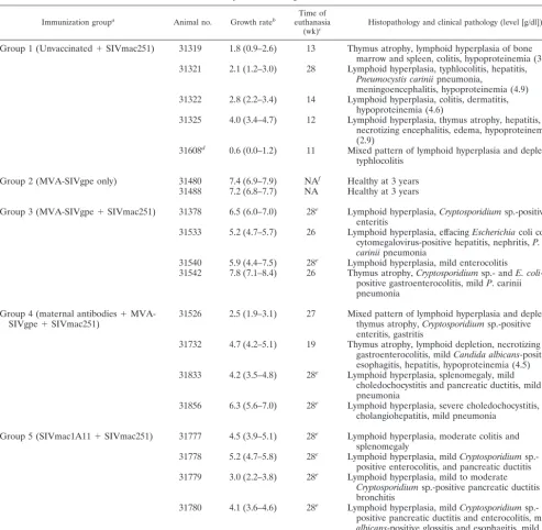
Immunization groupa Animal no. Growth rateb euthanasiaTime of
(wk)c Histopathology and clinical pathology (level [g/dl])
Group 1 (Unvaccinated⫹SIVmac251) 31319 1.8 (0.9–2.6) 13 Thymus atrophy, lymphoid hyperplasia of bone marrow and spleen, colitis, hypoproteinemia (3.5) 31321 2.1 (1.2–3.0) 28 Lymphoid hyperplasia, typhlocolitis, hepatitis,
Pneumocystis cariniipneumonia,
meningoencephalitis, hypoproteinemia (4.9) 31322 2.8 (2.2–3.4) 14 Lymphoid hyperplasia, colitis, dermatitis,
hypoproteinemia (4.6)
31325 4.0 (3.4–4.7) 12 Lymphoid hyperplasia, thymus atrophy, hepatitis, necrotizing encephalitis, edema, hypoproteinemia (2.9)
31608d 0.6 (0.0–1.2) 11 Mixed pattern of lymphoid hyperplasia and depletion, typhlocolitis
Group 2 (MVA-SIVgpe only) 31480 7.4 (6.9–7.9) NAf Healthy at 3 years 31488 7.2 (6.8–7.7) NA Healthy at 3 years
Group 3 (MVA-SIVgpe⫹SIVmac251) 31378 6.5 (6.0–7.0) 28e Lymphoid hyperplasia,Cryptosporidiumsp.-positive enteritis
31533 5.2 (4.7–5.7) 26 Lymphoid hyperplasia, effacingEscherichiacoli colitis, cytomegalovirus-positive hepatitis, nephritis,P. cariniipneumonia
31540 5.9 (4.4–7.5) 28e Lymphoid hyperplasia, mild enterocolitis 31542 7.8 (7.1–8.4) 26 Thymus atrophy,Cryptosporidiumsp.- andE. coli
-positive gastroenterocolitis, mildP. carinii pneumonia
Group 4 (maternal antibodies⫹
MVA-SIVgpe⫹SIVmac251) 31526 2.5 (1.9–3.1) 27 Mixed pattern of lymphoid hyperplasia and depletion,thymus atrophy,Cryptosporidiumsp.-positive enteritis, gastritis
31732 4.7 (4.2–5.1) 19 Thymus atrophy, lymphoid depletion, necrotizing gastroenterocolitis, mildCandida albicans-positive esophagitis, hepatitis, hypoproteinemia (4.5) 31833 4.2 (3.5–4.8) 28e Lymphoid hyperplasia, splenomegaly, mild
choledochocystitis and pancreatic ductitis, mild pneumonia
31856 6.3 (5.6–7.0) 28e Lymphoid hyperplasia, severe choledochocystitis, cholangiohepatitis, mild pneumonia
Group 5 (SIVmac1A11⫹SIVmac251) 31777 4.5 (3.9–5.1) 28e Lymphoid hyperplasia, moderate colitis and splenomegaly
31778 5.2 (4.7–5.8) 28e Lymphoid hyperplasia, mildCryptosporidium sp.-positive enterocolitis, and pancreatic ductitis 31779 3.0 (2.2–3.8) 28e Lymphoid hyperplasia, mild to moderate
Cryptosporidiumsp.-positive pancreatic ductitis and bronchitis
31780 4.1 (3.6–4.6) 28e Lymphoid hyperplasia, mildCryptosporidium sp.-positive pancreatic ductitis and enterocolitis, mildC. albicans-positive glossitis and esophagitis, mild pneumonia
aVaccine was administered in two doses; at birth and 3 weeks later. Animals of groups 1, 3, 4, and 5 were challenged orally at 4 weeks of age with SIVmac251-5/98.
bAverage weight gain in grams/day (with 95% confidence interval in parentheses) between 28 and 74 days of age, as determined by regression analysis.
cAge (weeks) at time of euthanasia.
dGroup 1 animal 31608 was born with maternally derived anti-SIV antibodies.
eAnimal was clinically stable at time of euthanasia; all other animals were euthanized due to life-threatening disease prior to or at 28 weeks of age.
fNA, not applicable.
on November 8, 2019 by guest
http://jvi.asm.org/
[image:4.603.45.539.79.561.2]cantly lower than those of the controls (group 3 versus group 1, P⫽0.0005; group 4 versus group 1,P⫽0.0941; and group 5 versus group 1,P⫽0.0012).
Levels of infectious cell-associated SIV in blood of all 17
SIV-infected infants showed considerable variation among an-imals within each group and temporal variation for individual animals, and thus there were no significant differences among the experimental groups in cell-associated infectious virus lev-els at the time of peak viremia or thereafter (Fig. 2). Cell-associated SIV levels in blood peaked at 1 or 2 weeks after oral challenge (460 to 21,530 TCID50 per million PBMC) and
thereafter ranged from 10 to 2,150 TCID50per million PBMC. Quantitative and qualitative comparison of antiviral
anti-body responses in infant macaques. Total antiviral IgG
anti-body responses were quantified by whole-SIV ELISA. Gag-and Env-specific antibodies were measured by using protein-based ELISA assays. For those samples with detectable Env-specific antibodies, measurements of antibody avidity and con-formational dependence were determined as described in Materials and Methods.
Except for maternally derived antibodies in animal 31608, all five SIVmac251-infected animals of group 1 had undetectable, or else low and transient de novo IgG responses, to whole SIV Gag and Env. The inability of these infants to mount or main-tain antiviral antibody responses is a result of the rapid immu-nosuppression induced by the highly virulent SIVmac251 iso-late.
[image:5.603.65.257.67.200.2]The two animals of group 2, which were immunized with MVA-SIVgpe but were not challenged with SIVmac251, mounted a rapid anti-SIV IgG response by 4 weeks of age (titer of 1:25,600), and the antibodies persisted throughout the observation period (Fig. 3). This observation demonstrates the immunogenicity of this vaccine in infant macaques. However, FIG. 1. Comparison of survival for vaccinated and unvaccinated
infant rhesus macaques. The survival of unvaccinated (dashed line) and vaccinated (solid lines) animals by age (weeks) is shown. The comparison of survival curves was performed by using the log-rank test. The median survival time for unvaccinated animals was 13 weeks of age. The survival curves for unvaccinated animals were statistically different from animals vaccinated with SIVmac1A11 (P⬍0.0049 [F])
and all eight animals vaccinated with MVA-SIVgpe (P⬍0.0093 [tri-angles]). There was no statistically significant difference in the survival curves among the different groups of vaccinated animals (MVA-SIVgpe immunized, median survival⫽27 weeks [ƒ]; MVA-SIVgpe
plus maternal SIV antibodies, median survival ⫽ 27.5 weeks [];
SIVmac1A11-immunized, all animals healthy at 28 weeks).
FIG. 2. Virus levels in blood. Levels of SIV RNA in plasma (top panel) were measured by bDNA assay, whereas cell-associated virus levels (bottom panel) were measured by limiting dilution assay. Vaccine (V) was administered within 3 days after birth and 3 weeks later. All monkeys were challenged (C) orally with SIVmac251 at 4 weeks of age.
on November 8, 2019 by guest
http://jvi.asm.org/
[image:5.603.45.539.418.689.2]SIV Env-specific endpoint titers were low at 4 weeks of age and became undetectable at 16 to 28 weeks of age. In contrast, SIV Gag-specific antibody responses continued to increase during the 28 weeks. Because MVA has been shown to have limited replication in human cells in vitro (3), the slow increase in SIV Gag-specific antibody responses in these infant macaques
sug-gest that immunization with MVA-SIVgpe may result in a low level of residual replication with preferential expression ofgag epitopes.
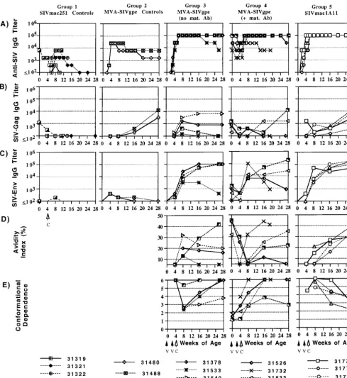
[image:6.603.47.536.71.603.2]At birth, prior to the immunizations, the four infants of group 4 already had moderate to high levels of maternally derived whole SIV-specific antibodies, moderate levels of SIV FIG. 3. Measurements of quantitative and qualitative antibody responses in infant macaques orally inoculated with SIVmac251. Quantitative levels of SIV whole virus (A) and gag-specific (B) antibodies were determined in a standard ELISA. SIV Env-specific antibody endpoint titers (C), avidity (D), and conformational dependence (E) were determined in a ConA ELISA. All procedures are described in Materials and Methods.
on November 8, 2019 by guest
http://jvi.asm.org/
Gag-specific antibodies, and except for animal 31732, detect-able levels of anti-SIV Env antibodies (Fig. 3). At 4 weeks of age (i.e., after the two immunizations with MVA-SIVgpe or SIVmac1A11) all animals of groups 3, 4, and 5 had moderate to high antibody titers against whole SIV (titers of 6,400 to 102,400) but low levels of antibodies to SIV Gag and Env. After oral inoculation with SIVmac251 at 4 weeks of age, antibody titers followed a similar pattern: despite individual variation within each group, the immunized animals of groups 3, 4, and 5 mounted similar levels of whole SIV-, SIV Gag-, and SIV Env-specific antibodies during the first 4 weeks after SIV infection. Later, there was more variation in SIV-specific an-tibody levels. In general, antiviral anan-tibody titers, in particular the Gag-specific antibody titers, declined for animals that de-veloped AIDS within the 28 weeks of observation period (group 3, animals 31533 and 31542; group 4, animals 31526 and 31732) but remained stable or increased for animals that were clinically still asymptomatic at 28 weeks of age (including all of the group 5, SIVmac1A11-immunized animals). The maternal antibodies in group 4 animals did not have any detectable inhibitory effect on the levels of SIV-specific antibodies pro-duced during the first 4 to 12 weeks of SIVmac251 infec-tion. In summary, the immunizations with MVA-SIVgpe or SIVmac1A11 primed animals in groups 3, 4, and 5 to produce similar levels of SIV-specific antibodies early after SIVmac251 infection.
In contrast, there were marked differences among animals of groups 3, 4, and 5 in the quality of SIV-envelope specific antibodies as assessed by avidity and conformational depen-dence. In group 3 (MVA-SIVgpe immunized), SIV Env anti-bodies at the time of oral challenge had low or undetectable avidity but high conformation ratios (indicating reactivity to native envelope glycoprotein). For three of these four animals, avidity index values peaked by 4 weeks after SIV infection but remained low (ⱕ32%) and declined afterward; the conforma-tion ratio in these animals showed a pattern opposite to avidity, in that the conformation ratio dropped sharply 4 weeks after SIV infection but then increased again to higher levels, indi-cating preferential recognition of native envelope glycoprotein. Animal 31542 was the only animal for which the SIV envelope antibody avidity index increased gradually to intermediate lev-els (42%) by the onset of AIDS at 26 weeks of age and for which the conformational dependence remained remained high and showed little variation over time.
For the group 4 animals (MVA-SIVgpe-immunized infants with maternally derived SIV antibodies at birth) anti-SIV Env antibodies were detectable in only three of four animals at birth; the anti-env antibodies from these three animals had a moderate avidity index (32 to 45%) and a low conformational dependence (between 1 and 2; Fig. 3) that is consistent with a mature antibody response in their immunized mothers (6, 8, 29). For these three animals, there was a sharp decrease in avidity index after MVA-SIVgpe immunization (i.e., prior to SIVmac251 inoculation). After a further decrease for two an-imals during the first 4 weeks of SIV infection, there was a slower increase in avidity index compared to group 3 animals (which already had SIV Env antibodies with detectable avidity 4 weeks after oral SIVmac251 challenge; Fig. 3). This suggests that the de novo anti-SIV Env antibody response of these infants that was clearly detectable 4 weeks after SIV infection
(Fig. 3C) consisted mainly of antibodies with very low avidity. In contrast, the fourth animal (i.e., animal 31732), which did not have detectable maternally derived anti-env antibodies at birth, mounted anti-env antibodies with moderate avidity within 4 weeks after SIV infection, a finding similar to the response of the group 3 animals that lacked maternal antibod-ies. For all four animals, the conformational dependence in-creased after SIV infection but then dein-creased again for ani-mal 31856.
For the group 5 animals (SIVmac1A11 immunized), SIV-env antibody levels at 4 weeks of age were only detectable at sufficient levels in one animal (animal 31777) to allow mea-surements of antibody quality; the anti-env antibodies in this animal were found to have low avidity and high conformational dependence (Fig. 3). After SIVmac251 infection, SIV-env tibody avidity values for all four SIVmac1A11-immunized an-imals increased gradually to intermediate levels (30 to 41%) and were higher than those of most of the SIVgpe-immunized animals of groups 3 and 4. In contrast to the MVA-SIVgpe-immunized animals in groups 3 and 4, the conforma-tional dependence of the anti-SIV Env antibodies of group 5 animals was high (⬎4) shortly after oral challenge with SIV-mac251 but then decreased until 28 weeks of age, a finding consistent with previous reports of antibody maturation result-ing from intravenous attenuated SIV infection (6, 8, 29).
In summary, there was a correlation between the develop-ment and maintenance of high antiviral antibody titers and lower viremia and delayed disease course. In contrast, the time course of the parameters of antibody quality was more com-plex, and there was no clear correlation of antibody quality with virus levels or survival. The initial mode of exposure to antigen (type of vaccine and the presence or absence of ma-ternal antibodies) appeared to have modulated the param-eters of SIV-specific antibody quality early during infection, and the subsequent changes in SIV envelope-specific anti-body quality reflect most likely a complex process of antianti-body maturation in the presence of various degrees of immunosup-pression.
Quantification of virus-specific IFN-␥-secreting
lympho-cytes. The presence of virus-specific IFN-␥-secreting cells in
PBMC was measured by an ELISPOT assay by using stimula-tion with overlapping peptides of the p24 Gag region. For all animals, cryopreserved PBMC samples that were collected at 4, 5, 6, and 8 weeks of age (i.e., the day of oral SIVmac251 challenge and 1, 2, and 4 weeks afterward) were tested. All PBMC samples collected at the time of the oral SIVmac251 challenge had levels of SIV-specific IFN-␥-secreting cells be-low the cutoff value (see Materials and Methods). After SIV-mac251 infection, only two samples had detectable levels of virus-specific IFN-␥-secreting cells: group 4 animal 31833 (week 8 of age, 80 SFC/million PBMC; medium-control wells, 0 SFC/million PBMC), and group 5 animal 31777 (week 5 of age, 128 SFC/million PBMC; medium control wells, 43 SFC/ million PBMC). Both of these animals developed moderate viremia (viral RNA levels at 12 to 16 weeks were between 107
and 108copies per ml of plasma). None of the animals that
developed low viremia (⬍107) had detectable virus-specific
IFN-␥-secreting cells at these early time points after infection. For the two MVA-SIVgpe-immunized animals of group 2,
on November 8, 2019 by guest
http://jvi.asm.org/
which did not receive SIVmac251 challenge, no IFN-␥ -secret-ing cells were detected at these same time points.
Immune response to cholera toxin and tetanus toxoid.None
of the 19 neonates in the study had any maternal antibodies against cholera toxin at birth. After the first cholera toxin subunit B immunization at 2 weeks of age (2 weeks prior to oral SIV inoculation), all 19 animals developed a strong pri-mary antibody response (cholera toxin-specific IgG titer of ⱖ25,600 at 8 weeks of age), demonstrating competence of infant macaques to respond immunologically to this antigen. All animals also made an anamnestic response (ⱖ4-fold in-crease in IgG titer; data not shown) after the second cholera toxin subunit B immunization at 10 weeks of age (6 weeks after oral challenge). These cholera toxin subunit B antibody re-sponses show that even in the unimmunized, SIV-infected con-trol animals (group 1), primary and secondary antibody re-sponses to non-SIV antigens were not impaired at these stages of SIV infection.
Most animals had maternally derived antibodies to tetanus toxoid that declined after birth with a half-life of ca. 2 weeks. At the time of the first immunization (6 weeks of age), tetanus toxoid-specific IgG titers ranged from 1:100 to 1:6,400. For the animals with high anti-tetanus toxoid titers (1:1,600 to 1,601: 6,400), an absence of a further decline of tetanus toxoid-spe-cific IgG was therefore interpreted as evidence of a primary immune response. Except for animals 31319 (group 1) and 31526 (group 4), all animals made a primary tetanus toxoid-specific antibody response. Animals received a booster immu-nization with tetanus toxoid at 14 weeks of age (10 weeks pc). In group 1, only animal 31321 was still alive and made a weak (⬃2-fold increase in titer) secondary tetanus toxoid response. For groups 2 to 5, there was a correlation between the degree of the booster response with clinical outcome; animals 31732 and 31533 (which both developed AIDS at 19 and 26 weeks of age, respectively) had a weak increase (2- to 4-fold), whereas all other animals had a ⱖ4-fold increase in tetanus toxoid-specific IgG titers.
Lymphocyte subset populations. Absolute lymphocyte
counts were quite variable over time in all animals, even for animals that were not SIV infected (group 2), and absolute counts of the T- and B-lymphocyte subsets showed similar levels of variability (Fig. 4). The percentages of cell types were more reliable parameters for comparing the animal groups.
Similar to humans, infant macaques have age-related changes in their numbers of total lymphocytes and lymphocyte subsets (12). In particular, newborn animals have a high per-centage of CD4⫹ T lymphocytes and a low percentage of CD8⫹T lymphocytes, resulting in a high CD4/CD8 ratio (⬎3). During the first few months of age, there is a gradual decrease in CD4⫹T lymphocytes and an increase in CD8⫹T lympho-cytes (Fig. 4), resulting in a decrease of the CD4/CD8 ratio to ca. 2 to 3. There is also an age-related increase in CD20⫹B lymphocytes (Fig. 4). Lymphocyte subsets of the unchallenged MVA-SIVgpe-immunized animals (group 2) were indistin-guishable from those of normal, age-matched control animals. For the other animals, all of which became infected after oral SIVmac251 inoculation at 4 weeks of age, some interesting observations were made. Except for the SIVmac1A11-immu-nized animals, the majority of the other animals had at least a 35% drop in absolute lymphocyte counts at 1 week
postchal-lenge. Whereas the unimmunized SIVmac251-infected animals showed absolute counts and percentages of CD20⫹B lympho-cytes near or below the range for uninfected animals, most immunized animals (groups 3 to 5), in particular the SIVmac1A11-immunized infants (group 5), had B lymphocytes within the normal range.
Unexpectedly, the unimmunized SIV-infected animals (group 1) showed percentages and numbers of CD4⫹T lym-phocytes that were generally in the range of the uninfected animals (Fig. 4). In contrast, most animals of groups 3, 4, and 5 had a sudden decrease in percent CD4⫹T lymphocytes 2 weeks after SIVmac251 inoculation, which slowly recovered but did not reach preinfection baseline values. Absolute CD4⫹-T-lymphocyte counts in groups 3 to 5 were within the normal range (⬎1,000/l) during the first few months after SIV infection, but total lymphocyte counts were also often elevated (Fig. 4). The numbers of CD4⫹ T lymphocytes de-clined in seven animals of groups 3 to 5 byⱖ16 weeks of age to ⬍1,000/l, concomitant with a decline in absolute total lymphocyte counts.
For the majority of animals, SIV infection was associated with increased and more variable percentages of CD8⫹T lym-phocytes. An exception were the SIVmac1A11-immunized an-imals (group 5) and the MVA-SIVgpe-immunized anan-imals of groups 3 and 4 that had slower disease progression (e.g., ani-mals 31540 and 31856), for which the percentage of CD8⫹T lymphocytes showed a minor increase after SIVmac251 infec-tion and remained more stable throughout the observainfec-tion period (Fig. 4).
As a result of these opposite changes in the percentages of CD4⫹ and CD8⫹T lymphocytes, the CD4/CD8 ratios were essentially unchanged in most of the unimmunized SIV-mac251-infected animals (group 1) but were reduced in all immunized SIVmac251-infected groups (groups 3 to 5), espe-cially in the MVA-SIVgpe-immunized animals (groups 3 to 4), with a nadir occurring 2 weeks after SIVmac251 infection (Fig. 4).
DISCUSSION
The primary goal of the present study was to determine whether SIV vaccines given as early as possible after birth could rapidly induce immunity against oral challenge with pathogenic SIVmac251. The second objective was to examine whether maternally acquired anti-SIV antibodies affected the generation of immune responses and the efficacy of active immunization of the infants.
Infant macaques were inoculated orally with SIVmac251 at 4 weeks of age. Four of the five unimmunized control animals exhibited persistently high virus levels and poor antiviral im-mune responses, and they developed fatal immunodeficiency within 11 to 14 weeks of age (i.e., 7 to 10 weeks after SIV-mac251 infection). This rapid disease course in the majority of unimmunized animals is indistinguishable from our previous observations when animals were inoculated with SIVmac251 at birth (25, 46, 49). The observation that anti-SIV immune re-sponses in these SIV-infected animals were very weak, whereas these animals were able to make immune responses to other non-SIV antigens, is in agreement with the recent observation that HIV preferentially infects HIV-specific CD4⫹T
on November 8, 2019 by guest
http://jvi.asm.org/
FIG. 4. Lymphocyte subsets in peripheral blood as measured by flow cytometric analyses. The percentages are expressed as fractions of the total number of lymphocytes.
on November 8, 2019 by guest
http://jvi.asm.org/
cytes, and thus antiviral immune responses are even sup-pressed prior to the development of generalized immunodefi-ciency (13).
We demonstrated that both vaccines, MVA-SIVgpe and SIVmac1A11, elicited rapid immune responses in infant ma-caques. Thus, the immune system of newborn monkeys is com-petent to make immune responses, a finding in agreement with the results of HIV-1 vaccine trials in human neonates (4, 26). Neither of the two vaccines in our study prevented SIV-infec-tion after oral challenge at 4 weeks of age. However, both vaccines primed the infant macaque immune system such that, after challenge with pathogenic SIVmac251, vaccinated ani-mals (i) mounted rapid, high SIV-specific antibody responses; (ii) more effectively controlled virus replication; and (iii) had longer disease-free survival than the unvaccinated infants. Sim-ilar results were reported for juvenile macaques immunized with MVA expressing SIV antigens and challenged intrave-nously or intrarectally with pathogenic SIV (18, 32, 35, 37). The efficacy of the MVA-SIVgpe vaccine is due to the expres-sion of SIV proteins and not simply to infection with MVA vector, since immunization of newborn macaques with MVA expressing measles antigens did not affect viremia or disease course after SIVmac251 challenge (unpublished observations). We previously reported that two of five macaques infected with SIVmac1A11 in utero or at birth were protected from oral SIVmac251 challenge (33). That study differed from the present one, however, in that monkeys were challenged 1 year after SIVmac1A11 infection, which allowed more time for immune responses to mature.
In the current study, both MVA-SIVgpe and SIVmac1A11 primed the immune system of the infant rhesus macaques to make more rapid antiviral immune responses after SIVmac251 infection. Both group 3 (MVA-SIVgpe) and group 5 (SIVmac1A11) animals had similar virus levels after SIV in-fection. However, the four SIVmac1A11-immunized animals remained healthy at 28 weeks of age, and they had minor histopathological evidence of opportunistic infections; in con-trast, two of the four MVA-SIVgpe-immunized animals had developed simian AIDS by 28 weeks. The maturation of the antibody responses also differed among these two groups. Three of four MVA-SIVgpe-immunized animals (group 3) had, after an initial peak at 8 weeks of age, a gradual decrease in antibody avidity; all four animals had, after an initial decline early after SIV infection, a progressive increase in tional dependence (indicating more recognition of conforma-tional epitopes than of linear epitopes). This pattern displayed by the MVA-SIVgpe-immunized group 4 monkeys is similar to that previously observed for some HIV-infected people (6). In contrast, the development of a high anti-SIV Env antibody response with a gradual increase in avidity and decrease of the conformational dependence observed for all four SIVmac1A11-immunized animals is consistent with that re-ported previously for juvenile and adult macaques infected with other attenuated or pathogenic strains of SIV (6, 8) and indicates that antibody responses are broadening to also in-clude fewer conformational epitopes (i.e., including linear epitopes) (7). Despite these different patterns, there was no detectable correlation between these changes in antibody qual-ity and virus levels or disease progression, a finding that sug-gests that the role of antibody quality on virus replication and
the disease course in infants is not clear. It is possible that these distinct temporal patterns of antibody quality between animals immunized with the two different SIV vaccines reflect a combination of initial differences in SIV vaccine epitopes or their expression level during the priming of the immune sys-tem, followed by differences in SIV-induced immunosuppres-sion (8).
The second objective of this study was to determine the effect of maternally derived SIV antibodies on the efficacy of SIV vaccines. One group of newborn macaques (group 4) was born to SIV-immunized macaques and therefore had maternal antiviral antibodies. We immunized these newborns with MVA-SIVgpe and then challenged them orally with SIV-mac251. Compared to the results of a similar challenge by using MVA-SIVgpe-vaccinated infant macaques born without maternal antibodies (group 3), both groups of MVA-SIVgpe-immunized infants had similar clinical outcomes (two of four animals in each group were healthy at 28 weeks of age). Both groups also developed similar levels of SIV-specific antibodies. This finding is consistent with the antibody responses observed in infants, born to HIV-infected women, who were given an HIV-1 recombinant gp120 vaccine (26). In a measles model in macaques, immunization of infant (51) or juvenile monkeys (39) with an MVA-based measles vaccine was effective in the presence of passively acquired neutralizing antibody. In addi-tion, a trial in which human newborns with maternal antibodies received an accelerated immunization schedule with recombi-nant HIV-1 gp120 also found that the presence of maternal antibodies did not inhibit a de novo antibody response (26). In the present study in infant macaques, although no effect on vaccine efficacy was observed, our observations suggest that maternal antibodies caused differences in the antibody quality, in particular with respect to avidity, after MVA-SIVgpe immu-nization (Fig. 3). The data suggest that, in the presence of maternal antibodies, the expression of SIV antigens by MVA-SIVgpe may have induced the clearance of high-avidity anti-bodies from the circulation and that these maternal antianti-bodies may have masked particular SIV epitopes needed to prime the induction of high-avidity SIV antibodies; thus, infants only started to make a primary immune response to these epitopes after SIVmac251 infection and when maternally derived SIV antibody levels were reduced. Although these measures of antibody quality did not show a detectable correlation with virus levels and survival in the SIV-infected infant macaques in the present study, these observations suggest that data regard-ing antibody quality derived from HIV vaccine trials in unin-fected adults cannot necessarily be extrapolated to infants who have maternally derived HIV antibodies. This underscores the urgent need for HIV vaccine studies that are targeted specif-ically for infants born to HIV-infected women.
These results appear to contrast with those of our previous studies that indicated an important role for anti-SIV IgG in preventing SIV infection in neonatal macaques (44, 49). How-ever, these studies are not directly comparable because of differences in the experimental design (transplacental transfer or passive immunization with pooled serum versus active im-munization), differences in measures of antibody quality (avid-ity and conformational dependence), and different ages at the time of virus challenge (neonates versus 4-week-old infants).
In the present study, immunization with MVA-SIVgpe or
on November 8, 2019 by guest
http://jvi.asm.org/
SIVmac1A11 at birth and 3 weeks of age did not give any detectable levels of virus-specific IFN-␥-secreting PBMC by 4 weeks of age, when the animals were exposed orally to SIV-mac251. The 4-week time period of immunizations in the present study may have been too short to induce detectable levels in peripheral blood, especially for newborn animals. Al-though immunization of human newborns with optimal dosage regimens of recombinant HIV-1 gp120 in adjuvant induced proliferative responses in PBMC at week 4, results from these different assays cannot be compared directly (4). In addition, peripheral blood samples are probably not representative of cell-mediated immune responses that occur in lymphoid tissues or mucosal sites (1, 17). It is unknown whether virus-specific IFN-␥-secreting cells were present in the mucosa and lymphoid tissues that drain the upper orogastrointestinal tract, where SIVmac251 infection was initiated. After infection with SIV-mac251, only two animals had detectable levels of virus-specific IFN-␥-secreting PBMC at a single time point within the first 4 weeks of infection, and neither of these animals was able to control viremia to low levels. Although further research on the early development of cell-mediated immunity is needed, these findings are in agreement with observations of infrequent de-tection of HIV-specific effector CD8⫹-T-cell activity in HIV-infected infants under 3 years of age (36).
Flow cytometric determination of lymphocyte subsets re-vealed some interesting trends. The lymphopenia during acute infection and subsequent increase in CD8⫹ T lymphocytes were expected findings based on prior experiments. Surprising findings were that the unimmunized SIV-infected control an-imals (group 1) maintained a relatively normal percentage of CD4⫹ T lymphocytes compared to the immunized animals after oral SIV challenge. Several of these group 1 animals showed histopathological evidence of lymphoid hyperplasia at the time of necropsy (Table 1). Altogether, these findings sug-gest that the rapid immunosuppression and rapid disease course in the group 1 animals was more likely due to a func-tional defect in CD4⫹T helper cells than to a direct effect of depletion. In contrast, most immunized animals had a reduc-tion in the percentage of CD4⫹T lymphocytes and an increase in the percentage of CD8⫹T lymphocytes in peripheral blood early after SIVmac251 infection, which is probably due to a combination of changes in cell death or proliferation and re-distribution among lymphoid compartments (5, 28).
In summary, the present study underscores the relevance of the SIV infant macaque model for evaluating strategies to prevent maternal transmission of HIV. In developing coun-tries, there is a critical need for a vaccine that can protect babies born to HIV-infected women from infection acquired via breast-feeding. In the present study, immunization with MVA-SIVgpe or SIVmac1A11 did not protect infant ma-caques against oral infection with virulent SIVmac251 and did not modulate the specific SIV variants that established sys-temic infection (unpublished data). However, vaccinated in-fant macaques had lower viral levels, enhanced immune responses, and longer disease-free survival times than unvac-cinated infants, demonstrating that both SIV vaccines substan-tially altered the disease course. A vaccine capable of inducing more effective immune responses rapidly, especially near the site of initial exposure to virus, is needed to prevent viral infection.
ACKNOWLEDGMENTS
We thank N. Aguirre, S. Au, D. Bennet, D. Brandt, I. Bolton, K. Bost, L. Brignolo, K. Christe, L. Hirst, A. Spinner, K. Schmidt, W. von Morgenland, and the California National Primate Research Center Colony Services for expert technical assistance. We thank Shilpa Hat-tangadi and Lynn Frampton for construction of recombinant MVAs; the NIH AIDS Research and Reference Reagent Program, Division of AIDS, NIAID, NIH, for providing the SIV p55 Gag protein and the 20-mer peptides of the p24 Gag region; and R. Desrosiers (New England Regional Primate Research Center) for the plasmid containing the SIV-mac239envgene. Neil Willits, Statistical Laboratory, University of Cali-fornia at Davis, provided expert assistance for statistical analyses.
This work was supported by Public Health Science grant RR00169 from the National Center for Research Resources, NIH/NIAID grants AI39109 (M.L.M.), AI46320 (M.L.M.), and AI47758 (K.S.C., J.D.S., and R.C.M.), and Elizabeth Glaser Scientist award 8-97 (M.L.M.) from the Elizabeth Glaser Pediatric AIDS Foundation.
REFERENCES
1. Abel, K., M. J. Alegria-Hartman, K. Zanotto, M. B. McChesney, M. L. Marthas, and C. J. Miller.2001. Anatomic site and immune function cor-relate with relative cytokine mRNA expression levels in lymphoid tissues of
normal rhesus macaques. Cytokine16:191–204.
2. Bertolli, J., M. E. St. Louis, R. J. Simonds, P. Nieburg, M. Kamenga, C. Brown, M. Tarande, T. Quinn, and C. Y. Ou.1996. Estimating the timing of mother-to-child transmission of human immunodeficiency virus in a
breast-feeding population in Kinshasa, Zaire. J. Infect. Dis.174:722–726.
3. Blanchard, T. J., A. Alcami, P. Andrea, and G. L. Smith.1998. Modified vaccinia virus Ankara undergoes limited replication in human cells and lacks several immunomodulatory proteins: implications for use as a human
vac-cine. J. Gen. Virol.79:1159–1167.
4. Borkowsky, W., D. Wara, T. Fenton, J. McNamara, M. Kang, L. Mofenson, E. McFarland, C. Cunningham, A.-M. Duliege, D. Francis, Y. Bryson, S. Burchett, S. A. Spector, L. M. Frenkel, S. Starr, R. Van Dyke, E. Jiminez, et
al.2000. Lymphoproliferative responses to recombinant HIV-1 envelope
antigens in neonates and infants receiving gp120 vaccines. J. Infect. Dis.
181:890–896.
5. Bucy, R. P., R. D. Hockett, C. A. Derdeyn, M. S. Saag, K. Squires, M. Sillers, R. T. Mitsuyasu, and J. M. Kilby.1999. Initial increase in blood CD4⫹
lymphocytes after HIV antiretroviral therapy reflects redistribution from
lymphoid tissues. J. Clin. Investig.103:1391–1398.
6. Cole, K. S., M. Murphey-Corb, O. Narayan, S. Y. Joag, G. M. Shaw, and R. C. Montelaro.1998. Common themes of antibody maturation to simian immunodeficiency virus, simian-human immunodeficiency virus, and human
immunodeficiency virus type 1 infections. J. Virol.72:7852–7859.
7. Cole, K. S., M. J. Paliotti, M. Murphey-Corb, and R. C. Montelaro.2000. Maturation of envelope-specific antibody responses to linear determinants in
monkeys inoculated with attenuated SIV. J. Med. Primatol.29:220–230.
8. Cole, K. S., J. L. Rowles, B. A. Jagerski, M. Murphey-Corb, T. Unangst, J. E. Clements, J. Robinson, M. S. Wyand, R. C. Desrosiers, and R. C. Montelaro.
1997. Evolution of envelope-specific antibody responses in monkeys exper-imentally infected or immunized with SIV and its association with the
de-velopment of protective immunity. J. Virol.71:5069–5079.
9. Dabis, F., P. Msellati, N. Meda, C. Welffens-Ekra, B. You, O. Manigart, V. Leroy, A. Simonon, M. Cartoux, P. Combe, A. Ouangre´, R. Ramon, O. Ky-Zerbo, C. Montcho, R. Salamon, C. Rouzioux, P. Van de Perre, L. Mandelbrot, et al.1999. Six-month efficacy, tolerance, and acceptability of a short regimen of oral zidovudine to reduce vertical transmission of HIV in breastfed children in Coˆte d’Ivoire and Burkina Faso: a double-blind
place-bo-controlled multicentre trial. Lancet353:786–792.
10. Datta, P., J. P. Embree, J. P. Kreiss, J. O. Ndinya-Achola, M. Braddick, M. Temmerman, N. J. D. Nagelkerke, G. Maitha, K. K. Holmes, P. Piot, H. O. Pamba, and F. A. Plummer.1994. Mother-to-child transmission of human immunodeficiency virus type 1: report from the Nairobi study. J. Infect. Dis.
170:1134–1140.
11. De Cock, K., M. G. Fowler, E. Mercier, I. de Vincenzi, J. Saba, E. Hoff, D. J. Alnwick, M. Rogers, and N. Shaffer.2000. Prevention of mother-to-child HIV transmission in resource-poor countries: translating research into policy
and practice. JAMA283:1175–1182.
12. DeMaria, M. A., M. Casto, M. O’Connell, R. P. Johnson, and M. Rosenz-weig.2000. Characterization of lymphocyte subsets in rhesus macaques
dur-ing the first year of life. Eur. J. Haematol.65:245–257.
13. Douek, D. C., J. M. Brenchley, M. R. Betts, D. R. Ambrozak, B. J. Hill, Y. Okamoto, J. P. Casazza, J. Kuruppu, K. Kunstman, S. Wolinsky, Z. Gross-man, M. Dybul, A. Oxenius, D. A. Price, and R. A. Koup.2002. HIV
pref-erentially infects HIV-specific CD4⫹T cells. Nature417:95–98.
14. Earl, P. L., L. S. Wyatt, D. C. Montefiori, M. Bilska, R. Woodward, P. D. Markham, J. D. Malley, T. U. Vogel, T. M. Allen, D. I. Watkins, N. Miller, and B. Moss.2002. Comparison of vaccine strategies using recombinant