A Dissertation On
“A COMPARITIVE STUDY OF OPEN CHOLECYSTECTOMY
VERSUS LAPAROSCOPIC CHOLECYSTECTOMY”
Submitted to
The Tamil Nadu Dr. M.G.R. Medical University
In fulfillment of regulations for the award of degree of
M.S., DEGREE EXAMINATION
BRANCH –I M.S., (GENERAL SURGERY)
GOVT.KILPAUK MEDICAL COLLEGE
THE TAMILNADU DR. M.G.R. MEDICAL UNIVERSITY
CHENNAI - 10
BONAFIDE CERTIFICATE
This is to certify that the dissertation entitled “COMPARITIVE STUDY OF
OPEN CHOLECYSTECTOMY VERSUS LAPAROSCOPIC
CHOLECYSTECTOMY” is a bonafide record of work done by Dr.A.Sekarin the Department of General Surgery, Government Kilpauk Medical College Hospital, Chennai-10.
PROF.P.RAMAKRISHNAN, MD., DLO., DEAN,
Government Kilpauk Medical College Chennai-10
Prof. D. NAGARAJAN, M.S., Prof.N.SHANMUGASUNDARAM, M.S., Professor of General Surgery, H.O.D. of General Surgery
DECLARATION
I
, Dr.A.Sekar,
solemnly declare that this dissertation, titled “ A COMPARITIVE STUDY OF OPEN CHOLECYSTECTOMYversus LAPAROSCOPIC CHOLECYSTECTOMY ” is a bonafide record of work done by me in the Department of General Surgery, Government Kilpauk Medical College, Chennai-10, during the period from July 2012 to Dec 2013 under the guidance of my Unit Chief Prof. D. Nagarajan M.S., Government Kilpauk Medical College Hospital, Chennai-600 010.
This dissertation is submitted to the Tamil Nadu Dr. M.G.R. Medical University, Chennai in fulfillment of the University regulations for the award of M.S. (General Surgery) Branch I, General Surgery Examination to be held in APRIL-2014
Place: Chennai Date:
ACKNOWLEDGEMENT
I wish to express my sincere gratitude and thanks to Prof. P. N. SHANMUGASUNDARAM, M.S., Professor & Head of the Department of General Surgery for having helped me at every step in completing this study successfully.
I wish to express my sincere gratitude to my Unit Chief Prof D.NAGARAJAN, M.S., Professor of Department of General Surgery for having helped me at every step in successfully completing this study. I also wish to express my sincere thanks to my Assistant Professors Dr.C.Malarvizhi, Dr.A.Rajeswaran for helping me to complete this study.
I also thank Professor and Head of the department of Pathology and all other teachers who helped me to do this study in time.
I am also very grateful to our Dean PROF.P.RAMAKRISHNAN, M.D.,DLO., for his permission to utilize the clinical material for this study.
I also extend my thanks to all my patients for their full co-operation in successful completion of this study.
CONTENTS
SL NO TOPIC
PAGENO
1
Introduction
2
Aim of study
3
Review of literature
A
Historical review
B
Embryology
C
Anatomy
D
Cholelithiasis
E
Cholecystectomy
F
Open Cholecystectomy
G
Laparoscopic Cholecystectomy
H
Complications
I
Comparison and treatment
4
Materials and methods
5
Analysis of results
6
Discussion
7
Conclusion
8
Bibliography
9
Annexure
ABSTRACT
TITLE: A COMPARITIVE STUDY OF OPEN CHOLECYSTECTOMY
VERSUS LAPAROSCOPIC CHOLECYSTECTOMY
Author: Dr.A.sekar. MS postgraduate. Dr. D. Nagarajan MS. Department of general Surgery , Govt.Kilpauk Medical College. Chennai -10
Introduction : Gall bladder disease is the most common curable disease in female of middle age .Laparoscopic cholecystectomy has rapidly become the choice of elective surgery for the treatment of Cholecystitis even though Open Cholecystectomy remains the main modality of surgery in many centres in India. But to become an alternative to open method, it should be safe, less morbid and it should have the possibility of early return to work better than that of open the procedure. This study compares the open
cholecystectomy and laparoscopic cholecystectomy with respect to duration of procedure post operative pain,wound infection,requirement of antibiotics and analgesics period of stay in hospital and return to work
half an hour prior to surgery is more enough to prevent infection except in conditions like diabetes mellitus association with it. Per rectal Paracetamol suppository is enough to relieve in both the groups . Time taken for
laparoscopic surgery is lesser than that for open procedure and the period of hospital stay is also less in laparoscopic surgery.
INTRODUCTION
It was a gynecologist who introduced the so called ‘endoprocedures’ in the field of surgery, his name was Kurt Semm, a German national who used his instruments for the removal of ovaries and myomas in 1970s. Only after a successful laparoscopic appendicectomy by Semm in 1982 the general surgeons realized the importance and started applying these instruments in the field of general surgery. The German surgeon Eric Muhe while pioneering cholecystectomy came to know that a modification of the instrument is needed for gall bladder removal. Since then rest of the world started doing laparoscopic cholecystectomies but, this was the time when abdominal open surgeries were becoming extensive operations with the concept of ‘bigger problems need bigger incisions’ and laparoscopic technique was criticized by
Aim of study
A Aim of study is to compare the open cholecystectomy and laparoscopic cholecystectomy with respect to
Duration of procedure
Post operative pain
Wound infection
Requirement of antibiotics and analgesics
Period of stay in hospital
Return to workREVIEW OF LITERATURE
Historical review
Founder of gall bladder surgery is Jean Louis Petit.
In 1420Antonio Benevieni, a pathologist gave the first description of gall stones in a woman who died of abdominal pain.
In 1687Stal Pert Von DerWeil explained gall stones during laparotomy done for an acute abdomen.
In 1743Petit removed gall bladder successfully for the first time.
In 1859 J.L.W Thudichum did a 2 staged cholecystectomy.
In 1895 Carl Johanan August Langenbuch did first open cholecystectomy On a 43 yr old male who suffered from gall bladder disease for more than 16 yrs.
In 1987Eric Muhe did the first laparoscopic cholecystectomy in Boblingen, Germany. [23]
In1988 Americans started doing lap cholecystectomies, since then it started its Journey to take over the conventional open technique. Only about 10% of cholecystectomies were done by laparoscopic method during 1990’s which accounted for 88 % during 2006, now almost replacing the open method except in some special situations like gall bladder carcinoma, conversion due to hemorrhage. Revolution of laparoscopy had led the surgeons to have a mobile set up with all the equipments and taking the assistants even to the remote areas to perform laparoscopic cholecystectomies.
Embryology of Gall bladder
Hepatic diverticulum arises from endodermal epithelium of distal end of the foregut during 3rd week of gestation. Proximal end of this diverticulum develops in to liver and the distal end develops in to gall bladder and cystic duct. By 5th week all the elements of biliary system are developed where common hepatic duct and cystic ducts are occluded initially which becomes recanalises later.
The stalk joining these 2 ducts to duodenum becomes common bile duct. By 6th week common bile duct rotates by 180 degrees around duodenum. Sphincter of oddi starts to develop by 10th week and completes by 28th week. Bile pigment starts to form at 13-16 weeks to reach duodenum.
[image:13.612.105.509.528.681.2]
Developmental anomalies of gall bladder:
Agenesis of the gall bladder [1] occurs due to failure of development of distal part of hepatic diverticulum. Atresia and Hypoplasia are due to maldevelopment and duplication of the gall bladder as well as due to defective revacuolization of the gall bladder which occurs in 1 in 4000 with definite chances of Cholecystitis is seen. Gall bladder Phrygian cap [2] occurs 1%-6% of the population.
Other anomalies are
Wandering GB
Multiseptate GB
Ectopic GB
Micro gall bladder
Cholecystomegaly.
Diverticula of gall bladder.
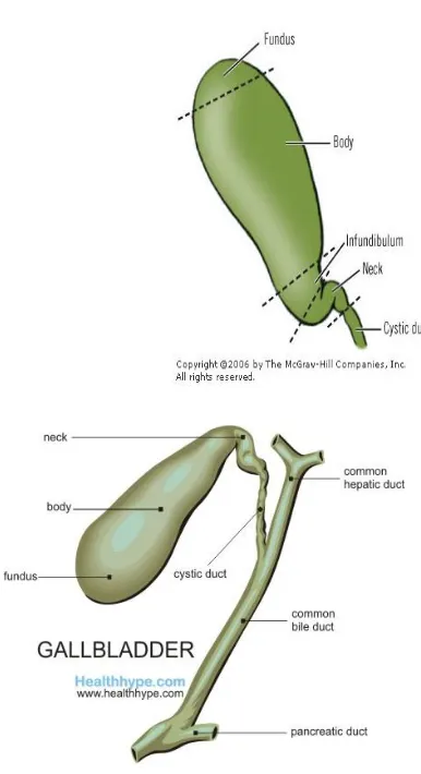
Anatomy of Gall Bladder
The knowledge about the anatomy of biliary system and its anomalies are essential for the safe execution of any surgical planning in gall bladder bed. The normal and abnormal variations of biliary system may lead to major post operative bile duct injuries, especially during laparoscopic procedures. Since, LC has become the gold standard procedure for Gall bladder disease which needs good exposure of calot’s triangle and failures are more common when compared to open procedures.
Gall bladder is a Pear shaped organ, located in the gall bladder fossa on the inferior surface of liver covered by peritoneum in all sides except on the fossa. It is 7 to 10cm long with an average capacity to hold 30 to 50 ml of bile as an “extrahepatic reservoir”.Cantlies line, dividing liver in to right and left runs through the gall bladder. It is mostly extra hepatic but may be intra hepatic in which case it is embedded in the liver parenchyma making the surgical procedure very difficult. Gall bladder is divided in to 4 parts namely,
1) Fundus
4) Neck
FIGURE: 2 & 3 ANATOMY OF GALL BLADDER
[image:16.612.216.409.102.457.2]Calot’s triangle [8]
: Also called as Hepatobiliary triangle this is bounded by
Right side- Proximal part of Gall bladder &Cystic duct
Left side- Common hepatic duct
Superiorly- Margin of the right lobe of the liver
[image:17.612.164.405.308.506.2]This triangle contains cystic artery and cystic lymph nodes
FIGURE: 4 CALOTS TRIANGLE
Bile ducts: These include
Right & Left Hepatic Ducts
Common Hepatic Duct
Cystic Duct
Common Bile Duct
Cystic duct starts from the neck of gall bladder with 3mm diameter and length of about 3cm which is variable. Its mucosa is arranged in a spiral fold known as the ‘valves of Heister’ and ‘sphincter of Lutkens’ which is a sphincteric structure to control bile flow out of gall bladder.
Right and left hepatic ducts joins together to form common hepatic duct which is of 2.5cm in length that finally joins with cystic duct at an acute angle to take part in the formation of CBD.
Common Bile Duct: The length of the CBD is 5.5cm to 15cm [3] and the diameter is 0.4mm to 1.4 cm in adult males and 5cm to 9.5 cm with the diameter between 4–8 mm in females. But the average length of CBD is 7.2cm and diameter of 5.25 mm and has been divided in to 4 parts namely,
Supra duodenal
Infra duodenal and Intra duodenal
Supra duodenal CBD: This portion lies along the right border of lesser omentum with portal vein as posterior relation and it is 0.5 to 2 cm in length[4]
Retro duodenal CBD: This part has portal vein on posterior side and common hepatic artery on the lateral side and more over it forms the anterior wall of the “Foramen of Winslow”.
Infra duodenal or Retro pancreatic CBD: This part passes through the ‘Quenu space’ which is bounded by 3 segments of the duodenum and SMV/PV
FIGURE 5: PARTS OF COMMON BILE DUCT Blood supply: The blood supply of gall bladder as follows
Gastroduodenal artery
Hepatic
Cystic
Celiac
Superior mesenteric arteries
cadaveric specimen that cystic artery is of 0.3mm in diameter and no artery with more than 0.3 mm in diameter in the calot’s triangle will be cystic artery.
Anatomical variants of cystic arteries:
small cystic artery branches rather than a single cystic artery seen in 2-6% of cases.
Left hepatic artery: The cystic artery can arise from the left hepatic artery, does not pass through the calot’s triangle seen in 1% of cases. Here cystic artery passes through the liver.
Low-lying cystic artery: Arising from gastroduodenal artery and passes inferior to the cystic duct through the cholecysto duodenal ligament outside the calot’s triangle. This rare anomaly is seen in 5% of the cases.
Clinical importance:
“Aberrant course of cystic artery is clinically important during cholecystectomy especially lap procedure, causing a massive hemorrhage that
could convert the procedure into open technique or causing great confusion to
the surgeon to delineate the arterial structures from the biliary radicals
leading to disastrous biliary leak, peritonitis and sepsis. Inadvertent right
hepatic artery ligation in cholecystectomy has been associated with liver
Venous drainage:
vein." Veins from the hepatic surface drain directly into the liver. Veins on the free surface open directly or follow the hepatic ducts into the liver.
[image:25.612.178.459.229.445.2]
Lymphatic Drainage: Subserosal and sub mucosal lymphatic’s of the gall bladder drain in to the cystic lymph node of Lund that lies in the junction between cystic duct and common hepatic duct. These lymphatic’s finally drain in to the hilum of liver that is acting as a channel for spread of carcinoma gall bladder.
[image:26.612.211.495.244.484.2]
FIGURE 10: LYMPHATIC DRAINAGE OF GALL BLADDER
1. Cholecystoretropancreatic pathway-
Is the main pathway which terminates in retroportal lymphnodes.
2. Cholecystoceliac pathway-
Terminates in celiac lymphnodes found on hepatoduodenal ligament.
3. Cholecystomesentric pathway –Passing left and front of portal vein
Physiology of bile secretion:
Components of bile:
Water 97%
Bile salts 0.7%
Bile pigments 0.2%
Cholesterol 0.06%
Inorganic salts 0.7%
Fatty acids 0.15%
Phosphatidylcholine 0.2%
Fat 0.1%
Alkaline phosphatase ….
Gall bladder disease:
Cholelithiasis –symptomatic
Acute Cholecystitis
Chronic Cholecystitis
Acalculous Cholecystitis
Mucocele of gall bladder
Cholelithiasis:
Stones are formed due to changes in the concentration process that increases the calcium and cholesterol components followed by decreased motility and bile stasis. The process of gall stone formation is complex and the pathophysiology is unclear. In India 80% of stones are pigment stones, especially brown pigment. Predisposing factors for the gall stone diseases are-
Obesity
Rapid weight loss
Pregnancy
Diabetes mellitus
TPN & Octreotide use
These conditions are associated with increased hepatic secretion of cholesterol
[4]
symptomatic or asymptomatic, when the symptoms are absent in the presence of documented gall stones.
Pathogenesis of gall stone formation:
Gall stone disease is polygenic and many factors contribute in its Pathogenesis, the primary factor is the excess biliary cholesterol in comparison to the solubilizing bile salts or phospholipids. Other factors are increased intestinal loss of bile salts and cholesterol. Genetics also plays a role in stone formation. Adenosine triphosphate–binding cassette (ABC) transporter which regulates the transport of cholesterol is located in the canalicular membranes of hepatocytes and the genetic disorders associated with this gene also leads to gall stone formation. One more genetic problem is increased expression of Niemann-Pick C1 –like protein which also plays a role in pathogenesis. A mutation in the ABCB11 may lead to progressive intrahepatic cholestasis and gall stone formation.
here.[15] Obesity, OCP and high caloric diet pour more amount of cholesterol in to the bile and ultimately all these lead to thick bile and dysmotility of gall bladder. So in gall stone disease removing the stones alone will not cure the disease unless gall bladder is removed.
Types of gall stones: There are three types namely
Cholesterol stones
Pigment stones
Mixed stones
Differences between cholesterol and pigment stones are:
Cholesterol stones Pigment stones >70% cholesterol <30% of cholesterol
Contains calcium salts, Bile acids ,
Bile pigments &Phospholipids
Calcium bilirubinate,
Calcium palmitate, Calcium stearate & Cholesterol
Formed in gall bladder Formed in bile duct
Pigment stones:
Two types of pigment stones are encountered, they are brown and black.
Brown pigment stones are more common in East Asia which are formed in the bile Ducts [1] because of bile stasis and bile infection and are usually less than 1 cm in diameter. They are brownish-yellow, soft, and often mushy. They are composed of calciumbilirubinate, [14]calcium palmitate and calcium stearate and varying amounts of cholesterol and protein and usually associated with chronic bacterial infection of the bile ducts and causing dangerous cholangitis. When the deconjugation of bilirubin deglucronide occurs this leads to stone formation by the action of Beta glucronidase which is a component of bacteria and deposition of insoluble unconjugated bilirubinateon the foreign bodies or parasites.
Bacteria & parasites forming stones are
Escherichia coli
Bacteroides
Clonorchis sinensis and
Black pigment stones are formed within the gall bladder and they are sterile and usually small, brittle, black, and sometimes speculated and composed of calcium bilirubinate mixed with calcium carbonate and calcium phosphate. In over all, about 20-30% of stones are black. Usually associated with chronic hemolytic disorders like sickle cell disease,[12] Hereditary spherocytosis but the gall bladder dysmotility & bile stasis do not play a role in the stone formation. It is more common in western countries. These stones are very rare in young age but, the incidence increases progressively as the age advances [3].
Cholesterol stones
lumen to become symptomatic. Stasis and bacterial invasion can cause infection.
Risk factors include:
Obesity
Pregnancy
Gallbladder stasis
Drugs
Heredity
The metabolic syndrome of truncal obesity, insulin resistance, type II diabetes mellitus, hypertension, and hyperlipidemia are associated with increased hepatic cholesterol secretion and is a major risk factor for the development of cholesterol gallstones.
Complications of gall stones:
In the gall bladder:
Silent stones
Acute Cholecystitis
Chronic Cholecystitis
Gangrene & perforation
Empyema
Mucocele
Carcinoma[16]
In the bile ducts:
Obstructive jaundice
Cholangitis
Acute pancreatitis
In the intestine:
inflammatory process with fibrosis the gall bladder shrinks. In the absence of resolution of inflammation gallbladder may perforate leading to peritonitis and empyema of gall bladder. If the stone is obstructing the bile duct which can be either due to a primary stone that is formed within the CBD or slipping from the cystic duct (secondary stone) may cause obstructive jaundice or inflammation extending in to intrahepatic biliary radicals leading to a devastating cholangitis.
Cholecystitis:
This is inflammation of the gall bladder due to various causes. Can be divided in to 1) Acute Cholecystitis 2) Chronic Cholecystitis 3) acute on chronic Cholecystitis
Acute cholecystitis
with fever, pain and tenderness in right subcostal region with positive “ Murphy’s sign”.
Chronic cholecyctits
This is “recurrent attacks of biliary colic due to cystic duct obstruction by a calculi but without causing acute cholecystitis can cause some inflammation and scarring the neck of the gall bladder and cystic duct”. Pain due to chronic cholecystitis generally does not last more than a few hours. If it exceeds 24 hrs with fever it suggests acute or acute on chronic cholecystitis.
patchy necrosis. In severe cases, about 5 to 10%, the inflammatory process progresses to ischemia and necrosis of the gallbladder wall. In some cases the gallstone is dislodged and the inflammation resolves. [18]
Courvoisier’s law:
Jaundice in a case of cholecystitis is rare and in the setting of obstructive jaundice if the gall bladder is palpable it is not due to stone disease rather may be due to a growth obstructing the carcinoma head of the pancreas or of CBD.
Clinical features:
Investigations :
USG abdomen:
Is the first choice of investigation and it is non invasive. It clearly defines the stone and its site with size within the biliary tree. Also gives details of gall bladder wall thickness and size of the CBD.
Endoscopic USG:
This is an ultrasound with an endoscope for detecting stones and growth in the periampullary region.
LFT:
Serum bilirubin, AST, ALT, Alkaline phosphatase and proteins. BT, CT with PT, INR.
MRCP:
ERCP:
Nowadays this is used mainly as a therapeutic aid to relieve obstruction in case of obstructive jaundice caused by stone in CBD and also in stricture. Because it causes acute cholangitis and pancreatitis, its usage as a diagnostic aid is restricted.
Operative Biliary Endoscope:
It is also called as ‘Choledochoscopy’ which is a flexible endoscope passed in to the CBD to identify stone and to remove it under direct vision.
Biliary Radionuclide Scanning (HIDA Scan)
Other investigations are :
Renal function test with serum electrolytes
Complete blood count with HB
CXR PA VIEW
TREATMENT
Treatment of gall stone diseases
All gall stones need not be treated by surgery.
I. Non operative medical treatment
II. SURGICAL TREATMENT
Medical management: Options available are dissolution with oral bile salt therapy, contact dissolution and ESWL.
Surgical options:
The most common indication for cholecystectomy are,
Biliary colic due to cholelithiasis and chronic cholecystitis
Obstruction of the cystic duct by a gallstone causing acute cholecystitis
Pancreatitis and Cholangitis caused by Stone obstructing the pancreatic duct and/or distal CBD and causing pancreatitis and Cholangitis.
Methods of Cholecystectomies
A. Open Cholecystectomy
Open Cholecystectomy:
Indications are
Poor cardiac or pulmonary reserve
Suspected gall bladder
General peritonitis
Penetrating trauma
Fistulous disease of gall bladder
Previous upper abdominal surgery
Cirrhosis of liver with PHT
3rd trimester of pregnancy
Incision:
Right Sub costal or Kocher ( most commonly employed) or
Right Transverse or
Mayo Robson incision
Position:
Methods:
Duct first method when calots triangle area is clear
Fundus first when the calots triangle is not clearly defined due to acute inflammatory pathology
Procedure:
After opening the abdomen it is explored to exclude other intra- abdominal pathology and a hand is passed over the right lobe of the liver to allow air to enter the subphrenic space. Moist pads are kept inferiorly to push colon downwards, one to push the stomach medially and one more pack for the assistant to give traction caudally so that the cystic duct is stretched and the calots triangle area is clearly visualized. When the gall bladder is tense with more bile it can be decompressed to avoid uncontrolled spillage of infected bile during dissection. The fundus of the gallbladder is grasped with a Kelly clamp and retracted caudally and an another to grasp Hartmann’s pouch and pulled away from the cystic duct.
The peritoneum overlying Calot's triangle is incised and the dissection is continued close to the gallbladder, displacing the cystic node and the fatty areolar tissues to expose the cystic duct and artery.
Picture: removed gall bladder
Laparoscopic Cholecystectomy
Laparoscopic Instrument:
High-quality video laparoscope
300-W light source
High-resolution monitors
Veress needle
High-flow carbon dioxide insufflator
Four Trocars (two 10-mm Trocars and two 5-mm Trocars)
Monopolar Electrode L-hook
Suction and Irrigation
Fine-tipped dissector
Two gallbladder graspers
Large gallbladder extractor
Pair of scissors
Clip Applicator
A 10-mm stone retrieval grasper
Bi polar cautery
Endo pouches ( to collect gall stones)
A good operating team members are very much needed to execute a successful laparoscopic cholecystectomy. It should include
A well trained surgeon
First assistant surgeon with equivalent skills
Camera operator
A scrub Nurse
Preoperative antibiotic: Inj. Cefotaxime 50mg /kg body weight, IV stat.
Anaesthesia : ETGA
Position of surgeons 1) Surgeon stands to the patient's left 2) First assistant stands to the patient's right 3) Camera operator stands at the surgeon's left Pneumoperitoneum : I) Closed method (veress needle)
II) Open method (Hasson method)
Carbon dioxide pneumoperitoneum at 15 mm Hg pressure is the agent of choice for many reason like low toxicity, low risk of gas embolism, rapid reabsorption, low cost and ease of use. Ideal insufflator should be able to deliver 8 to 10L/min with a minimum acceptable flow rate of 6L/min. It regulates flow rate, monitors intra abdominal pressure and stops delivering carbon dioxide whenever the pressure exceeds predetermined level of 12 to 15mm Hg.
Procedure :
abdominal cavity by direct vision and the trocar introduced through which carbon dioxide is insufflated to create pneumoperitoneum.
Placement of Trocars:
With the help of an assistant who lifts and holds the abdominal wall ,a 10 mm trocar introduced through umbilical wound in to the peritoneal cavity by screwing movement. Then either a 0-degree or 30-degree telescope is passed through this and thorough laparotomy of the abdomen including calots area is done. The advantages of 30 –degree telescope is that it can be used to view posterior wall of gall bladder, CBD and calots triangle. Next, an 10mm trocar passed through the epigastrium at the right of the falciform ligament. The third trocar is a 5-mm trocar is placed in the midclavicular line 2 to 3 cm below the costal margin and the fourth trocar in the anterior axillary line.
Dissection of calots triangle area
patients left side. Now the surgeon can dissect the peritoneum covering of the area by using a retract infundibulam with his left hand. The peritoneum is teased towards the common bile duct till cystic duct, cystic artery and calots lymph nodes are identified. If 30-degree telescope is used, dissection on the posterior aspect of the cystic duct is easier. Once this is done, two clips proximal and one clip distally close to the gall bladder is applied and transected with a scissors.
Removal of the Gallbladder:
Picture : showing removed gall bladder
CLOSURE:
Under the vision all the ports are removed one by one and the pneumo peritoneum is let out. The rectus sheath is closed with 2-0 Vicryl in ‘figure of 8 suture’ at 10mm port sites and skin closed with subcuticular stitch and sterile- strips placed.
Challenges
Hydrops of the Gall bladder: Some times the gall bladder may be too tense due to hydrops of the Gall bladder that cannot be grasped, in which it has to bed decompressed by cholecystostomy. Once reasonable quantity of bile is let out of it and sucked out the puncture site can be grasped with a grasper.
Wide mouthed cystic duct:
Other techniques for closing the cystic duct:
If the duct is long and wide, it can be transected and an Endo loop can be applied to the cystic duct stump.
Two ties can be passed around the cystic duct in continuity and secured with
Extra corporeal knotting techniques.
The cystic duct is transected with an endoscopic stapling device, or it
is simply divided and over sewn with an intra corporeal suturing
technique.
Post operative care:
Conversion to open method
The aim of conversion to an open method is to avoid complications and it must be decided before its occurrence. The rate of conversion is 5% in the hands of an experienced hepato biliary surgeon. Indications for conversion are
Dense adhesion on the calots area
Necrotic gall bladder
Carcinoma gall bladder
Difficult dissection of neck of gall bladder leading to hemorrhage
Delay in identifying the junction of gall bladder with cystic duct within 30 minutes due to any reason
Risk factors are
Male gender
Obesity
Cholecystitis
Choledochlithiasis
Complications of Laparoscopic Cholecystectomy
Veress needle or trocar injury to mesentery, duodenum or bowel
CBD injury causing stricture and obstructive jaundice
Bleeding
Bladder and Ureter injury
Bile leak
Infection
Cholangitis leading to septicemic shock
Infection
Subphrenic abscess
Gall stone spillage
Deep vein thrombosis
Pancreatitis
Conversion to open procedure
Ileus
Subcutaneous edema & emphysema
Biliary fistula
Port site hernia
How to manage complications?
Percentage of serious complications of laparoscopic cholecystectomy is less than 2%. They are
Intestinal injury –May be due to abdominal access, adhesiolysis or dissection of gall bladder away from duodenum. If accidental injury occurs due to veress needle (Incidence is 0.2%) it need not be repaired where as cautery injuries should be repaired in two layers.
Vasculo biliary injury – Occurs during abdominal access due to inadequate insufflations of CO2 or excessive thrusting of the trocar given by surgeon. If an unexplained hypotension or retroperitoneal hematoma is found that is an indication for immediate laparotomy.
Excessive bleeding from the porta hepatis- if it is minor bleeding clips can be applied where as major bleeding necessitates conversion to open method. Bleeding from the gall bladder bed can be controlled by fulguration of the bleeding site.
CBD injury:
Occurs commonly in laparoscopic procedure than in open method. The incidence during earlier period was more but now it has come down to 0.5%. The reasons for higher incidence in laparoscopic procedure are,
Incorrect traction forces to the gall bladder
Lack of knowledge about the anatomy of biliary system
Injudicious use of cautery on the calots triangle area
Steps taken to avoid CBD injury during the procedure:
Use of 30 degree telescope and a high quality imaging equipment.
‘Critical view of safety’ -all the tissues except for cystic artery and cystic duct should be cleared off in the triangle of calot so that these two structures entering the gall bladder can be clearly seen before ligating them.
Management of bile duct injury
Intra operative diagnosis
Class I & II injuries can be repaired primarily with or without T-tube drainage where as class III & IV are managed with Hepatico jejunostomy.
Post operative diagnosis
MATERIALS AND METHODS
Source:
The patients admitted in surgical wards in Govt.Kilpauk Medical College Hospitals including Govt.Royapettah Hospitals, Chennai with signs and symptoms of gall bladder diseases. This study was conducted from JULY 2012 to DECEMBER 2013.
Methods of collecting data;
Routine investigations;
Hemoglobin, White cell count, Differential count, Blood urea, Serum creatinine, Blood sugar, ECG in all leads, CXR and Urine examination were done for all cases.
Special investigations;
Liver function test-serum bilirubin, alkaline phosphatase, total protein, Ultra sonogram abdomen- for GB stones, wall thickness and to R/O CBD stones, MRCP- to know the aberrant biliary duct and cystic arteries and Lipid profile.
Inclusion criteria
Following patients are included for treatment,
Calculus cholecystitis
Acute cholecystitis
Chronic cholecystitis
Acalculous cholecystitis with physical status 1 and 2 (stable vitals)
All the patients with evidence of stone identified in USG, presenting with minimum of one episode of attack and fit to undergo elective surgery were included in this study group.
Exclusion criteria
Patients with following conditions were excluded from this study
History or investigations suggestive of CBD stones
Empyema of gall bladder
On immune suppressive drugs
Gross obesity
Mucocele of gall bladder
Malignancy of gall bladder
History of prior abdominal surgery
Age less than 30yrs
Patients age above 70yrs (symptom recurrence rate is only 15%)
Ruptured gall bladder
Sample size
Totally 65 patients divided in to two groups, 33 in Group A and 32 in Group B admitted from the period since July 2012 to December 2013.
GROUP A- Patients with open cholecystectomy
GROUP B- Patients with laparoscopic cholecystectomy
A day before surgery all the patients were kept NPO for about 10hrs, nasogastric tube insertion and bladder catheterization done according to the individual need. Then prophylactic antibiotic was given 30 minutes before the commencement of surgery. After anesthesia Cholecystectomy done and before extubation irrespective of the type of surgery all the patients were given MOL suppository 250mg per rectally for pain relief.
Comparision of results
Both the groups were analysed based on age, duration of surgery, post operative recovery , requirement of antibiotic and analgesics , complications and hospital stay.
Age analysis
Table:1 Age Distribution In The Study Group A
Age group (in years)
Number of subjects Percentage
30 – 40 8 24.2
40 – 50 10 30.3
50 – 60 5 15.2
60 – 70 10 30.3
In Group A, symptomatic gall stone disease patients underwent elective open cholecystectomies were between 40 and 50 yrs and 60 and 70 yrs. Where as in Group B patients between 30 to 40 yrs of age underwent laparoscopic cholecystectomies due to the fact that these patients were free of comorbidities and ability to withstand general anesthesia well when compared to patients above 50yrs of age.
Graph 1: Age Distribution of the Study Group
Table 2:Age distribution in Group B patients
Age group (in years)
Number of subjects Percentage
30 – 40 20 62.5
40 – 50 2 6.2
50 – 60 4 12.5
60 – 70 6 18.8
Total 32 100
GRAPH 2: Age distribution of Group A patients
0
5
10
15
20
25
30 - 40
40 - 50
50 - 60
60 - 70
Table 3: Gender Distribution in the Study Group A
Gender Number of subjects Percentage
Male 11 33.3
Female 22 66.7
Total 33 100
The gender distribution in our study shows 2/3rd of population belongs to female sex.
Graph 3: Gender Distribution in the Study Group A 22
11
GENDER DISTRIBUTION OF GROUP A
PATIENTS
Since gall bladder disease is more common in female than that of male gender, dominance of female observed in both the methods.
Table 4: Gender distribution of Group B patients
Gender Number of subjects Percentage
Male 8 25
Female 24 75
Total 32 100
Graph4: Gender Distribution in the Study Group B
24
8
GENDER DISTRIBUTION OF GROUP B
PATIENTS
Table 5: Comparison of duration of both surgeries
Surgery Mean
duration± SEM
T value P value
Open
Cholecystectomy
89.48 ± 3.61
6.08 0.001
Lap
Cholecystectomy
62.41 ± 2.57
[image:76.612.94.547.113.341.2]
Table 6: Comparison of post op recovery in both surgeries
Surgery Post op
recovery
Chi square value
P value Uneventful Eventful
Open
Cholecystectomy
21 12 0.98 0.321
Lap
Cholecystectomy
24 8
Graph 5: Post Op Recovery In Both Groups
The above mentioned stastical variable from our study does not show major significant difference in post operative recovery between open and laparoscopic procedures.
P = 0.321 (not significant)
0
10
20
30
40
OPEN LAPARASCOPY
Table 7: Comparison of antibiotic usage in both surgeries
Surgery Antibiotic used Chi square
value
P value Single dose Full course
Open Cholecystectomy 19 14 0.87 0.35
Lap Cholecystectomy 22 10
Graph 6: Post Operative Antibiotic Requirement In Both Groups
0
10
20
30
40
OPEN LAPARASCOPY
Since recommendation of antibiotic was based on the associated comorbidity our study shows no major difference in its usage in both the groups.
Table 8: Comparison of analgesic usage in both surgeries
Surgery Analgesic used Chi
square value
P value Perrectal
suppository
Injectable analgesics Open
Cholecystectomy
15 18 5.91 0.015
Lap
Cholecystectomy
24 8
Graph 7: Route of Analgesic Requirement
Table 9:Comparison of period of stay in both surgeries
Surgery Mean period of
stay
T value P value
Open
Cholecystectomy
7.88 ± 0.50 9.095 0.001
Lap
Cholecystectomy
2.95 ± 0.19
The mean period stay in the Hospital in Laparoscopic surgery was 2.95 days where as in open method it was 7.88 days
0% 50% 100%
OPEN
LAPARASCOPIC
per-rectal
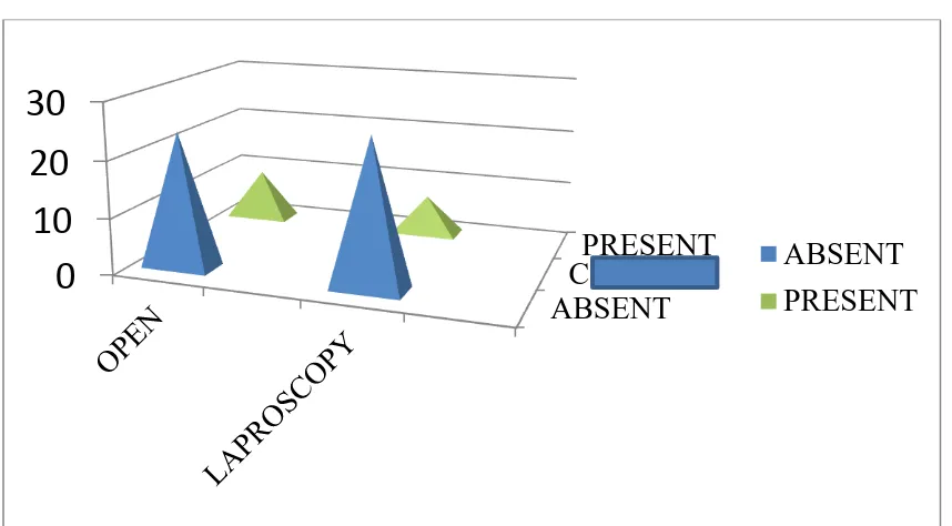
Table 10:Comparison of occurrence of complications in both surgeries
Surgery Complications Chi
square value
P value Present Absent
Open
Cholecystectomy
9 24 0.33 0.565
Lap
Cholecystectomy
7 26
P value < 0.05 is significant
[image:82.612.108.537.375.612.2]Analysis of the results
Total number of patients analysed for this study was 65 among which 33 taken for open cholecystectomy and 32 patients were taken up for laparoscopic cholecystectomy with 2 patients converted to open due to adhesions found in the calots region.
In Group A, symptomatic gall stone disease patients underwent elective open cholecystectomies were between 40 and 50 yrs and 60 and 70 yrs. Where as in Group B patients between 30 to 40 yrs of age underwent laparoscopic cholecystectomies due to the fact that these patients were free of comorbidities and ability to withstand general anesthesia well when compared to patients above 50yrs of age.
The gender distribution in our study shows 2/3rd of population belongs to female sex. Since gall bladder disease is more common in female than that of male gender, dominance of female observed in both the methods.
reason was its association with diabetes mellitus but for overall it does not significance between both the groups
The average time taken for laparoscopic cholecystectomy was much lesser (62.41 minutes) than that taken for the open method which is 89 minutes and48 seconds.
The mean period stay in the Hospital in Laparoscopic surgery was 2.95 days where as in open method it was 7.88 days. This is mainly due to the more trauma given to the adjacent organs by means of traction given in open method.
Discussion and Conclusion
This study proves that laparoscopic cholecystectomy is the surgical treatment of choice due to the following reasons
The time taken for laparoscopic procedure is less and in an experienced hand the entire procedure can be end within 30 minutes. So that prolonged anaesthesia and its complications like MI, Respiratory distress and ventilator support can be avoided.
Lesser need of analgesic especially powerful analgesics like diclofenac or tramadol in the form of Injectable.
If the associated co morbid illnesses like DM are absent the need of full course of antibiltics is definitely not needed.
Wound infection also very less compared to open procedure.
GROUP A
NAMES
AGE SEX IP NO Time taken Comorbidity Post op Recovery Antibiotic Analgesics period of
Stay Complications
Return to regular
work
Lilly 33 2 21777 120 DM 0 2 1 6 0 25
Ras avathy 45 2 22252 128 DM 0 2 1 5 1 20
Subhashini 31 2 23250 100 NIL 0 1 1 4 0 30
Velu 35 1 22212 105 NIL 0 1 1 8 0 18
Meenambigai 35 2 28667 70 NIL 0 1 2 10 0 21
TajilNisha 51 2 29741 110 DM/SHT/OBESITY 1 2 2 7 1 18
Mohan 31 1 31303 95 NIL 0 1 1 5 0 14
Dhanalakshmi 35 2 1301693 83 OBESITY 1 1 1 7 0 21
Jaghadhambal 44 2 1302111 125 DM/OBESITY 1 2 1 10 1 45
Krishnaveni 58 1 1304079 50 DM 1 2 1 5 0 30
Thangaiyan 65 1 1312361 77 SHT 1 1 1 5 0 21
Kumari 50 2 1330994 85 OBESITY 1 1 1 8 0 14
Narayanan 60 1 1333010 115 DM 1 2 2 13 1 20
Lakshmi 55 2 1311778 50 NIL 0 1 1 6 0 16
Anjali 70 2 1335242 68 DM 1 2 2 12 1 30
Geetha 45 2 1334959 74 HBsAg +ve 0 2 2 8 0 14
Sadhakdulla 61 1 1315079 85 DM/IHD 1 2 1 8 0 14
Radha 31 2 1326589 86 NIL 0 1 1 4 0 14
Saraswathy 47 2 1314568 65 NIL 0 1 1 4 0 21
Dhanalakshmi 41 2 3709 63 NIL 0 1 1 8 0 23
Palayam 65 1 3247 105 DM 0 2 2 10 1 30
Balamurugan 48 1 6144 100 NIL 0 1 1 7 0 18
Renuka 50 2 7041 88 OBESITY/DYSLIPEDEMIA 1 1 2 12 0 17
Philip Krishnan 64 1 110963 86 DM 1 2 1 11 1 15
Seema 45 2 111291 68 NIL 0 1 1 7 0 14
Muniyammal 60 2 104623 72 DM/OBESITY 0 2 1 14 0 35
Saradha 50 2 102881 75 NIL 0 1 1 6 0 16
Jayanthi 35 2 105968 78 NIL 0 1 1 5 0 18
Shanmugamaniya 61 1 110174 108 DM/HBsAg+ve 1 2 2 10 0 28
Rajeshwari 65 2 114805 111 DM/OBESITY 0 2 1 13 1 30
Kuppammal 65 2 3906 102 NIL 0 1 1 7 0 21
Selvathai 65 2 5794 98 OBESITY 0 1 1 10 1 28
NAME AGE SEX IPNO
Duration of
Surgery Comorbidity
Post op
recovery Antibiotic Analgesic
Period of
Hospital stay Complications
Return to
regular work conversion
Godhavari 68 2 1302995 77 DM/SHT 0 1 2 4 0 15
Sheela 37 2 1312361 78 NIL 0 1 1 3 0 12
Kavitha 36 2 1306910 66 DM 1 2 1 4 1 18
Lakshmi 37 2 1320370 40 NIL 1 1 1 2 1 10
Natarajan 70 1 1315201 54 DM 0 2 1 3 1 11 YES
Vatsala 37 2 1329343 62 FAMILY 0 1 1 2 0 10
Kauserbanu 35 2 1316044 65 NIL 0 1 2 2 0 7
Eswari 32 2 1322710 54 NIL 1 1 1 3 1 60
Sherkhan 53 1 1334655 63 DM/SHT 0 2 2 4 0 14
Hamsavalli 33 2 1731 55 NIL 1 1 1 2 0 14
Pushpa rani 65 2 10088 78 DM/OBESITY 1 2 2 5 1 12
Latha 32 2 847 90 OBESITY 0 1 1 2.5 0 10
Johnson 63 1 132462 65 SHT 0 1 1 3 1 14
Usharani 31 2 880 70 NIL 0 1 1 1.5 0 13
Selvi 32 2 113453 58 NIL 0 1 1 2 0 9
Mebin 31 1 1329345 42 NIL 0 1 1 3 0 10
Vimala 38 2 106107 62 DM/OBESITY 1 2 1 3 0 10 YES
Anjamma 45 2 106277 45 OBESITY 0 1 1 6 0 14
Arun 42 1 1323101 46 DM 0 2 2 4 1 13
Vinayagamurthy 62 1 108213 103 F/H OF CA GB 0 1 2 3 0 7
Kanchana 60 2 106330 66 DM 0 2 1 5 0 9
Loganayagi 60 2 102574 65 DM/OBESITY 0 2 2 3 0 8
Thamarai 33 2 106963 75 NIL 0 1 1 2 0 11
Seetha 55 2 108786 56 DM 0 2 1 3 0 10
Arunkumar 33 1 107787 67 NIL 0 1 2 2 0 7
Srinivasan 70 1 109033 55 SHT 0 1 1 1.5 0 7
Lakshmi 33 2 109331 77 NIL 0 1 1 2 0 7
Deepa 30 2 111125 45 NIL 0 1 1 3 0 7
Deepa 33 2 111107 48 OBESITY 0 1 1 3 0 8
Deivakani 35 2 112703 62 NIL 0 1 1 2 0 14
Malarvizhi 33 2 1549 70 OBESITY 1 1 1 3 0 11
Sridevi 32 2 3470 38 DM 2 1 3 0 10