EVALUATION OF THE EFFECT OF ADDITION OF
CLONIDINE TO 0.5% ROPIVACAINE IN
SUPRACLAVICULAR BRACHIAL PLEXUS BLOCK
Dissertation submitted to
The Tamilnadu Dr. M. G. R. Medical University
Chennai – 600032
With fulfillment of the regulations for the award of Degree
M.D. ANAESTHESIOLOGY
BRANCH – X
DEPARTMENT OF ANAESTHESIOLOGY,
K.A.P.V. GOVERNMENT MEDICAL COLLEGE,
TRICHY.
CERTIFICATE
This is certify that this dissertation titled
“EVALUATION OF
THE EFFECT OF ADDITION OF CLONIDINE TO 0.5%
ROPIVACAINE IN SUPRACLAVICULAR BRACHIAL PLEXUS
BLOCK”
is a bonafide work of
DR.D.ELAVARASAN.,
Post Graduate
in M.D. Anaesthesiology, Department of Anaesthesiology, K.A.P.V.
Government Medical College, Trichy and has been prepared by him
under our guidance. This has been submitted in partial fulfillment of
regulations of The Tamil Nadu Dr. M.G.R Medical University,
Chennai-32 for the award of M.D. Degree in Anaesthesiology.
Prof. Dr. N. JOTHI., MD, DA.
Professor and HOD
Department of Anaesthesiology
K.A.P.V.Govt.Medical College,
Trichy.
Prof. Dr.M.K.MURALIDHARAN, M.S,M.ch
DEAN,
DECLARATION
I
Dr. D. ELAVARASAN.,
solemnly declare that this dissertation
title, “
EVALUATION OF THE FFECT OF ADDITION OF
CLONIDINE TO 0.5% ROPIVACAINE IN SUPRACLAVICULAR
BRACHIAL PLEXUS BLOCK
” is a bonafide work done by me at
K.A.P.V. Government Medical College during 2014-2016 under the
guidance and supervision of
Prof. Dr. N. Jothi, MD.,
DA.,
Professor and Head of Department,
Department of Anaesthesiology.
The dissertation is submitted to the Tamilnadu Dr. M. G. R.
Medical University, towards the partial fulfillment of requirement for the
award of M.D. Degree in Anaesthesiology Branch X.
Place:
ACKNOWLEDGEMENT
I thank
Prof. Dr. M.K.Muralidharan, M.S., M.Ch.,
the
DEAN of
K.A.P.V. Govt. Medical College,
Trichy, for permitting me to conduct
this study in the Department of Anaesthesiology of K.A.P.V. Government
Medical College, Trichy. I thank
Prof. Dr. N. Jothi, MD, DA.
Head,
Department of Anaesthesiology,
for helping and guiding me during this
study.
My heartful gratitude to
Prof. Dr. R. Selvakumar MD, DA,
DNB, Prof. Dr. M. Suresh MD, DA.,
Prof. Dr. P.Elango MD and
Prof. Dr. G. Sivakumar MD, DA
and
for their esteemed guidance and
valuable suggestions.
It is my privileged duty to profusely thank my teacher, guide and
mentor
Prof. Dr. N. Jothi MD, DA.,
under whom I have the great
honour to work as a postgraduate student.
I am greatly indebted to my Assistant Professors who have put in
countless hours in guiding me in many aspects of this study and also in
honing my anaesthetic skills. I thank my fellow Post Graduates who
helped me in conducting the study. Last and most important, I am
thankful to my patients without whom this study could not have been
TABLE OF CONTENTS
S.
NO.
TITLE
PAGE NO.
1
INTRODUCTION1
2
AIMS AND OBJECTIVES4
3
REVIEW OF LITERATURE5
4
ANATOMY13
5
TECHNIQUE21
6
PHARMACOLOGY25
7
MATERIALSANDMETHODS39
8
OBSERVATIONSANDRESULTS46
9
DISCUSSION69
10
CONCLUSION74
11
SUMMARY75
12
BIBLIOGRAPHY77
LIST OF TABLES
S. No.
TABLE
PAGE NO.
1
RAMSAY SEDATION SCORE43
2
DEMOGRAPHIC PROFILE46
3
SENSORY ONSET48
4
MOTOR ONSET50
5
DURATION OF SENSORY BLOCK(IN MIN)52
6
DURATION OF MOTOR BLOCK(IN MIN)54
7
DURATION OF ANALGESIA(IN MIN)56
8
VAS SCORE58
9
INTRAOPERATIVE SEDATION SCORE59
10
PULSE RATE62
11
SYSTOLIC BP64
12
DIASTOLIC B.P66
LIST OF FIGURES
S. No.
FIGURE
PAGE NO.
1
M
OLECULARS
TRUCTUREO
FR
OPIVACAINE25
2
M
OLECULARS
TRUCTURE OFC
LONIDINEH
YDROCHLORIDE31
3
O
NSET OF
S
ENSORYB
LOCK(I
NM
IN)
47
4
O
NSETO
FM
OTORB
LOCK(I
NM
IN)
49
5
DURATION OF SENSORY BLOCK
(
IN MIN)
51
6
DURATION OF MOTOR BLOCK(
IN MIN)
53
7
DURATION OF ANALGESIA(
IN MIN)
55
8
VAS SCORE
57
9
INTRAOPERATIVE SEDATION SCORE60
10
PULSE RATE
61
11
SYSTOLIC BP63
12
DIASTOLIC B
.
P65
ABBREVATIONS
µg - MicroGram
ASA - American society of Anaesthesiologist CMax - Maximum Concentration
CNB - Central neuroaxial block CNS - Central nervous system CO - Cardiac out put
CVS - Cardio vascular system DBP - Diastolic Blood pressure ECG - Electrocardiogram GIT - Gastro intestinal system HR - Heart rate
I.M - Intramuscular I.V - Intravascular Inj - Injection
kg/Bw - Kilogram/ Body weight LA - Local anesthetics LFT - Liver function test
MAC - Minimum alveolar concentration . MHz - Mega hertz
mmHg- Milli meter of mercury NIBP - Non invasive blood pressure PNB - Peripheral neuroaxial block PR - Pulse rate
R - Ropivacaine
RC - Ropivacaine and Clonidine SBP - Systolic Blood pressure SD - Standard deviation .
Spo2 - Saturation of partial pressure of oxygen SVR - Systemic vascular resistance
INTRODUCTION
Supraclavicular Brachial plexus block is commonly practiced for upper limb surgeries. Once described as the “ spinal of arm” a supraclavicular block offers dense anesthesia for surgical procedures at sites at (or) distal to elbow, forearm & hand. It can be used as the sole anesthetic technique or in combination with general anesthesia for intraoperative &post operative analgesia. Supraclavicular block is a low cost anesthesia technique. It provides satisfactory/ optimal operative conditions due to both sensory &motor blockade without any systemic side effects.
Brachial plexus block also cause sympathetic block with resultant improvement in blood flow, reduction in vasospasm & edema which is more favorable for acute hand injury and reconstructive plastic surgery.
Common sites of approach to Brachial plexus block are Interscalene, supraclavicular, Infraclavicular, Axillary and posterior approach.
It is a must for all practicing anesthesiologist to be familiar with all the above approaches as well as each one’s advantages & limitations. Supraclavicular approach is one of the easiest and most consistent method for performing brachial plexus block.
Ropivacaine1 is an amino amide local anesthetic prepared as pure S– enantiomer. Ropivacaine has lesser lipid solubility and also produce less central nervous toxicity and cardio toxicity with less arrhythmogenic potential.
The purpose of adding an adjuvant to local anesthetics for peripheral nerve block is to have early onset of sensory and motor block and to prolong the duration of post operative analgesia with lesser adverse effect2.
Several clinical investigations have shown that Clonidine prolongs the post operative analgesia. Clonidine is an α 2-agonist. Although it had been used originally as an anti hypertensive agent, it has sedative, sympatholytic and analgesic property3.
Successful brachial plexus block depends on proper nerve localization, needle placement, local anesthetic injection i.e. right drug, right dose, placed in the right place, by the right technique.
Traditional land mark approach and elicitation of paraesthesia necessitates multiple attempts, resulting in procedure related complications such as pain, injury to blood vessels and pneumothorax.
AIMS AND OBJECTIVES
REVIEW OF THE LITERATURE
1.Quazi et al6 added 75µg of clonidine to Ropivacaine for supraclavicular brachial plexus for upper limb orthopedic surgeries. The study design was randomized, double blind placebo controlled study. The study group was divided into two categories:
Group A patients: received 30ml of 0.5% Ropivacaine& 0.5 ml of normal saline.
Group B patients: received 30ml of 0.5% Ropivacaine& 0.5 ml of (75µg) clonidine.
Aim of this study was to evaluate the effect of addition of clonidine to Ropivacaine in brachial plexus block (in supraclavicular approach). They also observed onset, duration, quality of sensory & motor block and also the duration of post operative analgesia and the complications.
2.Sidharth SR et al.7 did the study on orthopedic patients who underwent upper limb surgeries. The study population was divided into two groups. Each group consisting of 40 patients.
Here,
Group A receives 35 ml of 0.5% Ropivacaine with 150 µg of clonidine . Group B receives 35 ml of 0.5 % Ropivacaine with 1 ml of normal
saline.
In this study they wanted to compare the onset & duration of sensory & motor blockade, duration of post operative analgesia, sedation & observed for any side effect of drugs, complication of anesthetic procedure.
In conclusion of their study, addition of clonidine to Ropivacaine improves the quality & duration of supraclavicular block.
Clonidine not only enhance speed of onset , but also prolongs the duration of sensory & motor blockade & prolong the duration of analgesia in post operatively without major hemodynamic changes. Additional advantage of clonidine was more sedative potential, reduction in anxiety& better patient comfort in intra operative & post operative period.
3. Bafna et al8 reported that clonidine added as adjuvant to Ropivacaine in supraclavicular block for upper limb surgeries, gave significant prolongation of sensory & motor blockade.
Group I 40 patients received 28 ml of 0.5 % Ropivacaine & 2 ml of normal saline.
Group II 40 patients received 28 ml of 0.5 % Ropivacaine & 2 µgm/kgBWt of clonidine.
They assessed the onset of sensory & motor block, duration of sensory & motor block & observed the sedation score.
They concluded that clonidine was when added as adjuvant to 0.5% Ropivacaine in brachial plexus block; it increased the sensory & motor duration when compared to Ropivacaine alone, without side effects.
4.Gupta et al9 did a prospective randomized double blind clinical study .The study population divided into two groups .Each group consisted of 32 patients ASA I & II.
Group (R) received 19.8ml of 0.75% Ropivacaine & 0.2 ml Normal saline.
Group (RC) received 19.8ml of 0.75% Ropivacaine& 30µgm of clonidine
Block performed with ultrasound guided technique.
They observe the onset of sensory & motor block, duration of sensory & motor blockade & duration of postoperative analgesia.
Faster onset of sensory block: R group (2.5min) & RC group (2.36min) Faster onset of motor block: R(3.10 min) & RC (3.87 min)
& prolongation of duration of sensory& motor blockade & post operative analgesia without alteration of hemodynamic adverse effects.
5. Shobana Gupta et al10 studied the effect of clonidine as an adjuvant of 0.75% Ropivacaine (30ml) in supraclavicular plexus block in patients undergoing upper limb orthopedic surgeries. The study was done with 60 patients ASA I & II divided into two groups (R) & (RC).They monitored the onset of sensory & motor block, duration of sensory & motor block & observed the sedation score.
Thus they concluded that the Ropivacaine is long acting Local anesthesia with moderate onset period & longer post operative analgesia. The addition of Clonidine (150 µg) resulted in delay in the onset time of sensory & motor block & prolongation of motor block duration of analgesia, does not cause major adverse effect & complication.
Group I : Received 0.75% Ropivacaine 30ml
Group II : Received 0.75% Ropivacaine 29ml + Inj. Tramadol (50mg) 1 ml
Group III: Received 0.75% Ropivacaine 29ml + Inj. Fentanyl (50mg) 1 ml
They assess the onset, quality & duration of sensory & motor block with Ropivacaine & compare the additive (Tramadol & Fentanyl).Observation of their study was Onset time of sensory & motor blockade was not significantly different between control & test groups.
Average onset of sensory block 5 min , motor block 14 min , duration of sensory block (9hrs) , motor block (8hrs) was significant .additive group was compared with control group (sensory 6 hr, motor 5 hrs ).Their conclusion was that addition of opioids to Ropivacaine had additive effect in prolongation of postoperative analgesia .
7. The study done by Saritha.S.Swamiet al12 in, orthopedic upper limb surgical patients showed that addition of Clonidine &Dexmeditomidine in supraclavicular brachial plexus block result in prolongation of sensory & motor blockade in post operative analgesia. The study was done in 60 ASA I & II patients of either sex, aged 18-60 years & divided them into two groups.
Group C Bupivacaine 0.25% (35cc)+Clonidine 1 µg /kg BW
They compared the onset time of sensory & motor blockade, duration of sensory & motor blockade, duration of analgesia in postoperative period between the groups. They observed that the onset of sensory block was faster in Group D than Group C.
Motor block was faster in Clonidine than Dexmeditomidine. Duration of sensory block increased in Group D (413 min) than Group C (227 min). Duration of motor block 292 min (C Group) compared with 472 min in D Group. Post operative analgesia duration in D Group was 456 min when compared with C Group (289 min).They concluded that Dexmeditomidine prolongs the duration of sensory & motor block and enhances the quality of block compared with Clonidine when added as adjuvant to Bupivacaine in peripheral nerve block.
8.Birbalbaj et al13 did a comparative study of effect of Clonidine added to Ropivacaine & plain Ropivacaine during supraclavicular block for patients undergoing upper limb surgeries. Their study population (60 patients) allocated into two groups, each comprising 30 patients.
Group Ireceive 30 ml of coded drugs.
Group II receive 30 ml of 0.75% Ropivacaine& Clonidine 60 µg /Kg. They used paraesthesia eliciting technique for anesthetizing the supraclavicular brachial plexus.
They conclude that when was Clonidine when added to Ropivacaine for brachial plexus block, almost same onset time of sensory & motor block was obtained as compared to plain Ropivacaine ,but the duration of both sensory & motor block was increased. Postoperative analgesia duration was significantly prolonged in Clonidine group .Clonidine produced sedation in some patients that was statistically insignificant. Clonidine & Ropivacaine does not cause any adverse effect in supraclavicular brachial plexus block.
9.Bansal et al14.conducted a prospective randomized double blind study among 60 patients belonging to ASA I & II scheduled for forearm & hand surgery. They were divided into two groups, each comprising 30 patients.
Group I (n=30) received 35 ml of 0.5% Ropivacaine + 1 ml of Normal saline.
Group II (n=30) received 35 ml of 0.5% Ropivacaine + 1 ml of (150 µg) clonidine.
10.Santvana Kohli et al15.Performed a prospective randomized double blinded study for Clonidine as adjuvant in two different doses added to Bupivacaine (in supraclavicular block) for patients undergoing upper extremity surgery.
60 patients (18-65 years were included both ASA I & II .The study population divided into two groups.
Group I (n=30) received 30 ml of 0.5% Bupivacaine + 1 µgm/kg of Clonidine
Group II (n=30) received 30 ml of 0.5% Bupivacaine + 2 µgm /kg of Clonidine
ANATOMY
Thorough knowledge about anatomy of brachial plexus is essential for performing successful brachial plexus block16.
Brachial plexus consists of Roots
Trunks Divisions Cords.
ROOTS:
It derived from the anterior primary rami of the spinal nerves 5th, 6th, 7th, 8th cervical nerve and 1st thoracic nerve and also from the C4 to T2.The formation of brachial plexus may be one segment upwards (or)one segment downwards resulting in prefixed or post fixed plexus respectively.
In Prefixed plexus C4 is large and T2 contribution is absent.
In Post fixed plexus the contribution by T1 is large, T2is always present ,C4 is absent and C5 is reduced in size.
TRUNKS:
DIVISIONS:
Trunks are bifurcated into anterior division & posterior division, which are situated in between lateral border of first rib and posterior to clavicle and then descend into axilla.
CORDS:
The six divisions make up the lateral, medial and posterior cords. The cords are named according to relationships to the axillary artery.
Lateral cord -Formed by the union of ventral division of the upper & middle trunks.
Medial cord - Continuation of anterior division of lower trunk.
Posterior cord- Formed by the union of posterior division of all the three trunks.
TERMINAL BRANCHES:
Lower down in the axilla the cord gives rise to terminal branches namely the ulnar, medial and radial nerves.
(1). Branches of the roots:
1. Nerve to serratus anterior (Long thoracic nerve of bell) C5,C6,C7 2. Nerve to rhomboids (Dorsal scapular nerve)
(2) Branches of the trunk: These arise only from upper trunk which gives rise to two branches.
(3) Branches of the cord:
1. Lateral cord:
(1).Lateral pectoral nerve (C5, C6,C7) (2).Lateral root of Median nerve (C5,C6,C7) (3).Musculocutaneous nerve (C5, C6,C7)
2. Medial cord:
(1).Medial pectoral nerve C8, T1)
(2).Medial cutaneous nerve of arm (C8, T1) (3).Medial cutaneous nerve of forearm (C8, T1) (4).Ulnar nerve (C8, T1)
(5). Medial root of Median nerve (C8, T1)
3. Posterior cord:
(1).Upper subscapular nerve (C5, C6) (2). Lower subscapular nerve (C5, C6)
(3). Nerve to Lattismus dorsi (C6,C7,C8) (Thoraco dorsal nerve) (4).Axillary nerve (C5, C6)
In addition to branches of brachial plexus:
1. Upper limb also supplied near trunk by supraclavicular branch of cervical plexus.
2. By the intercosto brachial nerve of 2ndintercostal nerve.
RELATIONSHIP OF THE BRACHIAL PLEXUS
ROOTS:
The roots are exists between the anterior scalenus and medius scalenus muscles. It is situated cephalo posterior to the second part of subclavian artery. This is the important landmark for Classical interscalene block.
TRUNKS:
In the posterior triangle, upper and middle trunks lies above the subclavian artery as they pass across the first rib, but the lower trunk passes behind the artery. The trunks are superficially covered by the skin, platysma and deep fascia. Trunks are crossed by external jugular vein, inferior belly of omohyoid and supraclavicular nerves. The trunks are easily identified by palpation. This is the landmark for perivascular approach of brachial plexus block.
DIVISIONS:
The divisions originates from the trunks at the level of lateral edge of first rib and lies behind the clavicle, and then descends into axilla. This is the land mark for blockade using rib hitching technique.
CORDS:
cords lies lateral to the artery. The infraclavicular approach causes the blockade at the junction of divisions & cords.
TERMINAL BRANCHES:
SONOANATOMY OF BRACHIAL PLEXUS.
Ultrasound is a recently growing popular technique for regional anesthesia. Ultrasound guided peripheral nerve blockade first demonstrated in supraclavicular region by La Grange and colleagues in 1978. Later developed by Kapral et al 1994.
Advantages of supraclavicular block are that brachial plexus compact (proximal trunks and distal division) structures are shallow and easily visible4,5.
Advantages of Ultrasound:
1. Enabling real time visualization of (1)plexus (2)rib (3)pleura (4)pulsating subclavian artery.
2. Increase in safety because of ability to visualize the needle placement and local anesthetic spread observed during the injection and enables further needle repositioning if required.
3. Increase in speed of onset of nerve block during drug deposition near the plexus.
4. Lesser volume drug is needed.
Structural characteristic in ultrasound:
1. Subclavian artery pulsation should be appreciated. 2. First rib is seen as linear hyperechoeic structure.
4. Lung tissues are deep to the plexus.
TECHNIQUE
Divided into a series of steps (four “P” s) 1. Preparation
2. Positioning 3. Projection 4. Puncture
PREPARATION:
Informed consent must be obtained with adequate documentation of the risk and complications.
PREPARE THE O.T:
I. Anesthesia machine check.
II. Resuscitation equipment, laryngoscope, endotracheal tube, gum elastic bougies (GEB) and LMA.
III. Keep ready the emergency drugs (preloaded syringes) like 1. Inj.Adrenaline
2. Inj.Atropine 3. Inj.Midazolam 4. Inj .Dopamine 5. Inj.Dobutamine
6. Inj.Thiopentonesodium and general anesthesia drugs.
PROCEDURE TABLE:
1. Sterile bowl- 3 2. Sterile drape sheet 3. Pair of sterile gloves 4. Betadine solution
5. Sterile transparent sheet for covering the ultrasound probe. 6. Sterile jelly
7. Local anesthetic agent
8. Syringes 10ml X 2No.s,5 ml X 2 No.s,2ml X 1 No, 5cmX 2 9. 22 gauge needle
Following should be loaded in to an identical Syringe. 1. 35 ml of 0.5 % Ropivacaine
2. Clonidine 1ml (150 mcg) 3. Normal saline.
PREPARE THE PATIENT:
o Preoperative assessment
o Premedication
POSITIONING:
Position should allow comfortable placement of patient in supine position in O.T table with arm placed by side. Head is positioned without head rest and head turned 45 degree opposite side. Using high frequency (7.5 MHz) Ultrasound transducer, needle advancement is done.
PROCEDURE:
Goal: Placement of needle into brachial plexus sheath near the subclavian artery. Ultrasound visualization of the local anesthetic spread and displacement of trunk and divisions.
1. After proper positioning, skin preparation done with betadine and draping with sterile transparent sheet, Transducer is placed in coronal plane just above the clavicle at approximately its midpoint.(Land mark: subclavian artery, scalenus muscle, first rib).
2. The transducer should be targeted acutely down the neck, as if scanning the image deep to the thorax. Do not aim with probe across neck.
4. Brachial plexus is situated posterior and lateral to the artery (or) superior to the artery, looks like bunch of grapes, hypoechoeic structure encases hyperechoeic fascia.
5. Before insertion of needle, change to color Doppler to differentiate blood vessel (either artery or vein) and visualize the needle pathway. 6. Inplane technique, needle placed medial to lateral (or) lateral to medial
towards and underneath the transducer.
7. Needle should be advanced at the junction of the artery and rib. Ensure needle does not cross beyond the hyperechoeic line (pleura, rib).
8. After the Local anesthetic injection, plexus will separate away from the artery and is displaced.
PHARMACOLOGY
ROPIVACAINE
It is a newer amide, chemical congener to Bupivacaine and Mepivacaine, long acting, belongs to Local anesthetic group, the pipecoloxylide, propyl group in piperidine N2 atom compared to Bupivacaine –butyl group. Pure S-enantiomer and is mainly used for intraoperative anesthesia and post operative analgesia17,18,19.
PHYSICAL PROPERTIES:
The drug solution is a sterile, isotonic, isobaric, aqueous solution, free from preservatives, stereospecific structure.
Ropivacaine - Pure S enantiomer, differ from Bupivacaine
Molecular formula : C17H26N2O Molecular weight: 274.4 g/mo Pka 8.07
Figure-1:Molecular Structure of Ropivacaine
Rotation of the plane of a polarized light into,
(R+) - Clock wise rotation (Dextro rotatory stereoisomers). (S-)-counter clock wise rotation (Levo rotatory stereoisomers).
Ropivacaine also has less lipid solubility reduce CVS and CNS toxicity.
MECHANISM OF ACTION:
Ropivacaine causes reversible blockade of impulse propagation along the nerve fibre by preventing the inward movement of sodium channel through cell membrane. This action is also augmented by dose dependent inhibition of potassium channels. Ropivacaine has less lipid solubility than Bupivacaine.
Ropivacaine has a differential blocking effect on nerve fibres when lowest concentration is used, good differentiation between sensory and motor blockade. Motor blockade is of slower onset and shorter duration of as compared to Bupivacaine.
PHARMACOKINETICS:
All metabolites of Ropivacaine possess a local anesthetic effect with much lesser potency and shorter duration when compared to the parent compound.
The plasma concentration depends on the, 1. Injection of total dose
2. Route of injection
Ropivacaine follows linear pharmacokinetics and the C max is proportional to dose. Ropivacaine inject into the epidural space absorption is biphasic and complete with approximate half-life 14min and slower phase T 1/2 4.2 hours.
Absorption is the rate limiting factor in elimination of Ropivacaine. The elimination half time life is longer after epidural route than after intravenous route. Terminal half-life is1.8 hour after Intravenous administration.
Ropivacaine is mainly bound to α 1 acid glycoprotein in plasma (94%) and unbound fraction 6 %.
METABOLISM AND EXCRETION:
Ropivacaine is extensively metabolized in liver.
Mainly aromatic hydroxylation to 3-OH- Ropivacaine by cytochrome P450 enzyme.
N-dealkylation to 2’6 pipecoloxylide, 4 Hydroxy– de alkylated account for 1-3% conjugated and unconjugated 3-OH Ropivacaine.
Ropivacaine is mainly excreted via the renal system after a single Intravenous dose.
PHARMACODYNAMICS:
Ropivacaine is less lipid soluble than Bupivacaine and has stereoselective property. It has lesser cardio toxicity and CNS toxicity in
Other effects: It inhibit platelet aggregation in plasma concentration 3.75–1.88mg/ml. It has antibacterial activity in vitro.
POTENCY:
Lipid solubility is the important factor responsible for potency of drug. Ropivacaine has similar potency as compare with Bupivacaine at higher doses and lesser potency in lower doses.
TOLERABILITY:
Ropivacaine is well tolerated. Tolerability does not depend on route of administration.
CVS adverse event may be age related. Patient age >60years in epidural route increases the incidence of bradycardia.
Inadvertent I.V injection of Ropivacaine causes lower incidence of CVS & CNS toxicity as compared to Bupivacaine.
DRUG INTERACTION:
Ropivacaine extensively metabolized by cytochrome P450 1A2to 3OH Ropivacaine.
Competitive inhibition occurs with concurrent administration of Theophylline and Imipramine. So Ropivacaine should be used cautiously when used with cytochrome P 450 enzyme inhibitors.
DOSAGE:
In peripheral nerve block , Concentration 0.5% (or) 0.75% can be used . Volume 0.6 ml/kg
Dose 3 mg/kg
INDICATIONS:
Surgical anesthesia
o Epidural blocks
o Intrathecal block
o Major nerve blocks
o Field blocks. Acute pain management:
o Continuous epidural infusion and intermittent bolus administration for postoperative pain and labour analgesia.
ADVERSE EFFECTS:
Most common side effects are Hypotension
CLONIDINE HYDROCHLORIDE
It is the first α adrenoreceptor agonist tested as nasal decongestant. Used as an antihypertensive agent in late 1960.
Clonidine Hcl is a selective α 2 adrenergic agonist, central sympatholytic agent. It decreases sympathetic output from CNS via agonist action on α 2 receptor.α 2: α1 selectivity is of the ratio 200:117
CHEMISTRY:
It is an Imidazolone derivatives existing as a mesomeric compound. It is an odourless, bitter, white crystalline substance soluble in water & alcohol.
Molecular formula : C9H9Cl2N3.Hcl Molecular weight: 266.56
Pka 8.2
STORAGE:
The dose should be kept in closed container, protected from sunlight.
PHARMACOKINETICS:
After oral absorption Bioavailability 75-100% because of ether lipid solubility.
VD: 2.0-3.42L/kg Bwt.
Peak plasma concentration attain in 60-90 min.
Described by two compartment models 1. Rapid distribution phase 10 Min 2. Slow elimination phase 8-13 Hr Total body clearance 3.05-4.85ml /kg/min
After absorption, 50% metabolism takes place in the liver.
Clonidine is converted into an inactive metabolite (Hydroxy clonidine) and 40-50% is eliminated via the kidney in the unchanged form.
Lipid solubility Clonidine penetrates BBB, Extravascular site and fetal circulation.
Plasma binding capacity 20-40%.
TRANSDERMAL PATCHES:
MECHANISM OF ACTION:
Clonidine was originally used in hypertensive treatment. Nowadays, it is extensively used as an adjuvant in anesthesia practice in Central neuro axial block and peripheral nerve blockade.
CNS: Clonidine has agonist action on α 2 receptor in pons (locus ceruleus).
There are three type of alpha 2 receptors.
Alpha 2 ASedation, analgesia & sympatholysis. Alpha 2 BVasoconstriction and Antishivering effect. Alpha 2 CStartle-response.
ClonidineStimulates α 2 adrenergic -inhibitory neurons in vasomotor centre, resulting in reduce central sympathetic outflow to the peripheral tissues and causes peripheral vasodilatation ,resulting in decreased SBP, HR and CO.
Clonidine modifies the K+ channel function in neuron, thereby decreasing anesthetic requirement.
CARDIOVASCULAR EFFECT OF CLONIDINE:
Decreases SBP> DBP
Decreases CO initial treatment –return to normal in pre drug level. Systemic vascular resistance is little affected.
Homeostatic cardiovascular reflexes - maintained by Clonidine resulting in reduced incidence of orthostatic hypotension during exercise.
RESPIRATORY EFFECT:
Incidence of respiratory depressant effect is less in Clonidine. It does not potentiate the opioid induced respiratory depression.
ANESTHETIC APPLICATION OF CLONIDINE:
Clonidine is widely used as an adjuvant in Central neuroaxial block & peripheral nerve block.
REGIONAL ANESTHESIA(CNB):
Addition of clonidine 75-100 µg -with Tetracaine (or) Bupivacaine placed in Intrathecal space.
Shorten the onset time of sensory & motor block and prolongs the duration of both sensory & motor block.
Inhibit spinal substance P release and nocioceptive neuron firing to noxious stimulus.
MECHANISM:
It enhances the post synaptic action of α2 receptor in substantia gelatinosa of spinal cord.
PERIPHERAL NERVE BLOCK:
Clonidine acts as an adjuvant with Ropivacaine LA agent & improves the quality of nerve block.
Mechanism of action:
1. VasoconstrictionIncreases the concentration in nerve fibres exposed & lowers the plasma concentration.
2. Spinal action caused by retrograde axonal transport of nerve impulse. 3. It enhances the Na+ channel blocking action of Local anesthesia by
opening K+ channel. Hyperpolarisation of membrane results in unresponsiveness to excitatory input.
ADVANTAGES OF ORAL CLONIDINE:
At a dose of 180-200mg given 1Hr before spinal anesthesia with Tetracaine and Lidocaine, results in prolonged duration of sensory & motor block.
Mechanism of oral clonidine that prolongs the spinal anesthesia remain unclear .Clonidine premedication increases the risk of clinically significant hypotension & bradycardia.
Premedication dose: 5 µg/kg BWt.
1. Attenuates the intubation response (tachycardia and hypertension) during laryngoscopy and intubation to trachea.
3. Reduces plasma catecholamine level resulting in reduced surgical stress response.
4. Reduces MAC (inhaled anesthetic drug requirement)
5. Clonidine prolongs the postoperative analgesia – intrathecal morphine with local anesthesia without increasing the side effect of opioid .
Clonidine is used to control perioperative shivering by inhibiting central thermoregulatory temperature control and also inhibiting hypothermia induced vasoconstriction.
DOSAGE:
Premedication dose: 5 µg/kg BWt. Epidural 5 µg/kg BWt.
Spinal wide range 15-150 µg/kg BWt. Peripheral nerve block 1 µg/kg BWt.
DRUG INTERACTION:
Tricyclic antidepressant – Phenothiazine and butyrophenones interfere with clonidine action.
SIDE EFFECTS:
Monitor Heart rate, In patients on calcium channel blockers, β-blockers, digoxin, severe bradycardia can develop.
ADVERSE EFFECTS:
Most common symptom is dry mouth, constipation,dizziness, sedation, fatigue, fever, pallor, weakness, withdrawal syndrome, weakly positive Coombs test and increase sensitivity to alcohol.
CVS: Bradycardia, CCF, Junctional bradycardia, high degree AV block
ORTHOSTATIC SYMPTOMS:
Palpitation, syncope, tachycardia, Raynauds phenomenon.
CNS:
Anxiety, agitation, delirium, hallucinations (Auditory & visual), insomnia, mental depression.
DERMATOLOGICAL:
Alopecia, pruritus, rash, utricaria GIT:
HEMATOLOGICAL:
Thrombocytopenia
METABOLIC :
Gynaecomastia, transient elevation of blood glucose level.
GENITOURINARY:
Decrease sexual activity Erectile dysfunction Loss of libido Nocturia
Urinary retention
OPTHALMOLOGICAL:
Dry eye Blurred vision
MATERIALS & METHODS
After getting approval from the Institutional ethical committee of Mahatma Gandhi memorial Government Hospital, 60 patients were selected. They were randomly divided into two groups of 30 patients each.
The patient’s aged between 18-60yrs with American Society of Anesthesiologist grade I & II, scheduled for various elective surgeries lower arm, at the level of elbow, forearm & hand were included in the study.
The study was designed as a prospective randomized double blind placebo controlled study.
Exclusion criteria:
1. Patient refusal.
2. Patient age < 18 years (or)> 60 years. 3. Patient under anticoagulant drugs. 4. Coagulopathy bleeding diathesis. 5. H/o allergy to study drugs. 6. H/o hypertension.
7. H/o peripheral neuropathy.
8. H/o brachial plexus injury.
Patients were allocated into two groups by computer randomization table.
Group RC (n=30) received 175 mg of 0.5% Ropivacaine (35 ml)+ 1 ml of
Normal saline (36ml )
Group R (n=30) received 175 mg of 0.5% Ropivacaine (35 ml)+
150 µgClonidine (1 ml) (36ml )
(Clonidine150 µg ampoule, preservative free, Neon lab).
The study drug solution was prepared by my assistant professor who was not involved in this study.
Clonidine ampoule 1 ml contains 150 µgm was mixed with 35 ml of 0.5 % Ropivacaine to get final volume of 36 ml (B group).
1 ml of normal saline added to 35 ml of 0.5 % Ropivacaine to get total volume of 36 ml (A group).
Both anesthesiologist & patients were blinded to the study drugs.
Anesthesia technique:
Under strict aseptic precautions supraclavicular brachial plexus block performed by ultrasound guided approach in plane technique.
After real time visualization of brachial plexus by ultrasound, needle was placed near the plexus, following negative aspiration of blood, drug solution was injected around the brachial plexus.
Time at the end of drug injection was taken as zero min.
Assessment of sensory block in the cutaneous distribution of Musculocutaneous, Radial, median & Ulnar nerves was assessed every 3 min by pin pricking method.
Sensory block onset was tested by using 23 G needle every 3 min until the feeling of dull sensation to pinprick. Complete sensory block known as Total loss of sensation to pin prick.
Grading of sensory block20:
Grade 0: Sharp pin felt
Grade 1: Analgesia, dull sensation felt. Grade 2: Analgesia, no sensation felt.
Motor blockade was assessed every 3 min by “3 point modified bromage
scale’’ for upper limb.
Modified Bromage scale for upper limb20: ( 3 point scale)
Grade 0: Normal motor function with full extension & flexion of elbow, wrist, finger
Grade 2: Complete motor block with inability to move finger.
If any one of the nerve segment supply (Median, Radial, Ulnar Musculocutaneous nerves) did not get blocked even after 30 min after drug injection, the block was considered incomplete. These patient supplemented with Inj.Fentanyl 2 µgm/kg, Inj.Midazolam 0.03 µgm/kg and proceeded to surgery.
If remain more than one nerve segment was not anesthetized, the block considered as failed block. These patients under went general anesthesia.
Hemodynamic parameters such as Blood pressure, heart rate, Oxygen saturation every 15 min during the surgery and every 60 min postoperatively were monitored.
The blood loss & fluid status were assessed and replaced during the surgery.
Table 1: Ramsay Sedation Score
Score -To awake:
1 Anxious, agitated
2 Oriented and Cooperative
3 Responds to verbal commands
Score -To sleep:
4 Brisk response to light glabellar tap
5 Poor response to light glabellar tap 6 No response
Patients were assessed during surgery & post operatively by anesthesiologist who was unaware of the drugs given.
Post operative pain was assessed as per 22Visual analogue score using word scale.
Visual Analogue Score:
Score
0 : Patients does not complaining of pain 1 -3 : Patients complaining of mild pain 4-6 : Patients complaining of moderate pain 7-8 : Patients complaining of severe pain
(VAS) score recorded every 60min after the surgery (post operative period) till the score reached 4. If the score reached 4, Rescue analgesia was given in the form of Inj.Diclofenac 1.5 mg/ kg i.m.
During intra operative & postoperative period all the patients were observed for any side effects like dry mouth, nausea, vomiting, sedation & complications like Pneumothorax, Local anesthesia toxicity, hematoma at the site of injection .
Sensory block duration: from the time of injection of study drug solution to complete sensory recovery of all nerves.
Motor block duration: Time interval between the injection of study drug solution to complete recovery of motor function of hand & forearm.
Primary outcome:
Onset & duration of sensory and motor block. Duration of analgesia.
Secondary outcome:
Sedation Score
Statistical Analysis:
The observed data were recorded in excel sheet .Statistical Analysis was done with (Statistical package social science) SPSS-16 version.
Qualitative data like Categorical data (sex, ASA status) and side effects including dry mouth, nausea, vomiting and head ache were calculated as percentage and proportions. These data were analyzed by Chi-square test .
Quantitative data like time of onset of sensory & motor blockade ,duration of sensory and motor blockade and duration of analgesia were expressed as mean ±SD .These data were analyzed by unpaired student “ t ” test .
Sample size calculation:
Sample size was calculated using prior power analysis. We did a pilot study in which the average mean in the onset of sensory blockade for Ropivacaine plus clonidine and Ropivacaine plus normal saline were
6.55 min and 15.85 min respectively. Average mean difference was found to be 9.3 min and the standard deviation was 5.7.
OBSERVATIONS AND RESULTS
Demographic profile:
[image:68.595.88.513.302.553.2]Demographic characteristics like Age, Sex, Weight and ASA status were comparable among the two groups. There was no statistically significant difference noted between these demographics.
TABLE 2: DEMOGRAPHIC PROFILE BETWEEN RC AND R GROUP
Demographic profile Group RC Group R P value
Sex(Male: Female) 18:12 20:10 0.437
Mean age(Years) 36.77±11.913 39.20±12.169 0.599
Mean weight(Kg) 59.33±9.245 55.47±8.244 0.068
ONSET OF SENSORY BLOCK
[image:69.595.106.524.280.539.2]The mean time of onset of sensory block in group RC was 5.8 ± 2.72 min and in group R was 7.7±2.53 min.
TABLE 3: SENSORY ONSET
Group Mean±SD T Value P value Significance
RC Group 5.8±2.72
2.298 0.007 HS R Group 7.70±2.53
ONSET OF MOTOR BLOCK:
[image:71.595.105.511.231.506.2]The mean time of onset of motor block in group RC was 9.3 ± 2.72 min and in group R was 10.63 ±2.785min.
FIGURE 4: ONSET OF MOTOR BLOCK (in min)
Mean±SD 8
8.5 9 9.5 10 10.5 11
RC Group
R Group M
e a n
Motor onset
RC Group
TABLE 4: MOTOR ONSET
The statistical analysis by student’s unpaired “t” test showed that the motor onset in RC group was much earlier than R group, which was statistically significant (P value <0.05).
Group Mean±SD T Value P value
RC Group 9.03±2.72
2.086 0.05
DURATION OF SENSORY BLOCK:
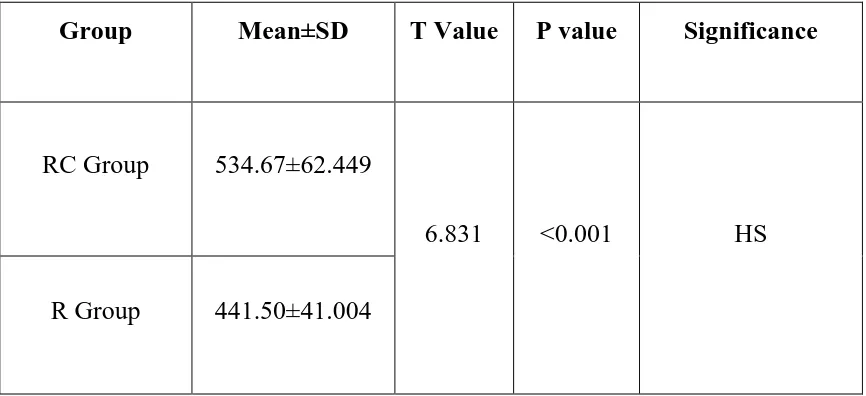
[image:73.595.92.533.214.552.2]The mean duration of sensory block in group RC was 534.67 ± 62.449 min and in group R was 44.50 ± 1.004 min.
FIGURE 5: DURATION OF SENSORY BLOCK .
0 100 200 300 400 500 600
RC Group
R Group M
e a n
Duration of sensory block(in min)
RC Group
TABLE 5: DURATION OF SENSORY BLOCK (in min)
Group Mean±SD T Value P value Significance
RC Group 534.67±62.449
6.831 <0.001 HS
R Group 441.50±41.004
DURATION OF MOTOR BLOCK:
[image:75.595.107.528.220.552.2]The mean duration of motor block in group RC was 498.00 ± 53.233 min and in group R was 400.67 ± 38.200 min .
FIGURE 6: DURATION OF MOTOR BLOCK
0 50 100 150 200 250 300 350 400 450 500 RC Group R Group M e a n
Duration of motor block (in min)
RC Group
TABLE 6: DURATION OF MOTOR BLOCK (in min)
Group Mean±SD T Value P value Significance
RC group 498.00±53.233
8.136 < 0.001
HS R Group 400.67±38.200
DURATION OF ANALGESIA:
[image:77.595.101.520.304.627.2]Duration of analgesia was significantly prolonged in RC group (Ropivacaine and Clonidine) 656.7±86.256 min than control R group (Ropivacaine and NS) 502± 53.169 min, which was statistically highly significant (P< 0.001).
FIGURE 7: DURATION OF ANALGESIA
0 100 200 300 400 500 600 700 RC Group R Group M e a n
Duration of analgesia(in min)
RC Group
TABLE7: DURATION OF ANALGESIA (in min)
After the VAS score 4, Inj. Diclofenac 1.5 mg/Kg Bwt was given as rescue analgesia for all patients.
Group Mean±SD T Value P value Significance
RC Group 656.7±86.256
FIGURE 8: VAS SCORE
The pain score was observed at the end of surgery. At 2 hours the mean VAS score in both groups were zero. After 8 hours, the mean VAS score in RC group which was not statistically significant.
0 0.5 1 1.5 2 2.5 3 3.5 4 4.5 5
2hrs 4 hrs 6 hrs 8 hrs 10 hrs 12hrs 14hrs 16hrs 20hrs 22hrs
M
e
an
Time of assessement
VAS Score
RC
TABLE 8:VAS SCORE
VAS
Score
RC R T
value
P
value
Significance
Mean SD Mean SD
2hrs 0.000 0.000 0.000 0.000 0.000 0.000 NS
4 hrs .27 .640 .57 .728 1.696 0.086 NS
6 hrs 1.20 1.031 1.70 1.179 1.749 0.472 NS
8 hrs 3.57 .817 4.53 .571 5.310 0.001 HS
10 hrs 3.73 .980 3.20 .845 .705 0.001 HS
12hrs 2.93 1.098 2.43 1.040 .724 0.001 HS
14hrs 3.21 .814 3.60 .759 2.461 0.017 HS
16hrs 2.27 1.388 3.47 1.074 3.745 0.001 HS
20hrs 2.03 1.474 3.43 1.074 4.207 0.001 HS
INTRA OPERATIVE SEDATION SCORE:
Sedation score was expressed as mean ±2SD and analyzed by unpaired student “t” test .Sedation score of patients was maximum at 30min in RC group 2.37±.669 and in R group 1.70± 0.466. There after the sedation score was decreased. The statistical analysis showed significant difference.
TABLE9: INTRAOPERATIVE SEDATION SCORE
Time of
assessment
RC R T value P value Significance
Mean SD Mean SD
5 min .90 .481 .50 .572 2.931 0.005 HS
10min 1.87 .507 1.03 .850 4.610 0.001 HS
15 min 2.03 .556 1.50 .630 3.477 0.001 HS
30 min 2.37 .669 1.70 .466 4.480 0.001 HS
45 min 2.17 .699 1.63 .490 3.422 0.001 HS
60 min 2.07 .640 1.57 .504 3.363 0.001 HS
75min 1.83 .648 1.50 .572 2.112 0.039 HS
90min 1.70 .466 1.27 .583 2.115 0.039 HS
105min 1.70 .466 1.27 .583 3.176 0.002 HS
FIGURE 9: INTRAOPERATIVE SEDATION SCORE
0 0.5 1 1.5 2 2.5
5 min 10min 15 min 30 min 45 min 60 min 75min 90min 105min 120min
M
e
an
Time of assessment
sedation score
RC
HEMODYNAMIC VARIABLES:
[image:83.595.108.535.289.588.2]Pulse rate, Systolic BP, Diastolic BP and Oxygen saturation were recorded at 0min, 5min, 15min, 30min, 45min, 60min, 2hrs, 6hrs, 12hrs and 24hrs after giving the block, throughout the surgery and postoperative period. These parametric data expressed as mean ± 2SD and further analyzed by student unpaired “t” test.
FIGURE 10: PULSE RATE
65 70 75 80 85 90 95
0min 5 min 15 min 30 min 45 min 60 min 2hrs 6 hrs 12 hrs 24 hrs
Pu
lse
rate
Pulse rate between RC and R group
RC
The mean pulse rate (Fig.10) at 30 min in RC group and R group were 76.17.and85.13 respectively whereas the mean pulse rate at 2 hours in RC and R groups were 75.23 and 84.53 respectively. Pulse rate was lower in RC group as compared with R group and statistically significant in 15min,30min,45min,60min, 2hrs, 6hrs 12hrs and 24hrs.
The statistical analysis showed significant difference, but none of the patients in RC group required Inj. Atrophine to maintain pulse rate
TABLE10: PULSE RATE
Time of assessment
RC R T value P value Significance
Mean SD Mean SD
0min 85.23 9.726 88.93 5.265 1.832 0.072 NS
5 min 82.63 11.586 87.23 5.244 1.981 0.052 NS
15 min 78.13 11.135 85.13 5.740 3.896 0.001 S
30 min 76.17 11.876 85.13 5.740 3.723 0.001 S
45 min 74.70 11.441 84.73 5.740 4.328 0.001 S
60 min 74.27 10.719 85.00 6.103 4.766 0.001 S
2hrs 75.23 10.183 84.53 5.812 4.344 0.001 S
6 hrs 77.20 9.532 85.20 5.774 3.932 0.001 S
12 hrs 78.07 8.008 84.03 5.082 3.446 0.001 S
SYSTOLIC BP:
The mean systolic BP in RC group was between 114.07±9.318 mmHg to 124.93 ± 12.281 mmHg and in R group 113.33±6.065 mmHg to123.00±12.077 mmHg, the statistical analysis showed no significant difference among both groups examined (RC and R).
Mean systolic B.P at30 min in RC group was 114.07 mmHg. Mean systolic B.P at30 min in R group was 113.47 mmHg.
Mean systolic B.P at 2hours in RC and R groups were 115.27 and 115.33 mmHg respectively.
[image:85.595.91.539.387.726.2]The statistical analysis showed no significant difference.
FIGURE 11: SYSTOLIC B.P (mmHg)
106 108 110 112 114 116 118 120 122 124 126
0min 5 min 15 min 30 min 45 min 60 min 2hrs 6 hrs 12 hrs 24 hrs
M
e
an
B
P
Time of assessment
Systolic BP(mmHg)
RC
TABLE 11: SYSTOLIC B.P(mmHg)
Time of
assessment
Systolic BP
RC
R
T
value
P
value
Significance
Mean
SD
Mean
SD
0min
85.23 9.726
88.93 5.265 1.832 0.072
NS
5 min
82.63 11.586 87.23 5.244 1.981 0.052
NS
15 min
78.13 11.135 85.13 5.740 3.896 0.001
S
30 min
76.17 11.876 85.13 5.740 3.723 0.001
S
45 min
74.70 11.441 84.73 5.740 4.328 0.001
S
60 min
74.27 10.719 85.00 6.103 4.766 0.001
S
2hrs
75.23 10.183 84.53 5.812 4.344 0.001
S
6 hrs
77.20 9.532
85.20 5.774 3.932 0.001
S
12 hrs
78.07 8.008
84.03 5.082 3.446 0.001
S
DIASTOLIC BP:
The mean diastolic BP in RC group was between 69.37±8.130mmHg to 80.10 ±7.818mmHg and in R group 70.33±8.503mmHg to78.33±8.339mmHg. The statistical analysis showed no significant difference among both groups examined (RC and R).
Mean Diastolic B.P at30 min in RC group was 69.37 mmHg. Mean Diastolic B.P at30 min in R group was 71.17 mmHg.
[image:87.595.92.537.375.682.2]Mean Diastolic B.P at 2hours in RC and R groups were 72.00 and 70.33 mmHg respectively. The statistical analysis showed no significant difference.
FIGURE 12: DIASTOLIC BP
64 66 68 70 72 74 76 78 80 82
0min 5 min 15 min 30 min 45 min 60 min 2hrs 6 hrs 12 hrs 24 hrs
M e an B P le
Diastolic BP(mmHg)
RC
TABLE 12: DIASTOLIC B.P
Time of
assessment
Diastolic BP mmHg
T
value
P
value
Significance
RC
R
Mean
SD
Mean
SD
0min
80.10
7.818
78.33
8.339
.847
.401
NS
5 min
71.13
9.372
73.00
9.523
.765
.447
NS
15 min
72.07
9.724
72.33
7.739
.818
.416
NS
30 min
69.37
8.130
71.17
8.477
.839
.405
NS
45 min
71.63
8.348
70.33
8.503
.598
.552
NS
60 min
71.97
8.560
72.00
7.611
.016
.987
NS
2hrs
72.00
8.867
70.33
8.503
.743
.460
NS
6 hrs
71.67
12.617 72.33
11.043 .363
.718
NS
12 hrs
71.33
8.604
72.33
11.043 .391
.697
NS
OXYGEN SATURATION
[image:89.595.94.545.234.598.2]Maximum and minimum oxygen saturation in RC group were found as follows 99.80±099.80% and 99.60± 0.675%, in R group was 99.33±0.606% and 99.47±0.507% respectively.
FIGURE 13: OXYGEN SATURATION
50 60 70 80 90 100 110
0min 5 min 15 min 30 min 45 min 60 min 2hrs 6 hrs 12 hrs 24 hrs
M
e
an
SPO
2
Time of assessment
SPO2
RC
Table 13(a) : SIDE EFFECTS IN RC GROUP:
In RC group, 10 patients had dry mouth. None of the patient had headache, nausea or vomiting. Incidence of dry mouth was 16.4% in RC group which was highly significant.
Side effect RC Group Percent (%)
Dry Mouth 10 16.4%
In R group, only 3 patients had head ache, Nausea and Vomiting noted in 1 patient only. Incidence of Headache, Nausea and Vomiting were 4.9%, 1.6% and 1.6% respectively in R group which was not significant.
Table 13(b): SIDE EFFECTS IN RGROUP
Side effect R Group Percent (%)
Headache 3 4.9
Nausea 1 1.6
[image:90.595.157.439.204.292.2] [image:90.595.158.439.470.622.2]DISCUSSION
In our study, supraclavicular brachial plexus block was performed under ultrasound guidance. All the patients had successful brachial plexus block and hence satisfactory surgical anesthesia .
The real time ultrasound imaging showed better visualization of the brachial plexus, accuracy of the needle placement and spreading of Local anesthesia around the brachial plexus. Identification of the adjacent structures like blood vessels (Subclavian artery and vein), first rib and pleura was helpful to avoid procedure related complications.
We observed that 150 µg of Clonidine added to 175 mg of 0.5% Ropivacaine, resulted in excellent quality of supraclavicular brachial plexus block for upper limb surgeries.
The advantage of Clonidine added as an adjuvant to Ropivacaine was rapid onset and prolonged duration of sensory and motor blockade .It also prolonged the duration of postoperative analgesia.
Many adjuvants like Neostigmine, Opioids, Dexamethasone, Hyaluronidase were used with Local anesthesia in various peripheral nerve blocks to prolong the duration of analgesia, but the results have been inconclusive because of its doubtful efficacy and side effects.
group (7.7±2.5) min. Sidharth SR et al. showed the mean onset time of sensory blockade in their study group was 10.44 ± 5.7 min and in control group was 15.85 ± 6.55 min7.The delayed onset of sensory block in the study by
Sidharth et al inspite of adding clonidine would have been due to the landmark technique used in administering the block. In our study we administered the block under ultrasound guidance which has helped in deposition of the local anesthetic in close proximity to the plexus contributing to the early onset of the sensory block.
The mean onset time of motor blockade in RC group was (9.3±2.72) min. as compared to R group (10.63±2.785) min which was statistically significant. Quazi et al .showed the mean onset time of motor blockade in study group (Ropivacaine 0.5% + 75 mcg clonidine) was 13 ± 3.69 min and in control group was 15.05 ± 4.21 min6.
The reason for early onset of motor blockade in our study would have been due to accuracy of needle placements close to the plexus ,higher volume of local anesthetic (35ml instead of 30ml) and higher dose of clonidine ( 150 µg instead of 75 µg).
The duration of motor block was less than the sensory block due to increased requirement of local anesthetic for larger motor fibre than small sensory fibre. Gupta et al. have shown earlier onset of sensory and motor blockade and prolonged duration of sensory and motor blockade with Ultrasound Versus other nerve localization techniques9.
The combined administration of clonidine and ropivacaine local anesthetics (synergistic mechanism) results in prolonged effect of sensory and motor blockade. This could be the only possible mechanism which explains the long lasting sensory and motor blockade23.
Duration of analgesia was significantly prolonged in RC group than control R group which was statistically highly significant. Gupta et al showed the duration of analgesia in study group was 956.47 ± 38 min and in control group was 736.53± 47 min9.
The mean duration of post operative analgesia in our study in RC group (656.7±86.256) min as compared with R group (502±53.169) min which was statistically significant. The prolonged duration of analgesia in RC group was due to synergistic effect of clonidine and ropivacaine.
. “Sia and Lepri” observed the synergistic mechanism between clonidine
and ropivacaine. This could be the probable cause of extended duration of analgesia in post-operative period24.
Mechanism of action of Clonidine to enhance the peripheral nerve block by “Vasoconstriction theory”23α 2 Adrenergic stimulation causes decreased systemic absorption of Local anesthetics (Ropivacaine) and also Ropivacaine has an intrinsic vasoconstriction effect .This intrinsic vasoconstriction effect of Ropivacaine is not enhanced by Epinephrine23.Clonidine has direct-action on A delta and C fibers and inhibits the nerve conduction, which further augments conduction block of local anesthetics8.
“Dalle et al” explained that clonidine augments the action of
hyper polarization by Na+/K+pump for the period of repetitive stimulation, increases threshold for initiation of action potential, resulting in slowing or blockade of conduction24.
VAS score in post-operative period up to 6hrs was comparable in both groups. RC group reached maximum VAS score at 10hrs and R group at 8hrs, showing extended duration of analgesia in RC group. Quazi et al also has similar results6.
Hemodynamic parameters such as Pulse rate, SBP, DBP, and Spo2 were comparable in both groups; the mean pulse rate in RC group was significantly lower than in R group at regular intervals. None of the patients had significant bradycardia and hypotension. Our observation concur with those obtained by Sidharth et al 7.
Dry mouth was observed in Clonidine group (16%) whereas it was nil in Ropivacaine group. Nausea, Vomiting, Headache was present only in R group. Similar results were obtained by other investigators6. No other complications were present in both the groups.
CONCLUSION
SUMMARY
This prospective Randomized double blind placebo controlled study was done in Mahatma Gandhi Memorial Government Hospital, Trichy from the period of 2014 to 2015.
A total of 60 patients belonging to ASA I& II, aged between 18 to 60 years were scheduled for upper limb surgeries were included in the study. Patients receiving anticoagulants, H/O coagulopathy and peripheral neuropathy, and age < 18 years, > 60 years, H/O uncontrolled hypertension, refusal of patient’s participation were excluded from this study.
Patients were divided into two groups, each group consisting of 30 patients (n=30).
1. RC Group: Patient receiving 1 ml of Clonidine (150 µgm) +35 ml of 0.5% Ropivacaine (175 mg).
2. R Group: Patient receiving 1 ml of Normal saline +35 ml of 0.5% Ropivacaine (175 mg). The parameters observed were
Time of onset of sensory and motor block, Duration of sensory and motor block Duration of postoperative analgesia
In this study following results were obtained
Rapid onset of sensory and motor block
Extended duration of sensory and motor block Extended duration of postoperative analgesia
Better operating conditions without major hemodynamic alterations and side effects in RC Group when compared with R Group.
BIBLIOGRAPHY
1. Khanduri KC; Regional anesthetic techniques for orthopedic surgeries. Med J Armed Forces India, 2008; 64:109
2. Akerman B, Hellberg IB: Primary evaluation of the local anesthetic properties of the amino amide agent Ropivacaine. Acta Anaesthesiology sc and., 1988;32:571-578
3. Singh, S., &Aggarwal, A. A randomized controlled double-blinded prospective study of the efficacy of clonidine added to bupivacaine as compared with bupivacaine alone used in supraclavicular brachial plexus block for upper limb surgeries. Indian journal of anesthesia. 2010; 54(6): 552.
4. Gray, A. T. Atlas of Ultrasound-Guided Regional Anesthesia: Expert Consult-Online and Print. Elsevier Health Sciences. 2012; 74:76
5. Ultrasound Guided Supraclavicular Block By Alan Macfarlane BSc (Hons), MBChB, MRCP, FRCA, Richard Brull MD, FRCPC, Department of Anesthesia, University of Toronto, Toronto Western Hospital, 399 Bathurst Street, Toronto, The journal of New York school of regional anesthesia .
6. Indian Journal of Anaesthesia, Vol. 58, No. 6, November-December, 2014, pp. 709-713 Clinical Investigation, Efficacy of clonidine as an adjuvant to ropivacaine in supraclavicular brachial plexus block: QaziEhsan Ali, Syed Hussain Amir, ShaistaJamil, L Manjunatha, Abdul Quadir
8. Bafna U, Yadav N, Khandelwal M, Mistry T, Chatterjee C S, Sharma R. Comparison of 0.5% ropivacaine alone and in combination with clonidine in supraclavicular brachial plexus block .Indian J Pain. 2015;29:4145.
9. Gupta, K., Tiwari, V., Gupta, P. K., Pandey, M. N., Singhal, A. B., &Shubham, G. Clonidine as an