A
COMPARATIVE
STUDY
OF
PULMONARY
FUNCTION
TEST
ABNORMALITIES
IN
RHEUMATOID
ARTHRITIS-TREATMENT
NAIVE
VERSUS
PATIENTS
ON
TREATMENT
DISSERTATIONSUBMITTEDFOR
M.D GENERAL MEDICINE
BRANCH – I
APRIL 2015
THE
TAMILNADU
DR.M.G.R.
MEDICAL
UNIVERSITY
CHENNAI,
CERTIFICATE FROM THE DEAN
This is to certify that the dissertation entitled
“
A
COMPARATIVE
STUDY
OF
PULMONARY
FUNCTION
TEST
ABNORMALITIES
IN
RHEUMATOID
ARTHRITIS-TREATMENT
NAIVE
VERSUS
PATIENTS
ON
TREATMENT
” is the bonafide work of Dr. ARUN KUMAR.A., in partial fulfillment of theuniversity regulations of the Tamil Nadu Dr. M.G.R. Medical
University, Chennai, forM.D General Medicine Branch I examination to
beheldinApril 2015.
Capt.Dr.B.SANTHAKUMAR
M.Sc ( F.Sc)., M.D (F.M), PGDMLE,
Dip. N.B (F.M)
CERTIFICATE FROM THE HOD
This is to certify that the dissertation entitled
“
A
COMPARATIVE
STUDY
OF
PULMONARY
FUNCTION
TEST
ABNORMALITIES
IN
RHEUMATOID
ARTHRITIS-TREATMENT
NAIVE
VERSUS
PATIENTS
ON
TREATMENT
” is the bonafide work of Dr. ARUN KUMAR.A., in partial fulfillment of theuniversity regulations of the Tamil Nadu Dr. M.G.R. Medical
University, Chennai, forM.D General Medicine Branch I examination to
beheldinApril 2015.
Dr. S.Vadivel Murugan, M.D
Professor and HOD,
Department of General Medicine, Madurai Medical College,
CERTIFICATE FROM THE GUIDE
This is to certify that the dissertation entitled
“
A
COMPARATIVE
STUDY
OF
PULMONARY
FUNCTION
TEST
ABNORMALITIES
IN
RHEUMATOID
ARTHRITIS-TREATMENT
NAIVE
VERSUS
PATIENTS
ON
TREATMENT
” is the bonafide work of Dr. ARUN KUMAR.A., in partial fulfillment of theuniversity regulations of the Tamil Nadu Dr. M.G.R. Medical
University, Chennai, forM.D General Medicine Branch I examination to
beheldinApril 2015.
Dr.M.Natarajan, M.D.
Professor,
DECLARATION
I, Dr.ARUN KUMAR.A., solemnly declare that, this dissertation
“
A
COMPARATIVE
STUDY
OF
PULMONARY
FUNCTION
TEST
ABNORMALITIES
IN
RHEUMATOID
ARTHRITIS-TREATMENT
NAIVE
VERSUS
PATIENTS
ON
TREATMENT
” is a bonafide record of work done by me at the Department of GeneralMedicine, Govt. Rajaji Hospital, Madurai, under the guidance of Dr.M.NATARAJAN, M.D, Professor, Department of General Medicine, MaduraiMedicalCollege,Madurai.This dissertation is submitted to The Tamil Nadu Dr. M. G. R.
Medical University, Chennai in partial fulfillment of the rules and
regulations for the award of M.D Degree General Medicine Branch-I;
examination to be held in April 2015.
Place: Madurai
Date:
ACKNOWLEDGEMENT
I would like to thank Capt. Dr.B. SANTHAKUMAR M.Sc
(F.Sc)., M.D (F.M), PGDMLE, Dip. N.B (F.M) Dean, Madurai Medical
College, for permitting me to utilize the facilities of Madurai Medical
College and Government Rajaji Hospital facilities for this dissertation.
I wish to express my respect and sincere gratitude to my beloved
teacher and Head of the Department, Prof.Dr.S.VADIVELMURUGAN,
M.D., Professor of Medicine for his valuable guidance and
encouragement during the study and also throughout my course period.
I would like to express my deep sense of gratitude, respect and
thanks to my beloved Unit Chief and Professor of Medicine,
Prof.Dr.M.NATARAJAN, M.D., for his valuable suggestions, guidance
and support throughout the study and also throughout my course period.
I am greatly indebted to my beloved Professors,
Dr.V.T.PREMKUMAR, M.D., Dr.R.BALAJINATHAN, M.D.,
Dr.G.BAGHYALAKSHMI, M.D., Dr.J.SANGUMANI, M.D.,
Dr.C.DHARMARAJ, M.D., and Dr.R.PRABHAKARAN, M.D., for
their valuable suggestions throughout the course of study.
facilities in the Department for the purpose of this study and guiding me
with enthusiasm throughout the study period.
I express my special thanks to Dr.MOSES.K.DANIEL, M.D.,
Retired Professor of Medicine for his guidance and support during the
study.
I am extremely thankful to Assistant Professors of Medicine of my
Unit, Dr.K.MURALIDHARAN, M.D., Dr.B.PALANIKUMAR, M.D.,
Dr.P.K.GANESH BABU., M.D., and Dr.G.SELVARANI, M.D., for
their valid comments and suggestions.
I sincerely thank the Assistant Professors of Thoracic Medicine,
Dr.K.BHARATHI BABU, M.D. (Chest) and Dr.R.PRABHAKARAN,
M.D. (Chest) for their guidance and suggestions in my dissertation work.
I express my special thanks to the Assistant Professor of Medicine
Dr.S.C.VIVEKANANDHAN, M.D., D.T.C.D., for his suggestions and
guidance.
I sincerely thank all the staffs of Department of Medicine and
Department of Thoracic Medicine for their timely help rendered to me,
whenever and wherever needed.
I extend my thanks to all my friends, batch mates and my junior
Finally, I thank all the patients, who form the most vital part of my
work, for their extreme patience and co-operation without whom this
project would have been a distant dream and I pray God, for their speedy
CONTENTS
S.NO CONTENTS PAGE
NO.
1. INTRODUCTION 1
2. AIM OF STUDY 5
3. REVIEW OF LITERATURE 6
4. MATERIALS AND METHODS 60
5. RESULTS AND INTERPRETATION 63
6. DISCUSSION 82
7. CONCLUSION 97
8. SUMMARY 99
9. ANNEXURES
BIBLIOGRAPHY
PROFORMA
ABBREVATIONS MASTER CHART
ETHICAL COMMITTEE APPROVAL LETTER
ABSTRACT
A COMPARATIVE STUDY OF PULMONARY FUNCTION TEST ABNORMALITIES IN RHEUMATOID ARTHRITIS- TREATMENT NAIVE VERSUS PATIENTS ON TREATMENT
Background:
A variety of pulmonary manifestations are associated with Rheumatoid
arthritis; lung disease is the second most common cause of death (18%)
after infection (27%) in patients with RA. Pulmonary function test
abnormalities in RA can be restrictive (19-44%) if there is pleural or
parenchymal involvement, or obstructive (16-38%) if there is
obliterative bronchiolitis, bronchiectasis or cricoaryetenoid arthritis. The
incidence of pulmonary function test abnormalities in various studies done
in patients with RA varies widely.
Objective:
1. To assess the proportion of pulmonary function test (PFT)
abnormalities in patients with rheumatoid arthritis.
2. To categorize the PFT abnormalities as obstructive or restrictive and
further quantify them as mild, moderate or severe.
3. To compare PFT abnormalities between treatment naïve and
Methods:
A pretested semi structured questionnaire was administered to newly
diagnosed and those patients on treatment for rheumatoid arthritis. They
underwent routine historical evaluation, physical examination and detailed
respiratory and musculoskeletal examination. Then they underwent
pulmonary function testing, chest radiography and pulse oximetry. The
parameters recorded in spirometry were FEV1, FVC, PEF and
FEF25-75%. The spirometer was selected and calibrated in accordance
with ATS guidelines. The criteria for obstruction and restriction were
selected in accordance with ATS guidelines
Results:
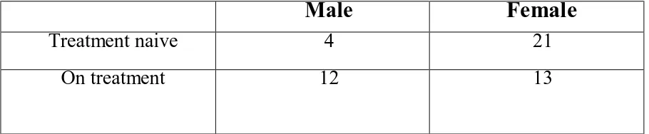
In our study fifty patients were included, 25 newly diagnosed and 25
patients on treatment for minimum 3 years. The male to female ratio in
this study was 1:2. The mean age of patients was 46.5±10.5 years.
Rheumatoid factor was positive in 35 (70%) patients. 16% of patients had
respiratory symptoms. Incidence of radiological abnormality was 10.7%.
Fifty patients underwent pulmonary function testing. 56% patients had
abnormal PFTs 16% had obstructive and 40% had restrictive pattern in
treatment naive and 64% patient had abnormal PFTs 20% had obstructive
The incidence of PFT abnormality in the age group of 50-59 was
highest. This was not statistically significant. The number of patients
with restrictive PFTs increased after three years of disease duration when
compared with newly diagnosed patients. The number of patients with
obstructive PFTs also increased when the disease duration was more than
3 years. No statistical correlation was found between the severity of
PFT abnormality (both obstruction and restriction) and the disease
duration. The number of abnormal PFTs increased as the functional
class of RA increased.
Interpretation and conclusion:
The number of abnormal PFTs increased as the disease duration increased.
There was no correlation between the disease duration and the severity of PFT
abnormality. PFT abnormalities were encountered with greater frequency in
patients of higher functional class of disability in rheumatoid arthritis. There
is increased incidence of PFT abnormality in on treatment group when
compared to treatment naive group this may be explained by the type of lung
involvement (ex. If the patient had usual interstitial pneumonia the lung
disease may progress or remain static) but a longitudinal study is warranted.
Pulmonary function testing when combined with chest radiography is cost
KEYWORDS:
INTRODUCTION
Rheumatoid arthritis (RA) is a chronic multi-system disease of
unknown cause. It is a systemic inflammatory disease and affects 1-2%
of the general population. The characteristic feature of RA is
inflammatory synovitis that is persistent and involves most of the
peripheral joints depending on the duration of disease causing
cartilaginous and bony erosions. In general population, RA is found in
0.8% of people and its prevalence increases with advancing age 1.The
onset of RA is more common in the age group of 40- 50 years
The mortality and morbidity of RA is mostly attributed to its
extraarticular involvement. Extra-articular manifestations are seen in
nearly 50% of patients with RA, the commonly affected sites being the
skin, eye, heart, and lungs4. In 40% of patients the prevalence of
extraarticular manifestation are so severe clinically.
A wide range of pulmonary manifestations are seen in patients
with RA. The involvement of lung is the second most common cause of
death (18%) after infection (27%) in patients with RA 6. In men pleuro-
pulmonary manifestations are more common compared to women 7.
common intrathoracic manifestation of rheumatoid arthritis 8.Pleural
effusion is clinically evident in only 5% of patients with rheumatoid
pleurisy9.
Similarly, the incidence of pleural effusion depends on the duration of
disease. Pleural effusions seen most frequently with longstanding active
articular disease. These patients also have rheumatoid nodules 10,11.
Prospective studies using high resolution computed tomography
[HRCT] of the lungs demonstrated that in 20% of patients, there is
associated fibrosing alveolitis 12.Both ILD and pleural effusion can
precede articular symptoms.
Parenchymal involvement in RA may be in the form of rheumatoid
nodules, Caplan syndrome and interstitial lung disease. The only specific
pulmonary lesion in these patients were rheumatoid nodules. They are
single or multiple nodules seen more commonly in males. Similarly
nodules are common among those with subcutaneous nodules and extra-
articular manifestations. Caplan’s syndrome is one such seen in silica
exposed patients with bilateral pulmonary nodules who have rheumatoid
The incidence of interstitial lung disease [ILD] in RA has been
found to be19- 44%in a study conducted by Dawson et al It is more
common in men who are seropositive and aged between 50-60 years of
age16.
RA can affect airways of all diameter and can cause varying disease
manifestation17-19. Upper airway involvement includes cricoarytenoid
arthritis, rheumatoid nodules in the vocal cords and vocal cord paresis.
Lower airway involvement includes bronchiectasis, bronchiolitis with or
without organizing pneumonia. There is a high incidence of radiographic
bronchiectasis, up to 30% in some HRCT studies 20-22.Pulmonary
involvement can be studied using HRCT and DLCO for anatomical
and physiological abnormalities respectively .However these diagnostic
tests may not be widely available and cost effective. Pulmonary function
testing using an office spirometer is a simpler, cheaper and more widely
available tool for screening patients with RA for pulmonary
involvement.
Pulmonary function test abnormalities in RA can be restrictive
(19-44%) if there Is pleural or parenchymal involvement, or obstructive
The incidence of pulmonary function test abnormalities in various
studies done in patients with RA varies widely.
Although the patients with RA have varying degree of severity in
the manifestation of pulmonary function, the progression of the disease
severity may or may not be altered by the immunosuppressive therapy.
The prognosis in these patients are guarded.The purpose of this study is
to assess the incidence and type of PFT abnormality in patients with
rheumatoid arthritis presenting to a tertiary care hospital and also to
compare PFT abnormalities between treatment naïve and on treatment
AIMS AND OBJECTIVES
• To assess the proportion of pulmonary function test (PFT)
abnormalities in patients with rheumatoid arthritis.
• To categorize the PFT abnormalities as obstructive or
restrictive and further quantify them as mild, moderate or severe.
• To compare the PFT abnormality in treatment naïve and patients
REVIEW OF LITERATURE
Rheumatoid arthritis (RA) is one of the functionally disabling
disease that is common among general population and various genetic
and environmental contributes to its development. It occurs in
approximately 1% of the population. The disease is characterized by
inflammation of synovial membranes and articular structures, which are
the primary target of the inflammatory process. Inflammation and
degeneration along with proliferation of synovial membrane typify the
disease. Joint deformities and disability result from the e rosion and
destruction of synovial membranes and articular surfaces 24.
RA usually affects the joints in a symmetric manner. The disease
is polyarticular and involves most of the small joints with exception to
the distal interphalangeal (DIP) joints 25. The joints most commonly
affected are Wrists, proximal interphalangeal, metacarpophalangeal,
followed by metatarsophalangeal and shoulders.
The least commonly affected are the hips and spine.
involvement, the other joints on the same side of the body may be
affected. The arthritis in RA predominantly involves peripheral joints.
Early wrist and metatarsophalangeal joint involvement is an indicator of
progression to severe RA26
There is also involvement of extra-articular structures in RA. The
predominant one among this is subcutaneous nodules and pleuro-
pulmonary manifestations. Other organs to be affected are cardiovascular and
nervous system. Vasculitis and Felty's syndrome are rare manifestations but
may be severe enough to cause disability 27. The occurrence and course of
the extraarticular manifestations of RA does not always parallel joint
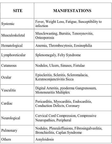
disease28. Extraarticular manifestations of RA are described in Table1.
Most common cause of death in patients with RA includes mostly
cardiac followed by pulmonary complication. Mortality due to pulmonary
complications is about 10 to 20% in patients with RA 28,30,31. Although
pulmonary infection and/ or drug toxicity are frequent complications, lung
disease directly related to underlying RA is more common. The mortality
ratio of patients with RA is about 2.5 to 5% when compared with
control28,30.
The majority of lung disease occurs within the first 5years after the
pulmonary vasculature and pleura can all be involved, with variable
[image:22.595.115.544.157.705.2]clinical features. [Table2]
Table1.Extra-articular manifestations of RA
SITE
MANIFESTATIONS
Systemic Fever, Weight Loss, Fatigue, Susceptibility to infection
Musculoskeletal Musclewasting, Bursitis, Tenosynovitis, Osteoporosis
Hematological Anemia, Thrombocytosis, Eosinophilia
Lymphoreticular Splenomegaly, Felty Syndrome
Cutaneous Nodules, Ulcers, Sinuses, Fistulae
Ocular Episcleritis, Scleritis, Scleromalacia, Keratoconjunctivitis Sicca
Vasculitis Digital Arteritis, pyoderma Gangrenosum, Mononeuritis Multiplex
Cardiac Pericarditis, Myocarditis, Endocarditis, Conduction Defects, Coronary
Neurological Cervical Cord Compression, Compressive Neuropathies, Peripheral
Pulmonary Nodules, Pleuraleffusions, Fibrosingalveolitis, Bronchiolitis, Caplan Syndrome
Table2.Pleuropulmonary manifestations of rheumatoid arthritis
SITE
MANIFESTATIONS
PLEURA
Pleuritis, Pleural effusion, Empyema
Pneumothorax
INTERSTITIUM Organizing Pneumonia, Usual interstitial Pneumonia
AIRWAYS
Constrictive Bronchiolitis (bronchiolitis Obliterans)
ti
VASCULAR Pulmonary Hypertension, Pulmonary
Vasculitis
PLEURAL INVOLVEMENT
Pleuritis and pleural effusions:
Pleuritic type of chest pain occurs in 25% of patients with RA 32.Five
percent of patients with RA develop pleural effusions which are small to
moderate, unilateral being more common than bilateral 33.Pleural disease
is more common in middle aged men. Pleural effusions are more common
in patients with longstanding active articular disease and rheumatoid
nodules34,35. Rarely , pleural effusion may precede joint disease 36.Small
effusions usually resolve spontaneously within weeks; however, they
may persist and present as chronic pleural effusion .Large effusions
need therapeutic aspiration and tend to recur after aspiration.
The triad of multinucleated macrophages, large elongated
macrophages, and under a background of granular debris is characteristic
of the pleural fluid cytology in rheumatoid effusions 37-39. Pseudochylous
effusions can also occur with RA35,40. Rheumatoid effusions are usually
exudative with very low glucose levels and are lymphocyte
In upto 20% of patients with rheumatoid arthritis and associated
pleural effusion, prospective studies show fibrosing alveolitis on HRCT.
Rarely, pneumothorax or pyothorax can occur from ruptured necrotic
rheumatoid nodule45.
The most common pulmonary function abnormality in rheumatoid
pleural effusion is restriction which worsens when the patient develops
fibrothorax46. The restriction is associated with paradoxically increased
DLCO which distinguishes it from interstitial lung disease where the
DLCO is reduced.
PARENCHYMAL INVOLVEMENT
Pulmonary parenchymal involvement can develop in RA during the
course of the disease or it may be the initial manifestation occurring in
very early stages of disease.
Rheumatoid nodules:
20% of patients47,48. They are either single or multiple in number usually
measuring 1-3cm, although nodules measuring upto 10cm may be seen 49.
Rheumatoid nodules are seen more commonly in males. Nodules are
commonly found sub-pleurally in the upper and Mid zones of the lung;
rarely they can occur endobronchially. In some case multiple widespread
nodules are described.
In Caplan syndrome, pneumoconiosis and RA are synergistic and
produce as severe fibroblastic reaction with obliterative granulomatous
fibrosis. Pathologically, the granulomas consist of collections of
lymphocyte, macrophages, plasmacells and histiocytes under the
background of necrotic debris. 52. These nodules must be differentiated
from malignant ones which is done by HRCT and biopsy. These nodules
when subpleural can lead to empyema or pneumothorax on rupture. Other
complication is presenting as hemoptysis. Spirometric evaluation in
rheumatoid nodules is reveals restriction 54. In some patients similar to
ankylosing spondylitis apical fibrobullous disease and aspiration
Interstitial Lung Disease:
The incidence of ILD in RA has been found to be 19-44% in a study
conducted by Dawson et al. It occurs more frequently in seropositive men
aged between 50-60years16. High titres of RF and presence of rheumatoid
nodules are associated with increased prevalence of pulmonary fibrosis in
RA55,56.
Active or previous tobacco smoking and rheumatoid factor (RF)
seropositivity are risk factors for the development and severity of ILD in
patients with RA57-60. A correlation has been proposed between the habit
of cigarette smoking and the presence of HLA-DRB1" shared
epitope"(SE), anticyclic citrullinated peptide antibody(anti- CCP), and the
development of RA61. The presence of smoking with associated two
copies of the HLA-DRSE genes increased the risk for RA 21-fold
compared non smokers with absent gene.
In some patients smoking has been independentally associated with
ILD. Patient with more than 25 pack years have 3.76 % incidence of 95%
The increased reactivity of mesenchymal cells in RA is postulated
as the cause of pulmonary fibrosis. In this disorder when there is acute
insult to lung parenchyma, there is activation of fibroblast which causes
chronic inflammatory change leading to the endpoint of pulmonary
fibrosis and tissue d estruction66.
Modes of presentation of RA associated ILD:
1. ILD may be incidentally detected on chest radiograph or abnormal
screening spirometry in an aymptomatic patient with RA.
2. Diagnosis may occur during screening for high-risk occupational exposure,
such as asbestosis.
3. Clinically overt disease may present insidiously and progress slowly
[chronic ILD]. It may also present acutely,sub-acutely or have a relapsing
remitting course. Disorders with chronic, insidious,and slowly progressive
courses are those that most resemble IPF and usually share a common
pathology (ie,UIP). Pulmonary fibrosis in RA can occur as a side effect of
Cryptogenic organizing pneumonia[COP]:
Cryptogenic Organising pneumonia is seen more common in
rheumatoid arthritis when compared to other connective tissue
disorders67.It usually involves terminal bronchioles and distal air spaces. It
is characterized by presence of plugs of granulation tissue in the airspaces.
The main pathophysiology of COP is lymphocytic infiltration within
bronchiolar walls and surrounding interstitium it usually shows multifocal
consolidation on computed tomography when compared to bronchiolitis
obliterans PFT shows restrictive pattern. 68. It responds better to
corticosteroids. The clinical presentation of COP initially is similar to a
flu like illness, which is gradually progressive in nature. It takes weeks or
months to develop a full blown disease characterized by dyspnea or
exercise intolerance. The disease course may vary from spontaneous
remission to progressive disorder. Steroids are the mainstay of therapy and
disease may reccur on withdrawal of drug. Some patients may progress to
Acute Interstitial pneumonia:
Occasionally ILD in RA can present acutely. The most common
pattern with this presentation is AIP. It is an idiopathic form of lung
disease which is more severe in presentation. It has a similar
histopathology as that of adult respiratory distress syndrome (ARDS)
with diffuse alveolar damage(DAD). These patients usually have no no
antecedent history. These patients progress rapidly to respiratory failure.
These patients does not respond to any treatment steroids or
immunosuppressive therapy. Clinical finding is similar to idiopathic
pulmonary fibrosis. They are usually cyanotic with fine end-inspiratory
pulmonary rales (Velcro rales). .Digital clubbing may accompany many of
these disorders. They may present with right heart failure in advanced
cases. Of all the histological patterns of ILD, the non specific interstitial
pattern is the most prevalent 69although in one study, most common pattern
is usual interstitial pneumonia.
Usually the prognosis of interstitial lung disease in RA is good.
There is slow deterioration of lung function in RA associated ILD.
pulmonary fibrosis, survival very similar to what is observed in patients
with idiopathic pulmonary fibrosis 71.The investigation of choice to detect
interstitial pneumonia is HRCT. It is abnormal in upto 80% of patients The
prevalence of radiological evidence of pulmonary fibrosis in patients who
have rheumatoid arthritis ranges from 2-10% 72. The most representative
data has come from a study in which chest radiographs of 309 patients who
have rheumatoid disease were compared with those of age and sex matched
controls73.In this study a reticulonodular pattern consistent with fibrosis
was seen in 4.5% of patients with rheumatoid disease v/s 0.3% of
controls.The pattern and distribution of fibrosis on both CXR and HRCT
are indistinguishable from those of IPF. In early stage, the radiographic
appearance consists of irregular linear opacities causing a fine reticular or
reticulonodular pattern 74. The abnormality usually involves the lower lung
zones75.
Pulmonary function tests(PFTs) with the diffusing capacity of the
lung for carbon monoxide (DLCO) are sensitive tests to detect RA
ILD. Evidence of restriction on lung function testing is found in 30-
40% of all patients with RA associated ILD 77.The important functional
defect is impairment of alveolo-capillary gas exchange with reduced
diffusion capacity best measured utilizing single breath carbon monoxide
diffusion capacity. Even though the prevalence of a restrictive defect in
consecutive patients was not high (5-15%) 78-81 reduced DLCO was
observed in more than 50% of patients with rheumatoid arthritis 82,84 and
reduced DLCO was suggested to be the most sensitive marker of interstitial
pneumonia on high-resolution computed tomography (HRCT).
In a study conducted by Laitinen O et al, vital capacity (VC) and
single-breath diffusing capacity for carbon monoxide of the lungs (DLCO)
were measured and chest X-ray evaluated in 129 patients with rheumatoid
arthritis (RA)83.Findings in the123 cases were observed as follows : in one
of the lung function tests or X-ray examinations , 35%; abnormal X-
rays,18%; reduced VC or Dco , 28% ; simultaneously low VC and Dco,
7%; and pathological findings in all three tests, 2%.The patients with
abnormal X-rays showed extremely low VC and Dco values .Changes in
respiratory function involved restrictive impairment and diffusion defects,
rheumatoid lung .The disparity abnormal findings in chest radiographic
changes and lung function tests suggests that, both radiographic methods
and pulmonary function tests should be used for evaluating pulmonary
manifestations in patients with RA.
In a study by Gabbay et al in 1997,abnormalities consistent with ILD
were found in one or more investigation in 58% of patients with RA.(PFT
in 22%,DTPA nuclear scaning 15%, HRCT in 33% and chest radiograph in
6%.Hence,PFT was a sensitive measure in picking up pulmonary
abnormalities85
Fibrosing alveolitis is a common serious complication of RA. In a
study conducted by Dawson et al,19% had fibrosing alveolitis ;most
frequently reticular pattern on HRCT, 14% had restrictive PFT 16 A
distinctive manifestation of rheumatoid lung disease is progressive upper
lobe fibrosis and cavitation .Patients with this disease may be
AIRWAY DISEASE IN RA:
RA can cause upper, lower, and small, distal airway disease 17-19.
Upper airway disease:
Cricoarytenoid arthritis is a frequent manifestation of RA that presents
with symptoms of foreign body sensation in throat, soreness, and throat pain
which radiates to the ears. Some patients may present with dysphonia,
dysphagia and stridor. In a study conducted by Hayakawa.H et al, nearly
26%of patients with RA had cricoaryetenoid arthritis 86. Usually the
clinically diagnosis is based on direct or indirect laryngoscopy. The finding
includes inflammatory changes of the arytenoids with reduced motility.
Diagnosis is usually confirmed by CT
In some cases, ankylosis of the cricoarytenoid joint may induce an
upper airway obstruction with a characteristic pattern on the flow-volume
curve. In these patients with dyspnea, surgery is indicated to treat ankylosis.
Small airway disease:
Small airway disease with physiologic obstruction is common 87.
And these patients usually have non productive cough, dyspnea on exertion
or wheezing. The diagnosis is made by HRCT. It usually shows involvement
of small airway with centrilobular nodules, heterogenous air trapping and
hyperinflation. Pathologically, both fibrosing (obliterative or constrictive
bronchiolitis) and cellular (diffuse panbronchiolitis and follicular
bronchiolitis) have been well described 88.
“Controlled studies of lung function in patients with RA demonstrate
an increased prevalence of chronic airway obstruction (16–38%) 89 and
increased bronchial reactivity to methacholine (55%). In a PFT survey of
patients with rheumatoid arthritis, airway obstruction was observed in9-
37%, even in non-smoking patients 90. In PFT, constrictive bronchiolitis
manifests as severe irreversible airway obstruction with hyperinflation.
Obliterative bronchiolitis:
It is a progressive condition and death from respiratory failure occurs
wall destruction with effacement of lumen by granulation tissue and
eventual replacement of bronchiolar wall by fibrous tissue. The histologic
picture is usually preceded by exudation of inflammatory substrates. The
progression of this disease is heterogeneous. It is more in patients having
indolent disease. The prevalence of unsuspected OB is uncertain. Patients
may benefit from treatment with inhaled corticosteroids and
bronchodilators. In 50% of patients the prognosis is poor. PFT shows
severe airflow obstruction with reduced DLCO. Pencillamine use has been
related to this condition 92.
Follicular bronchiolitis:
Follicular bronchiolitis is characterized by external compression of
bronchioles by hyperplastic lymphoid follicles with variable lymphocytic
infiltration of the bronchiolar wall. FB is more commonly seen in RA
compared to other connective tissue disorders. It is unclear if follicular
bronchiolitis predisposes to OB. Isolated FB simulates ILD with reticular
or reticulonodular abnormalities on CXR. The PFT abnormality may be
Restrictive or obstructive 93. It is more responsive to corticosteroid
Lower airway disease:
Bronchiectasis is a common manifestation of RA .Clinically
significant bronchiectasis is less frequent and involving 1–5% of Patients
with RA. Bronchiectasis is more common in women than in men (male to
female ratio of 1:2.8)
In some studies, RA appears at a younger age in patients with
bronchiectasis (46vs51 years).Symptoms are identical to other causes of
bronchiectasis and include cough, sputum production, frequent episodes of
infection, and hemoptysis. The co-existence of RA and bronchiectasis is
associated with an alteration of lung function tests and a poor 5-year
survival .In most patients (90%), bronchiectasis precedes the development
of RA by 25–30years .HRCT studies demonstrate that 20–35% of patients
with RA have bronchiectasis (associated with interstitial changes in one-
third of the cases)94.
However, clinically significant disease is much less frequent.
Secondary development of bronchiectasis after 7–10years of evolution of
In a case–control study, patients with RA and bronchiectasis were 7.3 times
more likely to die than the general population,5.0 times more likely than
patients with RA and 2.4 times more likely than patients with
bronchiectasis without RA. An increased risk of death within the RA and
bronchiectasis group was associated with a history of smoking, moresevere
RA and steroid usage. In this study, 60% of the mortality was due to
infections and acute respiratory failure.
Bronchiectasis is one of the predisposing factor to lung infections. It
also increases the postoperative morbidity in patients with RA. The
pathogenesis of bronchiectasis is poorly understood. These patients usually
have decreased humoral immunity which may be the reason for increased
risk of infection. Some patients with RA have recurrent bronchial infection,
lymphedema , pleural effusions and typical nail changes which constitutes
yellow nail syndrome.
VASCULAR INVOLVEMENT:
The involvement of lung vasculature is rare in RA. It presents as
secondary to pulmonary vasculitis. This presentation is correlated with the
presence of antineutrophil cytoplasmic antibodies 95.
DRUG INDUCED LUNG DISEASE
Several drugs used for the treatment of RA have been associated with
drug-induced lung disease. Methotrexate, goldsalts, D-penicillamine ,and
nonsteroidal anti- inflammatory drugs are associated with repiratory adverse
effects97. Two percent of patients with RA develop an acute respiratory
infection requiring hospitalization annually. Pneumonitis as a consequence
of treatment with methotrexate is now a frequent feature as the use of this
agent increases. “The reported incidence of methotrexate pneumonitis in RA
varies from 0.86% to 6.9%98” .The risk is maximal in the first year of
treatment. Mortality from methotrexate pneumonitis is around 20% in most
series99.
Patients with underlying lung disease and who are smokers are at
increased risk for pneumonitis. It is important to diagnose patients with
prior predisposing condition so that it may help in alternative treatment
regimen. The single most predictive test helpful to identify the risk for
lesser risk of developing infection than who are not on the drug. The major
cause of death is failure to mount immune response in this patients.
A survey from Japan reports an increased risk of acute pneumonitis
in leflunomide treated patients with an incidence of 0.5% 100. In Indian
studies, there is no evidence that combination of leflunomide with
methotrexate increases the risk of pneumonitis beyond that which
methotrexate itself carries.
Infliximab infusions administered for active articular disease have
caused acceleration of underlying rheumatoid interstitial lung disease in
few patients101. Recent data on patients receiving etanercept for RA
indicate that this agent can potentiate an acute pneumonitis with ground
glass shadowing on HRCT and histological evidence of granulomatous
pneumonitis.
This is distinct from Methotrexate pneumonitis and is almost
exclusively confined to patients with prior significant lung disease, patients
CLINICAL STUDIES–PULMONARY INVOLVEMENT IN RA
In a study conducted in 1997 by Cortet-Bernardet, sixty eight patients
(54 women,14men), a significant decrease of FEV1/FVC, FEF25%,
FEF50%, FEF75%, FEF25-75%, and TLCO was observed(p<0.05) and
13.2% of the patients had a small airways involvement defined by a decrease
of FEF25-75% below 1.64SD102. The most frequent HRCT findings were:
bronchiectasis(30.5%), pulmonary nodules (28%), and air trapping(25%).
The patients with small airways involvement had a high frequency of
recurrent bronchitis (75%v34%,p=0.05) and bronchiectasis(71% v23%,
p=0.019). The patients with bronchiectasis had low values of FEV 1, FVC,
FEF25-75%, and TLCO (p<0.01). This study suggests a significant
association between small airways involvement on PFT and bronchiectasis
on HRCT in asymptomatic patients with RA.
In a study conducted in 2004 by Terasakietal, PFT and HRCT were
done in 34 patients103. Bronchial wall thickening was detected in 85%,
small nodules in 71% and bronchial dilatation in 62%. The extent of
In another study conducted in 1998 by Perez et al where 50 patients
with RA (9malesand41females), included 39 non-smokers and 11
smokers104. Airway obstruction (reduced FEV1/FVC) was found in 18%
of patients. Small airway disease, reflected by reduced FEF25-75 was seen
in 8% of patients. The airway obstruction and small airway disease
correlated well with the presence of bronchiectasis, bronchial wall
thickening and with bronchial infection on HRCT.
A study was conducted in 2004 by Doyle et al to determine the
prevalence of airway hyperreactivity(AHR) in patients with newly
diagnosed rheumatoid arthritis (RA)who had received no disease-
modifying anti-rheumatic drugs (DMARD) and to characterize the
spectrum of lung diseases identifiable in these patients at the time of
presentation105.The pulmonary abnormalities included two patients with
hypoxia (12%), 2withobstruction (12%), 3withrestriction(18%) and
4withAHR(23%). Their data also suggested a strong association between
pulmonary diseases in RA and cigarette smoking. Although no single
characteristic lung disease such as AHR was identified in patients
presenting with RA, the association between pulmonary. Involvement in
A study was conducted by Vergnenegre et al in 1997, to assess the
percentage of respiratory disorders and airway obstruction in patients with
rheumatoid arthritis. They compared lung function test results between
patients with rheumatoid arthritis and control subjects with other
rheumatological conditions 90.
A prospective case-control study of respiratory symptoms and lung
function abnormalities was performed in a series of 100 patients with
rheumatoid arthritis. Eighty eight patients with other rheumatological
diseases served as controls. Diagnosis of respiratory disorders was based
on clinical, radiological and spirometric findings. Airway obstruction was
determined from predicted values. The number of symptoms, respiratory
disorders (including bronchiectasis) and lung function abnormalities was
higher in patients with rheumatoid arthritis than in controls.
In a study conducted by Radoux.V.etal, after excluding smokers, the
proportion of airway obstruction in patients with rheumatoid arthritis was
16% (versus0%incontrols), although the patients with rheumatoid arthritis
The Chi-squared test did not identify an relationship between airway
obstruction, duration of rheumatoid arthritis and type of treatment. The
study concluded that respiratory disorders (including bronchiectasis) and
airway obstruction are more frequent among patients with rheumatoid
arthritis than in rheumatological controls.
In a study by Fuld et al in 2003, a longitudinal study of pulmonary
function in asymptomatic, non-smoking patients with active RA requiring
DMARD(19), 134 temporal change in lung function was looked for that
would predict subsequent development of PFT abnormality or respiratory
symptoms106. The prevalence of PFT abnormality was higher than
expected when compared with the reference population but there was no
significant increase in number over 10 years (8.7%in1990and 8.8%
in2000). Reduced DLCO and increased RV/TLC were the only
abnormalities to develop over study period. Rates of change of pulmonary
function variables were not significantly different from zero.
EVALUATION
RA-ILD is usually when RA patient develops dyspnea, cough,
includes a combination of laboratory testing, PFTs, imaging, and sometimes
bronchoalveolar lavage or lung biopsy. These tests are designed to
characterize the presence, pattern, and severity of ILD, and also to exclude
differential diagnoses.
A key component of the evaluation is determination of the type of
ILD, as all of the histopathologic types of idiopathic interstitial lung disease
can occur in the context of RA . Often the cause and type of ILD can be
determined by the combination of clinical presentation, PFTs, and HRCT.
In a minority of cases, when these features are not typical for a given type
of ILD and the patient is symptomatic, fit for surgery, and the biopsy would
change the therapeutic approach , characterization of the ILD by lung
biopsy is often appropriate .
It is important to determine whether the patient is experiencing a first
presentation of new interstitial disease, an exacerbation of previously
unknown interstitial disease (usually UIP pattern), or one of these
possibilities combined with a superimposed comorbid disease not directly
of RA are designed to exclude the possibility that another lung disease or
extra-pulmonary process is etiologic, such as:
●Infection
●Drug-induced lung
●Hypersensitivity pneumonitis due to inhalational
●A new or intercurrent ILD, such as acute interstitial pneumonitis or
vasculitis, if symptoms are rapidly progressive
●Heart failure, pulmonary embolism, cancer, or recurrent gastroesophageal
aspiration
Laboratory testing —
For patients with (or without) RA who present with diffuse lung
disease, we generally obtain a complete cell count and differential to look
for leukocytosis (infection), leukopenia (immune suppression due to
medication), or eosinophilia (possible drug reaction). A serum natriuretic
peptide level is measured to screen for heart failure or pulmonary
hypertension. Most patients have already had serologic testing for
rheumatoid factor (RF) and anti-cyclic citrullinated peptide antibodies
and also cryoglobulins to assess for co-existent rheumatic disease that may
be contributory in the appropriate clinical setting, such as when purpura,
Raynaud phenomenon, skin ulcers, or renal disease are present.
Rheumatoid factor may be present in high titer in patients with ILD .
ACPA positivity also correlates with the presence of RA-ILD and higher
titers of ACPA may be associated with more severe ILD .
While the sedimentation rate (ESR) and C-reactive protein (CRP)
correlate with activity of RA joint disease, their role in the evaluation of
lung disease is unclear.
Pulmonary function tests :
Complete lung function testing (spirometry, lung volumes, diffusing
capacity) and pulse oximetry are obtained in all patients with suspected ILD
to assess the pattern, severity, and progression of respiratory impairment.
Abnormalities associated with ILD include reductions in lung volumes and
diffusing capacity for carbon monoxide , oxygen desaturation during
exercise, and in late disease, resting hypoxemia. In a study of 81 patients
<80 percent of predicted, while only 14 percent had symptoms . When
assessing changes over time, changes that are considered clinically important
include a decrease in forced vital capacity of ≥10 percent or a decrease in
DLCO of ≥15 percent.
Among patients with RA, restrictive abnormalities on pulmonary
function tests are common even in the absence of symptoms and may reflect
poor muscle strength or kyphosis due to osteoporosis rather than ILD. The
association of restrictive abnormalities and evidence of abnormal gas
exchange (eg, reduced DLCO, low pulse oxygen saturation) favor the
diagnosis of ILD.
Arterial blood gases are obtained to corroborate abnormal pulse oxygen
saturation or DLCO findings.
Imaging studies —
In patients with RA, a chest radiograph is typically obtained to assess
complaints of dyspnea or abnormal findings on lung examination. Further
imaging depends on the chest radiograph findings and severity of symptoms.
●
Chest radiograph –opacities, reticular and nodular opacities, and honeycombing. Late in the
course of the disease, changes suggestive of pulmonary hypertension (eg,
enlargement of central pulmonary arteries, attenuation of peripheral vessels)
may be detectable
●High resolution computed tomography –
High resolution computed tomography (HRCT) is obtained in almost all
patients with symptoms, PFT findings, or chest radiograph abnormalities
suggestive of diffuse parenchymal disease. Both prone and supine views are
obtained to avoid misinterpretation of gravity-induced opacities in
dependent areas. HRCT detects abnormalities earlier than chest radiography
and may reveal a range of parenchymal abnormalities . In one study of 20
non-smoking patients with RA and normal chest radiographs, five patients
had basilar bronchiectasis and one had mild ILD by HRCT . In a review of
84 patients with longstanding RA, 29 percent of asymptomatic and 69
percent of symptomatic patients had abnormalities on HRCT. These
findings included bronchiectasis or bronchiolectasis in the absence of
fibrosis (19 percent); ground glass attenuation (14 percent); nonseptal linear
distinguish a predominantly ground glass pattern from reticular changes and
honeycombing, which is helpful in differentiating among the various types
of ILD. As examples:
•Ground glass opacification is consistent nonspecific interstitial pneumonia
(NSIP), acute interstitial pneumonia, and desquamative interstitial
pneumonia (DIP).
•Reticular changes, traction bronchiectasis, and honeycombing are more
typical of usual interstitial pneumonia (UIP) . Infrequently, however, the
HRCT may suggest UIP, but NSIP will be identified by biopsy •Persistent
areas of subpleural consolidation are more suggestive of organizing
pneumonia [7].
Review of previously performed CT images, including abdominal CTs with
views that include the lung bases, may identify a pre-existing ILD. In
addition, review of older images can help determine the rate of progression
of ILD and whether the timing of changes in CT findings over time
●Nuclear imaging –
Nuclear imaging with gallium and technetium-99m diethylene
triamine penta-acetic acid (Tc-99m DTPA) may be abnormal in RA-ILD.
However, the role of these studies in diagnosis or prognosis of RA-ILD has
not been defined.
Bronchoalveolar lavage —
The main role for bronchoalveolar lavage (BAL) in patients with an
acute onset of respiratory symptoms or fever and radiographic
abnormalities is to exclude diffuse lung diseases other than RA-ILD, such
as acute eosinophilic pneumonia, alveolar hemorrhage, malignancy, or
opportunistic or atypical infection. BAL is frequently abnormal in patients
with RA-ILD, but the findings are nonspecific.
Abnormalities in cellular constituents and mediators found on BAL are
not useful for differentiating among the types of RA-ILD or predicting
prognosis or response to therapy. As a result, BAL is not considered to be a
routine part of the diagnostic approach to RA-ILD. The following BAL
●
In patients with clinical evidence of RA-ILD, total cells, neutrophils, andoccasionally eosinophils are elevated .
●In the absence of symptoms, lymphocytosis is more common . This
finding may be associated with a better prognosis, as evidenced by the
subclinical nature of the lung disease.
●Increases in the production of tumor necrosis factor (TNF) alpha by
macrophages and the levels of superoxide anion, fibronectin, and
collagenase activity in BAL have been noted in patients with RA-ILD .
Lung biopsy —
As HRCT patterns have been found to correlate reasonably closely
with ILD histopathologic patterns, lung biopsy is rarely required in most
patients with RA-ILD. However, when the results of the above evaluation
do not allow the clinician to make a confident diagnosis of a given type of
ILD (eg, UIP) and the patient’s lung disease is clinically
bronchoscopy is usually inadequate for diagnosis, so lung biopsy is
typically performed by either video-assisted thoracoscopy (VATS) or open
thoracotomy. The decision about whether a lung biopsy should be
performed should be made on a case-by-case basis, taking into account the
patient's clinical condition and the impact of the results on the patient's
management. As an example, lung biopsy may be warranted in younger
patients in whom lung transplantation might be considered eventually.
Serum markers —
No serum markers have demonstrated clinical utility for the diagnosis
of RA-associated ILD, although some may be promising. Increased serum
concentrations of KL-6, a glycoprotein found predominantly on type II
pneumocytes and alveolar macrophages, have been reported in patients with
interstitial pneumonia . As an example, one study assessed the potential role
of serum KL-6 for the diagnosis of ILD associated with systemic
inflammatory disorders in 57 patients, 22 of whom had known ILD .
Patients with ILD had significantly higher KL-6 values than those without
lung disease, with the sensitivity and specificity ILD estimated at 61 and 99
percent, respectively, in this selected population. Measurement of serum
Another report noted that serum anti-interleukin-1-alpha antibody
titers were significantly higher in patients with RA and ILD, in comparison
to patients with RA, but not ILD, and to controls. Higher titers were
associated with higher serum lactic dehydrogenase (LDH) concentrations
and larger alveolar to arterial oxygen gradients .
In a case series (58 patients with RA-ILD; 27 with RA but no ILD),
serum antibodies to citrullinated Hsp90 appeared specific (>95 percent),
although not sensitive for RA-ILD . Anti-citrullinated Hsp90 antibodies
were not found in 41 patients with mixed connective tissue disease or 33
patients with idiopathic pulmonary fibrosis, further suggesting specificity.
The role of these autoantibodies to citrullinated-Hsp90 in identifying
patients with RA-associated ILD needs validation in other groups of
patients with RA. In a separate study, a stronger association was observed
between the number of anti-citrullinated peptide antibodies (ACPA) and
radiographic usual interstitial pneumonia than with non-specific interstitial
pneumonia . If confirmed this would be a very useful test to help
DIAGNOSIS —
The diagnosis of RA-ILD is generally based on the combination of
compatible clinical features, pulmonary function testing (eg, restrictive
changes and a gas transfer abnormality), and high resolution computed
tomography (HRCT) findings (eg, reticular, ground glass, or consolidative
changes), and also exclusion of other processes, such as infection, drug-
induced pulmonary toxicity, and malignancy.
Determination of the underlying pattern of RA-ILD may be based on a
typical HRCT pattern or on lung biopsy findings.
DIFFERENTIAL DIAGNOSIS —
In patients with RA, the differential diagnosis of diffuse lung disease
includes drug-induced lung toxicity, opportunistic infection, heart failure,
recurrent aspiration, malignancy, and other inflammatory causes of ILD. In
addition, patients presenting with new respiratory symptoms with evidence
of ILD may have an exacerbation of previously unknown ILD. In the latter
situation, obtaining old computed tomography images, even if performed for
●Drug-induced lung toxicity –
Drug-induced lung toxicity has been associated with most of the
medications used to treat RA, including the nonsteroidal anti-inflammatory
drugs (NSAIDs) , methotrexate , leflunamide , gold, penicillinamine,
and biologic agents (eg, tumor necrosis factor inhibitors tocilizumab ,
rituximab) . Toxicity has rarely been reported with anakindra and not
with abatacept. Also, no reports of drug-induced ILD have been published
since approval of the Janus kinase (JAK) enzyme inhibitor tofacinib for use
in RA. An essential step in the evaluation of possible drug-induced lung
toxicity is to stop any implicated medication(s) and observe for
improvement over the next few days to weeks.
Development of a sarcoid-like reaction in the lungs has been reported
with infliximab , adalimumab , entanarcept and appears to be a class effect
of anti-tumor necrosis factor-alpha (anti-TNF-alpha) agents .
Patients may present with dry cough, night sweats, and weight loss .
Onset of disease ranges from 1 to 50 months after initiation of the anti-TNF
series, the serum angiotensin converting enzyme level was elevated in 48
percent . Cessation of the anti-TNF-alpha agent generally leads to
resolution of the granulomas over several months . Recurrences have been
reported when the same anti-TNF-alpha agent is resumed, but appears less
common when an alternate agent is used .
●Opportunistic infection –
Opportunistic infections are well-known complications of
immunosuppressive therapies used to treat RA. The diagnosis of
opportunistic infection typically requires special stains and culture of
induced sputum and/or bronchoalveolar lavage specimens.
Pneumocystis (jirovecii) pneumonia (PCP) is associated with all of the
immunosuppressive agents, particularly when the patient is receiving a
glucocorticoid dose equivalent to ≥20 mg of prednisonedaily for one month
or longer in addition to a second immunosuppressive agent or taking an
anti-TNF-alpha agent in combination with other intensive
immunosuppression. PCP should be in the differential of new, recent onset
Anti-TNF-alpha agents also increase the risk for new and reactivation
of latent fungal infections, such as histoplasmosis, coccidioidomycosis,
cryptococcosis, and other invasive fungal infections.
Mycobacterial disease (both tuberculous and nontuberculous) is well-
described complication of anti-TNF-alpha agents.
●Hypersensitivity pneumonitis –
The clinical, imaging, and histopathologic characteristics of chronic
hypersensitivity pneumonitis are similar to those of the UIP pattern of RA-
ILD. The radiographic findings typical of subacute hypersensitivity
pneumonitis (eg, diffuse micronodules, ground glass attenuation) are also
seen in some patients with RA and organizing pneumonia.
●Other causes –
Heart failure is generally excluded based on physical examination,
natriuretic peptide measurement, and echocardiogram.
Recurrent aspiration typically affects the lower lobes; swallowing
difficulties provide a clue to the diagnosis, although they are not always
The optimal treatment for RA-ILD has not been determined, but
generally parallels the treatments that have been used for the underlying
type of interstitial pneumonia, whether that pattern is diagnosed by lung
biopsy or presumed based on clinical presentation and high resolution
computed tomography (HRCT). Certainly patients who are current cigarette
smokers should be encouraged to stop smoking. Case series and clinical
experience suggest a benefit to systemic glucocorticoids and
immunosuppressive agents in selected patients .
As with the idiopathic interstitial pneumonias, the decision to treat the
various histopathologic forms of RA-ILD needs to weigh prognosis,
likelihood of response to therapy, and potential benefits of early therapy (ie,
before fibrosis is established) against the potentially significant adverse
effects
of treatment
(eg, uncontrolled diabetes, immunosuppression,osteoporosis). Abnormalities in any single pulmonary function test are
common in patients with RA. Thus, the diagnosis of clinically significant
disease that warrants further monitoring or treatment is based upon the
severity of impairment, rate of progression, and pattern of abnormalities
identified by the investigations described above, rather than results of a
As a way to guide treatment and monitoring strategies, a newer
approach used in guidelines for idiopathic interstitial pneumonias has been
to categorize the disease behavior as self-limited, reversible, stable,
progressive, or irreversible, with or without the potential for long-term
stabilization with therapy. This means that predictors of survival, such as a
low diffusing capacity and extensive fibrosis on the HRCT, linked with
observed rate of progression may better guide treatment in the face of
infrequent pathological confirmation, heterogeneous outcomes, and little
data to guide treatment other than clinical behavior
Patients who can be monitored without specific treatment —
Asymptomatic patients and those with mild RA-ILD are monitored
with clinical assessment, pulmonary function tests (PFTs), and a chest
radiograph at six to twelve month intervals, or sooner if symptoms worsen.
Similarly, patients with a usual interstitial pneumonia/idiopathic pulmonary
fibrosis (UIP/IPF) pattern and stable disease by symptoms, PFTs, and
HRCT are monitored without specific therapy (other than treatment of their
articular disease), as no therapy has been shown to improve this type of
lung disease. Typically, these latter patients are older, and their RA-ILD is
Therapy of their joint disease continues as indicated, although any drugs
that are associated with lung toxicity are discontinued.
Indications for treatment —
Features that suggest that treatment of RA-ILD is likely to be
beneficial include younger age, histopathologic patterns other than UIP, and
worsening of symptoms, PFTs, or HRCT over the preceding three to six
months. The decision to commence therapy is also influenced by the
presence of comorbid disease that might increase the risk of adverse effects
(eg, diabetes mellitus, osteoporosis).
Some clinicians would also treat selected patients with a radiographic
UIP pattern of RA-ILD who are young, have a shorter duration of ILD, and
deteriorating lung function, but no significant comorbid problems. This
approach was addressed in a retrospective study of 144 patients with RA-
UIP of whom 41 percent received immunosuppressive treatment due to
poor initial lung function or ILD progression.
After a median follow up of 33 months, 50 percent of those treated
outcome between the treated and untreated groups, despite worse initial
lung function in the treatment group. This study, while not randomized,
would suggest that the outlook with treatment is better in RA-UIP than IPF
or that the clinical diagnosis of RA-UIP is not accurate and may include
patients with RA-NSIP, shown to have a better prognosis and response to
treatment. Thus, treatment with a goal of slowing disease progression may
reasonably be considered in such patients, while awaiting further data.
Patients with the organizing pneumonia, nonspecific interstitial pneumonia,
and lymphocytic interstitial pneumonia histopathologic types of RA-ILD
are believed to be likely to respond to glucocorticoid/ immune-
suppressive therapy based on experience with the idiopathic forms of these
ILDs and on clinical reports.
Initiation of glucocorticoid therapy —
Glucocorticoid therapy produces variable subjective and
objective improvement in the treatment of RA-ILD, although some of the
reported variability in response may be due to a lack of precision in
determining the histopathologic subtype . As with the idiopathic interstitial
For symptomatic patients with RA-ILD, evidence of progressive
respiratory impairment, an amenable histopathologic type (ie, non-UIP
based on HRCT or biopsy), and no evidence of lung infection, we suggest
initiating therapy with oral prednisone at a dose of 0.5 mg/kg per day, based
on ideal body weight as a single morning dose . A maximum dose of
60 mg/day should not be exceeded, as there is no clear benefit but
significant risk above this level. If a response is going to occur, it is usually
seen within one to three months. The prednisone dose should be slowly
reduced to a maintenance dose of 10 mg/day once a response occurs, using
symptomatic response and pulmonary function tests to monitor disease
activity.
In severe, rapidly progressive disease, after excluding infection,
glucocorticoids are administered intravenously, as described for fulminant
disease.
Fa