Copyright © 2004, American Society for Microbiology. All Rights Reserved.
Role for CCR5
⌬
32 Protein in Resistance to R5, R5X4, and X4 Human
Immunodeficiency Virus Type 1 in Primary CD4
⫹
Cells
Lokesh Agrawal,
1Xihua Lu,
1Jin Qingwen,
1Zainab VanHorn-Ali,
1Ioan Vlad Nicolescu,
1David H. McDermott,
2Philip M. Murphy,
2and Ghalib Alkhatib
1*
Department of Microbiology and Immunology and Walther Oncology Center, Indiana University School of
Medicine, Indianapolis, Indiana 46202,1and Laboratory of Host Defenses, NIAID,
National Institutes of Health, Bethesda, Maryland 208922
Received 30 June 2003/Accepted 11 November 2003
CCR5⌬32is a loss-of-function mutation that abolishes cell surface expression of the human immunodefi-ciency virus (HIV) coreceptor CCR5 and provides genetic resistance to HIV infection and disease progression. Since CXCR4 and other HIV coreceptors also exist, we hypothesized thatCCR5⌬32-mediated resistance may be due not only to the loss of CCR5 function but also to a gain-of-function mechanism, specifically the active inhibition of alternative coreceptors by the mutant CCR5⌬32 protein. Here we demonstrate that efficient expression of the CCR5⌬32 protein in primary CD4ⴙcells by use of a recombinant adenovirus (Ad5/⌬32) was able to down-regulate surface expression of both wild-type CCR5 and CXCR4 and to confer broad resistance to R5, R5X4, and X4 HIV type 1 (HIV-1). This may be important clinically, since we found that CD4ⴙcells purified from peripheral blood mononuclear cells of individuals who were homozygous forCCR5⌬32, which expressed the mutant protein endogenously, consistently expressed lower levels of CXCR4 and showed less susceptibility to X4 HIV-1 isolates than cells from individuals lacking the mutation. Moreover, CD4ⴙcells from individuals who were homozygous for CCR5⌬32expressed the mutant protein in five of five HIV-exposed, uninfected donors tested but not in either of two HIV-infected donors tested. The mechanism of inhibition may involve direct scavenging, since we were able to observe a direct interaction of CCR5 and CXCR4 with CCR5⌬32, both by genetic criteria using the yeast two-hybrid system and by biochemical criteria using the coimmunoprecipitation of heterodimers. Thus, these results suggest that at least two distinct mechanisms may account for genetic resistance to HIV conferred byCCR5⌬32: the loss of wild-type CCR5 surface expression and the generation of CCR5⌬32 protein, which functions as a scavenger of both CCR5 and CXCR4.
Human immunodeficiency virus type 1 (HIV-1) infection is mediated by binding of the viral envelope protein gp120 to two proteins on the surfaces of target cells, namely CD4 and a coreceptor. The coreceptor is almost always a chemokine re-ceptor, typically either CCR5 or CXCR4 (reviewed in refer-ence 1). HIV-1 strains can be divided into three major groups based on their coreceptor specificity as follows: R5 (CCR5-tropic), X4 (CXCR4-(CCR5-tropic), and R5X4 (able to use either CCR5 or CXCR4).
The importance of CCR5 in HIV infection and pathogenesis was revealed by the discovery of theCCR5⌬32allele, a 32-bp deletion in the portion of the human CCR5 open reading frame (ORF) that encodes the second extracellular loop be-tween transmembrane domains four and five of the seven-transmembrane domain architecture (16, 21, 25, 37, 50).
CCR5⌬32encodes a truncated protein, designated⌬32 in this study, that is not detected on the cell surface and therefore is not functional as a coreceptor (16, 21, 25, 37, 50). The CCR5⌬32 mutant protein has 215 amino acids and an appar-ent molecular mass of 30 kDa, while wild-type (wt) CCR5 has 352 amino acids and an apparent molecular mass of 46 kDa.
CCR5⌬32is common among Caucasians (⬃10% allele
fre-quency in North America) but is absent or present at a very low frequency in native African and Asian populations (16, 25, 37, 50). According to the Hardy-Weinberg test, it has no effect on reproductive fitness; moreover, the homozygotes that have been evaluated appear healthy. Mice lacking CCR5 have been prepared by gene targeting and they too appear healthy (48). In rare cases,CCR5⌬32homozygosity has been associated with HIV-1 infection (reviewed in references 26 and 30), but in these cases the mechanism of infection has not been defined. Heterozygous individuals (⫹/⫺) are not protected against in-fection, but once they are infected, the progression to AIDS is slightly delayed (16, 25, 37, 50), indicating that partial resis-tance can occur in the presence of a single copy ofCCR5⌬32. An analysis of T cells from heterozygous (⫹/⫺) individuals revealed markedly reduced CCR5 expression compared to cells from homozygous (⫹/⫹) individuals (42, 45).
In vitro expression studies have demonstrated that mutant CCR5⌬32 protein is retained in the endoplasmic reticulum and exerts a transdominant negative (TDN) effect on wt CCR5, impairing its transport to the cell surface (8, 13). These findings suggested that in addition to reduced gene dosage, CCR5-⌬32 heterodimerization may be a molecular mechanism for slower progression to AIDS in CCR5⌬32heterozygotes. However, the extent to which this occurs in primary CD4⫹cell targets of HIV-1 has not been analyzed, nor has it been deter-mined whether the mutant protein affects CXCR4 expression and function. Here we address both of these issues. Our results suggest that resistance to HIV-1 infection in CCR5⌬32
ho-* Corresponding author. Mailing address: Department of Microbi-ology and ImmunMicrobi-ology, Walther OncMicrobi-ology Center, Indiana University School of Medicine, 635 Barnhill Dr., Rm. 420, Indianapolis, IN 46202. Phone: (317) 278-3698. Fax: (317) 274-4090. E-mail: galkhati@iupui .edu.
2277
on November 8, 2019 by guest
http://jvi.asm.org/
mozygotes may result from both the genetic loss of CCR5 on the cell surface and the active down-regulation of CXCR4 expression by the mutant CCR5⌬32 protein. We also demon-strate for the first time that the expression of recombinant CCR5⌬32 protein in primary CD4⫹cells confers broad pro-tection against R5, R5X4, and X4 HIV-1 infection.
MATERIALS AND METHODS
Cells and viruses.HeLa cells purchased from the American Type Culture Collection (Rockville, Md.) were cultured in Dulbecco’s modified Eagle’s me-dium (Quality Biologicals, Gaithersburg, Md.) containing 10% fetal bovine se-rum (FBS) (HyClone, Logan, Utah), 2 mML-glutamine, and antibiotics.
Recom-binant viruses vCB-21R (pT7-LacZ), vTF7-3 (T7 polymerase), vCB-16 (Unc), vCB-43 (BaL), and vCB-41 (LAV) have been previously described (3). The HIV-1 isolates IIIB, Ba-L, and 89.6 were obtained from the NIH AIDS Reagent Program, Rockville, Md.
Recombinant vaccinia virus encoding CCR5 was obtained from Chris Broder (Uniformed Services University of the Health Sciences, Bethesda, Md.). Con-struction of a recombinant vaccinia virus encoding the CCR5⌬32 protein was performed as previously described (12). Briefly, the cDNA fragment encoding the CCR5⌬32 protein was inserted into p1107 under the control of the vaccinia virus 7.5 promoter and was transfected into thymidine kinase-negative cells that were infected with the Western Reserve wt strain of vaccinia virus. The Western Reserve virus does not encode any foreign proteins and was used as a negative vector control. Positive plaques were identified by dot blot hybridization using 32P-labeled CCR5⌬32 cDNA. After three cycles of plaque purification on thy-midine kinase-negative cells, virus stocks were prepared by infection of HeLa cells and were frozen at⫺70°C.
Peripheral blood mononuclear cells (PBMCs) from healthy anonymous blood donors who were homozygous forCCR5⌬32were collected at the Division of Transfusion Medicine, Warren Grant Magnuson Clinical Center, according to an NIH institutional review board approved protocol. A detailed description and an analysis of these samples have been previously described (50). PBMCs from all donors were either used as a total population or to purify the CD4⫹fraction by
positive selection using microbeads coated with antibodies against CD4 accord-ing to the instructions provided by the manufacturer (Miltenyi Biotec, Auburn, Calif.). Briefly, the PBMCs were magnetically labeled with CD4 microbeads and the cell suspension was loaded onto a column that had been placed in the magnetic field of a magnetic cell-sorting separator. The magnetically labeled CD4⫹cells retained in the column were separated from the magnetic beads by
removal of the column from the separator (removes the magnetic field) and placement of the column on a suitable tube. The CD4⫹cells were eluted from
the column by use of a plunger.
PBMCs or purified CD4⫹ cells were activated with phytohemagglutinin
(PHA) (10g/ml) (Sigma Chemicals, St. Louis, Mo.) and 100 U of recombinant human interleukin-2 (rIL-2) (NIH AIDS Reagent Program)/ml for 3 days before use. PBMCs from two HIV-infected (⫺/⫺) individuals were obtained from H. Naif, Sydney, Australia, and H. Sheppard, San Francisco, Calif. The genotype of these samples was confirmed by reverse transcription (RT)-PCR. RT-PCR was performed on the total RNA (0.5g) isolated from uninfected and infected (⫺/⫺) PBMCs. Forward and reverse oligonucleotide primers for amplification were 5⬘-TGTGAAGCAAATCGCAGCCC-3⬘and 5⬘ATGGTGAAGATAAGA GCCTCACAGCC-3⬘, respectively. The primers were designed to amplify a 616-bp CCR5⌬32 fragment and a 648-bp CCR5 fragment.
Antisera against the CCR5⌬32 carboxy terminus.To obtain antisera specific for the CCR5⌬32 protein, rabbits were immunized with a peptide corresponding to the carboxy terminus of the protein (IKDSHLGAGPAAACHGHLLLGN-PKNSASVSK) conjugated to keyhole limpet hemocyanin. The carboxy-terminal region was chosen because this region has no shared amino acid homology with CCR5, and antibodies against this domain are specific for the CCR5⌬32 protein. The immunoglobulin G fraction was purified from crude antisera by using pro-tein G-Sepharose, and further purification was performed by using a CCR5⌬ 32-specific peptide affinity column. The peak fraction containing affinity-purified antibodies was stored at⫺70°C until use.
FACS analysis.Cells were washed twice in fluorescence-activated cell sorting (FACS) buffer (supplemented with 0.5% FBS and 0.02% sodium azide), resus-pended in 100l of FACS buffer at 107/ml, and incubated with a 1:200 dilution of monoclonal antibodies (MAbs) raised against the different coreceptors at 4°C for 30 min. All MAbs and matched isotype controls were purchased from Pharm-ingen Inc., San Diego, Calif. Cells were then washed twice, resuspended in 100 l of ice-cold FACS buffer in the presence of phycoerythrin (PE)-conjugated
anti-mouse immunoglobulin G, and incubated at 4°C for 30 min. Finally, cells were washed twice, resuspended in 500-l of ice-cold FACS buffer, and analyzed in a FACScan cytometer (Becton Dickinson).
Fresh PBMCs from 10 different donors were isolated by Ficoll centrifugation. Three-color flow cytometry was performed on unstimulated PBMCs or on cells stimulated with either PHA plus IL-2 or␣CD3 antibody plus IL-2 for 3 days. A PE-conjugated MAb against CXCR4 (12G5), a fluorescein isothiocyanate-con-jugated MAb against CCR5 (2D7), a cychrome-conisothiocyanate-con-jugated MAb against CD4 (RPA-T4), and a cychrome-conjugated CD3 MAb (HIT3Aa) were obtained from Pharmingen. Staining was performed by incubating 106PBMCs in phos-phate-buffered saline (PBS) containing 1% bovine serum albumin with the re-spective MAb(s) alone or in combination depending on the gate used (i.e., either CD3 or CD4 gated). Incubations were done for 30 min at 4°C and reaction mixtures were washed three times before analysis in a FACScan apparatus (Becton Dickinson).
Construction of recombinant vectors encoding CCR5 or CCR5⌬32. DNA fragments encoding either CCR5 or CCR5⌬32 were subcloned into pMCV-SV24 under control of the major late promoter of adenovirus type 2. The plasmid construct and method used to generate recombinant adenoviruses encoding measles virus hemagglutinin (MVHA) have been previously described (2). We have used this method to generate recombinant adenoviruses carrying several genes of measles virus (2, 5, 6). Briefly, CCR5 or CCR5⌬32 cDNA was inserted by homologous recombination into the early region of the adenovirus type 5 (Ad5) genome, replacing the E1 region (reviewed in references 10 and 18). The resulting E1-deficient viruses are defective for replication and are propagated to high titers in human 293 complementation cells by providing the missing E1 gene products intrans(19). Efficient expression of CCR5⌬32 or CCR5 by Ad5/⌬32 or Ad5/CCR5 is accomplished by infecting primary cells at a high multiplicity of infection, since these recombinant Ad5 vectors are replication defective in cells other than 293 cells.
HIV-1 Env-mediated fusion using Ad5/CCR5-infected target cells.The basic features of the fusion assay were developed by using the HIV-1 Env-CD4 inter-action of two different populations of cells, with one expressing CD4 and the other expressing the HIV-1 envelope glycoprotein (Env) (29). Target cells (pu-rified CD4⫹cells) were coinfected with Ad5/⌬32, Ad5/CCR5, Ad5 vector, or
Ad5/MVHA and Ad5Pol3. Ad5Pol3 was obtained from Frank Graham, McMas-ter University, Hamilton, Ontario, Canada. Ad5Pol3 is a recombinant adenovirus that encodes T7 RNA polymerase. HeLa cells coinfected with vCB-21R (LacZ under control of the T7 promoter) and one of the HIV-1 Env proteins served as effector cells. After mixing of the effector and target cell populations and incu-bation at 37°C for 2.5 h, the fusion specificity was measured by-galactosidase (-Gal) production in a colorimetric lysate assay. In experiments shown in Fig. 6, 293-CD4 cells were used to analyze CCR5⌬32 protein interactions and the protein’s effect on Env-mediated cell fusion. 293-CD4 cells were coinfected with either Ad5/CCR5 plus Ad5/⌬32 or Ad5CCR5 plus Ad5 and Ad5Pol3 and were challenged with HeLa cells expressing HIV-1 Env and LacZ under control of the T7 promoter.
Western blot analysis.For Western blotting, cell lines or primary CD4⫹cells
purified fromCCR5⫹/⫹PBMCs were infected with Ad5/⌬32, and the expression
of CCR5⌬32 was monitored by immunoblotting. For CCR5⌬32 protein detec-tion in CCR5⌬32⫺/⫺ individuals, primary CD4⫹ cells were purified from CCR5⌬32⫺/⫺PBMCs. Cell lysates were prepared, fractionated by sodium
dode-cyl sulfate–12.5% polyacrylamide gel electrophoresis (PAGE), and immunoblot-ted onto polyvinylidene difluoride membranes (Millipore). After blocking, mem-branes were reacted with polyclonal antibodies directed against the N terminus of CCR5 (5) or with affinity-purified CCR5⌬32-specific antibodies at a 1/100 dilution, washed, and incubated with horseradish peroxidase-conjugated second-ary antibodies. Protein bands were detected by the addition of a substrate.
Analysis of CCR5⌬32 protein interactions.The yeast Matchmaker two-hybrid system 3 (GAL4-based) was utilized to study protein-protein interactions as suggested by the manufacturer (Clontech). The system provides a transcriptional assay for the detection of protein interactions in vivo in the yeastSaccharomyces cerevisiae. The CCR5⌬32 ORF was expressed as a fusion to the GAL4 DNA-binding domain (DNA-BD), while CCR5 or CXCR4 was expressed as a fusion to the GAL4 activation domain (AD). The DNA-BD is amino acids 1 to 147 of the yeast GAL4 protein, which binds to the GAL upstream activation sequence upstream of the reporter genes. If CCR5 or CXCR4 interacted with CCR5⌬32, AD amino acids 768 to 881 act as a transcriptional activator to drive expression of the reporter genes. Strain AH109, which uses the reporters Ade2, HIS3, and LacZ under the control of distinct GAL4 upstream activity sequences, was used to study coreceptor-CCR5⌬32 interactions. At medium stringency, HIS3 was used as a reporter gene, with 10 mM 3-amino-1,2,4-triazole (3-AT) as a com-petitive inhibitor.
on November 8, 2019 by guest
http://jvi.asm.org/
Complex formation, or heterodimerization, between CCR5⌬32 and either CCR5 or CXCR4 was analyzed in 293-CD4 cells that had been coinfected with Ad5Pol3 and either Ad5/CCR5 plus Ad5/⌬32 or Ad5/CCR5 plus Ad5. At the appropriate time after infection, a portion of infected cells was used in a cell fusion assay as described above, and the other portion was lysed in RIPA buffer (0.5% Triton X-100, 250 mM NaCl, 50 mM Tris-HCl [pH 7.4], 0.2 mM phenyl-methylsulfonyl fluoride) containing 0.1% bovine serum albumin, rabbit poly-clonal anti-CCR5⌬32 serum, and protein A-Sepharose. For resolution of the protein complexes, the precipitated proteins (complexed to protein A-Sepha-rose) were washed three times in RIPA buffer and resuspended in perfluoro-octanoic acid (PFO) loading buffer (50 mM Tris base [pH 8.0], 4% [wt/vol] NaPFO, 10% glycerol, and 0.005% bromophenol blue). Samples were fraction-ated in a freshly poured 10% Tris-glycine-acrylamide gel without sodium dodecyl sulfate. The running buffer contained 25 mM Tris, 192 mM glycine, and 0.5% PFO. It was previously demonstrated that the detergent PFO protects interac-tions within protein oligomers (34). The fractionated proteins were transferred onto a polyvinylidene difluoride membrane by blotting (Millipore). After blot-ting, the membrane was treated with 5% skim milk powder in PBS for 2 h, incubated with anti-CCR5⌬32 antibody for 16 h at 4°C, and washed with PBS containing 0.1% Triton X-100. Bound antibodies were detected by incubation with horseradish peroxidase-conjugated secondary antibodies and the addition of a substrate after washing. The membrane was stripped and used to react with an anti-CCR5 MAb (CTC-6; Protein Design Labs). After detection, the membrane was stripped again and reacted with antibodies specific to CXCR4 (17).
HIV-1 infection of wt primary CD4ⴙcells transduced with Ad5/⌬32.
PHA-plus-IL-2-activated CD4⫹cells isolated from an HIV-seronegative (⫹/⫹) donor
were infected with either Ad5/⌬32 or Ad5/CCR5 at a multiplicity of infection of 50 PFU/cell. Infected cells were incubated for 2 days to allow the expression of recombinant proteins (CCR5 or CCR5⌬32 protein) and then were infected with either IIIB (X4), Ba-L (R5), or 89.6 (R5X4) HIV-1. The virus was adsorbed for 2 h, and cells were then washed three times with PBS and maintained in RPMI 1640 supplemented with 10% FBS, PHA, and IL-2. The culture fluid (50l) was harvested after cell resuspension every 3 days and was replaced with fresh medium. The amount of p24 antigen in the cell-containing supernatants was measured by using an enzyme-linked immunosorbent assay kit purchased from DuPont (Wilmington, Del.).
RESULTS
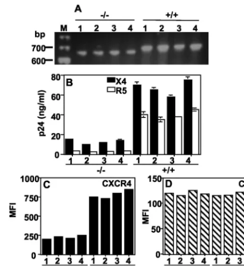
Reduced susceptibility to X4 HIV-1 infection of primary CD4ⴙcells isolated from CCR5⌬32homozygous individuals. A biological activity of CCR5⌬32 protein has been described as downmodulating CCR5 at the cell surface in a TDN manner (8, 13). The recent isolation of X4 (27) and R5X4 (28) from infected (⫺/⫺) individuals suggested that viral transmission occurred through CXCR4. However, why CXCR4 is rarely used in uninfected (⫺/⫺) individuals remains an unresolved question. To investigate this issue, we first compared the sus-ceptibility to X4 HIV-1 infection of CD4⫹cells purified from four HIV-uninfectedCCR5⌬32homozygotes (⫺/⫺) and from individuals carrying the homozygous wt CCR5allele (⫹/⫹). The genotypes of these individuals were confirmed by RT-PCR amplification of the fragment spanning the 32-bp deletion (Fig. 1A). CD4⫹cells purified from⫺/⫺or⫹/⫹individuals will be referred to as⫺/⫺or⫹/⫹CD4⫹cells. The⫹/⫹CD4⫹cells supported high levels of both X4 and R5 HIV infection. The ⫺/⫺ CD4⫹ cells did not support R5 HIV infection, as ex-pected, but unexpectedly were also markedly deficient com-pared to⫹/⫹cells in the ability to support X4 HIV infection (Fig. 1B). This correlated with a marked reduction of CXCR4 expression on the surfaces of⫺/⫺versus⫹/⫹cells by FACS, whereas CD4 levels were similar (Fig. 1C and D). We hypoth-esized thatCCR5⌬32exerts a TDN effect on CXCR4 expres-sion through expresexpres-sion of the CCR5⌬32 protein.
Stimulation of PBMCs reduces the percentage of CCR5ⴙ CXCR4ⴙ double-positive cells. To exert a TDN effect on
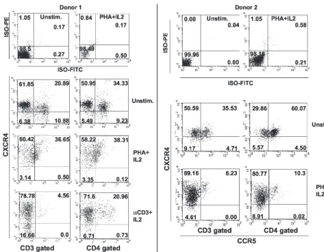
[image:3.603.301.541.68.328.2]CXCR4, the mutant CCR5⌬32 protein must be coexpressed in CXCR4⫹cells. Previous studies reported a low percentage of CCR5⫹ CXCR4⫹cells in CD3-gated, PHA-plus-IL-2-stimu-lated T lymphocytes (11). To readdress this issue, we per-formed three-color immunofluorescence flow cytometry on PBMCs from 10 healthy donors and found that the percentage of cells coexpressing CCR5 and CXCR4 is four- to fivefold higher in unstimulated PBMCs than in PHA and IL-2 blasts (Fig. 2 and Table 1). The percentage of double-positive cells depended on the cell population gated and the method of stimulation. For all donors tested, the percentage of double positives in CD3-gated cells was lower than in the CD4-gated cell population (Fig. 2 and Table 1). The reduction was donor dependent and also depended on the cell activation method. For seven donors, PHA plus IL-2 stimulation resulted in the redistribution of coreceptor expression, causing a dramatic reduction in the percentage of double-positive cells. All 10 donors tested showed a dramatic reduction in double-positive cells (CD3 gated) upon anti-CD3 plus IL-2 stimulation. The results demonstrate that the percentage of CCR5⫹CXCR4⫹ cells was consistently higher in unstimulated CD4-gated cells (34.33 to 76.5%) than in CD3-gated cells (16 to 43%). Table 1 also shows the percentages of CD4⫹CXCR4⫹cells that coex-press CCR5. These values may represent the percentages of CD4⫹ CXCR4⫹ cells that could potentially be affected by
FIG. 1. CD4⫹ cells from individuals homozygous for CCR5⌬32
(⫺/⫺) are resistant to infection with both X4 and R5 HIV. (A) The genotypes of the PBMC samples were verified by RT-PCR amplifica-tion of a fragment that spans the 32-bp deleamplifica-tion. CD4⫹cells purified
from four⫺/⫺and four⫹/⫹individuals were stimulated with PHA plus IL-2 for 3 days and were used in an HIV-1 infection assay (B) or for FACS analysis of CXCR4 (C) or CD4 (D). The p24 values in panel B represent amounts produced at day 9 postinfection. The results shown are from a single experiment representative of four indepen-dent experiments. MFI, mean fluorescence intensity; M, DNA size marker.
on November 8, 2019 by guest
http://jvi.asm.org/
coexpression of the CCR5⌬32 protein in⫺/⫺individuals. The results of this analysis demonstrate that the percentage of double-positive cells (CXCR4⫹ CCR5⫹) is higher than was previously thought and may explain the three- to fourfold drop in sensitivity to X4 virus infection shown in Fig. 1B.
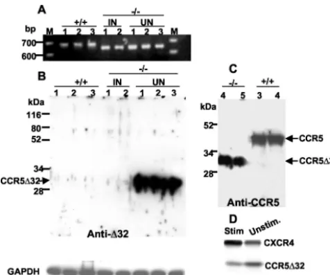
Endogenous expression of the truncated CCR5⌬32 protein. The genotypes of PBMCs isolated from⫺/⫺and⫹/⫹ individ-uals were confirmed by RT-PCR analysis using primers that span the 32-bp deletion (Fig. 3A). To analyze the potential protective effects of the CCR5⌬32 protein, we examined whether the truncated protein is expressed in CD4⫹cells pu-rified from ⫺/⫺ or⫹/⫹ PBMCs by using a polyclonal anti-serum raised against the frame-shifted 31 amino acid residues found in CCR5⌬32 but not in CCR5. The analysis revealed a high level of expression of the CCR5⌬32 protein, which ap-peared as a 30-kDa band on a Western blot of CD4⫹cells purified from PBMCs isolated from five different⫺/⫺ individ-uals (Fig. 3B and C). The mutant CCR5⌬32 protein was also detected by using polyclonal antibodies against the N-terminal
region of CCR5 (common to CCR5 and CCR5⌬32) (4) (Fig. 3C). Interestingly, the CCR5⌬32 protein was not significantly detected in CD4⫹cells purified from two different unrelated infected⫺/⫺individuals (Fig. 3B). As expected, we were
un-FIG. 2. CCR5⫹CXCR4⫹double-positive cells in unstimulated and stimulated PBMCs. Three-color flow cytometry of freshly isolated PBMCs
[image:4.603.54.527.64.432.2]was performed on either unstimulated cells or cells stimulated with PHA plus IL-2 or␣CD3 antibody plus IL-2. PBMCs were gated according to forward and side scatter and with either CD3-cychrome (HIT3Aa) or CD4-cychrome (RPA-T4) staining. The two-dimensional plots show expression of CCR5 (fluorescein isothiocyanate conjugate; 2D7 MAb) versus CXCR4 (PE conjugate; 12G5 MAb). Percentages of cells that are positive in the respective quadrants are indicated. Results for the respective isotype control antibodies are shown in the top two plots for each donor. The cell treatment is indicated to the right of each pair of plots, and gating is indicated at the bottom of each column of plots. The figure shows primary data from two different individuals and Table 1 summarizes the results from these two donors and eight others.
TABLE 1. Distribution of cells expressing both CCR5 and CXCR4 in resting and activated PBMCsa
Treatment
Mean % (range) of indicated cells in PBMC subset
CCR5⫹CXCR4⫹ CCR5⫹
CD3 gated CD4 gated CD4⫹gatedCXCR4⫹
Unstimulated 28.8 (16–43) 67.1 (34.33–76.5) 71.8 (40.26–81.25) PHA plus IL-2 14.8 (5–36.65) 20.4 (10.3–38.31) 22.6 (11.3–39.85) Anti-CD3 plus IL-2 5.6 (3–9.4) 14.3 (10.6–21.4) 15.8 (11.4–23.9)
aTen PBMC samples were used in a three-color staining procedure to
exam-ine the percentages of CCR5⫹CXCR4⫹cells in CD3- versus CD4-gated cells.
The races and identities of volunteers are unknown.
on November 8, 2019 by guest
http://jvi.asm.org/
[image:4.603.300.543.608.699.2]able to detect the protein in cells from any of the⫹/⫹subjects tested (Fig. 3B and C).
To examine the effect of cell activation on protein expres-sion, we compared the levels of the CCR5⌬32 protein made in PHA-plus-IL-2-stimulated cells to those in unstimulated CD4⫹ cells. The results indicated down-regulation of CCR5⌬32 ex-pression and up-regulation of CXCR4 exex-pression upon stim-ulation (Fig. 3D).
Construction of recombinant adenoviruses encoding either CCR5 or CCR5⌬32.For analysis of the apparent TDN effect of the CCR5⌬32 protein in more detail, the cDNA fragment encoding either CCR5 or CCR5⌬32 protein was inserted into the genome of Ad5 (Fig. 4A). We used this expression system because Ad5 viruses are replication defective in PBMCs and do not cause the cytopathic effect associated with the wt virus and because primary cells infected with these vectors can be infected with HIV-1 and monitored for p24 production. To demonstrate that these vectors can deliver the CCR5 or CCR5⌬32 gene into most of the infected cells, we used a similar Ad5/GFP vector encoding green fluorescent protein (GFP). We used 293 cells as a target for either infection with Ad5/GFP (10 PFU/cell) or transfection with a plasmid carrying
GFP under cytomegalovirus promoter. The results demon-strated that infection resulted in efficient GFP expression in most infected cells, whereas only 10% of the transfected cells expressed GFP (data not shown).
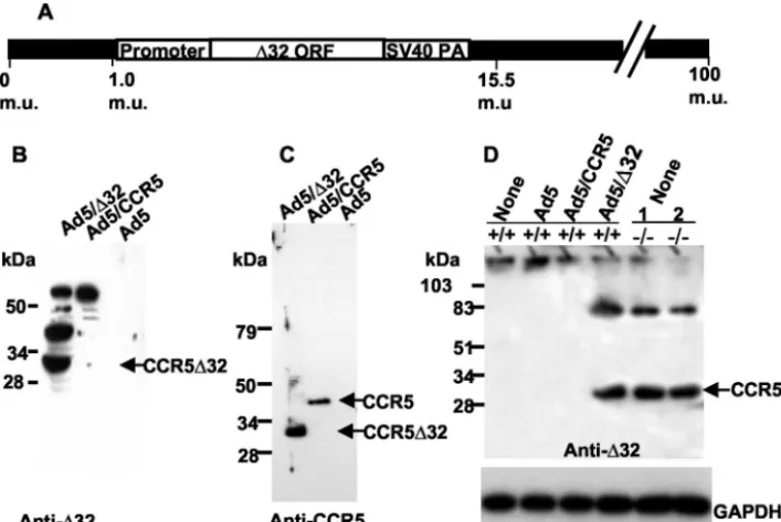
We found that the CCR5 protein encoded by Ad5/CCR5 is a functional receptor in terms of its coreceptor activity, as demonstrated by cell fusion assays. Immunoblot analysis of cellular lysates prepared from Ad5/⌬32-infected 293 cells re-vealed expression of the 30-kDa CCR5⌬32 protein, as detected with either CCR5⌬32-specific antibodies (Fig. 4B) or CCR5 antibodies directed against the common N terminus (Fig. 4C). Protein bands above the 34-kDa marker band were sometimes detected in CCR5⌬32-expressing 293 cells (Fig. 4B). These protein species could represent complexes between CCR5⌬32 molecules and cellular proteins or oligomers of the CCR5⌬32 protein itself. In order to determine that our adenovirus vector does not result in the overexpression of CCR5⌬32 protein, we demonstrated that the levels of Ad5-encoded CCR5⌬32 are comparable to those endogenously made in CD4⫹cells puri-fied from ⫺/⫺ PBMCs (Fig. 4D). As expected, uninfected CD4⫹cells or cells infected with Ad5/CCR5 or Ad5 did not show any protein band corresponding to the CCR5⌬32 pro-tein.
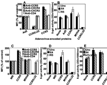
trans-downmodulation and impairment of HIV coreceptor activity.For examination of the TDN effect of Ad5-encoded CCR5⌬32 protein, CD4⫹cells from healthy donors (wt CCR5) were infected with wt Ad5, Ad5/⌬32, or Ad5/CCR5 and stained with MAbs against CCR5, CXCR4, CXCR2, or CD4. FACS analysis demonstrated that primary CD4⫹cells express-ing recombinant CCR5⌬32 protein, but not any other protein, showed specific downmodulation of CCR5 and CXCR4 (75 to 80% reduction) (Fig. 5A). Interestingly, expression of the CCR5⌬32 protein had no significant effect on cell surface CD4 or CXCR2, indicating the specificity of the downmodulation effect (Fig. 5A). In other experiments, we found that expres-sion of the CCR5⌬32 protein did not affect CCR4 or CCR2 expression (data not shown). These downmodulation results were confirmed by using CD4⫹ cells purified from PBMCs from 10 different donors.
[image:5.603.45.283.69.267.2]For analysis of the effect of CCR5⌬32-induced downmodu-lation on CCR5 coreceptor activity, the same CD4⫹cells used for Fig. 5A were mixed with HeLa cells expressing either R5 or X4 HIV-1 Env. The extent of Env-mediated fusion was mea-sured by a reporter gene activation assay (-Gal production). The results indicated that CCR5⌬32 expression impaired the ability of R5 and X4 Env proteins to fuse with CD4⫹cells (Fig. 5B). As expected, R5 and X4 Env-mediated fusion was not reduced by the expression of Ad5 proteins (Ad5), CCR5, or MVHA. To exclude the possibility that the TDN effect was due to the Ad5 vector system, we repeated the analysis with a recombinant vaccinia virus vector encoding the CCR5⌬32 pro-tein. The results confirmed those obtained with the Ad5 sys-tem (Fig. 5C and D). In contrast, the vaccinia virus-encoded CCR5⌬32 protein had no effect on fusion mediated by human T-cell leukemia virus type 1 Env, from a closely related retro-virus that recognizes a wide range of mammalian cell targets (41) (Fig. 5E). Thus, we were able to demonstrate the speci-ficity of the TDN CCR5⌬32 activity against CCR5 and CXCR4 in two different vector systems.
FIG. 3. Detection of endogenous CCR5⌬32 protein in unstimu-lated primary CD4⫹cells from CCR5⌬32 homozygotes. (A)
Geno-types were confirmed by RT-PCR amplification of the appropriate fragment of CCR5 (648 bp) or CCR5⌬32 (616 bp) from purified CD4⫹
cells from⫺/⫺and⫹/⫹individuals.⫺/⫺, the genotype of individuals homozygous for the CCR5⌬32 allele;⫹/⫹, individuals homozygous for the wt CCR5 allele. The CD4⫹ cells were purified from five
HIV-uninfected (⫺/⫺) donors (identified as UN 1, 2, and 3 in panel B and 4 and 5 in panel C), two different HIV-infected (⫺/⫺) individuals (labeled IN 1 and 2), and three HIV-uninfected individuals lacking CCR5⌬32. The CCR5⌬32 protein was detected by using anti-CCR5⌬32 (anti-⌬32), a polyclonal rabbit antiserum directed against the novel frame-shifted amino acids that specifically recognizes the mutant protein (B), or antibodies against the common N terminus of CCR5 and CCR5⌬32 proteins (C). Probing similar blots with preim-mune serum did not show any protein bands specific for the CCR5⌬32 protein (not shown). (D) Up-regulation of CXCR4 and down-regula-tion of CCR5⌬32 proteins upon PHA plus IL-2 stimulation of CD4⫹
cells (Stim). Unstim., unstimulated. The numbers at the left in panel C indicate the positions of protein standards. kDa, kilodaltons; M, DNA size marker.
on November 8, 2019 by guest
http://jvi.asm.org/
Analysis of CCR5⌬32 interaction with CCR5 and CXCR4. The yeast two-hybrid system has been used as a powerful genetic tool to rapidly select previously uncharacterized pro-teins and to identify novel components of signaling pathways. The expression of functional CXCR4 in yeast has recently been described (35). The yeast two-hybrid system has been previ-ously used to demonstrate CCR5⌬32 protein interaction with wt CCR5 (8). To control for nonspecific interactions, we used either empty vectors (Table 2, rows 1, 3, 4, and 5) or a vector that expressed a nonrelevant protein (pGADT-7-MVHA). A positive interaction was detected upon cotransformation with pGBKT7-53 plus pGBKT7-TAg (positive control), pGBKT-7-CCR5⌬32 plus pGADT-7-CCR5, and pGBKT7-CCR5⌬32 plus pGADT7-CXCR4, but not with pGBKT7-CCR5⌬32 plus pGADT7-CXCR2 (under high-stringency conditions), indicat-ing the specificity of the CCR5⌬32 protein interaction with the HIV coreceptors. A strong positive interaction under condi-tions of medium stringency had a colony count of⬎1,000. The colonies obtained with medium stringency were further analyzed, and the interaction was confirmed by using a -Gal colony-lift filter assay (high-stringency conditions). Strongly positive colonies appeared dark blue within 30 min. These results imply that the CCR5⌬32 protein binds directly with CXCR4 and CCR5.
To confirm the yeast two-hybrid system results, we analyzed CCR5⌬32 protein-coreceptor interactions by coimmunopre-cipitation. In order to detect CCR5⌬32-coreceptor hetero-dimers, we used 293-CD4 cells, which constitutively express CXCR4, coinfected with Ad5/⌬32 plus Ad5/CCR5 or Ad5/
[image:6.603.116.470.68.304.2]CCR5 plus Ad5. The cells were first examined for the CCR5⌬32 effect on HIV-1 Env fusion, and as expected, R5 and X4 fusion was dramatically inhibited by CCR5⌬32 protein expression but not by wt CCR5 expression (Fig. 6A). After confirming the
FIG. 4. Expression of CCR5⌬32 protein in 293 cells. (A) Structural map of a recombinant adenovirus (Ad5) encoding the⌬32 ORF. The CCR5⌬32 ORF, cloned from an HIV-seronegative CCR5⌬32 homozygote (⫺/⫺), is under the control of the major late promoter of adenovirus type 2 and the simian virus 40 poly(A) 3⬘(SV40 PA) processing signal. The adenovirus genome is represented as 100 map units (m.u.), with 365 bp/m.u. (B and C) Detection of Ad5-encoded CCR5⌬32 protein in infected 293 cells. Ad5-encoded CCR5⌬32 protein was detected by using anti-CCR5⌬32 antisera (anti-⌬32) directed against the frame-shifted domain of CCR5⌬32 (B) or antisera directed against the CCR5 N terminus (anti-CCR5) (C). Note that bands above the 34-kDa marker band are frequently detected with anti-CCR5⌬32 but not with anti-CCR5 antibodies. (D) Comparison of CCR5⌬32 protein levels made in Ad5/⌬32-infected CD4⫹cells with those endogenously made in⫺/⫺CD4⫹cells. The blot
was reprobed with antibodies against GAPDH to control for gel loading. PBMC samples from individuals homozygous for the CCR5⌬32 allele are referred to as⫺/⫺, and those from individuals homozygous for the wt CCR5 allele are referred to as⫹/⫹. Samples labeled as 1 and 2 are the same as samples 1 and 2 used in other figures. The numbers at the left indicate the positions of protein standards. kDa, kilodaltons.
TABLE 2. Interaction of CCR5⌬32 protein with CCR5 and CXCR4
DNA transformed (0.1g each)
Interaction under conditions indicateda
Medium
stringency stringencyHigh
pGBKT7-Lam plus pGADT7-T
(negative control) ⫺ ⫺
pGBKT-7-53 plus pGADT-7-T-Ag
(positive control) ⫹⫹⫹⫹⫹⫹ ⫹⫹⫹
pGBKT-7 plus pGADT-7-CXCR4 ⫺ ⫺
pGBKT-7 plus pGADT-7-CCR5 ⫺ ⫺
pGBKT-7-CCR5⌬32 plus pGADT-7 ⫺ ⫺ pGBKT-7-CCR5⌬32 plus pGADT-7-MVHA ⫺ ⫺ pGBKT-7-CCR5⌬32 plus pGADT-7-CCR5 ⫹⫹⫹⫹⫹ ⫹⫹ pGBKT-7-CCR5⌬32 plus pGADT-7-CXCR4 ⫹⫹⫹ ⫹ pGBKT-7-CCR5⌬32 plus pGADT-7-CXCR2 ⫾ ⫺
aA strong positive interaction (⫹⫹⫹⫹⫹⫹or⫹⫹⫹⫹⫹) at medium
strin-gency had a colony count of⬎1,000, whereas⫹⫹⫹indicates colony counts of ⬎500. The⫾plate had 25 colonies total, with 24 appearing on one plate and only 1 appearing on the duplicate plate. The colonies obtained at medium stringency were further analyzed, and interactions were confirmed by using a-Gal colony-lift filter assay (high stringency). Strongly positive colonies (⫹⫹⫹) appeared dark blue within 30 min.⫹⫹and⫹, colonies turned dark blue within 1 and 1.5 h, respectively. CXCR2 is the IL-8 receptor that shares 35% amino acid homol-ogy with CXCR4.
on November 8, 2019 by guest
http://jvi.asm.org/
[image:6.603.301.541.503.651.2]FIG. 5. Exogenous CCR5⌬32 specifically inhibits endogenous CCR5 and CXCR4 expression and HIV coreceptor activity in primary CD4⫹
cells. (A) Purified CD4⫹cells were infected with Ad5 vector, Ad5/⌬32, or Ad5/CCR5 and then stained for cell surface CCR5, CXCR4, CXCR2,
or CD4. Staining of cells infected with Ad5 was considered 100% staining. The matched isotype staining was calculated to be⬍3%. (B) Purified CD4⫹cells were infected with Ad5 vectors encoding the proteins indicated on thexaxis, challenged with HeLa cells expressing either the control
Unc, LAV (X4), or Ba-L (R5) HIV-1 Env, and examined for the extent of cell fusion by measuring-Gal production. (C) Purified CD4⫹cells were
infected with WR (a vaccinia virus vector control that does not encode any foreign protein) or recombinant vaccinia viruses encoding the proteins indicated on thexaxis and then were stained for cell surface CCR5, CXCR4, CCR2, or CD4. (D) Separate samples of CD4⫹cells were infected
with recombinant vaccinia viruses encoding the proteins indicated on thexaxis, challenged with HeLa cells expressing the same HIV-1 Env proteins as in panel B, and examined for-Gal production. (E) Some targets used in panel C were challenged with HeLa cells expressing the human T-cell leukemia virus type 1 Env, and the extent of cell fusion was measured similarly. In all of these experiments, fusion with Unc Env represents the background signal resulting from nonspecific cell fusion. The error bars represent replicates of the same experiment. The results shown represent 1 of 10 independent experiments using 10 different donors. MFI, mean fluorescence intensity.
FIG. 6. Heterodimerization of CCR5⌬32 protein with CCR5 and CXCR4. (A) 293-CD4 cells were coinfected with either Ad5/CCR5 plus Ad5 or Ad5CCR5 plus Ad5/⌬32 and challenged with HeLa cells expressing the indicated HIV-1 Envs. The error bars represent replicates of the same experiment. The results shown represent one of three independent experiments. (B) Coimmunoprecipitation analysis. 293-CD4 cells were infected with adenovirus encoding CCR5 (CCR5) plus adenovirus encoding CCR5⌬32 (⌬32) or control adenovirus (Ad5), as indicated at the top of each pair of lanes. Coinfected cells were lysed and immunoprecipitated with anti-CCR5⌬32 antibodies (anti-⌬32). Immunoprecipitates were resuspended in native PFO loading sample buffer and subjected to PFO–10% PAGE and sequential immunoblot analysis with antibodies specific for CCR5⌬32, CCR5, and CXCR4, as indicated at the bottom of each pair of lanes. wt CCR5 was detected with CTC-6 MAbs that do not react with CCR5⌬32 protein. The numbers on the left indicate the positions of molecular weight markers. kDa, kilodaltons. The identity of each band is indicated to the right.
on November 8, 2019 by guest
http://jvi.asm.org/
[image:7.603.112.470.489.640.2]CCR5⌬32 effect in our fusion assay, cell lysates were prepared and immunoprecipitated with anti-CCR5⌬32 antibodies and subjected to PFO-PAGE and blotting. Sequential probing with anti-CCR5⌬32, stripping and reprobing with anti-CCR5 (CTC-6; Protein Design Labs), and stripping and reprobing with anti-CXCR4 antibodies revealed the specific detection of high-molecular-weight bands that do not correspond to the sizes of CCR5⌬32, CCR5, and CXCR4 monomer bands (Fig. 6B). Detection of the same high-molecular-weight protein bands with different antibodies against CCR5⌬32, CCR5, and CXCR4 proteins suggested that complex formation occurred between CCR5⌬32 and either CCR5 or CXCR4. The results provide biochemical evidence for the interaction of the CCR5⌬32 protein with CCR5 and CXCR4.
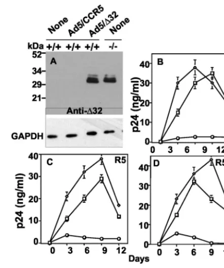
Expression of CCR5⌬32 protein confers resistance to di-verse HIV-1 strains. To show efficient gene delivery by our adenovirus vector system, we used a similar recombinant ade-novirus, Ad5/GFP, encoding GFP under the control of a cyto-megalovirus promoter. The infection of primary CD4⫹cells at 25 to 50 PFU/cell resulted in efficient GFP expression (green fluorescence) in most of the infected CD4⫹ cells (data not shown). This high efficiency of gene delivery allowed a detailed analysis of the biological activities of the CCR5⌬32 protein in primary cells. In order to examine⌬32 effects on HIV-1 infec-tion, healthy seronegative (⫹/⫹) CD4⫹cells were transduced to express Ad5-encoded CCR5 or Ad5-encoded CCR5⌬32 protein. The expression of CCR5⌬32 protein in this experi-ment was verified by Western blotting to show that the Ad5-encoded CCR5⌬32 protein was expressed at levels comparable to those endogenously made in ⫺/⫺ CD4⫹ cells (Fig. 7A). Purified CD4⫹cells expressing wt CCR5 protein were sensitive to HIV-1 infection and showed the expected levels of p24 protein. In contrast, CD4⫹ cells expressing Ad5-encoded CCR5⌬32 protein showed a dramatic inhibition of productive infection by X4 (Fig. 7B), R5 (Fig. 7C), and R5X4 isolates (Fig. 7D). Since primary⫹/⫹cells express endogenous CCR5, the expression of Ad5-encoded CCR5 was verified by FACS analysis of infected cells. The results indicated a twofold aug-mentation of CCR5 surface expression in Ad5/CCR5-infected cells (data not shown). These results are consistent with the CCR5⌬32 effects on Env fusion and provide evidence for a broad protective CCR5⌬32 effect in primary cells.
DISCUSSION
Our data demonstrate that the truncated mutant protein encoded by CCR5⌬32is endogenously expressed in PBMCs isolated fromCCR5⌬32homozygotes and is biologically active in primary CD4⫹cells. In particular, the protein exerts a dom-inant-negative effect on the expression of wt CCR5 and CXCR4, with the consequence that cells expressing it become less susceptible to infection with prototypic R5, R5X4, and X4 strains of HIV. The dominant-negative mechanism appears to involve direct binding of CCR5⌬32 to wt CCR5 and CXCR4. Taken together, our data suggest thatCCR5⌬32may geneti-cally restrict HIV pathogenesis in at least two ways, (i) by reducing in heterozygotes or preventing in homozygotes the production of normal CCR5 and (ii) by encoding a biologically
active mutant protein that is able to scavenge normal CCR5 and CXCR4.
[image:8.603.310.531.68.332.2]This is the first study to test the biological effects of recom-binant CCR5⌬32 protein in primary CD4⫹cells. The finding that CXCR4 expression is downregulated by CCR5⌬32 protein activity is relevant to disease progression and pathogenesis since CXCR4-using viruses emerge at the late symptomatic stage of AIDS. The novelty of our approach is the use of an efficient vector system that delivers the CCR5⌬32 gene to all cells under analysis. The observed downmodulation of CXCR4 is unlikely to be due to an artifact of massive, unregulated overexpression of the CCR5⌬32 protein, since firstly, this vector system is replication defective in primary cells, and secondly, our analysis indicated that the levels of CCR5⌬32
FIG. 7. Gene delivery of CCR5⌬32 into CD4⫹ cells from
HIV-seronegative CCR5 (⫹/⫹) individuals confers resistance to R5 and X4 HIV-1 infection. (A) Expression of CCR5⌬32 protein in CD4⫹cells.
Lysates of cells from individuals homozygous for wt CCR5 (⫹/⫹) or CCR5⌬32 (⫺/⫺) that had been infected with no virus (none) or ade-novirus encoding CCR5 (Ad/CCR5) or CCR5⌬32 (Ad5/⌬32) were analyzed by Western blotting using anti-CCR5⌬32-specific antiserum (anti-⌬32). Note that the levels of exogenous and endogenous CCR5⌬32 protein were similar in appropriate samples. GAPDH was monitored to assess equivalent loading of samples. The numbers at the left indicate the positions of protein standards. kDa, kilodaltons. (B to D) HIV replication kinetics in CD4⫹cells. Control cells were
incu-bated with no virus (none; open squares) and compared to cells in-fected with either Ad5/⌬32 (open circles) or Ad5/CCR5 (open trian-gles) at 50 PFU/cell for each virus with regard to productive infection by the X4 HIV-1 strain IIIB (B), the R5 strain Ba-L (C), and the R5X4 strain 89.6 (D). The amount of p24 antigen in the cell-containing supernatants was measured over time by enzyme-linked immunosor-bent assay. A zidovudine control infection resulted in p24 values below 1 ng/ml (not shown). The results shown are the means ⫾standard errors of the means from one experiment representative of five inde-pendent experiments using five different donors.
on November 8, 2019 by guest
http://jvi.asm.org/
protein driven by our vector were comparable to those endo-genously made in⫺/⫺CD4⫹cells.
The dominant-negative effect we describe can only occur in cells that coexpress CCR5 and CXCR4. Previous studies have demonstrated that CCR5 and CXCR4 are expressed pre-dominantly on CD4⫹ CD45RO⫹ and CD4⫹ CD45RA⫹ cells, respectively, and that CXCR4 is widely expressed on PBMCs, whereas CCR5 is more restricted, suggesting that the CCR5⌬32 protein would be expressed in only a small propor-tion of CXCR4⫹cells (11, 24). The results, however, depended on the analysis of a limited number of donors. We have found that donor variability and the method of stimulation are im-portant factors that influence the abundance of CXCR4⫹ CCR5⫹double-positive cells. Our study considered both CD3-and CD4-gated cells to show that different donors responded differently to cell activation, leading to dramatic differences in the percentages of double-positive cells. Donor variability in coreceptor expression has also been demonstrated in unstimu-lated PBMCs (23, 24). We consistently found that the CD4-gated cell population expressed a significantly higher percent-age of double-positive cells.
Identifying changes in CXCR4 levels in cells that coexpress CCR5 may be difficult since CXCR4 is widely expressed on human lymphocytes. A comparative analysis of CXCR4 ex-pression in a significant number of unstimulated CD4⫹cells isolated from ⫺/⫺ and ⫹/⫹ individuals has not previously been reported. Previous studies that performed CXCR4 stain-ing in⫺/⫺cells used total PBMCs from either one (46) or two (45) individuals. Although⫺/⫺PBMCs express CXCR4, the level of expression may vary with cell activation, and this may affect the degree to which the cells support X4 HIV replica-tion. In particular, we have shown that cell activation causes up-regulation of CXCR4 and reciprocal down-regulation of CCR5⌬32 protein in⫺/⫺CD4⫹T cells. Down-regulation of CCR5 and up-regulation of CXCR4 in wt CCR5 PBMCs have previously been reported to occur as a result of cell activation (11).
Previous studies reported different results in terms of HIV-1 X4 infection of⫺/⫺PBMCs. For example, reduced CXCR4 expression (compared to healthy wt CCR5 individuals) has been reported for two different⫺/⫺individuals and one⫹/⫺ individual (45). The lower level of CXCR4 staining correlated with resistance to X4 infection by the two⫺/⫺PBMC samples (45). The same ⫺/⫺ PBMC samples (45) were shown to be partially permissive for X4 infection upon challenge with a high-input X4 virus (15). Although CXCR4 staining has not been compared in CD4⫹cells from⫺/⫺individuals, the liter-ature contains a significant number of studies showing the resistance of⫺/⫺PBMCs to X4 infection (15, 33, 36, 40, 46). Other studies reported lower X4 infectivity in PBMCs isolated from two exposed, uninfected (⫺/⫺and⫹/⫺) individuals than in those from two unexposed, uninfected (⫹/⫹and ⫹/⫺) in-dividuals (45). Additionally, Salkowitz et al. (37) reported that the concentration of PHA used for PBMC activation is critical for the observation of resistance to the X4 NL4-3 isolate. Taken together, these studies indicated that individual vari-ability, virus input, and the method of cell activation have significantly contributed to the previously reported results re-garding X4 infection of⫺/⫺PBMCs.
None of the previous studies that analyzed the mechanism of
the⌬32 effects had utilized primary CD4⫹cells as targets for CCR5⌬32 protein expression (8, 13, 42). Those studies utilized different cell lines in their analyses of the TDN effect and reported opposite results (8, 13, 42). While Benkirane et al. (8) and Chelli et al. (13) reported a partial TDN effect for the mutant CCR5⌬32 protein against R5 infection and/or fusion, Venkatesan et al. (42) reported no such effect. We believe that these discrepant findings were probably a consequence of vari-able transfection efficiencies that resulted in a large number of cells lacking expression of mutant CCR5⌬32 proteins but ex-pressing abundant levels of coreceptors. Therefore, examining the inhibitory effect of CCR5⌬32 under these conditions may not reveal the actual activity of the mutant protein. The two studies that reported CCR5⌬32 protein activity against R5 did not detect the TDN effect against X4 (8, 13). The reasons for this could be poor expression of the mutant protein by trans-fection and the use of cell lines that express endogenous CXCR4. Low levels of exogenous mutant protein expression may be insufficient to induce a detectable TDN effect on en-dogenous CXCR4.
All G-protein-coupled receptors (GPCRs) are predicted to share a similar molecular structure characterized by the pres-ence of seven transmembrane helices connected by three in-tracellular and three exin-tracellular loops (9). Functionally de-fective, naturally occurring mutations in GPCRs, including CCR5⌬32, have been reported to cause impaired processing and intracellular retention. Previous studies on members of this family have suggested that 7TM receptors in some cases may exist in oligomeric forms (14, 20, 32, 44). Zhu et al. have demonstrated that the truncated mutant vasopressin receptors are able to form heterodimers with the full-length receptor and that this complex formation inhibited wt receptor function (49). The same study found that truncated vasopressin recep-tor mutants specifically inhibited the function and cell surface trafficking of the coexpressed full-length vasopressin receptor but had no effect on the function of other GPCRs. An analysis of the domains of CCR5 and CXCR4 that interact with the mutant protein will probably lead to the identification of com-mon epitopes that will have implications for the design of drugs that down-regulate the major HIV coreceptors.
Our study suggests that CXCR4 expression is limiting for viral entry (Fig. 5) and that there may be an optimum number and density of coreceptor molecules for HIV-1 infection, which is in agreement with studies from Kabat’s group (22). By ex-tension, the CCR5⌬32/CXCR4 and CCR5⌬32/CCR5 ratios may be critical determinants for the inhibition of X4 and R5 HIV infection, respectively, by the mutant protein. Previous studies have reported that protection against HIV-1 in het-erozygotes might depend on the ratio of wt to mutant CCR5 mRNA (31). However, detailed analysis of the CCR5⌬32/ CXCR4 and CCR5⌬32/CCR5 protein ratios in these individ-uals will be necessary to confirm such findings.
Despite the absence of CCR5 in⫺/⫺individuals, CXCR4 is rarely used as an alternate coreceptor to infect CD4⫹ cells. Macrophages have classically been regarded as relatively resis-tant to X4; however, some primary X4 isolates are able to infect macrophages via CXCR4 (38, 43). Recently, Naif et al. described an R5X4 isolate capable of infecting ⫺/⫺ macro-phages through CXCR4 (28). Furthermore, a number of pri-mary syncytium-inducing HIV-1 strains were found to be of the
on November 8, 2019 by guest
http://jvi.asm.org/
R5X4 type, which can utilize both CCR5 and CXCR4 and have the ability to infect primary macrophages in vitro (7, 39, 47). Our results indicate that two of these infected⫺/⫺individuals lacked expression of the CCR5⌬32 protein. We hypothesize that the absence of detectable CCR5⌬32 protein in infected ⫺/⫺individuals resulted in the loss of the TDN effect against CXCR4, leading to X4 infection. The lack of CCR5⌬32 pro-tein expression may be explained by a defect either in tran-scription or translation. These possibilities are currently under investigation.
In summary, this study provides the first evidence that ex-pression of recombinant CCR5⌬32 protein in human PBMCs confers broad protection against R5 and X4 strains of HIV-1. We have shown that the truncated CCR5⌬32 protein can act as a negative regulator of wt CCR5 and CXCR4. The dominant-negative activity of the mutant CCR5⌬32 protein correlated with its ability to reduce the cell surface expression of the major HIV coreceptors and to form heterodimeric complexes with CCR5 and CXCR4. Understanding the molecular nature of the protective effects of the CCR5⌬32 protein may lead to new insights into the interrelationships of these molecules that are employed by HIV-1 and perhaps to the development of novel therapeutic agents capable of conferring broad antiviral effects. New insights gained in the mechanism of action of this naturally occurring protein may be incorporated into new ap-proaches to induce resistance to HIV-1.
ACKNOWLEDGMENTS
We thank Hal Broxmeyer, S. Spinola, and A. Srivastava for their comments on the manuscript, Hassan Naif, Haynes Sheppard, and Susan Buchbinder for providing PBMC samples from infected⫺/⫺ individuals, and Frank Graham for providing Ad5Pol3. The following reagents were obtained through the AIDS Research and Reference Reagent Program, Division of AIDS, NIAID, NIH: (i) recombinant human IL-2 from Maurice Gately, Hoffmann-La Roche, Inc.; (ii) HIV-1 89.6 from Ronald Collman; (iii) HIV-1 Ba-L from Suzanne Gartner, Mikulas Popovic, and Robert Gallo; and (iv) HIV-1 IIIB from Robert Gallo.
This study was supported by NIH grant A152019-01A1 to G.A. PBMC samples were obtained under Indiana University IRB study 9812-05.
REFERENCES
1. Agrawal, L., and G. Alkhatib.2001. Chemokine receptors: emerging oppor-tunities for new anti-HIV therapies. Expert Opin. Ther. Targets5:303–326. 2. Alkhatib, G., and D. J. Briedis.1988. High-level eucaryotic in vivo expression of biologically active measles virus hemagglutinin by using an adenovirus type 5 helper-free vector system. J. Virol.62:2718–2727.
3. Alkhatib, G., C. Combadiere, C. C. Broder, Y. Feng, P. E. Kennedy, P. M. Murphy, and E. A. Berger. 1996. CC CKR5: a RANTES, MIP-1alpha, MIP-1beta receptor as a fusion cofactor for macrophage-tropic HIV-1. Sci-ence272:1955–1958.
4. Alkhatib, G., M. Locati, P. E. Kennedy, P. M. Murphy, and E. A. Berger. 1997. HIV-1 coreceptor activity of CCR5 and its inhibition by chemokines: independence from G protein signaling and importance of coreceptor down-modulation. Virology234:340–348.
5. Alkhatib, G., B. Massie, and D. J. Briedis.1988. Expression of bicistronic measles virus P/C mRNA by using hybrid adenoviruses: levels of C protein synthesized in vivo are unaffected by the presence or absence of the up-stream P initiator codon. J. Virol.62:4059–4069.
6. Alkhatib, G., C. Richardson, and S. H. Shen.1990. Intracellular processing, glycosylation, and cell-surface expression of the measles virus fusion protein (F) encoded by a recombinant adenovirus. Virology175:262–270. 7. Bazan, H. A., G. Alkhatib, C. C. Broder, and E. A. Berger.1998. Patterns of
CCR5, CXCR4, and CCR3 usage by envelope glycoproteins from human immunodeficiency virus type 1 primary isolates. J. Virol.72:4485–4491. 8. Benkirane, M., D. Y. Jin, R. F. Chun, R. A. Koup, and K. T. Jeang.1997.
Mechanism of transdominant inhibition of CCR5-mediated HIV-1 infection by ccr5delta32. J. Biol. Chem.272:30603–30606.
9. Berger, E. A., P. M. Murphy, and J. M. Farber.1999. Chemokine receptors as HIV-1 coreceptors: roles in viral entry, tropism, and disease. Annu. Rev. Immunol.17:657–700.
10. Bett, A. J., W. Haddara, L. Prevec, and F. L. Graham.1994. An efficient and flexible system for construction of adenovirus vectors with insertions or deletions in early regions 1 and 3. Proc. Natl. Acad. Sci. USA91:8802–8806. 11. Bleul, C. C., L. Wu, J. A. Hoxie, T. A. Springer, and C. R. Mackay.1997. The HIV coreceptors CXCR4 and CCR5 are differentially expressed and regu-lated on human T lymphocytes. Proc. Natl. Acad. Sci. USA94:1925–1930. 12. Broder, C. C., and P. L. Earl.1999. Recombinant vaccinia viruses. Design,
generation, and isolation. Mol. Biotechnol.13:223–245.
13. Chelli, M., and M. Alizon.2001. Determinants of the trans-dominant nega-tive effect of truncated forms of the CCR5 chemokine receptor. J. Biol. Chem.276:46975–46982.
14. Chidiac, P., M. A. Green, A. B. Pawagi, and J. W. Wells.1997. Cardiac muscarinic receptors. Cooperativity as the basis for multiple states of affinity. Biochemistry36:7361–7379.
15. Connor, R. I., W. A. Paxton, K. E. Sheridan, and R. A. Koup.1996. Macro-phages and CD4⫹T lymphocytes from two multiply exposed, uninfected
individuals resist infection with primary non-syncytium-inducing isolates of human immunodeficiency virus type 1. J. Virol.70:8758–8764.
16. Dean, M., M. Carrington, C. Winkler, G. A. Huttley, M. W. Smith, R. Allikmets, J. J. Goedert, S. P. Buchbinder, E. Vittinghoff, E. Gomperts, S. Donfield, D. Vlahov, R. Kaslow, A. Saah, C. Rinaldo, R. Detels, and S. J. O’Brien.1996. Genetic restriction of HIV-1 infection and progression to AIDS by a deletion allele of the CKR5 structural gene. Hemophilia Growth and Development Study, Multicenter AIDS Cohort Study, Multicenter He-mophilia Cohort Study, San Francisco City Cohort, ALIVE Study. Science 273:1856–1862. (Erratum,274:1069.)
17. Feng, Y., C. C. Broder, P. E. Kennedy, and E. A. Berger.1996. HIV-1 entry cofactor: functional cDNA cloning of a seven-transmembrane, G protein-coupled receptor. Science272:872–877.
18. Graham, F. L.1990. Adenoviruses as expression vectors and recombinant vaccines. Trends Biotechnol.8:85–87.
19. Graham, F. L., J. Smiley, W. C. Russell, and R. Nairn.1977. Characteristics of a human cell line transformed by DNA from human adenovirus type 5. J. Gen. Virol.36:59–74.
20. Hirschberg, B. T., and M. I. Schimerlik.1994. A kinetic model for oxotremo-rine M binding to recombinant porcine m2 muscarinic receptors expressed in Chinese hamster ovary cells. J. Biol. Chem.269:26127–26135.
21. Huang, Y., W. A. Paxton, S. M. Wolinsky, A. U. Neumann, L. Zhang, T. He, S. Kang, D. Ceradini, Z. Jin, K. Yazdanbakhsh, K. Kunstman, D. Erickson, E. Dragon, N. R. Landau, J. Phair, D. D. Ho, and R. A. Koup.1996. The role of a mutant CCR5 allele in HIV-1 transmission and disease progression. Nat. Med.2:1240–1243.
22. Kuhmann, S. E., E. J. Platt, S. L. Kozak, and D. Kabat.2000. Cooperation of multiple CCR5 coreceptors is required for infections by human immuno-deficiency virus type 1. J. Virol.74:7005–7015.
23. Lee, B., B. J. Doranz, S. Rana, Y. Yi, M. Mellado, J. M. Frade, A. C. Martinez, S. J. O’Brien, M. Dean, R. G. Collman, and R. W. Doms.1998. Influence of the CCR2-V64I polymorphism on human immunodeficiency virus type 1 coreceptor activity and on chemokine receptor function of CCR2b, CCR3, CCR5, and CXCR4. J. Virol.72:7450–7458.
24. Lee, B., M. Sharron, L. J. Montaner, D. Weissman, and R. W. Doms.1999. Quantification of CD4, CCR5, and CXCR4 levels on lymphocyte subsets, dendritic cells, and differentially conditioned monocyte-derived macro-phages. Proc. Natl. Acad. Sci. USA96:5215–5220.
25. Liu, R., W. A. Paxton, S. Choe, D. Ceradini, S. R. Martin, R. Horuk, M. E. MacDonald, H. Stuhlmann, R. A. Koup, and N. R. Landau.1996. Homozy-gous defect in HIV-1 coreceptor accounts for resistance of some multiply-exposed individuals to HIV-1 infection. Cell86:367–377.
26. Michael, N. L., and J. P. Moore.1999. HIV-1 entry inhibitors: evading the issue. Nat. Med.5:740–742.
27. Michael, N. L., J. A. Nelson, V. N. KewalRamani, G. Chang, S. J. O’Brien, J. R. Mascola, B. Volsky, M. Louder, G. C. White II, D. R. Littman, R. Swanstrom, and T. R. O’Brien.1998. Exclusive and persistent use of the entry coreceptor CXCR4 by human immunodeficiency virus type 1 from a subject homozygous for CCR5⌬32. J. Virol.72:6040–6047.
28. Naif, H. M., A. L. Cunningham, M. Alali, S. Li, N. Nasr, M. M. Buhler, D. Schols, E. de Clercq, and G. Stewart.2002. A human immunodeficiency virus type 1 isolate from an infected person homozygous for CCR5⌬32 exhibits dual tropism by infecting macrophages and MT2 cells via CXCR4. J. Virol. 76:3114–3124.
29. Nussbaum, O., C. C. Broder, and E. A. Berger.1994. Fusogenic mechanisms of enveloped-virus glycoproteins analyzed by a novel recombinant vaccinia virus-based assay quantitating cell fusion-dependent reporter gene activa-tion. J. Virol.68:5411–5422.
30. O’Brien, S. J., and J. P. Moore.2000. The effect of genetic variation in chemokines and their receptors on HIV transmission and progression to AIDS. Immunol. Rev.177:99–111.
31. Ometto, L., M. Zanchetta, A. Cabrelle, G. Esposito, M. Mainardi, L. Chieco-Bianchi, and A. De Rossi.1999. Restriction of HIV type 1 infection in
on November 8, 2019 by guest
http://jvi.asm.org/
macrophages heterozygous for a deletion in the CC-chemokine receptor 5 gene. AIDS Res. Hum. Retrovir.15:1441–1452.
32. Potter, L. T., L. A. Ballesteros, L. H. Bichajian, C. A. Ferrendelli, A. Fisher, H. E. Hanchett, and R. Zhang.1991. Evidence of paired M2 muscarinic receptors. Mol. Pharmacol.39:211–221.
33. Quillent, C., E. Oberlin, J. Braun, D. Rousset, G. Gonzalez-Canali, P. Me-tais, L. Montagnier, J. L. Virelizier, F. Arenzana-Seisdedos, and A. Beretta. 1998. HIV-1-resistance phenotype conferred by combination of two separate inherited mutations of CCR5 gene. Lancet351:14–18.
34. Ramjeesingh, M., L. J. Huan, E. Garami, and C. E. Bear.1999. Novel method for evaluation of the oligomeric structure of membrane proteins. Biochem. J.342:119–123.
35. Sachpatzidis, A., B. K. Benton, J. P. Manfredi, H. Wang, A. Hamilton, H. G. Dohlman, and E. Lolis.2003. Identification of allosteric peptide agonists of CXCR4. J. Biol. Chem.278:896–907.
36. Salkowitz, J. R., S. F. Purvis, H. Meyerson, P. Zimmerman, T. R. O’Brien, L. Aledort, M. E. Eyster, M. Hilgartner, C. Kessler, B. A. Konkle, G. C. White II, J. J. Goedert, and M. M. Lederman.2001. Characterization of high-risk HIV-1 seronegative hemophiliacs. Clin. Immunol.98:200–211. 37. Samson, M., F. Libert, B. J. Doranz, J. Rucker, C. Liesnard, C. M. Farber,
S. Saragosti, C. Lapoumeroulie, J. Cognaux, C. Forceille, G. Muyldermans, C. Verhofstede, G. Burtonboy, M. Georges, T. Imai, S. Rana, Y. Yi, R. J. Smyth, R. G. Collman, R. W. Doms, G. Vassart, and M. Parmentier.1996. Resistance to HIV-1 infection in Caucasian individuals bearing mutant al-leles of the CCR-5 chemokine receptor gene. Nature382:722–725. 38. Simmons, G., J. D. Reeves, A. McKnight, N. Dejucq, S. Hibbitts, C. A. Power,
E. Aarons, D. Schols, E. De Clercq, A. E. Proudfoot, and P. R. Clapham. 1998. CXCR4 as a functional coreceptor for human immunodeficiency virus type 1 infection of primary macrophages. J. Virol.72:8453–8457. 39. Simmons, G., D. Wilkinson, J. D. Reeves, M. T. Dittmar, S. Beddows, J.
Weber, G. Carnegie, U. Desselberger, P. W. Gray, R. A. Weiss, and P. R. Clapham.1996. Primary, syncytium-inducing human immunodeficiency virus type 1 isolates are dual-tropic and most can use either Lestr or CCR5 as coreceptors for virus entry. J. Virol.70:8355–8360.
40. Sol, N., F. Ferchal, J. Braun, O. Pleskoff, C. Treboute, I. Ansart, and M. Alizon.1997. Usage of the coreceptors CCR-5, CCR-3, and CXCR-4 by primary and cell line-adapted human immunodeficiency virus type 2. J. Virol. 71:8237–8244.
41. Sutton, R. E., and D. R. Littman.1996. Broad host range of human T-cell
leukemia virus type 1 demonstrated with an improved pseudotyping system. J. Virol.70:7322–7326.
42. Venkatesan, S., A. Petrovic, D. I. Van Ryk, M. Locati, D. Weissman, and P. M. Murphy.2002. Reduced cell surface expression of CCR5 in CCR5⌬32 heterozygotes is mediated by gene dosage rather than receptor sequestra-tion. J. Biol. Chem.277:2287–2301.
43. Verani, A., E. Pesenti, S. Polo, E. Tresoldi, G. Scarlatti, P. Lusso, A. G. Siccardi, and D. Vercelli.1998. CXCR4 is a functional coreceptor for infec-tion of human macrophages by CXCR4-dependent primary HIV-1 isolates. J. Immunol.161:2084–2088.
44. Wreggett, K. A., and J. W. Wells.1995. Cooperativity manifest in the binding properties of purified cardiac muscarinic receptors. J. Biol. Chem.270: 22488–22499.
45. Wu, L., W. A. Paxton, N. Kassam, N. Ruffing, J. B. Rottman, N. Sullivan, H. Choe, J. Sodroski, W. Newman, R. A. Koup, and C. R. Mackay.1997. CCR5 levels and expression pattern correlate with infectability by macrophage-tropic HIV-1, in vitro. J. Exp. Med.185:1681–1691.
46. Xiao, L., S. H. Weiss, S. H. Qari, D. Rudolph, C. Zhao, T. N. Denny, T. Hodge, and R. B. Lal.1999. Partial resistance to infection by R5X4 primary HIV type 1 isolates in an exposed-uninfected individual homozygous for CCR5 32-base pair deletion. AIDS Res. Hum. Retrovir.15:1201–1208. 47. Yi, Y., S. Rana, J. D. Turner, N. Gaddis, and R. G. Collman.1998. CXCR-4
is expressed by primary macrophages and supports CCR5-independent in-fection by dual-tropic but not T-tropic isolates of human immunodeficiency virus type 1. J. Virol.72:772–777.
48. Zhou, Y., T. Kurihara, R. P. Ryseck, Y. Yang, C. Ryan, J. Loy, G. Warr, and R. Bravo.1998. Impaired macrophage function and enhanced T cell-depen-dent immune response in mice lacking CCR5, the mouse homologue of the major HIV-1 coreceptor. J. Immunol.160:4018–4025.
49. Zhu, X., and J. Wess.1998. Truncated V2 vasopressin receptors as negative regulators of wild-type V2 receptor function. Biochemistry37:15773–15784. 50. Zimmerman, P. A., A. Buckler-White, G. Alkhatib, T. Spalding, J. Kubofcik, C. Combadiere, D. Weissman, O. Cohen, A. Rubbert, G. Lam, M. Vac-carezza, P. E. Kennedy, V. Kumaraswami, J. V. Giorgi, R. Detels, J. Hunter, M. Chopek, E. A. Berger, A. S. Fauci, T. B. Nutman, and P. M. Murphy. 1997. Inherited resistance to HIV-1 conferred by an inactivating mutation in CC chemokine receptor 5: studies in populations with contrasting clinical phenotypes, defined racial background, and quantified risk. Mol. Med.3:23– 36.