Saxitoxin binding proteins: biological perspectives
242
0
0
Full text
(2) SAXITOXIN BINDING PROTEINS: BIOLOGICAL PERSPECTIVES. Thesis submitted by ALISON ROBERTSON BSc (Hons). In March, 2005. For the degree of Doctor of Philosophy. In the School of Pharmacy and Molecular Sciences JAMES COOK UNIVERSITY Townsville, Queensland, Australia.. In collaboration with: THE AUSTRALIAN INSTUTUTE OF MARINE SCIENCE Marine Biotechnology Townsville, Queensland, Australia.. Jl/O//.
(3) STATEMENT OF ACCESS. I, the undersigned author of this work, understand that James Cook University will make this thesis available for use within the University Library and, via the Australian Digital Theses network, for . use elsewhere. I understand that, as an unpublished work, a thesis has significant protection under the Copyright Act and;. I do not wish to place any further restriction on access to this work. :Z07 N Ociober .?W5 Signature. Date.
(4) ELECTRONIC COPY. I, the undersigned, the author of this work, declare that the electronic copy of this thesis provided to the James Cook University Library, is an accurate copy of the print thesis submitted, within the limits of the technology available.. Signature. Date.
(5) STATEMENT OF SOURCES DECLARATION. I certify that this thesis is my own work and has not been admitted in any other form for another degree or diploma at any university or other institution of tertiary education. Information derived from the published or unpublished works of others has been acknowledged in the text and a list ofreferences is provided.. ALISON ROBERTSON. iii.
(6) DECLARATION ON ETHICS The research presented and reported in this thesis was conducted within the guidelines for research ethics outlined in the Joint NHMRCIAVCC Statement and Guidelines on Research Practice (1997), the James Cook University Policy on Experimentation Ethics, Standard Practices and Guidelines (2001), and the James Cook University Statement and Guidelines on Research Practice (2001).. All components of the project and reporting procedures were in compliance with the Australian Code of Practice for the Care and Use of Animals for Scientific Purposes, and the Queensland Animal Care and Protection Act (2001). The research methodology received clearance from the James Cook University Experimentation Ethics Review Committee (approval number A745-02).. ALISON ROBERTSON. IV.
(7) STATEMENT ON THE CONTRIBUTION OF OTHERS. Financial. I am very grateful for the considerable financial support that was provided by the following organisations over the course of my candidature:. An Australian Postgraduate Award (APA), granted by James Cook University (JCU) and financed by the federal Department of Science, Education and Training (DEST), provided a generous stipend and was greatly appreciated.. I am indebted to the School of Pharmacy and Molecular Sciences, JCU, who financed bench fees, transport and overheads incurred from my fulltime placement at the Australian Institute of Marine Science (AIMS), allowing me the opportunity to work in a world class marine research institution.. Finally I would like to express my appreciation to AIMS for providing cutting edge research facilities, and a considerable postgraduate research award which covered research consumables and equipment purchases over the course of candidature.. Collaborations. I am sincerely grateful to Dr. David Stirling from the Institute of Environmental Science & Research Limited, Porirua, New Zealand for his collaboration and insights. David performed preliminary MS analysis of several octopi extracts and contributed to an accepted manuscript as detailed in Chapter 4.. I am also grateful and humbled by the generosity of Dr. David Rowell from the Australian National University (ANU) who provided his time, transport, permits and experience to assist me on a field collection of log dwelling invertebrates from the Tallaganda State forest in NSW.. v.
(8) Bio-molecular Analysis Services •. The Australian Proteome Analysis Facility (APAF) performed tryptic digests of purified saxitoxin binding protein and subsequent peptide sequencing by manual QTOF mass spectrometry, on a fee for service basis.. •. Likewise, nucleotide sequencing was performed by Dr. Lynn Woodward from the Genetic Analysis Facility (GAF) in the Advanced Analytical Centre (AAC) at JCU.. All other personal and scientific contributions are listed as acknowledgements for each relevant thesis chapter.. ALISON ROBERTSON. VI.
(9) ACKNOWLEDGEMENTS I would like to thank my supervisors Dr. Lyndon Llewellyn and Dr. Andrew Negri from the Australian Institute of Marine Science and Prof. Jim Burnell from James Cook University for providing me with the opportunity to embark upon this PhD research and for believing in me. Thanks to Lyndon who's greatest attribute is his love of science. I appreciate you letting me in to the world of toxins and proteins and hope that one day we can solve this saxiphilin riddle. Thanks also to Jim for administrative assistance, encouragement and molecular biology input. Special thanks to Andrew, who was a continual source of friendship, motivation, advice and encouragement. Thanks for holding the torch at the end of the long winding tunnel, whenever I lost sight of the goal.. I would also like to acknowledge the laughs, friendship, support and encouragement of everyone in the Marine Biotechnology group at AIMS. It was a wonderful opportunity to work with and learn from such a great group of people. To my office buddies, Carsten, Neil, Enrique and Carlos for the laughs and friendship.. I would like to thank Dr. Cherie Motti for being a impartial sounding board to new ideas, for helping me troubleshoot new chemistry methods and equipment and for reviewing some of my early manuscripts with humility. I would also like to thank Dr. Diane Tapiolas who has been a constant source of encouragement and support and for urging me to seek out post doctoral opportunities overseas. Thanks to Dr. Rick Willis who invited me into the world of Mass Spectrometry and was always willing to have a bash at some new and interesting samples. Special thanks to Dr. Cedric Robillot for his friendship, interest and support of my project and training and assistance with newly commissioned LC and LCMS equipment.. Thanks to Dr. Kate Wilson for her SUppOlt, ideas, molecular biology advice and wealth of experience which really transformed my approach to the partial sequencing of saxiphilin and to Jennie Swan for showing me that molecular. vii.
(10) biology could be fun. Thanks also to Lesa, Enrique, Dave and Nicole for fielding my many questions on molecular strategies. I am grateful to Jason, Lyndon and Rick who enthusiastically provided me with a steady stream of toads when I needed them and to all who assisted with field collections. Particular thanks go to Mr. Carsten Wolff (AIMS), Dr. John Collins (JCU), Dr. Mark Norman and Mr. Julian Finn who assisted with taxonomy following extensive field collections.. Appreciation is extended to all staff from AIMS including members from science, IT, administration, stores, purchasing, accommodation, security, finance, marine operations, library, registry, and human resources for their kindness, laughs and assistance in day to day activities. I would particularly like to acknowledge the assistance of Gavin, Carsten and Darren who helped me resurrect my PC and sanity through four successive hard drive crashes and a multitude of computer related dramas.. I deeply appreciate the support and friendship of Dr. Anna Marie Babey. Thanks for the independent advice and perspective during tough times.. To my beloved family Heather, Jim and Bruce, thank you all for your unwavering love and encouragement which has made the vast distances between us disappear.. Last, but certainly not least, I would like to thank Terry who has been by my side for the whole journey, through the highs and lows with his unconditional love and support. Life has hit you with some tough blows but I am constantly amazed at you inner strength and determination. There are only great things to look forward to from now on.. viii.
(11) PUBLICATIONS ARISING FROM THESIS Each research chapter of this thesis will stand alone as at least one peer reviewed publication. as. detailed. below.. Additional. publications. and. conference. presentations indirectly relating to my thesis research are detailed as additional outcomes in Chapter 6.. Thesis Research Papers Robertson, A., Stirling, D., Robillot, C., Llewellyn, L., Negri, A.P. (2004). First report of saxitoxin in octopi. Toxicon, 44, 765- 771.. Robertson, A., Llewellyn, L. E., Negri, A. P. (2005) Development of a. robust radio-receptor assay for discovery and screening of novel saxitoxin binding. proteins. and. paralytic. shellfish toxins.. Target Journal:. Environmental Science & Technology (in preparation).. Robertson, A., Negri, A.P., Llewellyn, L.E. (2005) Extending the. phylogenetic distribution of saxitoxin binding proteins in search of functionality. Target Journal: Toxicon (in preparation).. Robertson, A., Llewellyn, L.E., Negri, A.P. (2005) Survey of a tropical. saxitoxin "hotspot": Port Hedland, Western Australia. Target Journal:. Marine Biology (in preparation).. Robertson, A., Swan, J., Negri, A. P., Burnell, J., Llewellyn, L., Wilson, K. (2005) Purification and partial sequencing of a saxitoxin binding. protein from Bufo marinus plasma. Target Journal: Biochemistry (in preparation).. Robertson, A., Motti, C.A., Negri, A. P., Llewellyn, L.E. (2004) Evidence. for endogenous ligands of saxiphilin in Bufo marinus. Target Journal:. Analytical Chemistry (in preparation).. IX.
(12) Thesis Conference Abstracts Robertson, A. (2002) Paralytic shellfish toxin "hotspots" The Port. Hedland Story. Oral presentation at 4th Workshop of the Australian. Research Network for Algal Toxins (ARNAT), Townsville, July 2002. (PhD research).. Robertson, A. and Llewellyn, L. E. (2002) Saxitoxin Binding Proteins: A. Unique Suite of Receptors. Poster presented at ComBio 2002, Sydney, October 2002. (PhD research).. x.
(13) ABSTRACT Saxitoxin binding protein (STXBP) is a functional classification which describes all proteins capable of binding to the paralytic shellfish toxin (PST), saxitoxin (STX). Based on this functionality, this group includes the voltage gated sodium channels (VGSCs), pufferfish STX and tetrodotoxin (TTX) binding proteins (PSTBPs) and saxiphilin (SXPN) which was been isolated from the amphibian. Rana catesbeiana. Various activities and relationships of bullfi'og SXPN have been elucidated including the ability to inhibit papain, human cathepsin Band L and the substantial homology of the amino acid sequence to transferrins (TFs). However, the biological role of SXPN has not been thoroughly examined and remains a mystery. It is likely that a detoxification mechanism exists in animals exposed to PSTs, and may explain the defined STX binding activity of soluble STXBPs. Therefore, the main objective of this thesis was to examine various aspects of the biological relationship between STX and soluble STXBPs to determine whether these proteins provide a defensive arsenal against PST intoxication.. Preliminary studies indicated that current methods for the detection of STXBPs were problematic and time intensive therefore several radio-receptor assays were developed and trialled to identify a suitable primary screening regimen for the detection and characterisation of these proteins. Assays utilising anion and cation exhange methods, protein binding and traditional charcoal radio-receptor methods were compared to published formats. A receptor binding filtration assay incorporating protein binding membranes of mixed cellulose esters (MCE) proved to be a robust method for the sensitive and accurate detection of STXBPs. This assay was easily converted for use as a PST screening tool and was validated in subsequent chapters using Bufo marinus plasma which is readily obtainable source of STXBPs.. With the aid of this optimised assay method, the diversity of soluble STX-specific receptors was investigated to extend previous phylogenetic surveys and identify any commonality between species that contain STXBPs. More than 1000 extracts, representing over 200 different species from the marine, freshwater and terrestrial. Xl.
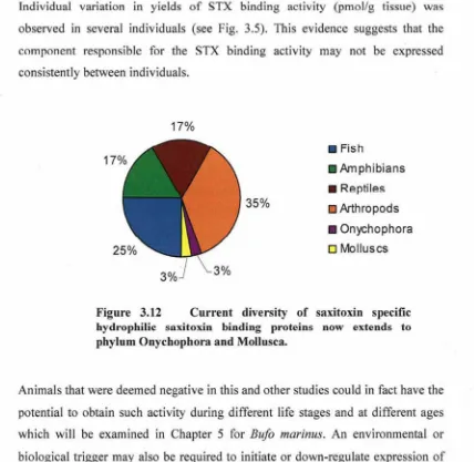
(14) environment were investigated, resulting in the discovery of eight novel STXBPs extending the known phylogenetic diversity of STXBPs to include species from Onychophora and Mollusca. Seven of these species were characterised as STX specific hydrophilic receptors based on their ability to exclusively bind STX. In addition, a STXBP likely to belong to the PSTBP group was identified in the toad fish, T. pleurogramma.. Further examination of species collected from a verified STX "HotSpot" resulted in the identification of one additional STXBP in the extracts from the crab, Lophozozymus octodentatus. The occurrence of PSTs and diversity STX sources. and vectors at this site were examined by means of radioreceptor assays including the centipede SXPN assay, rat brain synaptosome assay, liquid chromatographyfluorescence detection with post column oxidation, in addition to confirmatory mass spectrometric analysis. This study confirmed 3 new sources of PSTs in benthic food web of Port Hedland including the macroalgae, Sargassum sp. Jania sp.2 and Jania sp.3. However, the STX levels in these species did not explain the. extreme levels of STX observed in some vectors. A number of new PST vectors were identified from the bivalves Tridacna squamosa, Pinctada albina sugilata, Saccostrea glomerata, Malleus regula and the first incidence of STX in the. octopi, Octopus (Abdopus) sp. 5, was reported. The lack of widespread STXBPs in the intertidal STX "Hot Spot" did not conclusively support the toxin defence hypothesis as a likely biological role of STXBPs.. Finally, an animal model, B. marinus, was selected for an in depth analysis of STXBPs. STXBP levels in the toad were ubiquitous across all life stages and within all tissues with the exception of the venom glands, and reflected previous reports of saxiphilin distribution in the bullfrog R .. catesbeiana. Interestingly, a positive correlation was demonstrated between environmental temperature and levels of STX binding activity in toad plasma. The STXBP isolated from B. marinus plasma was successfully purified, revealing an estimated protein size of. 93 kDa and 3 peptide sequences which facilitated degenerate PCR experiments. Cloned STXBP-specific fragments of cDNA from toad liver were then cloned and the corresponding translated amino acid sequences revealed homology to the C-. xii.
(15) lobe of both saxiphilin from R. catesbeiana (also known to contain the STX binding site) and a variety ofTFs.. The biological role of soluble STXBPs remains a mystery but substantial advances have been made in terms of the diversity, function and relationships of these proteins. The potential application of STXBPs uncovered during this research, in both medical and research applications ofPSP treatment and detection is substantial and the wealth of data collected will promote several new directions of research in this area.. Xlll.
(16) TABLE OF CONTENTS Statement of Access Statement of Sources Declaration on Ethics Statement on the Contribution of Others Acknowledgements Publications Arising from Thesis Abstract Table of Contents List of Tables. ii iii iv v Vll. ix xi xiv xvi. ~~~~. ~. Abbreviations. XXIV. CHAPTER 1. 1. Theoretical Background and Objectives 1.1 Introduction 1.2 Paralytic Shellfish Toxins 1.3 Sources of Paralytic Shellfish Toxins 1.4 Chemistry and Toxicity 1.5 Voltage Gated Sodium Channels 1.6 Regulatory Limits and Detection ofPSTs 1.7 Saxitoxin Resistance 1.8 Saxitoxin Binding Proteins 1.9 Saxiphilin 1.10 Thesis Aims and Objectives References. 1 1 3 5 7 9 12 15 16 21 26. CHAPTER 2 Development of a Universal Receptor Assay for the Detection of Soluble Saxitoxin Binding Proteins 2.1 Introduction 2.2 Materials and Methods 2.3 Results 2.4 Discussion 2.5 Conclusions References CHAPTER 3 Diversity and Functional Classification of Hydrophilic Saxitoxin Binding Proteins 3.1 Introduction 3.2 Materials and Methods 3.3 Results 3.4 Discussion 3.5 Conclusions 3.6 Acknowledgements References. xiv. 42. 42 51 60 67 70 71 76. 76 79 90 102 108 109 109.
(17) CHAPTER 4 Occurrence of Paralytic Shellfish Toxins and Saxitoxin Binding Proteins in a Tropical Saxitoxin "Hot Spot" 4.1 Introduction 4.2 Materials and Methods 4.3 Results 4.4 Discussion 4.5 Conclusions 4.6 Acknowledgements References. 114. CHAPTER 5 Characterisation, Purification and Partial Sequencing of a Saxitoxin Binding Protein from Bufo marinus 5.1 Introduction 5.2 Materials and Methods 5.3 Results 5.4 Discussion 5.5 Conclusions 5.6 Acknowledgements References. 157. CHAPTER 6. 198. General Conclusions and Perspectives 6.1 Research Overview 6.2 The biological Role of STXBPs 6.3 Research Applications 6.4 Future Directions 6.5 Additional Outcomes References. 198 200 202 203 204 206. APPENDICES Appendix 1: Multiple sequence alignment of saxitoxin binding proteins. Appendix 2: Species collection for the phylogenetic survey of sources of STXBPs and PSTs from QLD and NSW sites. Appendix 3: STX concentrations in crabs and octopi collected from Port Hedland Appendix 4: BLASTX results of partial experimental sequences fi'om B. marinus. xv. 114 119 127 143 151 151 152. 157 160 173 188 194 195 195. A-I A-2. A-7 A-9.
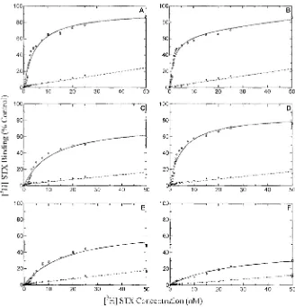
(18) LIST OF TABLES CHAPTERl Table 1.1. Reported dinoflagellates and cyanobacterial sources of paralytic shellfish toxins in the marine and freshwater environment.. Table 1.2. Structure and toxicity of known paralytic shellfish toxins.. Table 1.3. Regulatory limits for the safe consumption of shellfish required by various countries around the globe.. Table 1.4. Comparison of a variety of reported methods for the detection of PSTs.. Table 1.5. Comparison of reported biochemical and pharmacological parameters of native bullfrog, recombinant saxiphilin and the recombinant C-lobe of the protein.. Table 1.6. Comparative biological and pharmacological parameters of known saxitoxin binding proteins.. CHAPTER 2 Table 2.1. Comparison of radio-receptor assay methods trialled in the development of a primary screening assay for the detection of STX binding activity.. Table 2.2. Summary of performance of all radio-receptor methods assessed in development of a detection assay for STX binding activity.. Table 2.3. Optimised assay conditions for the MCE protein binding assay.. CHAPTER 3 Table 3.1. Summary of phylogenetic diversity survey of soluble saxitoxin binding activity reported by Llewellyn et al. (1997b).. Table 3.2. Overview of 203 species collected from a variety of QLD and NSW sites and sources within Australia for screening of STX binding activity and PST analyses. Table 3.3. Summary of ATCC requirements, specifications and characteristics of ZFL~cellline from Brachydanio rerio.. Table 3.4. Characterisation of novel saxitoxin binding activity by saturation binding experiments in extracts from a variety of vertebrate and invertebrates. XVI.
(19) Table 3.5. Characterisation of novel saxitoxin binding activity by saturation binding experiments in extracts from a variety of vertebrate and invertebrates.. Table 3.6. Structure-activity relationships between STX derivatives and concentration standardised protein extracts of animals identified with STX binding activity as determined by competitive binding with 2nM eH] STX in the MCE receptor assay.. Table 3.7. Molecular weight based ultrafiltration of pooled extracts of species identified with saxitoxin binding activity.. Table 3.8. Toxin concentration of individual AOAC extracts defined as toxic by at least one method.. CHAPTER 4 Table 4.1. Species collection summary from Cooke Point, Port Hedland, Western Australia.. Table 4.2. Sodium channel (NaCh) and centipede saxiphilin (SXPN) radioreceptor assay and LC-FD quantification of toxins identified in extracts from algae and molluscs from Port Hedland collection A.. Table 4.3. Paralytic shellfish toxin composition of extracts from a variety of algae, molluscs and crustaceans collected from Cooke Point, Port Hedland.. Table 4.4. Observed mass losses from Octopus (Abdopus) sp. 5 (extract 29) mlz 300 daughter ion mass spectrum.. Table 4.5. Calculations of gross amount of PST source that a specimen of Atergatis floridus would be required to consume to acquire concentrations of 11,336 fJgll OOg tissue as observed from Port Hedland during this study.. Table 4.6. Toxin profiles of 3 common PST-producing dinoflagellates from Asia Pacific region.. CHAPTERS Table 5.1. PCR cycling conditions used in verification of cDNA prepared from B. marinus liver with 18S forward and reverse primers.. Table 5.2. Degenerate primer combinations used in polymerase chain reaction of cDNA from the liver of B. marinus.. xvii.
(20) Table 5.3. Polymerase chain reaction thermo-cycling conditions used in preliminary and final experiments using degenerate primers designed from B. marinus STXBP peptide sequences and cDNA derived from B. marinus liver.. Table 5.4. Thermo-cycle conditions employed for PCR amplification of cloned inserts from JMl09 high efficiency competent cells with USP and RSP primers.. Table 5.5. Tissue distribution of saxitoxin binding activity in Bufo marinus.. Table 5.6. Peptide sequences resulting from MS-MS analysis of purified B. marinus STXBP tryptic digests.. Table 5.7. Degenerate forward and reverse oligonucleotides designed from back translated peptide sequences from purified B. marinus STXBP incorporating codon frequencies for B. marimlS.. Table 5.8. Summary of BLASTX results of final nucleotide sequences resulting from cloning of degenerate PCR fragments into pGEM®T Easy vector.. Table 5.9. Comparison of characteristics between saxiphilin from Rana catesbeiana and STXBP from Bufo marinus as determined during this chapter.. xviii.
(21) LIST OF FIGURES CHAPTERl Fig. 1.1. Structure of saxitoxin (A) and tetrodotoxin (B) illustrating the position of positively charged guanidinium moieties on each molecule.. Fig. 1.2. Photographs of toxic dinoflagellate (A) and cyanobacterial (B) blooms.. Fig. 1.3. Structure and toxicity of known paralytic shellfish toxins.. Fig. 1.4. Schematic model of the secondary structure of the voltage gated sodium channel highlighting the arrangement of alpha helices within the phospho-lipid membrane.. Fig. 1.5. Characteristic ribbon structure of transferrins obtained from Baker (2003).. Fig. 1.6. Protein sequence alignment of Rana catesbeiana saxiphilin, human lactoferrin and human serum transferrin.. Fig. 1.7. Schematic representation of the linear sequence of bullfrog saxiphilin showing the location of conserved disulphide bonds and N- and C-Iobe structural domains predicted on the basis of sequence alignment with human lactoferrin as presented by Morabito et al (1995).. Fig. 1.8. Schematic outline of the thesis research plan and relationships of research objectives.. CHAPTER 2 Fig. 2.1. Schematic diagram depicting separation of protein bound and free radioligand using cationic exchange resin AG50W-X2 (Dowex).. Fig. 2.2. Schematic diagram of the separation of protein bound and free radio ligand in centipede saxiphilin assay on polyethylimine pretreated glass fiber type B filters.. Fig. 2.3. Representative saturation binding curves.. Fig. 2.4. Representative competitive binding curve.. Fig. 2.5. Schematic diagram depicting separation of protein bound and free radioligand using a negatively charged phosphocellulose cation exchange membrane.. . xix.
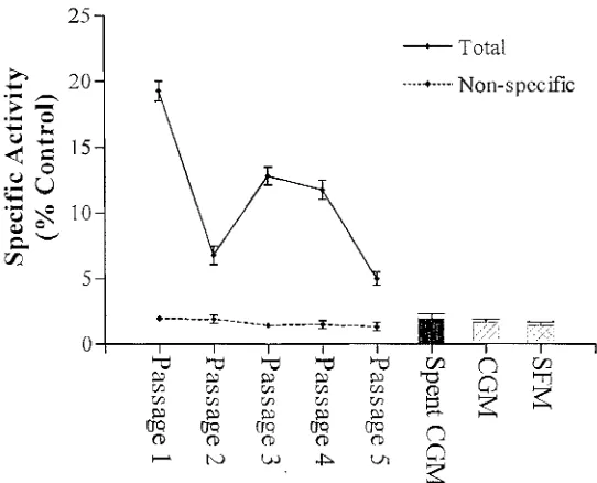
(22) Fig. 2.6. Schematic diagram of separation of protein bound and free radioligand using a positively charged diethylaminoethyl anion exchange membrane.. Fig. 2.7. Schematic diagram depicting separation of protein bound and free radioligand using high protein binding membranes such as mixed cellulose esters and polyvinylidene difluoride.. Fig. 2.8. Schematic diagram depicting separation of protein bound and free radioligand using activated charcoal and centrifugation.. Fig. 2.9. Receptor assay trial.. Fig. 2.10. Binding site titrations of STXBP extracts from Bufo marinus, Ethmostigmus rubripes, Brachydanio rerio, and Lophozozymus pictor.. Fig. 2.11. eH] STX saturation binding experiments performed using the MCE STXBP assay.. Fig. 2.12. Effect of increasing salt concentrations on the performance of the MCE STXBP assay.. Fig. 2.13. pH titrations of equilibrium levels of eH] STX bound by Bufo marinus plasma, Ethmostigmus rubripes extract and Brachydanio rerio extract performed with the MCE STXBP assay.. CHAPTER 3 Fig. 3.1. Queensland collection sites for the phylogenetic survey of STX binding activity extended from Cardwell to Mackay in marine, freshwater and terrestrial locations.. Fig. 3.2. Several log-dwelling invertebrates were collected from Tallaganda State Forest (35 0 35'S, 1490 27'E), N.S.W.. Fig. 3.3. Schematic diagram of a high performance liquid chromatography system coupled to a post-column reactor and fluorescence detector as used for the analysis of paralytic shellfish toxins.. Fig. 3.4. Photographs of Laternula elliptica depicting whole animal subsequent to collection (A) and prior to collection in Antarctic sediments showing siphons (B).. Fig. 3.5. Individual variation in specific activity from protein extracts of organisms exhibiting novel saxitoxin binding activity.. xx.
(23) Fig. 3.6. Saturation binding curves of pooled extracts from Gambusia hamiltoni, Liocheles waigiensis, Lycosa Jurcillata, Cormocephalus sp., Epiperipatus rowelli and Laternula elliptica.. Fig. 3.7. Saxitoxin binding activity of media and cell extracts from zebrafish liver cell (ZFL) from Brachydanio rerio.. Fig. 3.8. Basic structure of saxitoxin highlighting functional groups RI-R4 and pKa of each guanidinium residue. Fig. 3.9. Competition binding curve of Torquiguener pleurogramma extract showing inhibition of eH] STX binding by tetrodotoxin.. Fig. 3.10. Correlations between sodium channel, saxiphilin assay and HPLC analysis for the calculation of STX equivalents ().lg STXeq/ 100g tissue).. Fig. 3.11. Comparison of the number of species examined during two phylogenetic surveys of saxitoxin binding activity.. Fig. 3.12. Current diversity of saxitoxin specific hydrophilic saxitoxin binding proteins extends to phylum Onychophora and Mollusca.. CHAPTER 4 Fig. 4.1. Representative marine food web.. Fig. 4.2. Map of Western Australian coastline with inset highlighting study site at Cooke Point, Port Hedland.. Fig. 4.3. Dorsal and ventral view of an octopus specimen collected from Cooke Point, Port Hedland, Western Australia identified as Octopus (Abdopus) sp. 5 (Norman, 2000).. Fig. 4.4. Photographs of crab species targeted in 2002 benthic collection from Cooke Point, Port Hedland.. Fig. 4.5. Assay calibration curves of competitive binding between STX and eH] STX to rat brain sodium channel and centipede saxiphilin.. Fig. 4.6. Correlation between sodium channel and centipede saxiphilin radio-receptor assays and HLPC analysis of the total toxin concentrations of extracts of algae, molluscs and crustaceans from Port Hedland.. Fig. 4.7. Several examples of LC-FD analyses of PSTs from organisms collected from Cooke Point, Port Hedland.. xxi.
(24) Fig. 4.8. Total toxin concentration of crustacean extracts of Pilumnus pulcher, Thalamita stimpsoni and Lophozozymus octodentatus as calculated from sodium channel receptor assay, centipede saxiphilin receptor assay and HPLC analysis (LC-FD).. Fig. 4.9. Total toxin concentration of extracts of Atergatis floridus and Octopus (Abdopus) Sp. 5 calculated from sodium channel receptor assay, centipede saxiphilin receptor assay and HPLC analysis.. Fig. 4.10. Tissue distribution of STX in Octopus (Abdopus) sp. 5 as determined by sodium channel and centipede saxiphilin radioreceptor assays.. Fig. 4.11. LC-FD chromatograms of calibrated standard STX injection compared to a toxic extract of Octopus (Abdopus) sp. 5. showing peak retention, spiking of the extract with an authentic STX standard, and with no post-column oxidation.. Fig. 4.12. LC-MS chromatograms of an authentic STX standard compared to extract of Octopus (Abdopus) sp. 5. measured in single ion monitoring mode (mlz 300) on a triple quadrupole ion trap mass spectrometer.. Fig. 4.13. Daughter ion mass spectra for authentic STX and an extract of Octopus (Abdopus) sp. 5. measured on a quadrupole ion trap mass spectrometer at m/z 100- 300 after fragmentation ofmlz 300.. Fig. 4.14. Relationship between levels of STXBPs and PSTs isolated from the Xanthid crab, Lophozozymus octodentatus, collected from Port Hedland.. Fig. 4.15. Binding site titration and saturation binding curve of protein extracts from Lophozozymus octodentatus collected from Cooke Point, Port Hedland.. CHAPTERS Fig. 5.1. Female and male adult specimens of the cane toad, Bufo marinus.. Fig. 5.2. Lifecycle of the cane toad, Bufo marinus.. Fig. 5.3. Inhibitory STX binding activity detected in MeOH extracts of B. marinus tissues and following fractionation of viscera extract on C 18 in the presence of B. marinus STXBP extract.. Fig. 5.4. Titration of 30% methanol fraction obtained following C18 SPE chromatography of B. marinus MeOH viscera extract in the presence of B. marinus STXBP extract.. xxii.
(25) stages. of. Fig. 5.5. Saxitoxin binding activity observed at various metamorphosis in the cane toad, Bufo marinus.. Fig. 5.6. Monthly and seasonal variation in saxitoxin binding activity from the plasma of the cane toad Bufo marinus compared to environmental temperature for Townsville (2002 to 2004).. Fig. 5.7. Protein separation of crude plasma from Bufo marinus by anion exchange chromatography on a column ofQ-Sepharose Fast Flow.. Fig. 5.8. Purification and size estimation of STXBP from B. marinus plasma by Superdex 200HR size exclusion chromatography.. Fig. 5.9. Silver stained 10% SDS-PAGE of STXBP purification from B. marinus plasma, performed under denaturing conditions.. Fig. 5.10. First strand cDNA synthesis from B. marinus liver compared to control cDNA from P. mono don and DNA ladder.. Fig. 5.11. peR amplification of cDNA from B. marinus liver with degenerate primer combinations in the presence of 5.5 mM Mg2+ and 6 mM. Mi+.. Fig. 5.12. Re-amplified peR products from degenerate peR of cDNA from B. marinus liver in the presence of 6mM Mg2+.. Fig. 5.13. Representative PGemr® Easy cloned inserts from degenerate PCR amplification of cDNA fro B. marinus liver, amplified with USP and RSP primers.. Fig. 5.14. Multiple nucleotide sequence alignment of partial STXBP sequences from Bufo marinus compared to saxiphilin from Rana catesbeiana.. Fig. 5.15. Protein sequence alignment of R. catesbeiana saxiphilin, X laevis serotransferrin (frog), G. gallus ovotransferrin (chicken) and human serum transferrin, lactotransferrin, melanotransferrin and selected experimentally determined amino acid sequences derived fromB. marinus (AR_SOI, AR_S05, AR_S06, AR_S07, AR_S08).. Fig. 5.16. Structure of purine (A) and pyrrolo (B) ring systems which form part of the STX parent structure (C).. Fig. 5.17. Chemical structures of the nitrogenous adenosine and theophylline and cardiac glycosides: marinobufagenin and bufotelin.. xxiii.
(26) ABBREVIATIONS. eH] STX AEX ANGIS ANOVA APAF ATCC BLAST bp cDNA CEX CHAR dcGTX2 dcGTX 3 DCM dcSTX DEAE DNA DTT EtBr GC 1 GC2 GC3 GAF-AAC GFIB GFIC GTX 1 GTX2 GTX3 GTX4 GTX5 GTX6 HAB HIC HPLC IC50 ICA IPTG. Kd kDa Ki LC-FD LC-MS LF LSC MCE Mol% MS-MS. tritiated saxitoxin anion exchange Australian National Genomic Information Service analysis of variance Australian Proteome Analysis Facility American Type Culture Collection Basic local alignment tool base pair complementary DNA cation exchange charcoal decarbamoyl gonyautoxin 2 decarbamoyl gonyautoxin 3 dichloromethane decarbamoyl saxitoxin diethy laminoethy1 deoxyribonucleic acid dithiothreitol ethidium bromide Guanidinium catenatum toxin 1 Guanidinium catenatum toxin 2 Guanidinium catenatum toxin 3 Genetic Analysis Facility- Advanced Analytical Centre glass fibre type B filters glass fibre type C filters gonyautoxin 1 gonyautoxin 2 gonyautoxin 3 gonyautoxin 4 gonyautoxin 5 gonyautoxin 6 harmful algal bloom hydrophobic interaction chromatography high performance liquid chromatography 50 % inhibition constant inhibitor of carbonic anhydrase isopropyl-beta-D-thiogalactoside dissociation constant kilo dalton inhibition constant liquid chromatography- fluorescence detection liquid chromatography- mass spectrometry lactoferrin liquid scintillation counting mixed cellulose esters percentage of moles mass spectrometry- mass spectrometry. XXIV.
(27) MTF MW neoSTX NSW. OT PBS PCR PEl PH pKa PSP PST PSTBPI PSTBP2 PVDF QLD RACE RNA RT SBE SDS-PAGE SEC SEM ST STX STXBP STXdiHCI SXPN TAE TF TTX VGSC X-Gal. melanotransferrin molecular weight neosaxitoxin New South Wales ovotransferrin phosphate buffered saline polymerase chain reaction polyethylene imine ph osph ocell ul ose ionisation constant paralytic shellfish poison paralytic shellfish toxin puffer-fish saxitoxin and tetrodotoxin binding protein 1 puffer-fish saxitoxin and tetrodotoxin binding protein 2 polyvinylidene difluoride Queensland papid amplification of cDNA ends ribonucleic acid room temperature saturation binding experiments sodium dodecyl sulphate- polyacrylamide gel electrophoresis size exclusion chromatography standard error of the mean Serotransferrin saxitoxin saxitoxin binding protein saxitoxin dihydrochloride saxiphilin Tris acetate buffer transferrin tetrodotoxin voltage gated sodium channel 5-bromo-4-chloro-3-indolyl-~-D-galactoside. xxv.
(28) CHAPTERl Theoretical Background and Objectives 1.1. Introduction. Saxitoxin binding protein (STXBP) is a functional classification of all proteins capable of binding to saxitoxin (STX; Fig. 1.lA), a potent neurotoxin from the paralytic shellfish toxin (PST) family. This protein group includes the membrane bound voltage gated sodium channels (VGSCs), puffer-fish STX and tetrodotoxin (TTX; Fig. l.lB) binding proteins (PSTBPs) and saxiphilin (SXPN). Despite this functional similarity, there is no significant protein sequence or structural homology between members of this unique suite of receptors (see Appendix 1). This thesis will focus on the investigation of soluble STX-specific STXBPs such as SXPN and the primary focal point will be the relationship between these mobile receptors and their only confirmed ligand, STX.. A N. B. pKa = 8.2. :FN~. G. N OH OH. pKa =8.8 H Figure 1.1. Structure of saxitoxin (A) and tetrodotoxin (B) illustrating the position of. positively charged guanidinium moieties on each molecule.. 1.2. Paralytic Shellfish Toxins. PSTs are produced by a variety of marine dinoflagellates and cyanobacteria and each species and strain produces a characteristic and complex mixture of toxins. Dense harmful algal blooms (RABs) of cyanobacteria can contaminate freshwater sources including dams, rivers, lakes, affecting both livestock and human health (Jones and Negri, 1997; Kaas and Henriksen, 2000; Negri et ai., 1995).. 1.
(29) CHAPTER! In affected inland waters, PSTs accumulate in benthic feeders such as mussels which can impact on freshwater aquaculture and recreational fishing of affected species (Negri and Jones, 1995; Vasconcelos, 1999). Likewise, in coastal waters toxic dinoflagellate blooms can accumulate in filter feeners such as oysters and. mussels (Blanco et al., 2003; Garate-Lizarraga et al., 2004; Oshima et al., 1987) and a variety of gastropod molluscs, crustaceans and fish (Chen and Chou, 2001;. Ito et al., 2004; Llewellyn and Endean, 1989). Consumers of these vector species can also be at considerable risk with PSTs transmitted to higher vertebrates. including humans, via the food web (Llewellyn et aI., 2002; Rodrigue et al., 1990) and pose a major threat. to. the viability of both wild shellfish fisheries and. shellfish farm ing operations.. Figure 1.2 Photographs of toxic dinoflagellate (A) and cyanobacterial (8) blooms. Image (A) was obtained and modified from www.pac.dfo-mpo.gc.ca!ops/fm/shelifishlBiotoxinsIPSPe.htm and. image. (8). which. illustrates. a. bloom. of Anabaena circinalis. (8). was. from. www.dlwc.nsw.gov.au .. PSTs are hydrophilic cyclic alkaloids and at least 24 known chemical STX derivatives have been reported (Hall et aI., 1990; Negri et al., 2003b; Shimizu, 1993). STX selectively blocks the inward sodium current in excitable cells (Catterall, 1980; Denac et al., 2000b; Fozzard and Lipkind, 1996) causing a syndrome known as paralytic shellfish poisoning (PSP). One of the most toxic PST derivatives is the parent molecule, STX, which was reported to have a 50% lethal dose for mice (IP) in the order of 10 !lg/kg (Carmichael, 1992). Based on this high potency, water solubility and reports of successful synthesis (Jacobi et al.. 1984; Tanino et al.. 1977). STX is recognised as a potential chemical weapon and is listed as a schedule one chemical alongside mustard and sarin gas by the. 2.
(30) CHAPTERl United Nations Chemical Weapons Convention (OPCW, 2002). PSP is characterised by neurological symptoms including dizziness, tingling and numbness in the face and extremities and gastrointestinal upset including nausea and vomiting due to a relaxant action on vascular smooth muscle cells (Falconer, 1993; Stommel and Watters, 2004; Watters, 1995). Onset of symptoms following consumption of contaminated shellfish or water have been reported to take from between 30 minutes and 12 hours (Lehane, 2000; Smart, 1995; van Dolah, 2000) and in severe intoxications, muscular paralysis, respiratory paralysis and death may result (Cheng et al., 1991; Llewellyn et al., 2002; Rodrigue et al., 1990). At present there is no antidote or treatment for PSP but supportive care such as artificial respiration is crucial for patient recovery (Stommel and Watters, 2004), allowing time until the toxins are eliminated from the body by renal clearance (Andrinolo et al., 2002; Andrinolo et al., 1999; Gessner et al., 1997).. 1.3. Sources of Paralytic Shellfish Toxins. In marine systems, PSTs naturally occur in a variety of bloom forming dinoflagellates from the genus Alexandrium, Pyrodinium and Gymnodinium (Table 1.1) and in freshwater environments, certain filamentous cyanobacteria including Anaebaena, Amphizomenon,. Cylindrospermospsis,. Lyngbya and. Planktothrix species have been reported to produce the deadly toxins (Table 1.1). Of these known PST producers, only six have been reported in Australian waterways including five marine dinoflagellates and one representative of the blue green alga, Anabaena circinalis (Hump age et al., 1994); see Fig. 1.2B). The global occurrences of saxitoxic cyanobacterial (Kaas and Henriksen, 2000; Pereira et al., 2000; Pomati et al., 2000) and dinoflagellate blooms (Hallegraeff, 1993; Quilliam, 2001) has been increasingly reported (Hallegraeff, 1993; Quilliam, 2001). The biological advantage of PST production in these sources has not been clarified but considerable efforts have been made towards the understanding of the biosynthetic pathways, metabolism and molecular mechanisms of these secondary metabolites in cyanobacteria and dinoflagellates (Pomati et al., 2001; Pomati et al., 2004b; Pomati and Neilan, 2004; Shimizu, 1986a; Shimizu, 1986b; Shimizu, 1988; Shimizu, 1996).. 3.
(31) CHAPTER 1 Table 1.1 Reported dinoflagellates and cyanobacterial sources of paralytic shellfish toxins in the marine and freshwater environment. Marine Species Dinoflagellates. Freshwater Reference. Species Cyanobacteria. Reference. Alexandrium andersoni. (Climiniello et a1., 2000). Anabaena circinalis*. (Humpage et al., 1994). A. catenella*. (Negri et a1., 2003a). Anabaena lemmermannii. A. excavatum. (Cembella et a!., 1993). A. fundyense. (Anderson et a!., 1990). (Kaas and Henriksen, 2000) (Mahmood and Carmichael, 1986) (Nogueira et aI., 2004). A. minutum*. (Hallegraeff et a!., 1988). Aphanizomenon jlosaquae Aphanizomenon issatschenkoi Aphanizomenon gracile. A. ostenjeldii. (MacKenzie et a1., 1996). Lyngbya wollei. (Carmichael et aI., 1997). A. tamarense *. (Oshima et aI., 1992). (Lagos et a!., 1999). A. tamiyavanichii Gymnodinium catenatum* Pyrodinium bahamanse var compressa. (Kodama et aI., 1988a) (Oshima et aI., 1987). Cylindrospermopsis raciborskii Planktothrix sp.. (Pereira et aI., 2004). (Pomati et aI., 2000). (Harada et aI., 1982). * Known occurrence in Australia according to (Negri et aI., 2003a) Several studies have suggested that PSTs may also be produced by bacteria associated with dinoflagellate cultures, including Moraxella sp. (Kodama et aI., 1990; Kodama et aI., 1988b) and Alteromonas sp. and Pseudomonas spp. (Gallacher et aI., 1997; Gallacher et aI., 1996; Gallacher and Smith, 1999). In addition, several marine bacteria from shellfish including Vibrio spp. and Pseudomonas spp. (Gallacher and Smith, 1999; Levasseur et aI., 1996) and even. enterobacter bacteria from the rumen of cattle (Sevcik et aI., 2003) have been reported to produce STXs. However, levels of PST production by both symbiotic and free living bacteria in each of these cases, does not account for the total toxin concentrations observed in dinoflagellates and chemical identification of STX derivatives has not been unambiguously confirmed. In fact, one report clearly demonstrated that several Pseudomonas spp. bacteria isolated from Alexandrium spp. produced fluorescent components that mimicked STX peaks during High. Performance Liquid Chromatography (HPLC) analysis (Baker et aI., 2003b). Nevertheless, mounting evidence on the effects of isolated bacteria in the biotranformations, elimination and metabolism ofPSTs from both dinoflagellates and benthic animals (Geier, 2003; Kotaki et aI., 1985; Smith et aI., 20(1), suggests that bacteria may have a contributory role in toxin pharmacokinetics. A discussion. 4.
(32) CHAPTER!. of sources and vectors identified and distributed in Australian waters is provided in Chapter 4.. 1.4. Chemistry and Toxicity. STX was the first isolated PST derivative and was purified from the Alaskan butter clam, Saxidomus giganteus from which its name was derived (Schantz et aI., 1957). It was not until more than 15 years later that the crystal structure of STX, also isolated from S. giganteus was elucidated (Schantz et aI., 1975). The isolation of STX on carboxylate resins (e.g. BioGel P2 and BioRex 70) which was devised in these early years (Oshima et aI., 1977; Shimizu, 1985) are still used routinely and efficiently for the purification of PSTs (Lippemeier et aI., 2003; Negri et aI., 1997). Each PST is characterised by a unique tricyclic ring system with two guanidinium groups at either side of the molecule (see Fig. 1.1). The pyrimidine guanidine has a highly basic pKa of approx. 11.3 for STX hydrate while the imidazole guanidine has a much lower pKa of approx. 8.2 (Rogers and Rapoport, 1980). Another key feature of the PSTs is the presence of a hydrated ketone at C12 (see structure in Table 1.2), which is also a critical element in mammalian toxicity (Hu et aI., 1987; Onodera et aI., 1997). At neutral pH these hydroxyl groups are protonated and have been correlated to an increase in the toxic action of these compounds (Hall and Reichardt, 1984).. The PSTs form a group of closely related derivatives which can be divided into five groups according to their structural differences (see Table 1.2). These groups include the i) carbamate (STX, neoSTX and gonyautoxins (GTXl-4)); ii) Nsulpho-carbamoyl (GTX5-6, Cl-4); iii) decarbamoyl (dc-) (dcSTX, dcneoSTX, dcGTXl-4); iv) deoxydecarbamoyl (do-) (doSTX, doneoSTX and doGTX1); and (V) hydroxybenzoates (GC 1- 3).. Each PST group differs in relative mammalian toxicity with the non-sulphated STX and neosaxitoxin (neoSTX) being the most potent, followed by the singly sulphated gonyautoxins (GTXs), and the mildly toxic C toxins (Oshima, 1995a) see Table 1.2). In natural sources of PSTs such as the dinoflagellate, A. minutum, a complex mixture of these derivatives may be present. These toxin profiles have been reported to have characteristic signatures depending on species, strains and. 5.
(33) CHAPTERl even geographical locations (Chang et aI., 1997; MacKenzie et aI., 1996; Taleb et aI., 2003) which can be useful in determining the likely causative organism.. R4. Table 1.2 Structure and toxicity of known paralytic shellfish toxins.. 17. H R1~. N. H N. 6. 7~. H2N.A~. 9 8. NH. +NH 2. 15. 13. 14. PST. Toxicityt MU/~mol. STX GTX2 GTX3 NEO GTX1 GTX4 GTX5 C1 C2 GTX6 C3 C4 dcSTX dcGTX2 dcGTX3 dcNEO dcGTX1 dcGTX4 doSTX doGTX2 doGTX3 GC3 GC1 GC2. 2483 892 1584 2295 2468 1803 160 15 329 33. 143 1872 1617 1872. Rl H H H OH OH OH H H H OH OH OH H H H OH OH OH H H H H H H. Toxicity values reported by (Oshima, 1995a) - Not tested. t. R2 H H OS0 3H H OS0 3H H OS0 3H H OS0 3H H OS03H H OSO; H H OS0 3' H H OS0 3-. R3 H OS03H H OS03H H OS03H H OS03H H OS03H H OS03H H OS03H H OS03-. H. R4. )-0 H2N. \ 0. HN)lO I. SO. I. 3-. HO-. H-. de 0. I. #. I. HO. 6.
(34) CHAPTERl However, in some locations this differentiation of strains and even species has proved challenging (Chou et aI., 2004), which is not surprising when toxicity and PST profiles of certain species may be affected by changes in growth conditions (Hwang and Lu, 2000; Hwang et aI., 2003; Lippemeier et aI., 2003). The discovery of genetic markers and molecular methods for identification of toxic species and strains is currently in rapid development (Galluzzi et aI., 2004; Godhe et aI., 2001; Guillou et aI., 2002; Pomati et aI., 2004a; Pomati and Neilan, 2004) and the potential for both sensitive detection and accurate identification of toxic algae by these methods is likely.. The majority of PSTs exist in ionised forms, are heat stable at acidic pH, are highly soluble in water, but insoluble in lipid solvents (Hall et aI., 1990). These qualities are significant in terms of seafood safety because the act of cooking contaminated shellfish would not be likely to eliminate the toxicity. In fact, cooking. may. increase. toxicity. by. hydrolysing. any. mildly. toxic. N-. sulphocarbamoyl derivatives (e.g. C 1-4, GTX 5, GTX 6), commonly found in a variety of Australian cyanobacteria and dinoflagellate species, to a desulphated form (GTX, STX, neoSTX). This has been demonstrated to cause dramatic increases in mouse toxicity (Hall and Reichardt, 1984). In alkaline conditions, PSTs are highly unstable and are easily oxidised and degraded which suggests that they would degrade in seawater (approx. pH 8) unless complexed with other stabilising molecules (Shimizu, 2000).. 1.5. Voltage Gated Sodium Channels. Consumption of PST contaminated shellfish can have marked physiological effects due to the rapidly reversible blockade (sec.- min.) of the VGSC at the neuromuscular junction in excitable cells (Denac et aI., 2000b). This blockade occurs at the extracellular side of the plasma membrane and impedes Na+ influx into the cell which is critical for depolarisation of the membrane and subsequent propagation of action potentials. The rapid onset of parasthesias, numbness and muscular and respiratory paralysis associated with PSP syndrome can be explained by this high affinity binding (dissociation constant Kd ~ 1-5 nM), with a single toxin molecule interacting with a single VGSC (Campbell and Hille, 1976; Moczydlowski et aI., 1984).. 7.
(35) CHAPTER 1. STX, TTX and their analogues have proved to be valuable tools in the physiological understanding ofVGSCs and have greatly facilitated examination of the shape and structure of the outer vestibule and in modelling of the TTX and STX binding site (Lipkind and Fozzard, 1994). Unfortunately due to the large size (approx. 300 kDa) and complexity of this trans-membrane protein (Sato et aI., 1998), the complete crystal structure of the VGSC is yet to be successfully elucidated, but extensive modelling from the encoded cDNA sequences and pharmacological examination of individually cloned subunits has provided a wealth of information on the structure and interaction of each subunit. In rat brain, the VGSC is reported to be a heterotrimeric protein containing subunits a, ~1. and. ~2,. (see Fig. 1.3) whereas in heart and skeletal muscle only the a and. ~1. subunits are present (Roberts and Barchi, 1987; Satin et aI., 1992a). The asubunits of many different species have now been cloned and sequenced revealing highly conserved regions of nucleotide sequence (Chen et aI., 1997; Noda et aI., 1986; Rogart et aI., 1989; Satin et aI., 1992b). The a-subunit has four repeating units, each consisting of six alpha-helical trans-membrane segments (see Fig. 1.3) which are thought to be arranged in a clockwise configuration around the central ion permeation site (Dudley et aI., 2000; Li et aI., 2000). The positively charged guanidinium groups of PSP toxins (see Fig. 1.1) interact with negatively charged carboxyl groups (Khan et aI., 2002) on the P-Ioops situated at the mouth of the channel between segments 5 and 6 of the a-subunit (see Fig. 1.3). Point mutation studies revealed that several glutamate residues corresponding to these P-Ioops are essential for effective inhibition of Na+ current by STX and included Glu 387 (Noda et aI., 1989), Glu 942, Glu 945 and Asp 384 (Lipkind and Fozzard, 1994; Satin et aI., 1992a; Terlau et aI., 1991). Binding studies with tritiated STX also suggest that the. ~ I-subunit. participates in forming the TTX/STX binding site. (Messner and Catterall, 1986).. 8.
(36) CHAPTERl Extracellular (l. NH. +. (. ~----------~------------. I. /I. III. IV. """. NH3. Intracellular Figure 1.3. Schematic model of the secondary structure of the voltage gated sodium. channel highlighting the arrangement of alpha helices within the phospholipid memhrane.. The alpha subunit consists of 4 homologous domains (I-IV) with 6 transmembrane segments and two beta subunits (~1, ~2). The polypeptide chain is represented by the solid line and cylinders represent a-helices. Red diamonds on the extracellular surface of the membrane represent the sites that are thought to interact with the positively charged guanidino groups on the STX molecule. Orange circles indicate sites of demonstrated protein phosphorylation and the inactivation gate is highlighted in pink. Schematic is based on model by Lipkind and Fozzard 1994.. 1.6. Regulatory Limits and Detection of PSTs. PSTs pose a serious public health threat due to their high potency and potential contamination to food and water supplies. In most countries, commercial shellfish harvesting is now regulated and levels of PSTs monitored closely to prevent contaminated stock reaching consumers (van Egmond et aI., 1992). At present most monitoring programs only require that filter feeding shellfish such as clams, mussels and oysters are tested routinely for PST contamination. However, repons of PSTs in animals higher in the food chain such as carnivorous gastropods (Hwang et aI., 1994; Ito et aI., 2004) and crustaceans (Baden, 1983 ; Brelz el aI., 2002), emphasises the importance of additional classes being considered for testi.ng prior to reaching consumers (Shumway, 1995).. The most widely accepted safe level of PST contamination is presently 80 Ilg STX equivalents/ 100 g tissue (see Table 1.3), which is not particularly conservative considering that illness has been reported at PSP doses as low as 144. 9.
(37) CHAPTER 1. Jlg per person and fatalities have been reported at 300 Jlg (van Egmond et aI., 1993). A single meal could easily reach these levels given that an average meal may consist of a 200-300 g serving of shellfish potentially contaminated with STX at the regulatory limit. If the lowest reported toxic dose of STX was used to establish a tolerance level, the current regulatory limits would need to be substantially reduced (van Egmond et aI., 1993).. Table 1.3 Regulatory limits for the safe consumption of shellfish required by various countries around the globe. Table was adapted from data reported by Lehane (2000). Country. Regulatory PST level (rz STXeq/ lOOg tissue)t. Australia New Zealand USA Norway Japan European Union Canada. 80 80 80 36 72. 80 80. t STXeq= saxitoxin equivalents; 0.18 Ilg STXeq/lOOg tissue = IMU. The most widely accepted method of detection of PSTs from shellfish is the mouse lethality assay which was established from the early work of Sommer and coworkers (Sommer and Meyer, 1937; Sommer, 1937). This was the first bioassay that made it possible to quantify the toxicity of STX and all of its analogues. The mouse bioassay is a standardised procedure in which the strain, size and condition of the mice are all critical to the accuracy of the toxicity calculation (AOACInternational, 1997). The basic protocol suggested by the Association of Official Analytical Chemists (AOAC) involves injecting an acid extract of shellfish (ImL) into the mouse (20 g) and then recording the time of death at the last breath. Standard dose response curves are constructed using calibrated STX standards and toxic sample extracts are diluted to ensure mortality within five to 15 minutes (AOAC-International, 1997). The AOAC method is still the dominant method used for detecting PSTs despite having a detection limit of only 40 )lg STXeq/1 00 g shellfish, which is quite poor compared to other methods that are available (see Table 1.4) and for these reasons is often coupled to analytical methods such as HPLC (Asp et aI., 2004; van Egmond et aI., 2004). The major drawbacks of the. 10.
(38) CHAPTERl mQuse lethality assay centre around the use of mice, which not only have considerable ethical considerations but need to be maintained in constant stocks weighing 19-22 g which can be labour intensive and expensive (AOACInternational, 1997; Lehane, 2000; van Egmond, 2004). The effects of the PSTs on the mice are both strain and colony dependent, which means that large numbers of mice are needed from each new colony, to produce a single dose response curve from which sample extracts can then be tested. Other reported problems inherent with this assay includes interference of salt which can suppress toxic effects at high concentrations (LeDoux and Hall, 2000) and extracts of oysters contaminated with zinc, have been reported to cause lethal effects even when sub-lethal doses ofPSTs have been injected (Aune et aI., 1998).. Many alternative PST detection methods have been developed including the in vivo locust lethality assay (McElhiney et aI., 1998); in vitro cell based (Jellett et. aI., 1992; Kogure et aI., 1988), hippocampal slice (Kerr et aI., 1999) and sodium channel assays (Doucette et aI., 1997; Ruberu et aI., 2003); biochemical methods such as enzyme-linked immunosorbent assays (ELISA) (Chu and Fan, 1985; Chu et aI., 1996; Usleber et aI., 2001) and centipede SXPN assay (Llewellyn et aI., 2001a; Llewellyn et aI., 1998); chemical analysis by HPLC (Oshima, 1995a; Oshima, 1995b; Sullivan and Iwaoka, 1983), capillary electrophoresis (Pleasance et aI., 1992; Quilliam, 1995) and mass spectrometry (Dahlmann et aI., 2003; Pleasance et aI., 1992; Quilliam et aI., 1989). Each method has inherent advantages and disadvantages with varying degrees of sensitivity to PSTs, but all eliminate the use of mammals and have lower detection limits that the mouse bioassay (see Table 1.4). In general, there is a distinct need for a detection method that meets several criteria:. Sensitive to all PST derivatives Accurate and low detection limits •. Robust to the effects of salt, metals and other contaminants Simple and inexpensive Correlates well to mammalian toxicity. •. Does not require specialist equipment or training. 11.
(39) CHAPTER 1. Table 1.4 Comparison of a variety of reported methods for the detection of PSTs. Detection Method. Mouse Mortality Bioassay. MIST-Alert Cell Bioassay Kit. RidaScreen STXELISA. Sodium Channel Assay. Centipede Saxiphilin Assay. Liquid chromatography. STX Detection Limie ~f!~ /100~~. 35-40. 2. 0.25. 0.2- 1. Disadvantages. References. Closely predicts human toxicity, sensitive to all derivatives. Ethical considerations; costly; time consuming, high variability; cannot confirm toxin identity or Erofile.. (Adams and Miescier, 1980; Salter et aI., 1989). Simplified cell bioassay, elimination of animals and radioligand, sensitive to all toxins. Expensive; limited shelf-life; cannot confirm toxin identity or profile.. (Jellett et aI., 2002; Llewellyn et aI., 2001 a; Mackintosh et aI.,2002). Rapid, simple, sensitive. Dependent on antibody specificity; cannot confirm toxin identity or profile.. (Usleber et a!., 2001). Advantages. High-throughput, Sensitive to all derivatives. 1- 2. High-throughput, Sensitive, distinguishes between TTX and STX-like activity. 0.01-1. High sensitivity, confirmation of toxin identity and profile.. Requirement of rat brain; reliance on radio ligand; cannot confirm toxin identity or Erofile. Centipede availability is limited; reliance of radioligand; cannot confirm toxin identity or profile; Low sensitivi!l: to C-toxins. Specialised equipment and training; requires calibrated toxin standards.. (Doucette et aI., 1997; Llewellyn et aI., 2001b; Ruberu et aI., 2003). (Llewellyn and Doyle, 2001; Negri and Llewellyn, 1998) (Lawrence et aI., 1991; Negri and Llewellyn, 1998; Oshima, 1995a). High sensitivity, (Pleasance et aI., structural Specialised equipment 0.001-1 1992; Quilliam, 2003; confirmation of toxin and training Quilliam et a!., 1993) identi~ and Erofile. Note: This list is not exhaustive and so does not include all reported methods available. t For comparison reported detection limits were converted to /lg STX eq. Mass Spectrometry. 1.7. Saxitoxin Resistance. Fossil evidence of dinoflagellate cysts suggest that these organisms have existed for at least a billion years (Graham and Wilcox, 2000) and are thought to be derived from a endosymbiotic event between a cyanobacterium and eukaryotic organism (Mordon and Sherwood, 2002). Based on its unique distribution as a bioactive secondary metabolite in both prokaryotes and eukaryotes, it has been. 12.
(40) CHAPTERl suggested that STX may have been acquired early in the evolution of these microorganisms (Shimizu, 1993). Reports of the red tide phenomenon and their toxic effects have been recognised for centuries and references in the Old Testament of the Bible are thought to relate to a dinoflagellate bloom in the Nile River, an event that has been dated back to approximately 1500BC by both archaeological and historical data (Rohl, 1999).. Exodus 7:20-21: "...... and the waters in the river were turned to blood And the fish in the river died; and the river became foul smelling, and the Egyptians could not drink its water. ". In light of this history of occurrence, it is not surprising that many benthic animals have developed mechanisms of resistance to some of the toxins produced by cyanobacteria and dinoflagellates. Several suggested mechanisms of STX resistance have been reported including nerve insensitivity (Daigo et aI., 1988; Kvitek and Beitler, 1991), STX induced proteins (Barber et aI., 1988; Smith et aI., 1989) and soluble STXBPs (Llewellyn, 1997). Each of these mechanisms, which have been observed in a variety of species, may afford the exposed animal protection from PST intoxication.. Marine species that have demonstrated resistance to STX generally accumulate this toxin at high concentrations without detriment as was demonstrated in the Xanthid crab Atergatis jloridus which is a well recognised vector of PSTs (Arakawa et aI., 1995; Raj et aI., 1983; Tsai et aI., 1995) and has a minimal lethal dose of 5,000-10,000 MU/ 20 g, equivalent to approximately 4500-9000 /lg STX/ 100 g (Koyama et aI., 1983). Crab resistance has been correlated with detectable levels of PSTs in a variety of crab species (Koyama et aI., 1983; Yasumoto et aI., 1981) and resistance has been attributed to the presence of PST insensitive nerves (Daigo et aI., 1988). Similar effects were observed in the mussel Mytilus edulis and a variety of other bivalve molluscs which accumulated large quantities of PSTs but had no observed reaction to the toxins unlike less toxic species (Twarog, 1974). Further studies from these animals demonstrated that resistance to STX was also due to PST insensitive nerves (Kvitek and Beitler, 1991; Twarog, 1974). In contrast, marine species susceptible to the effects of STX do not accumulate. 13.
(41) CHAPTER 1 excessive concentrations within their tissues and usually demonstrate typical PSP symptoms when exposed to PSTs (Adams et al., 1968; Koyama et al., 1983; Smith et al., 1989; Twarog, 1974).. Not all marine animals exposed to STX have nerves that are resistant to PSTs yet are able to survive and accumulate moderate levels of PSTs in these environments. For instance, the appearance of a STX induced protein in the crab, . Hemigrapsus oregonesis following STX injection was correlated to resistance in. this species (Barber et al., 1988). Further investigation of the anti-STX induced protein in extracts of a selection of bivalves suggested that a related protein may be present in those species (Smith et al., 1989). These proteins which were reported more than fifteen years ago has never been sequenced or fully characterised so the true nature of its functional relationship to PSTs or STX binding capability is not known. In another study hemolymph from several Xanthid crabs including Lophozozymus pictor, Liomera tristis, Chlorodiella nigra and Actaeodes tomentosus, were reported to contain STX -specific binding activity and it was suggested that the component involved may act as a STX resistance mechanism in these animals (Llewellyn, 1997; Llewellyn et al., 1997). However, as with nerve resistance, this STX-binding hemolymph factor was not present in known toxic species and in all non-toxic species and presence of the binding factor did not seem to correlate with increased PST accumulation (Llewellyn, 1997), although this was not conclusively examined.. STX resistance in terrestrial species has also been reported. One early report concluded that frogs were resistant to the effects of natural toxins contained in crude mussel extracts compared to mice and demonstrated a lethal dose 15 times that required to cause mouse mortality being required to induce PSP symptoms in frogs (Prinzmetal et al (1932) as reported by Mahar et al (1991». This resistance was confirmed in a subsequent study of the effects of pure STX in the leopard frog Rana pipiens (Kao and Fuhrman, 1967). A minimal lethal dose of 75 /-lg/kg was required in the frog compared to only 5 /-lg/kg in mice tested at that time, while the effect of TTX in both animals was similar (Kao and Fuhrman, 1967). Unlike several crab and mollusc species, this resistance in frogs could not be correlated to STX-insensitive nerves which have been clearly shown to bind both. 14.
(42) CHAPTER 1. STX and TTX with high affinity (Campbell and Hille, 1976; Hille et aI., 1975; Strichartz, 1984b). It has been postulated that the presence of SXPN, a 91 kDa soluble STXBP isolated from Rana catesbeiana, is responsible for the reduced effects of STX that have been observed in a variety of amphibian species (Mahar et aI., 1991). Similar STXBP activity has since been demonstrated in a variety of ectothermic vertebrates including fish, reptiles and amphibians and in a small number of invertebrates including several crustacean, insect and arachnid species (Llewellyn, 1997; Llewellyn et aI., 1997; Mahar et aI., 1991). The phylogenetic distribution of soluble STX binding activity is discussed in detail in Chapter 3.. 1.8. Soluble Saxitoxin Binding Proteins. Soluble STX binding activity from biological sources was first recognised in the clarified supernatant of frog heart homogenates (Doyle et aI., 1982; Tanaka et aI., 1984), an activity which was confirmed in an independent investigation of the distribution of VGSC subtypes in frog skeletal muscle (Moczydlowski et aI., 1988). From these early observations, a distinct class of binding sites were isolated from soluble muscle extracts of the North American Bullfrog, Rana catesbeiana, and had a high affinity (0.1 nM) for eH] STX but was insensitive to. TTX. (Moczydlowski. et. aI.,. 1988). and. on. this. basis. was. deemed. pharmacologically distinct from the VGSC. STX binding activity was attiibuted to a soluble plasma protein, which was aptly named SXPN based on its binding specificity for STX (Mahar et aI., 1991). A wealth of data has been reported for R. catesbeiana SXPN and the amino acid sequence from molecular cloning studies. revealed a high homology for the transferrin (TF) family of iron binding proteins (Li and Moczydlowski, 1991; Morabito and Moczydlowski, 1994; Morabito and Moczydlowski, 1995) which is discussed further in section 1.9.. Another class of soluble STXBPs was recently reported and was isolated from the plasma of pufferfish, Fugu pardalis (Yotsu-Yamashita et aI., 2001). The soluble nature of these proteins distinguished them from the VGSC, however the demonstrated co-affinity for TTX. (Kd~. 12 llM) and STX. (Kd~. 8.5 nM) suggested. that the proteins were also dissimilar to SXPN (Yotsu-Yamashita et aI., 2002). Subsequent cloning and amino acid sequencing revealed that these proteins, named PSTBPs, were most similar to the partially sequenced TTX binding. 15.
(43) CHAPTER 1 proteins (TTXBP), isolated from related pufferfish, Takifugu niphobles (Matsui et aI., 2000).. 1.9. Saxiphilin. SXPN is a 91 kDa plasma protein isolated from R. catesbeiana (Morabito and Moczydlowski, 1994). CH] STX specifically binds to a single site on the SXPN molecule with high affinity (Kd= 0.18 nM) and has varied affinity for other PSTs (Llewellyn and Moczydlowski, 1994; Mahar et aI., 1991) and is discussed further in Chapter 3. SXPN does not interact or bind TTX (Moczydlowski et aI., 1988), unlike the membrane-bound voltage gated sodium channels (VGSC) (Denac et aI., 2000a) and puffer-fish STX and TTX binding proteins (PSTBPs) (YotsuYamashita et aI., 2002; Yotsu-Yamashita et aI., 2001). Pharmacological characterisation studies revealed that SXPN binding of CH] STX to SXPN is partially inhibited by a variety of divalent metal and lanthanide cations and the carboxyl-methylating reagent trimethyloxonium (Llewellyn and Moczydlowski, 1994). The latter activity indicated that at least one carboxyl moiety is likely to be required within the STX binding on the molecule and it was postulated that this site was located in the C-terminus of the protein (Llewellyn and Moczydlowski, 1994).. The unique STX binding activity of the protein facilitated initial purification efforts which incorporated assay guided fractionation of crude plasma by size exclusion chromatography (SEC), cation exchange chromatography (CEX; SSepharose) and isoelectric focussing (Mahar et aI., 1991). While these trials resulted in 70 % loss of activity, estimates of size (74 ± 8 kDa), basic isoelectric point (approx. 10.5) and net positive charge of the molecule at pH 6, were crucial for the development of a successful SXPN purification strategy (Mahar et aI., 1991). Final purification was achieved by Heparin Sepharose affinity chromatography and chromatofocussing with PBE 94 anion exchanger gel (Li and Moczydlowski, 1991). This methodology resulted in a 436 fold purification of SXPN from plasma and produced a single Coomassie stained band following sodium dodecyl sulphate polyacrylamide gel electrophoresis (SDS-PAGE) with an estimated molecular weight of approx. 89 kDa (Li and Moczydlowski, 1991).. 16.
(44) CHAPTER! Purification and partial sequencing of native SXPN from bullfrog plasma identified a possible relationship between SXPN and TFs (Li and Moczydlowski, 1991) and facilitated the isolation of cDNA encoding SXPN (Morabito and Moczydlowski,. 1994;. Morabito. and. Moczydlowski,. 1995).. Further. immunochemical and biochemical evidence demonstrated that TF was in fact distinct from SXPN based on differences in size (- 78 kDa TF; 89 kDa SXPN), chromatographic behaviour (DEAE Sepharose), visible absorption spectra and ligand binding (Li et aI., 1993). The translated protein sequence of SXPN showed high amino acid sequence homology (30-70 % identity) to the TF family of Fe 3+ binding. proteins. (Morabito. and. Moczydlowski,. 1994;. Morabito. and. Moczydlowski, 1995).. Transferrins are a family of single-chain, gIycosylated proteins which include serum transferrin (ST), lactoferrin (L T), melanotransferrin and (MTF), inhibitor of carbonic anhydrase (leA) and ovotransferrin (OT) (for review see (Aisen and Leibman, 1972; Baker et aI., 2002; Lambert et aI., 2005). TFs are iron transport molecules which rapidly and efficiently mobilise iron from plasma to cells and regulate plasma iron levels. The majority of TFs consist of two homologous lobes. (N and C) connected by a short hinge region (see Fig l.4A; (Baker et aI., 2002). Each of the two lobes is able to reversibly bind a single FeJ + ion and an associated carbonate ion (RC03-; see Fig I.4B).. Jiinlle. Figure 1.4 Characteristic ribbon structure of transferrins obtained and from Baker, 2003. (A) The iron-bound fOlm of human lactoferrin illustrating the bilobed nature of the molecule which is common to transferrins and position of Fe3+ (red) and associated (orange) molecules. (B) The conformational change which occurs during iron-binding shown in the N-lobe of human transferrin.. cot. 17.
(45) CHAPTER 1 The binding interaction between Fe3+ and HC0 3- has been described as synergistic and binding studies have demonstrated that Fe3+ cannot effectively bind to TFs without the concomitant binding of HC03- and vice versa (Baker et aI., 1996). In fact both kinetic and structural data suggests that HC03- is required to bind to TF first to facilitate Fe3+ binding (Anderson et aI., 1989; Baker et aI., 1996). Fe 3+_ bound TF is internalised by binding to a cell surface receptor and is then propelled into the endosome compartment of the cell by endocytosis. The acidic environment of the endosome induces the release of Fe3+ allowing recycling of apotransferrin (TF minus Fe3+) to the cell surface (Baker et aI., 2003a; Baker and Baker, 2004).. Full amino acid sequencing of SXPN revealed that SXPN differs from TFs with substitutions at 9 of 10 highly conserved residues which are known to be directly involved in the two Fe 3+/HC03- binding sites (Morabito and Moczydlowski, 1994). These differences can be readily demonstrated by multiple protein sequence alignment with other TFs (see Fig 1.5). These differences are quite significant in terms of function and may account for the inability of SXPN to bind Fe3+ (Li et aI., 1993).. Another interesting homology, unrelated to TF, was the unique 143 amino acid insertion in the N-lobe of SXPN (see Fig 1.6). This insertion contains two homologous thyroglobulin type-1 domains (Thyr-1), which is unanimously absent in TFs but are present in diverse protein families including mosaic proteins such as thyroglobulin, membrane proteins such as the p41 invariant chain and insulinlike growth factor- binding proteins (Lenarcic and Turk, 1999; Shimasaki et aI., 1991; Turk et aI., 1999; Yamashita and Konagaya, 1996). Thyr-l modules have been shown to inhibit the papain family of cysteine proteinases (Galesa et aI., 2003; Lenarcic and Turk, 1999; Pungercic et aI., 2002; Turk et aI., 1999; Yamashita and Konagaya, 1996) and their presence in SXPN explains the potent inhibition of papain, human cathepsin B and cathepsin L that has been recently demonstrated (Lenarcic et aI., 2000).. 18.
(46) •. •. •. I s liR DamsililPs as ST --------------------VPD as-l.T ---------------------Il11. · PI ~11.nmr;i!lR-• • LVGSC llVPD---llC:::SX M"PTFQTAl.FFTIISLSFA"PlIAI · 1 _ . . . . . . . 10 . . . . . . . . ZO . . . . . . . . 30 .. . . . . . . 40 . . . . •. . . 50 . . . . . . . . 60 . . .. :~:::H .lIt~~~~~~;;~~;~~~~~~~~;~;~~~;~;;;it~;~~;~;~;~~;~~~;~~~~;;;;;~~~;~;~;;;i~;~~~~i;~;~~~~~;~;~~ 101 . . . . . . 110 . . . . . . . 120 . . . . . . . 130 . . . . . . . 140 .. . . . . . 150 . . . . . . . 160 . . . . . . . 170 . . . . . . . 1eo . . . . . . . 190 . . . . . . . .. as ST -----------------------------------------------------IIS:::l.T 11.C SX. as. 5T. LT RC - SX 1\1<.L~liJL';. liS. as ST as-l.T llC:::SX as ST !JEGTCPIZAP IIS-LT ~SE-E3VAAll~~-~ RC:::SX as_ST as l.T llC-SX as ST IIS-l.T llC:::SX liS ST as-l.T RC-SX. Figure 1.5 Protein sequence alignment of Raila catesbeilUla saxiphilin (RC_SX). human lactofe.-rin (HS_LF) and serum transferrin (HS_ST). The alignment was produced using CLUSTALW with BioManager hosted by Australian National Genomic Infonnation Service. Gaps ill the alignment appear as hyphens (-). Identical residues are highlighted in black and similar residues are in blue. The positions of 10 highly conserved residues of the Fe 3 +/ HC0 3• sites from the Nand C lobe of transferrins are indicated red circles and green arrows indicate conserved cysteine residues suggested to form disulphide bonds. Sequence highlighted in pink corresponds to a region of 143 amino acids from saxiphilin which is absent in transferrins. Figure based on sequence data presented in Morabito et ~11994 , 1995. )000<. \C.
(47) CHAPTERl. 143·residu$ insertion. TThyr-1A. 1-mrs. ThyH6. ~ I. N-lobe signal sequence. connecting peptide. C-Iobe. N1 01. N2 02. Figure 1.6 Schematic representation of the linear sequence of bullfrog saxiphilin (SXPN) showing the location of conserved disulphide bonds and N- and C-Iobe structural domains predicted on the basis of sequence alignment with human lactoferrin as presented by Morabito et at (1995). Numbers refer to the SXPN amino acid sequence (Morabito & Moczydlowski, 1994, 1995) and residues pairs a-f and a'-h' indicate predicted disulphide bonds. SXPN residues 90-232 containing two type 1 thyroglobulin domains (Thyr-IA and lB) are shown as an insertion in the NIobe. The bottom line shows the relative location of SXPN sequences corresponding to predicted sub-domains of the N-Iobe and C-Iobe, respectively. Figure taken from Morabito et a11995.. Further structural information on bullfrog SXPN was obtained following functionally expression and characterisation with a baculovirus expression (BVE) vector (Krishnan et ai., 2001; Morabito et ai., 1995). These studies demonstrated that the recombinant SXPN had retained functional activity and thus protein folding consistent with the native protein (Morabito et ai., 1995). A BVE vector was also constructed, encoding a 361 amino acid residue insert, homologous to the C-Iobe of TFs (Morabito et ai., 1995). This recombinant C-Iobe of SXPN demonstrated low affinity eH] STX binding activity (0.9 nM) with a faster dissociation rate and pH dependence closely resembling native SXPN (see Table 1.5) (Llewellyn and Moczydlowski, 1994; Morabito et ai., 1995). This evidence confirmed that STX binds to a single site in the C-Iobe of SXPN.. 20.
(48) CHAPTER 1 Table 1.5 Comparison of reported biochemical and pharmacological parameters of native bullfrog, recombinant saxiphilin and the recombinant C-Iobe of the protein. (see (Llewellyn and Moczydlowski, 1994; Morabito et aI., 1995)) Parametert Estimated Mass (kDa) STX Affinity (Kd) pH dependent binding (pH 0.5) Association Rate Kon (S-I M- 1) Dissociation Rate Korr(s-I). Native SXPN 91 0.16 5.7 8 xl 05 1.4 x 10⁻⁴. Recombinant SXPN 91 0.18 5.4 1.8 x 10⁶ 1.6 X 10⁻⁴. Recombinant C-Lobe 40 0.75 5.7 1.7 x 10⁶ 7 X 10⁻⁴. t [3H] STX binding assays were performed at pH 7.4 and 0 °C. Although some aspects relating to the properties of bullfrog SXPN have been described, the physiological function of SXPN remains a mystery. There is good evidence to suggest potential biomedical importance of the protein in terms of clinical treatment of PSP, but further research is required to investigate the ecological significance, molecular and biochemical interactions of the protein in the whole organism. As a unique member of the TF super-family, SXPN may aid in the elucidation of new functions facilitated by this group of proteins and provide a useful tool for the examination of PST's in biological systems.. 1.10. Thesis Aims and Objectives. Clearly at least three distinct classes of STXBPs are capable of specifically binding STX. Of these, only two are soluble plasma proteins (SXPN, PSTBP) and only one is unable to bind TTX (SXPN). The functional and biochemical differences between these three protein groups (see Table 1.6) may distinguish these three STXBP sub-types and thereby facilitate the tentative classification of soluble STXBPs from soluble extracts from a variety of animals.. 21.
Figure


+7
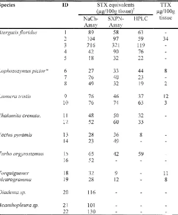
![Table 3.6 Structure-activity relationships between STX derivatives and concentration standardised protein extracts of animals identified with STX binding activity as determined by competitive binding with 2nM [3H] STX in the MCE receptor assay](https://thumb-us.123doks.com/thumbv2/123dok_us/228079.57276/124.843.101.745.140.392/structure-relationships-derivatives-concentration-standardised-identified-determined-competitive.webp)


Related documents
Political Parties approved by CNE to stand in at least some constituencies PLD – Partido de Liberdade e Desenvolvimento – Party of Freedom and Development ECOLOGISTA – MT –
In the previous sections, we dis- cuss the expectation that a neural network exploiting the fractional convolution should perform slightly worse than a pure binary (1-bit weights
• Our goal is to make Pittsburgh Public Schools First Choice by offering a portfolio of quality school options that promote high student achievement in the most equitable and
The largest transactions in the third quarter of 2015 on the Polish M&A market were the acquisition of PKP Energetyka SA by the CVC Capital Partners and the acquisition of
ter mean to the prototypes computed from the true labels of all the samples. Similar to the semi-supervised scenario, we use a PN trained in the episodic mode as the feature
We also deal with the question whether the inferiority of the polluter pays principle in comparison to the cheapest cost avoider principle can be compensated
Comments This can be a real eye-opener to learn what team members believe are requirements to succeed on your team. Teams often incorporate things into their “perfect team
During the thesis work, I measured six different parameters: the number of emergency processes, hash table entry number, caching replacement policy, cache entry
