Anti Retroviral therapy (ART)
Dissertation submitted in partial fulfilment of the
University regulations for the award of the degree of
Doctor of Medicine
(M.D General Medicine)
Branch I
Of
THE TAMILNADU Dr.M.G.R. MEDICAL
UNIVERSITY,
CHENNAI, INDIA.
DECLARATION
I solemnly hereby declare that this dissertation entitled
“Immune
Reconstitution Inflammatory Syndrome (IRIS) in HIV
positive patients on Anti Retroviral therapy (ART)”
has beenundertaken by me at the Madras Medical College, chennai during 2008 -
2011 under the guidance of Dr.E.Dhandapani, professor of medicine,
Dr. Ragunanthanan, Professor of Medicine and under the supervision of
Dr.Rajendiran, Professor and HOD, Department of Medicine, Madras
Medical College, Chennai in partial fulfilment of the university regulations
for the award of the degree of Doctor of Medicine (M.D. Medicine). This
has not been submitted previously by me to any other University.
Place : Chennai
At the outset, I thank Prof.MOHANASUNDARAM, MD, DNB,
Ph.D. Dean, Madras Medical College and Government General Hospital
Chennai-03 for having permitted me to use the hospital material for the
study
I would like to express my sincere gratitude to my teacher
Dr. E. Dhandapani, Professor of Medicine, Institute of Internal Medicine
and Dr.Ragunanthanan, Professor of Medicine, Institute of Internal
Medicine, Madras Medical College, chennai for their guidance,
encouragement and timely advice without which this work would not have
been possible.
My special thanks to Prof.Dr.C.Rajendiran, Director and Head of
Department, Institute of Internal Medicine, Madras Medical College,
Chennai for his constant encouragement and guidance.
I would also like to thank my Asst.Professors Dr.Alexander, M.D.
and Dr.Shanthi, M.D. Department of Medicine, Institute of Internal
Medicine and all my colleagues for their help during this study.
I am indebted to my family and friends who have never failed to
support me at all times.
My special thanks to all patients in the study, for their participation
This is to certify that Dr. Sivaprakash .v has undergone the
prescribed course of studies leading to the M.D Degree examination in
Medicine in accordance with the rules and regulations of the Dr.M.G.R.
University and the dissertation entitled
“Immune Reconstitution
Inflammatory Syndrome (IRIS) in HIV positive patients on
Anti Retroviral therapy (ART)”
is a bonafide work.PROF. DR C.RAJENDIRAN, M.D.,
Professor and Director, Institute of Internal Medicine, Madras Medical College,
Chennai – 03.
PROF.E.DHANDAPANI, M.D.,
Professor of Medicine,
Institute of Internal Medicine, Madras Medical College, Chennai – 03.
CONTENTS PAGE
1. INTRODUCTION 1
2. AIMS AND OBJECTIVES 9
3. REVIEW OF LITERATURE 10
4. MATERIALS AND METHODOLOGY 30
5. RESULTS & ANALYSIS 32
6. DISCUSSION 46
7. CONCLUSION 49
8. LIMITATIONS & RECOMMENDATIONS 51
9. BIBILOGRAPHY 53
10. ANNEXURE 1 : PROFORMA 62
11. ANNEXURE 2 : MASTER CHART – Cases 68
INTRODUCTION
Antiretroviral therapy (ART) has led to a significant decline in AIDS -
associated morbidity and mortality (1). These benefits are in part a result of
partial recovery of the immune system, manifested by increase in CD4
T-lymphocytes counts and decrease in plasma HIV-1 viral loads
(2). In some patients clinical deterioration occurs despite increased CD4
T-lymphocytes and decreased plasma HIV-1 viral loads
(3). This clinical deterioration is due to inflammatory response of the
immune system to both subclinical pathogens and residual antigens.
In the mid-1990s, clinicians noticed that certain patients deteriorated after
starting HAART despite having decreasing HIV-1 RNA levels and rising CD4
cell counts [2-5]. In these patients, receipt of HAART results in a pathological
inflammatory response to either previously treated infections or subclinical
infections [6-8]. This inflammation could result in deleterious clinical outcomes,
such as culture-negative meningitis or necrotizing lymphadenitis; it has been
labeled as immune reconstitution disease (IRD) or immune reconstitution
inflammatory syndrome (IRIS) [9-11].
Immune Reconstitution Inflammatory Syndrome (IRIS) is defined as a
attributable to the recovery of the immune response to latent or subclinical
infection or non-infectious processes.
Despite numerous descriptions of the manifestations its pathogenesis
remains speculative. Current theories concerning the pathogenesis of IRIS
involves a combination of underlying antigenic burden, a degree of immune
restoration following anti-retroviral therapy and host genetic susceptibility.
Immune Reconstitution Inflammatory Syndrome (IRIS) that occurs as a
result of “unmasking” of clinically silent infection is characterized by atypical
exuberant inflammation or an accelerated clinical presentation suggesting a
restoration of antigen-specific immunity.
Following anti- retroviral therapy an increase in memory CD4 cell types
is observed (4) possibly as a result of redistribution from peripheral lymphoid
tissue (5). This CD4 is primed to recognize previous antigenic stimuli and be
responsible for the manifestations of Immune Reconstitution Inflammatory
Syndrome (IRIS) seen soon after initiation of Anti Retroviral therapy.
The best described associations between particular infectious agents and
IRIS include ophthalmic cytomegalovirus (CMV) disease, disseminated
infection with Mycobacterium tuberculosis or Mycobacterium avium complex,
Risk Factors :
• Advanced HIV disease - CD4 counts <50
• Unrecognized Opportunistic infection or high microbial burden
• Early initiation of HAART
• ARV naïve
• Immune recovery with rapid fall in HIV RNA
• Genetic factors which can be pathogen specific
Mycobacteria – TNF-308*2, IL6 – 174*G
Antiretroviral Therapy Improves
Qualitative and Quantitative Immune
Defects
Immune suppression/deficiency ↑HIV replication ↑Immune activation ↑Qualitative/functio nal immune defects ↓Response to recallantigens
↑Quantitative immune defects
↓↓CD4 counts
Impaired pathogen -specific immunity OI HAART ↓HIV replication ↓Immune activation ↓Qualitative/function al immune defects
Reversal of anergy
↑Lymphocyte proliferative capacity
↓Quantitative immune defects
Redistribution, ↓death (HIV-, activation-induced),
↑production (peripheral expansion and thymic)
Improved pathogen-specific immunity Immune Reconstitution Improved immune control
ART and the treatment of OIs
Patient with OI Treated with ART
Asymptomatic immune recovery
Return of original
symptoms New Symptoms
ART with subclinical infection
ART in advanced
HIV disease
Asymptomatic Immune recovery
TB- IRIS :
• Well recognized phenomenon for decades
• Lymphadenitis (12 – 25 %),
• 1 – 6 months post initiation of therapy
• Pulmonary disease, central nervous system-new tuberculomas, fevers, ARDS
• 75% have worsening of original lesions
• Often required steroids
• Due to intensification of the cell mediated immune response and conversion of TST
• Concomitant rise in TNF levels
We have very few studies about Immune Reconstitution Inflammatory
Syndrome and most of it is Western Studies.
The incidence, risk factors and presentation of IRIS may differ in our
Indian context.
As we have very few Indian studies about IRIS, a study is planned to find
the clinical spectrum of IRIS in HIV positive patients visiting a tertiary care
centre at the chennai.
The present study was undertaken to determine the incidence of IRIS in
long-We hypothesized that patients who started HAART in closer proximity to
the diagnosis of their underlying opportunistic infection and who had a more
robust response to HAART in terms of increasing CD4 levels would be at an
Aim :
To study the profile of Immune Reconstitution Inflammatory Syndrome
(IRIS) in HIV positive patients on Anti Retroviral therapy (ART)
Objectives :
1. Type of Opportunistic Infections
2. Correlation with the CD 4 count
REVIEW OF LITERATURE
Immune Reconstitution Inflammatory Syndrome (IRIS)
o IRIS can be described as an adverse clinical phenomenon
following rapid restoration of immune function in a previously
severely immune-compromised individual.
o This is not specific to HIV positive persons on ART but can follow
recovery from neutropenia (chemotherapy/transplantation) or dose
reduction/withdrawal of steroids.
o The syndrome is well-described for many bacteria, virus, fungi,
protozoa, helminth, virus related malignancy and non-infectious
processes.
o In HIV-infected patients, IRIS can be described as a deterioration
of the opportunistic infection due to restoration of pathogen
specific immune responses.
DIAGNOSIS OF IRIS :
In India, the agreed practical definition of IRIS would be the “occurrence
or manifestations of new opportunistic infections or existing opportunistic
infections within six weeks to six months after initiating anti-retroviral
therapy with an increase in CD4 count”
‐ Typically, IRIS occurs within 2–12 weeks of the initiation of ART,
although it may present later (usually between 6 weeks to 6 months)
‐ The incidence of IRIS is estimated to be 10% among all patients in
whom ART has been initiated; and up to 25% among those who have
started ART and who have a CD4 cell count of below 50 cells/mm3
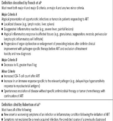
FIG 3. CASE DEFINITIONS OF IRIS
UNUSUAL PRESENATIONS OF IRIS :
o Unexpected localized disease, e.g. lymph nodes (appearance or
enlargement and/or suppuration), or involving liver or spleen
o Exaggerated inflammatory reaction, e.g. severe fever, with exclusion of
other causes
o Painful lesions
o Atypical inflammatory response in affected tissues, e.g. granulomas,
suppuration, necrosis
o Perivascular lymphocytic inflammatory cell infiltrate
o Progression of organ dysfunction or enlargement of pre-existing lesions
o Development or enlargement of cerebral space-occupying lesions after
treatment for cerebral cryptococcosis or toxoplasmosis
o Progressive pneumonitis or the development of organizing pneumonia
after treatment for pulmonary MTB or PCP
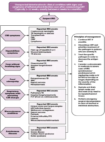
FIG 4. DIAGNOSTIC PROTOCOL FOR IRIS
FIG
5.
SUMMARY
OF
INCIDENCE
AND
RISK
FACTORS
OF
IRIS
IN
[image:22.612.132.496.71.547.2]
FIG
6.
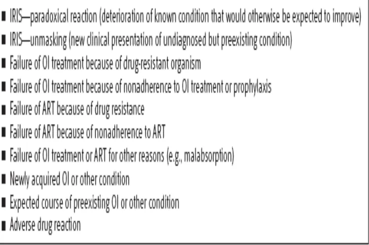
DIFFERENTIAL
DIAGNOSIS
OF
IRIS
[image:23.612.120.494.136.385.2]
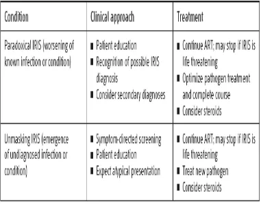
FIG
7.
GENERAL
APPROACH
TO
IRIS
[image:24.612.116.497.74.372.2]
FIG
8.
MANAGEMENT
OF
IRIS
[image:25.612.119.509.78.545.2]
Treatment of IRIS :
o There are no standard guidelines for the treatment of IRIS
o Milder forms of IRIS resolve with continuing anti-infective therapy and
anti-retroviral therapy
o In the majority of cases, ART can be safely continued and need not be
interrupted
o In general, most clinicians prefer to continue ART if the CD4 count is
below 100/mm3 or if the patient presents with IRIS months after the
initiation of ART
o However, the discontinuation of ART should be considered if the
inflammatory responses are considered life-threatening (e.g. intracranial
IRIS leading to encephalitis, cerebritis, perilesional cerebral oedema and
pulmonary IRIS with ARDS/acute respiratory distress syndrome), or are
unresponsive to steroids
o Discontinuation of the treatment should also be considered if the
pathogens involved are not amenable to specific antimicrobials (e.g.
Parvovirus B19, polyomavirus JC causing progressive multifocal
o Hepatitis →uncertainty about contribution of drug toxicity
o Skin eruptions – usually possible to differentiate cutaneous IRIS from
drug rash.
o Non-steroidal anti-inflammatory drugs (NSAIDs) are helpful in
controlling inflammation and fever associated with IRIS
o However, in severe IRIS, a short course of oral prednisolone is required
to alleviate the symptoms
o The dosage and duration of treatment required is variable and should be
judged clinically. Severe disease requires at least 1–2 mg of prednisolone
per kg body weight
o In a study conduted by Shelburne et all ,To determine whether patients
with IRIS require more interventions to prevent morbidity and mortality,
they collected data regarding invasive procedures and hospitalizations
during the first year following initiation of HAART as surrogate markers
for healthcare utilization.
o In the 12 months after starting HAART, patients with IRIS required
increased numbers of invasive procedures, such as lumbar punctures to
relieve increased intracranial pressure, and had a higher number of
intensification of their healthcare, thereby suggesting that preventive
strategies might be cost effective. Such strategies might be especially
effective in developing countries where coinfection with C. neoformans
or M. tuberculosis is relatively common and the ability to manage
complex paradoxical reactions readily may be limited [51].
Course of IRIS :
o Although there may be short-term morbidity associated with IRIS, these
patients appear to have comparably good long-term outcomes. After 24
months of HAART, patients with IRIS were more likely to have
successful viral suppression and immune reconstitution than patients
without the syndrome. In addition, there was no significant mortality
difference between the two groups of patients. In fact, the survival trend
was in favor of the IRIS patients, which is likely a reflection of the
durable viral suppression and immune reconstitution seen in these
Previous Studies :
In a study by I. Ratnam, C. Chiu, N.-B. Kandala, and P. J. Easterbrook
from Department of HIV/Genitourinary Medicine, King’s College London,
Guy’s, King’s College and St. Thomas’ Hospitals, London, United Kingdom did
a retrospective study of all patients starting HAART between 1 January 2000
and 31 August 2002at a human immunodeficiency virus (HIV) clinic in London
where a total of 199 patients were included, of whom 50.8% were male, 59.3%
were black African, 29.1% were white, and 10.5% were black Caribbean. The
median baseline CD4 cell count and HIV RNA load were174 cells/L and 37,830
copies/mL respectively. Forty-four patients (22.7%) experienced an IRIS event
at a median of 12 weeks after HAART initiation ; 22 events (50%) involved
genital herpes, 10 (23%) involved genital warts, 4 (9.0%) involved molluscum
contagiosum, and 4 (9.0%) involved varicella zoster virus infection. Five
patients had mycobacterial infections, 4 had hepatitis B, 1 had Pneumocystis
jirovecci infection, and 1 had Kaposi sarcoma. The strongest independent
predictors of IRIS were younger age at initiation of HAART (Pp.003), baseline
CD4 cell percentage (odds ratio [OR], 2.97) and ratio of CD4 cell percentage
to CD8 cell.
Murdoch and David M did a prospective surveillance cohort and nested
case-control study in a large university hospital-based antiretroviral therapy
patients were followed for signs and symptoms IRIS during the first 6 months of
ART which was published in Journal of International of AIDS Society. During
the first 6 months of ART, 44 (10.4%) patients experienced IRIS for an overall
incidence rate of 25.1 cases per 100 patient-years. Diagnoses included
tuberculosis (18/44, 41%), abscess formation and suppurative folliculitis
(8/44, 18.2%), varicella zoster (6/44, 13.6%), herpes simplex (4/44, 9.1%),
cryptococcal meningitis (3/44, 6.8%), molluscum contagiosum (3/44, 6.8%),
and Kaposi's sarcoma (2/44, 4.5%). Median IRIS onset was 48 days
(interquartile range, 29-99) from ART initiation. In comparison with controls,
IRIS cases had significantly lower CD4 cell counts at baseline (79 versus 142
cells/μl; P= 0.02) and at IRIS diagnosis (183 versus 263 cells/μl; P = 0.05), but
similar virological and immunological response to ART. In multivariable
analyses, higher baseline CD4 cell count was protective of developing IRIS (HR
0.72 per 50 cells/μl increase). Most IRIS cases were mild, with ART
discontinued in three (6.8%) patients, corticosteroids administered to four (9.1%)
patients, and hospitalization required in 12 (27.3%) patients. Two deaths were
attributable to IRIS.
Weerawat Manosuthi , Sasisopin Kiertiburanakul, Thanongsri Phoorisri,
Somnuek Sungkanuparph did a retrospective study in Bamrasnaradura Infectious
Diseases Institute and Ramathibodi Hospital, Thailand were 167 patients with a
months after TB treatment. IRIS was identified in 21 (12.6%) patients. Patients
with IRIS had a higher proportion of extrapulmonary TB than patients without
IRIS (P < 0.001). By multivariate analysis, extrapulmonary TB was a risk factor
for IRIS (odds ratio ¼ 8.225, 95% confidence interval ¼ 1.785e37.911, P ¼
0.007). Of 21 patients with IRIS, 15 patients developed IRIS within the first two
months of ART. The mortality rate in patients with and without IRIS was not
different.
Shelburne and Samuel did a retrospective cohort identified through a
city-wide prospective surveillance program where a retrospective chart review
was performed for 180 HIV-infected patients who received HAART and were
coinfected with Mycobacterium tuberculosis,Mycobacterium avium complex, or
Cryptococcus neoformans between 1997 and 2000. Medical records were
reviewed for baseline demographics, receipt and type of HAART, response to
antiretroviral therapy, development of IRIS, and long-term outcome. In this
cohort, 31.7% of patients who received HAART developed IRIS. Patients with
IRIS were more likely to have initiated HAART nearer to the time of
diagnosis of their opportunistic infection (P < 0.001), to have been
antiretroviral naive at time of diagnosis of their opportunistic infection (P <
0.001), and to have a more rapid initial fall in HIV-1 RNA level in response to
Lawn and Stephen did a retrospective analysis of a study cohort enrolled
over 3 years within a community-based ART service in South Africa. Patients
receiving treatment for TB at the time ART was initiated (n = 160) were studied.
Cases of TB-associated IRD during the first 4 months of ART were ascertained.
The median baseline CD4 cell count was 68 cells/μl and ART was initiated after
a median of 105 days from TB diagnosis. Although IRIS was diagnosed in just
12% (n = 19) of patients overall, IRIS developed in 32% (n = 12) of those who
started ART within 2 months of TB diagnosis. Pulmonary involvement was
observed in 84% (n = 16) and intra-abdominal manifestations were also
common (37%). Overall, 4% (n = 7) of the cohort required secondary level
health-care for IRIS and two (1%) patients died. In multivariate analysis, risk of
IRIS was strongly associated with early ART initiation and low baseline CD4
cell count. Of patients with CD4 counts < 50 cells/μl, the proportions who
developed IRD following initiation of ART within 0-30, 31-60, 61-90, 91-120
and > 120 days of TB diagnosis were 100%, 33%, 14%, 7% and 0%,
respectively.
Manabe, Yukari C, Campbell, James D, Sydnor, Emily, Moor and
Richard D did a study which was published in Journal of Acquired Immune
Deficiency Syndromes Dec 2001. Here patients from the Johns Hopkins HIV
Clinic who had IRIS were identified and matched with 4 controls without IRIS
HAART (range: 4 to 186 days). A multivariate analysis showed that the
development of IRIS was independently associated with using a boosted
protease inhibitor (BPI) (odds ratio [OR] = 7.41; P = 0.006), a nadir CD4
count <100 cells/mm3 (OR = 6.2; P < 0.001), and a plasma HIV viral RNA
decrease of more than 2.5 log at the time of IRIS compared with RNA levels
before the initiation of HAART. Incrementally greater decreases in viral loads
directly correlated with increased risk for the development of IRIS.
TB – IRIS :
• Paradoxical reactions have been seen in TB prior to HIV thus IRIS
phenomena in coinfected pts may have been under reported
• 29 - 36 % coinfected pts on TB Rx and HAART develop clinically
apparent IRIS
• Radiologic deterioration in 46%
• More frequent in HIV+ than HIV – patients
• 36% (12/33) Narita M, et al. AJRCCM 1998;158:157.
• 32% (6/19) Navas E, et al. ICAAC, 1999.
• 6% (6/82) Wendel K, et al Chest 2001;120:193.
• 30.2% (26/86) Shelburne S, et al AIDS 2005; 19:399
• Majority of cases of IRIS occurred in pts who were being treated for
TB when HAART initiated
• Duration of TB Rx median = 2 months prior to IRIS presentation
• Duration of HAART median = 1month prior to IRIS presentation
• 50% with undetectable HIV RNA at time of IRIS
• Median CD4# 205 from nadir of 51 ( 26 – 103 )
MATERIALS AND METHODOLODY
This study is a case control study done on HIV positive patients on
Antiretroviral Therapy at Government General Hospital at Chennai.
Immune Reconstitution Inflammatory Syndrome (IRIS) cases were
diagnosed according to the latest NACO guidelines.
The cases included HIV positive patients who developed Immune
Reconstitution Inflammatory Syndrome (IRIS) during Antiretroviral therapy
(ART) and the controls included patients who did not develop IRIS during the
ART.
Inclusion Criteria :
1). HIV positive patients on Anti Retroviral therapy of age > 18yrs with
an increase in CD4 count
2). Occurrence of opportunistic infection (New OI) or worsening of
symptoms (Existing OI) within 6 weeks to 6 months after initiation of
Exclusion Criteria :
1). Patient not on Anti Retroviral Therapy
2). Poor adherence
3). Defaulters
RESULTS AND ANALYSIS
This was a case control study where 50 IRIS cases were diagnosed in
HIV positive patients on Antiretroviral therapy based on the NACO guidelines
both retrospectively and prospectively which occurred between May 2008 to
May 2010 and compared with 80 controls who were HIV positive patients on
Antiretroviral therapy but did not develop IRIS.
The cases and controls were compared based on Age, Sex, Initial
CD4 count, Final CD4 count, CD4 rise, Duration of ART, Type of ART and
then the opportunistic infections and their relation to CD4 count was analysed.
CHART 1 : AGE DISTRIBUTION
• The mean age of the cases is 36
CHART 2 : SEX DISTRIBUTION
• In cases, 76% are males and 24% are females
•
In controls, 65% are males and 35% are females7
24
24
76
76
76
CHART 3 : CD4 DISTRIBUTION
In cases, the mean initial CD4 count is 135
the mean final CD4 count is 239
the mean CD4 count rise is 115
In controls, the mean initial CD4 count is 116
the mean final CD4 count is 222
the mean CD4 count rise is 103
115
CHART 4 : MEAN DURATION OF ART
In cases the mean duration of ART was 4 months
In controls the mean duration of ART was 4.5 months
3.6 3.8 4 4.2 4.4 4.6
CASES
CONTROLS 4
CHART 5 : TYPE OF ART
In cases - 64% were on L+S+N regimen
24% were on Z+L+N regimen
8% were on L+S+E regimen
4% were on Z+L+E regimen
In controls - 54% were on L+S+N regimen
46% were on Z+L+N regimen
64
8
24
CHART 6 : TYPE OF OPPORTUNISTIC INFECTION IN THE
DIAGNOSED IRIS PATIENTS
22% with Pulmonary tuberculosis
18 with Tuberculous lymphadenitis
16% with Pnuemocystis Jiroveci Pnuemonia
10% with Cryptococcal Meningitis
10% with Herpes Zoster
8% with Disseminated Tuberculosis
6% with Tuberculous Meningitis
2% with Cytomegalovirus retinitis, 2% with Oesophageal candidiasis, 2% with Aspergilloma, 2% with Non Hodgkins Lymphoma and2% with Acute Transverse Myelitis
0 5 10 15 20 25
PULMONARY TUBERCULOSIS TUBERCULOUS LYMPHADENITIS DISSEMINATED TUBERCULOSIS TUBERCULOUS MENINGITIS PNEUMOCYSIS JIROVECI PNEUMONIA HERPES ZOSTER CRYPTOCOCCAL MENINGITIS CYTOMEGALOVIRUS RETINITIS ASPERGILLOMA
ACUTE TRANSVERSE MYELITIS
CHART
7
:
TB
IRIS
AND
OTHER
OPPORTUNISTIC
INFECTIONS
• 54% of the diagnosed IRIS patients had tuberculosis
•
46% constituted rest of all the opportunistic infections
CHART 8 : SUBTYPES OF TB IRIS
• 41% with Pulmonary tuberculosis
• 33% with Tuberculous lymphadenitis
• 15% with Disseminated tuberculosis
•
11% with Tuberculous meningitis43
33
14
10
PULMONARY
TUBERCULOSIS
TUBERCULOUS
LYMPHADENITIS
DISSEMINATED
TUBERCULOSIS
TUBERCULOUS
MENINGITIS
41
15
11
CHART 9 : MEAN DURATION BETWEEN ATT AND ART OF
TB IRIS PATIENTS
•
The mean duration between ATT and ART of TB IRIS patients is 2.5months
0 0.5 1 1.5 2 2.5
2.5
DURATION
OF ART IN
MONTHS
INITIAL CD4 COUNT
CHART 10 : CORRELATION BETWEEN INITIAL
CD4 COUNT AND THE DEVELOPMENT OF IRIS
•
There is a weak correlation between the initial CD4 count and thedevelopment of IRIS
0 1 2 3 4 5 6 7
DURATION
OF ART
IN MONTHS
CD4 COUNT RISE
CHART 11 : CORRELATION BETWEEN THE CD4 COUNT
RISE AND DEVELOPMENT OF IRIS
• There is a weak correlation between the CD4 count rise and the
development of IRIS
0 1 2 3 4 5 6 7
RESULTS :
o The mean age of the IRIS patients was 36yrs.
o 76% of the patients were of male sex and the rest 24% were of
female sex.
o The mean initial CD4 count was 135.
o The mean final CD4 count was 239.
o The mean CD4 count rise was 115.
o The mean duration of ART in IRIS patients was 4 months.
o 64% of the IRIS patients were on Lamivudine + Stavudine +
Nevirapine Regimen, 24% of them on Zidovudine + Lamivudine
+Nevirapine regimen, 8% were on Lamivudine + Stavudine +
Efavirenz and 4% were on Zidovudine + Lamivudine + Efavirenz.
o The most common opportunistic infection was the Pulmonary
Tuberculosis, secondly Tuberculous Lymphadenitis and thirdly
Pneumocysitis Jiroveci Pneumonia.
o Among Tuberculosis patients 41% of the patients had Pulmonary
Tuberculosis, 33% of the patients had Tuberculous
Lymphadenitis, 15% had Disseminated Tuberculosis and 11% had
Tuberculous Meningitis.
o In TB IRIS patients the mean duration between ATT initiation and
ART initiation was 2.5 months.
o There is a weak correlation between initial CD4 count and the
development of IRIS.
o There is a weak correlation between CD4 count rise and the
development of IRIS.
DISCUSSION :
In this study a total of 50 cases of HIV postive patients on ART who
developed IRIS were identified and were matched with 80 controls of HIV
patients on ART who did not develop IRIS .
Following matching which was done for Age, Sex, CD4 count and Type
of ART the opportunistic infections and their relationship to CD4 count was
analysed.
o In this study the most common opportunistic infection was
Mycobacterium Tuberculosis followed by Pnuemocystis
infection. This is in accordance with the study by Narita M (8)
where Mycobacterium Tuberculosis is the most common
opportunistic infection in IRIS.
o In Tuberculosis, Pulmonary tuberculosis was the most common
opportunistic infection, secondly Tuberculous lymphadenitis
followed by Disseminated Tuberculosis. This is in accordance
with the study done by Lawn SD (9) where pulmonary tuberculosis
was the most common type of TB IRIS.
o In our study, patients diagnosed with IRIS initiated HAART in
compared with patients who did not develop IRIS. This is
consistent with a previous report of 17 HAART-treated patients
coinfected with M. tuberculosis and HIV [10]
o Biological reasons for this association are unclear at present,
although we speculate that patients who receive prolonged therapy
for their opportunistic infection prior to starting HAART will have
decreased microbial antigen burdens when HAART is initiated.
This, in turn, would provide less material to stimulate a
reconstituting immune system once HAART is begun. These
concerns may lend added support to the recent recommendations
to consider delaying HAART for 4-8 weeks after starting M.
tuberculosis therapy in coinfected patients [42].
o TB IRIS occurred most commonly 2.5 months after initiating
o There was weak correlation between a lower CD4 count or higher
CD4 rise and the incidence of IRIS. This result was in accordance
from the study by Shelburne, Samuel A(51) where a significant
association between CD4 cell count rise and the diagnosis of IRIS
was not seen until later in therapy .
o It has been noted that reductions in HIV-1 RNA levels in response
to HAART result initially in redistribution of memory CD4
lymphocytes
o This redistribution of activated CD4 lymphocytes may be,atleast
partly responsible for the manifestations of IRIS, which could
explain why the rise in CD4 cell count appears delayed compared
with the viral load decrease
o This result was not in accordance from the study by Lenzo N (11)
where there was a strong association between a lower CD4 count
CONCLUSION:
o In our study the most common opportunistic infection in Immune
Reconstitution Inflammatory Syndrome in HIV positive patients
on Antiretroviral therapy is Mycobacterium Tuberculosis
followed by Pnuemocystis infection.
o In Tuberculosis, Pulmonary Tuberculosis was the most common
opportunistic infection followed by Tuberculous lymbphadenitis.
o Here, TB IRIS most commonly occurred 2.5 months after
initiating of anti retroviral therapy.
o There is a weak correlation between the a lower CD4 count and
the development of IRIS.
o There is a weak correlation between the CD4 rise and the
development of IRIS. Hence it is vital to include HIV-1 RNA load
measurement to diagnose IRIS.
o IRIS is a syndrome that occurs because a patient develops an
o The inclusion of IRIS in the differential diagnosis of a patient who
presents with an inflammatory process after initiating HAART
allows for a focused approach to diagnosis and therapy.
o These patients often require significant interventions to minimize
short-term morbidity but their long-term outcome appears
relatively good.
o Further studies looking at how to decrease the rate of IRIS in
high-risk patients appear warranted by its prevalent nature and the
association of IRIS with increased hospitalizations and invasive
LIMITATIONS :
o This is a small scale study where only 50 diagnosed cases of IRIS
are analysed.
o The diagnosis of IRIS was based on only the rise in CD4 count and
did not include a fall in HIV RNA levels.
o Drug Resistance to antiretroviral therapy was not ruled out.
RECOMMENDATIONS :
o A large scale randomised controlled study is recommended.
o Diagnosis of IRIS should include a fall in the HIV RNA levels and a
rise in CD4 count.
o Resistance testing to antiretroviral therapy should be done before a
case is diagnosed as IRIS.
o
Drug resistant opportunistic infection should be ruled out beforediagnosing IRIS
.
o
Studies of early vs. deferred HAART in TB patients may providevaluable information on the optimal timing of HAART.
o Initiate HAART before CD4 drops verylow.
o Exclude OI before starting HAART.
BIBILIOGRAPHY:
1). Declining morbidity and mortality among patients with advanced human
immunodeficiency virus infection. HIV Outpatient Study investigators. (NEJM
1998, 338(13):853-860)
2). Immune reconstitution in HIV infection. (Aids 1999, 13 Suppl A: S25-38)
3). Immune restoration disease after the treatment immunodeficient HIV –
infected patients with highly active antiretroviral therapy. ( HIV Med 2000, I(2):
107-115)
4). Positive effects of combined antiretroviral therapy on CD4 T cell
homeostasis and function in advanced HIV disease. ( Science 1997, 277(5322):
112-116)
5). Initial increase in blood CD4(+) lymphocytes after HIV antiretroviral therapy
reflects redistribution from lymphoid tissues (J Clin Invest 1999,
103(10):1391-1398)
6). NACO – Antitetroviral therapy guidelines for the HIV infected adults and
adolescents and post exposure prophylaxis
7). Science and Treatment of HIV infection – Immune Reconstitution
8). Narita M, Ashkin D, Hollender ES, Pitchenik AE: Paradoxical worsening of
tuberculosis following antiretroviral therapy in patients with AIDS. Am J Respir
Crit Care Med 1998,158(1):157-161
9). Lawn SD, Bekker LG, Miller RF: Immune reconstitution disease associated
with mycobacterial infections in HIV-infected individuals receiving
antiretrovirals. Lancet Infect Dis 2005, 5(6):361-373
10). Navas E, Martin-Davila P, Moreno L, Pintado V, Casado JL, Fortun J,
Perez-Elias MJ, Gomez-Mampaso E, Moreno S: Paradoxical reactions of
tuberculosis in patients with the acquired immunodeficiency syndrome who are
treated with highly active antiretroviral therapy. Arch Intern Med 2002,
162(1):97-99
11). Lenzo N, French MA, John M, Mallal SA, McKinnon EJ, James IR, Price P,
Flexman JP, Tay-Kearney ML: Immune restoration disease after the treatment of
immunodeficient HIV-infected patients with highly active antiretroviral therapy.
HIV Med 2000, 1(2):107-115
12). Adult Prevention and Treatment of Opportunistic Infections Guidelines
Working Group. Guidelines for Prevention and Treatment of Opportunistic
Infections in HIV-Infected Adults and Adolescents [DRAFT]. June 18, 2008; pp
13). Karavellas MP, Plummer DJ, Macdonald JC, et al. Incidence of immune
recovery vitritis in cytomegalovirus retinitis patients following institution of
successful highly active antiretroviral therapy. J Infect Dis 1999;179:697-700.
14).Breton G, Duval X, Estellat C, et al. Determinants of immune reconstitution
inflammatory syndrome in HIV type 1-infected patients with tuberculosis after
initiation of antiretroviral therapy. Clin Infect Dis 2004;39:1709-1712
15). Battegay M, Nüesch R, Hirschel B, et al. Immunological recovery and
antiretroviral therapy in HIV-1 infection. Lancet Infect Dis 2006;6:280-287.
16). Shankar EM, Vignesh R, Velu V, et al. Does CD4+CD25+foxp3+ cell
(Treg) and IL-10 profile determine susceptibility to immune reconstitution
inflammatory syndrome (IRIS) in HIV disease? J Inflamm (Lond) 2008;5:2.
17). Boulware D, Meya D, Bergemann T, et al. Inflammatory biomarkers in
serum predict HIV immune reconstitution inflammatory syndrome and death
after cryptococcal meningitis. Sixteenth Conference on Retroviruses and
Opportunistic Infections. February 8-11, 2009, Montreal Canada.
18). Schiffer JT, Sterling TR. Timing of antiretroviral therapy initiation in
tuberculosis patients with AIDS: A decision analysis. J Acquir Immune Defic
19). French MA, Price P, Stone SF. Immune restoration disease after
antiretroviral therapy. AIDS 2004; 18:1615-1627.
20). Robertson J, Meier M, Wall J, et al. Immune reconstitution syndrome in
HIV: Validating a case definition and identifying clinical predictors in persons
initiating antiretroviral therapy. Clin Infect Dis 2006;42:1639-1646.
21).Lortholary O, Fontanet A, Mémain N, et al. for the French Cryptococcosis
Study Group. Incidence and risk factors of immune reconstitution inflammatory
syndrome complicating HIV-associated cryptococcosis in France. AIDS
2005;19:1043-1049.
22).Gray F, Bazille C, Adle-Biassette H, et al. Central nervous system immune
reconstitution disease in acquired immunodeficiency syndrome patients
receiving highly active antiretroviral treatment. J Neurovirol 2005;11(Suppl
3):16-22.
23). Breen RA, Smith CJ, Bettinson H, et al. Paradoxical reactions during
tuberculosis treatment in patients with and without HIV co-infection. Thorax
2004;59:704-707.
24). Phillips P, Bonner S, Gataric N, et al. Nontuberculous mycobacterial
immune reconstitution syndrome in HIV-infected patients: Spectrum of disease
25).Ramirez-Amador VA, Espinosa E, Gonzalez-Ramirez I, et al. Identification
of oral candidosis, hairy leukoplakia, and recurrent oral ulcers as distinct cases
of immune reconstitution inflammatory syndrome. Int J STD AIDS
2009;20:259-261.
26). Shelburne SA 3rd, Hamill RJ, Rodriguez-Barradas MC, et al. Immune
reconstitution inflammatory syndrome: Emergence of a unique syndrome during
highly active antiretroviral therapy. Medicine 2002;81:213-227.
27). . Bucy RP, Hockett RD, Derdeyn CA, Saag MS, Squires K, Sillers M, et al.
Initial increase in blood CD4(+) lymphocytes after HIV antiretroviral therapy
reflects redistribution from lymphoid tissues. J Clin Invest 1999; 103:1391-1398.
28). Improved outcomes of HIV-1-infected adults with tuberculosis in the era of
Highly active antiretroviral therapy. [AIDS. 2003]
29). Orlovic D, Smego RA Jr. Paradoxical tuberculous reactions in HIV-infected
patients. Int J Tuberc Lung Dis 2001; 5:370-375.
30). Chuck SL, Sande MA. Infections with Cryptococcus neoformans in the
acquired immunodeficiency syndrome. N Engl J Med 1989; 321:794-799.
31). . Wendel KA, Alwood KS, Gachhi R, Chaisson RE, Bishai WR, Sterling
TR. Paradoxical worsening of tuberculosis in HIV-infected persons. Chest 2001;
32). Price P, Mathiot N, Krueger R, Stone S, Keane NM, French MA. Immune
dysfunction and immune restoration disease in HIV patients given highly active
antiretroviral therapy. J Clin Virol 2001; 22:279-287.
33). Woods ML 2nd, MacGinley R, Eisen DP, Allworth AM. HIV combination
therapy: partial immune restitution unmasking latent cryptococcal infection.
AIDS 1998; 12:1491-1494.
34). Hirsch HH, Kaufmann G, Sendi P, Battegay M. Immune reconstitution in
HIV-infected patients. Clin Infect Dis 2004; 38:1159-1166.
35). DeSimone JA, Pomerantz RJ, Babinchak TJ. Inflammatory reactions in
HIV-1-infected persons after initiation of highly active antiretroviral therapy.
Ann Intern Med 2000; 133:447-454.
36). Cheng VC, Yuen KY, Chan WM, Wong SS, Ma ES, Chan RM.
Immunorestitution disease involving the innate and adaptive response. Clin
Infect Dis 2000; 30:882-892.
37). Carr A, Cooper DA. Restoration of immunity to chronic hepatitis B
infection in HIV-infected patient on protease inhibitor. Lancet 1997;
38). Immune reconstitution inflammatory syndrome of tuberculosis among
=IV-infected patients receiving antituberculous and antiretroviral therapy. Infect;
53(6):357-63. ">[J Infect. =006]
39). John M, French MA. Exacerbation of the inflammatory response to
Mycobacterium tuberculosis after antiretroviral therapy. Med J Aust 1998;
169:473-474.
40) Mocroft A, Ledergerber B, Katlama C, Kirk O, Reiss P, d'Arminio Monforte
A, et al. Decline in the AIDS and death rates in the EuroSIDA study: an
observational study. Lancet 2003; 362:22-29
41). Palella FJ Jr, Chmiel JS, Moorman AC, Holmberg SD. Durability and
predictors of success of highly active antiretroviral therapy for ambulatory
HIV-infected patients. AIDS 2002; 16:1617-1626.
42). American Thoracic Society/Centers for Disease Control and
Prevention/Infectious Diseases Society of America. Treatment of tuberculosis.
MMWR Recomm Rep 2003. 52:1-77.
43). Cinti SK, Armstrong WS, Kauffman CA. Case report. Recurrence of
increased intracranial pressure with antiretroviral therapy in an AIDS patient
44). Race EM, Adelson-Mitty J, Kriegel GR, Barlam TF, Reimann KA, Letvin
NL, et al. Focal mycobacterial lymphadenitis following initiation of
protease-inhibitor therapy in patients with advanced HIV-1 disease. Lancet 1998;
351:252-255.
45). Brandt ME, Hutwagner LC, Klug LA, Baughman WS, Rimland D, Graviss
EA, et al. Molecular subtype distribution of Cryptococcus neoformans in four
areas of the United States. Cryptococcal Disease Active Surveillance Group. J
Clin Microbiol 1996; 34:912-917.
46). Stone SF, Price P, Tay-Kearney ML, French MA. Cytomegalovirus (CMV)
retinitis immune restoration disease occurs during highly active antiretroviral
therapy-induced restoration of CMV-specific immune responses within a
predominant Th2 cytokine environment. J Infect Dis 2002; 185:1813-1817.
47). von Both U, Laffer R, Grube C, et al. Acute cytomegalovirus colitis
presenting during primary HIV infection: An unusual case of an immune
reconstitution inflammatory syndrome. Clin Infect Dis 2008;46:e38-e40.
48).Acosta RD, Mays BC, Wong RK. Electronic clinical challenges and images
in GI. CMV colitis with immune reconstitution syndrome. Gastroenterology
2008;134:e1-e2.
50). Safdar A, Rubocki RJ, Horvath JA, et al. Fatal immune restoration disease
in human immunodeficiency virus type 1-infected patients with progressive
multifocal leukoencephalopathy: Impact of antiretroviral therapy-associated
immune reconstitution. Clin Infect Dis 2002;35:1250-1257.
51). Incidence and risk factors for immune reconstitution inflammatory
syndrome during highly active antiretroviral therapy Shelburne, Samuel A;
Visnegarwala, Fehmida; Darcourt, Jorge; Graviss, Edward A; Giordano,
Thomas P; White, A Clinton Jr; Hamill, Richard J AIDS: 4 March 2005 -
Proforma :
1). Serial No:
2).I.P No :
3). Name:
4). Age:
5). Sex:
6). Occupation:
7). Place:
8). Duration of HIV +ve status:
9). Duration of ART:
10). Drugs :
Risk factors: Yes No
Male sex
Younger age
ART within 6 weeks
Low baseline CD4 count at ART initiation
Low CD4 cell percentage at ART initiation
Lower CD4:CD8 ratio at ART initiation
Prompt rise of CD4 count
High bacillary burden
Started on Protease Inhibitor (PI)
Disseminated TB/ Extrapulm TB/Advanced TB
CLINICAL FEATURES :
Fever :
Lymhadenopathy :
Skin lesions :
Cough :
Expectoration:
Hemoptysis :
Dyspnoea :
Chest pain:
Palpitation:
Pedal edema:
Abdominal distension:
Loose stools :
Urine output adequate /decreased
Headache :
Neck Stiffness:
Vomiting :
Altered sensorium :
Seizures :
Focal Deficits :
PERSONAL HISTORY:
Smoker yes/no alcohol yes/no
DM yes/no SHT yes/no
CLINICAL FINDINGS:
Pallor:
Ictreus :
Pedal edema:
Lymphnodes :
JVP
Pulse
BMI
CVS
RS
INVESTIGATIONS:
Blood sugar
Urea
creatine
LFT
CHG
T.Chol
LDL
TGL
HDL
ECG
ECHO
CT chest :
Sputum AFB x 2 :
Sputum C/S :
Others :
CD 4 count (Initial)
Clinical
Spectrum
:
1). Mycobacterium Tuberculosis
2). Pneumocystis Carinii Pneumonia
3). Cryptococcous
4). Toxoplasmosis
5). Cytomegalovirus
6). Herpes Virus (Herpes Zoster and Herpes Simplex)
7). Cryptospora and Isospora
8). Hepatits B virus or Hepatits C Virus
9). Progressive multifocal leucoencephelitis
10). Molluscum contagiosum and genital warts
11). Rheumatoid arthritis
12). Systemic Lupus Erythematosus
TREATMENT DETAILS :
ANNEXURE 2 : Masterchart Cases (50)
SR.
NO NAME AGE SEX
INITIAL
CD4
FINAL
CD4 CD 4 RISE
TYPE OF
ART
DURATION
OF ART
(months)
OPPORTUNISTIC
INFECTION
Duration if on
ATT (months)
1.
Kumaran 38 M 105 193 88
L+ S+ N
6
Pulmonary
Tuberculosis 3
2. Selvi 43 F 150 201 51 Z+ L+ N 5 Pulmonary
Tuberculosis 1
3. Babu 41 M 95 118 123 L+ S+ N 6 Pulmonary
Tuberculosis 2
4. Geetha 33 F 248 307 59 L+ S+ N 5 Pulmonary
Tuberculosis 4
5. Jagadesh 34 M 160 180 120 L+ S+ N 2 Pulmonary
Tuberculosis 3
6. Sheela 29 F 256 454 248 L+ S+ N 8 Pulmonary
Tuberculosis 3
7. Ganesh 35 M 42 145 103 Z+ L+ N 2 Pulmonary
Tuberculosis
8. Ayesha 31 F 139 330 191 Z+ L+ N 2 Pulmonary
Tuberculosis
9. Gopal 52 M 186 255 69 L+ S+ N 4 Pulmonary
Tuberculosis 2
10. Mohan 30 M 128 207 79 L+ S+ N 3 Pulmonary
Tuberculosis 2
11. Sampath 42 M 242 374 132 Z+ L+ N 4 Pulmonary
Tuberculosis 3
12. Vasantha 48 F 204 252 78 L+ S+ N 6 Tuberculous
lymphadenitis 4
13. Ashok 35 M 107 177 70 Z + L + N 4 Tuberculous
lymphadenitis
14. Asraf 28 M 124 225 101 L + S + E 6 Tuberculous
lymphadenitis 2
15. Selva 37 M 211 272 61 L+ S+ N 5 Tuberculous
16. Ravi 44 M 102 250 148 L+ S + E 2 Tuberculous lymphadenitis 1
17. Renuka 25 F 182 275 93 Z+ L+ N 6 Tuberculous lymphadenitis
18. Ganamurthy 42 M 122 304 182 L+ S+ N 2 Tuberculous lymphadenitis
19. Manohar 27 M 60 188 128 L+ S +N 2 Tuberculous lymphadenitis
20. Naveenbabu 45 M 102 202 100 L+ S+ N 6 Tuberculous lymphadenitis 1
21. Gajendiran 33 M 27 136 109 Z+ L+ N 4 Disseminated Tuberculous 3
22. Yasim 28 M 101 220 119 L+ S+ N 3 Disseminated Tuberculosis 2
23. Seetha 42 F 370 537 167 L+ S+ E 5 Disseminated Tuberculosis 2
24. Ganesan 40 M 210 315 105 L+ S N 5 Disseminated Tuberculosis 4
25. Partheeban 41 M 97 214 117 Z+ L+ E 4 Tuberculous Meningitis
26. Vinoth 32 M 82 193 111 Z+ L+ N 7 Tuberculous Meningitis
3
27. Pravin 34 M 40 150 90 L+ S+ N 2 Tuberculous Meningitis
28. Jagadesh 34 M 192 264 72 L+ S+ N 6 PCP Pneumonia
29. Ashiya 28 F 32 261 229 L+ S+ N 2 PCP Pneumonia
30. RamaKrishna 30 M 86 154 68 L+ S+ N 6 PCP Pneumonia
31. Narayanan 39 M 300 370 270 Z+ L+ N 2 PCP Pneumonia
32. Padmavathy 32 F 90 176 86 Z+ L+ N 3 PCP Pneumonia
33.
janardhanan 39 M 100 172 72 L+ S+ N 2 PCP Pneumonia
34. Aranya 34 F 160 294 134 L+ S+ E 5 PCP Pneumonia
35. Raman 38 M 145 250 105 L+ S+ N 4 PCP Pneumonia
36. Abdul
Khader
39 M 71 276 205 Z+ L+ N 3 Cryptococcal Meningitis
37. Harish 45 M 102 205 103 L+ S+ N 3 Cryptococcal Meningitis
38. Asif 31 M 34 144 110 L+ S+ N 2 Cryptococcal Meningitis
41. Ashok 37 M 177 265 88 L+ S+ N 2 Non Hogkins Lymphoma
42. Prema 33 f 125 199 74 L+ S+ N 2 Herpes Zoster
43. Suresh
Kumar
36 M 101 153 52 L+ S+ N 5 Herpes Zoster
44. Salim 27 M 108 240 132 L+ S+ N 4 Herpes Zoster
45. Abdul 31 M 145 268 123 Z+ l+ N 3 Herpes Zoster
46. Sarumathi 25 F 49 133 84 Z+ L+ E 8 Herpes Zoster
47. Ismail 45 M 112 132 120 L+ S+ N 6 CMV Retinitis
48. Satish 49 M 96 256 160 L+ S+ N 7 Aspergilloma
49. Sethu 34 M 258 310 52 L+ S+ N 1.5 Acute Transverse myelitis
ANNEXURE 3 – Masterchart Controls ( 80)
SR.NO
NAME AGE SEX INITIAL CD 4 FINAL CD 4 CD 4 RISE REGIMEN DURATION OF ART (months)
1. abdulla 50 M 164 268 104 3 6
2. venkatesh 40 M 189 298 193 3 9
3. mallika 30 F 139 332 119 3 9
4. devaki 30 F 103 279 76 3 3
5. sukanya 35 F 67 189 122 1 3
6. wilfred 35 M 87 171 84 3 3
7. janardhan 32 M 107 209 102 3 3
8. chandra 47 F 139 225 86 3 9
9. susheela 47 F 160 225 65 3 3
10. Kumari 46 F 50 170 120 3 3
11. rajkumar 53 M 144 212 68 3 3
12. Mani 54 M 181 279 98 3 5
13. Ashok 53 M 152 283 131 3 6
14. manohar 38 M 128 225 61 3 9
15. Harish 40 M 11 138 127 3 6
16. Anandan 38 M 153 369 216 3 6
17. Jagadish 33 M 90 160 70 3 6
18. Santhosh 37 M 139 220 81 3 6
19. George 53 M 142 212 68 3 9
20. Vasanthi 38 F 75 280 205 3 6
21. Janardan 36 M 126 235 99 3 3
22. Susheela 32 F 124 176 52 3 5
23. Jayaram 43 M 169 385 216 3 3
26. Chandravathi 30 F 165 323 158 3 6
27. Anandkumar 42 M 57 108 51 3 3
28. Jagannath 38 M 18 83 65 3 6
29. Balraj 34 M 175 262 87 3 6
30. Lakshmi 34 F 186 291 105 3 3
31. Sarojini 35 F 167 259 92 3 3
32. Venkatraman 47 M 64 180 116 3 3
33. Ganga 65 F 76 158 82 1 3
34. Shobana 36 F 169 228 59 1 9
35. Venkatesan 38 M 70 198 128 1 3
36. Salima 35 F 175 256 81 1 9
37. Narayanan 46 M 175 258 83 3 3
38. Ashok 30 M 71 138 67 3 3
39. Bashir 39 M 65 280 215 1 3
40. Mohammed 55 M 180 250 70 1 3
41. Palani 49 m 176 292 116 1 3
42. Ameer 46 m 193 249 56 3 6
43. Divya 58 f 114 178 64 3 3
44. Sita 66 f 168 238 70 3 3
45. Vimala 32 F 133 245 112 1 2
46. Karthick 44 M 89 254 165 1 2
47. Pari 38 M 120 222 102 1 3
48. Narayanan 39 M 79 189 110 1 3
49. Vinoth 27 M 144 235 91 1 4
50. Kavitha 45 F 65 189 124 1 1
51. Vincent 46 M 122 189 67 1 3
52. Abdulla 36 M 113 189 73 1 8
53. Sukumar 37 M 90 176 86 1 3
Regimens : 1- Lamivudine + Stavudine + Nevirapine
2 – Zidovudine + Lamivudine + Nevirapine
3 – Lamivudine + Stavudine + Efavirenz
56. Abdul Rahman 41 M 47 157 110 1 2
57. Rakesh 36 M 160 220 60 1 5
58. Partheeban 43 M 100 198 98 1 3
59. Moorthy 39 M 59 178 119 1 7
60. Jagadesh 39 M 97 178 81 1 4
61. Ashiya 33 F 89 190 101 1 6
62. RamaKrishnan 29 M 80 279 199 1 3
63. Narayanan 34 M 130 278 148 1 2
64. Padmavathy 43 F 120 220 100 1 4
65. Janardhanan 42 M 118 201 83 1 3
66. Abdul Khader 40 M 47 150 103 1 5
67. Harish 33 M 89 210 121 1 6
68. Asif ali 38 M 79 130 51 1 3
69. Surendaran 35 M 101 220 119 1 4
70. Parvathy 40 F 90 173 83 1 5
71. Dinesh 33 M 87 186 99 3 7
72. George 38 M 69 130 61 1 2
73. Harikumar 46 M 220 301 81 1 5
74. Lavanya 27 F 145 237 92 1 6
75. Manohar 37 M 92 176 84 1 3
76. Padmini 39 F 90 145 55 1 4
77. Ramanathan 33 M 110 230 120 1 4
78. Sathyanarayana 42 M 167 234 67 1 6
79. Rohini 39 F 88 277 129 1 4
ABBREVATIONS
IRSIS : Immune Reconstitution Inflammatory Syndrome
ART : Anti Retroviral therapy
CRP : C‐Reactive Protein
CMV : Cytomegalovirus
HSV : Herpes Simplex Virus
MAC : Mycobacterium avium complex
LDH : Lactate dehydrogenase
PCP: Pneumocystis Carinii Pneumonia
PCR : Polymerase Chain Reaction
Immune Reconstitution Inflammatory Syndrome (IRIS) in HIV +ve patients on Anti Retroviral therapy (ART)
Study centre :Institute of internal medicine / Madras Medical College- ART centre
Patients Name :
Patients Age :
Identification number :
Patient may check ( ) these boxes
I confirm that I have understood the purpose of procedure for the above study. I have the opportunity to ask question and all my questions and doubts have been answered to my complete satisfaction.
I understand that my participation in the study is voluntary and that I am free to withdraw at any time without giving reason, without my legal rights being affected.
I understand that sponsor of the clinical study, others working on the sponsor’s behalf, the ethics committee and the regulatory authorities will not need my permission to look at my health records both in respect of current study and any further research that may be conduction in relation to it,even I withdraw from the study I agree to this
access.However, I understand that my identity will not be revealed in any information released to third parties or published, unless as required under the law. I agree not to restrict the use of any data or results that arise from this study.
I agree to take part in the above study and to comply with the instructions given during the study and faithfully cooperate with the study team and to immediately inform the study staff if I suffer from any deterioration in my health or well being or any unexpected or unusual symptoms.
I hereby consent to participate in this study.
I hereby give permission to undergo complete clinical examination and diagnostic tests including hematological .biochemical, radiological tests.
Signature/thumb impression: place date
Patients Name and Address: