A
INFE
THE in partiD
THE TASTUD
ECTIO
E TAMILN
ial fulfilm
DM (NE
AMILNA
Y OF S
ONS IN
REC
Dissertat NADU DR.
ment of t
EPHRO
ADU DR.M CHEN AUGSYSTE
RENA
CIPIEN
tion subm .M.G.R. Mhe requir
LOGY)
M.G.R. M NNAI – 6
GUST - 2
EMIC F
AL TRA
NTS
mitted to MEDICAL rement f) - BRAN
MEDICAL 600032 2011
FUNGA
ANSPLA
o UNIVERSDECLARATION
I solemnly declare that the dissertation titled “A STUDY OF
SYSTEMIC FUNGAL INFECTIONS IN RENAL TRANSPLANT RECIPIENTS” is
done by me at the Department of Nephrology, Madras Medical
College & Govt. General Hospital, Chennai, during August 2008 –
December 2011 under the guidance and supervision of Prof.
Dr.N.GOPALAKRISHNAN.M.D., D.M., FRCP.
The dissertation is submitted to The Tamil Nadu Dr. M.G.R.
Medical University towards the partial fulfillment of requirements
for the award of D.M., degree in Nephrology.
Place: Chennai
Date:
Dr.N.D.SRINIVASA PRASAD,
Postgraduate Student, D.M. Nephrology,
Department of Nephrology,
Madras Medical College,
CERTIFICATE
This is to certify that the Dissertation
titled, “A STUDY OF SYSTEMIC FUNGAL INFECTIONS
IN RENAL TRANSPLANT RECIPIENTS” is the bonafide
record work done by Dr.N.D.SRINIVASA PRASAD, under our
guidance and supervision in the Department of Nephrology,
Government General Hospital, Madras Medical College,
Chennai, submitted as partial fulfilment for the requirements of
D.M. Degree examination Branch III NEPHROLOGY,
AUGUST 2011, under The Dr.M.G.R. Medical University,
Chennai.
Dr.V .KANAGASABAI, M.D.,DNB.,Ph.D., THE DEAN,
MADRAS MEDICAL COLLEGE, CHENNAI,
Dr.N.GOPALAKRISHNAN, M.D., D.M., FRCP,
ACKNOWLEDGEMENT
I sincerely thank the Dean, Dr.V.KANAGASABAI
M.D.,DNB.,Ph.D., for having permitted me to carry out this dissertation
work at Government General Hospital, Madras Medical College,
Chennai.
I have great pleasure in expressing my gratitude and respect to
PROF. Dr.M.JAYAKUMAR, M.D., D.M., former Professor and Head,
Department of Nephrology, Madras Medical College, Chennai, for his
valuable suggestions, kind guidance, constant supervision and moral
support without which this study would not have been possible.
I have great pleasure in expressing my gratitude and respect to
PROF. Dr.N.GOPALAKRISHNAN, M.D., D.M., FRCP., Professor and
Head, Department of Nephrology, Madras Medical College, Chennai,
who allowed me to continue my dissertation, for his valuable suggestions,
kind guidance, constant supervision and moral support without which this
study would not have been possible.
I am thankful to DR.T.BALASUBRAMANIAN, M.D., D.M.,
Associate Professors, Department of Nephrology, Madras Medical
I am thankful to Dr.R.VENKATRAMAN, M.D., D.M.,
Dr.S.JAYALAKSHMI, M.D., D.M., and DR.N.MALATHY, M.D.,
D.M., Assistant Professors, Department of Nephrology, Madras Medical
College, Chennai, for their valuable suggestions in writing up the
dissertation.
Last but not the least, my sincere thanks to the patients who
co-operated for this study, without whom the study could not have been
Sl.
No
Contents
Page
No
1.
Introduction 1
2.
Aim
3
3.
Review of Literature
4
4.
Materials & Methods
32
5.
Observation & Results
36
6.
Discussion 44
7.
Conclusion 49
8.
Bibliography 50
9.
Appendix
Appendix – 1: Consent Form
Appendix – 2: Proforma
Appendix – 3: Ethical Committee
Clearance
1
BACKGROUND:
Two major factors for successful organ transplantation are better
control of rejection and better prevention and treatment of infections. In
renal allograft recipients, immunosuppressive drug therapy is the major
cause of immunocompromised status and occurrence of infections, which
arise most commonly as a result of invasion by endogenous opportunists. It
may also follow colonization by exogenous environmental organisms and
via transfer of cytomegalovirus along with the transplanted kidney. The
overall incidence of opportunistic infections varies from center to center; up
to 15% of renal transplant recipients die of these infections. Clinical signs
and symptoms of infection in immunocompromised patients may be
concealed or imitated by the underlying disease, and a high index of clinical
suspicion is vital. The unusual pathogens encountered in these patients
demand thorough investigation. The success of management of
opportunistic infections depends on strong clinical suspicion, early
diagnosis, and prompt treatment. The challenges of early diagnosis of
opportunistic infections and prompt treatment are great; the rewards are even
2 Fungi are one of the important causes of opportunistic infection in
renal transplant recipients. Invasive fungal infections are among the most
important causes of mortality among transplant patients. The incidence of
these infections is increasing due to greater number of transplant surgeries
and usage of potent immunosuppression. Although fungal infections are less
common among kidney transplant recipients (1- 14%), they are responsible
for significant mortality in this group of patients. The occurrence of invasive
fungal infections is highest in the early post transplant period, when
immunosuppression is greatest. Prolonged antifungal therapy and surgical
intervention are needed for control of fungal infections. Early and prompt
diagnosis of the condition and intervention is required to prevent morbidity
3
AIM
To study the clinical profile, risk factors for acquiring fungal
infections, its outcome and the factors influencing outcome in living and
4
REVIEW OF LITERATURE
INTRODUCTION:
Despite technical, immunological, and therapeutic advances in the field
of renal transplantation, infections remain a major barrier to a successful
outcome. The high mortality associated with infections emphasizes the
negative impact of immunosuppression on the recipient's immune system.
More than 80% of renal transplant recipients suffer at least one episode of
infection within 1 year of transplantation.3
Fungal infections after solid organ transplantation, despite a lower
incidence than bacterial and viral infections, remain a major cause of
morbidity and mortality. Fungal infections in different types of solid organ
transplantation show various incidences, underlying pathogenesis, and
modes of clinical presentation.2 As many as 14% of renal allograft
recipients, 32% of heart recipients, 35% of heart-lung, 38% of pancreas
recipients, and 42% of liver recipients have been reported to develop
clinically significant fungal infections.4 Among fungi, the responsible
pathogens include Cryptococcus neoformans, Aspergillus species, Candida
species, Coccidioidomyces immitis, Histoplasma capsulatum, and
5 WHAT MAKES FUNGAL INFECTIONS SPECIAL?
Fungi are eukaryotic organisms like humans. They have a more
complicated genome and they synthesize proteins in a complex manner.
Their replication process is also complex. And also fewer medications are
available for their treatment. Even though they are less invasive, they are
ubiquitous. Because of their complex structure, their eradication is difficult
requiring long duration of medical therapy and surgical debridement. There
is no vaccine available to prevent fungal infection.6
The humans are protected from fungal invasion by Th1 helper cell
response and macrophage activation. The human immune system mounts a
type IV reaction against the fungal pathogen. The human body is not
protected by Ab response. And antibody titre is not useful in diagnosis. But
antigen detection is useful in diagnosis.
RISK FACTORS FOR THE DEVELOPMENT OF FUNGAL INFECTION6
• Anti Thymocyte Globulin (ATG) usage
• Multiple anti rejection therapies
6
• Cytomegalo virus (CMV), Hepatitis C Virus (HCV) and Epstein Bar
Virus (EBV) infections
• Graft dysfunction (serum creatinine > 2 mg/dl)
Invasive fungal infection has a high mortality rate, because the infection
is often advanced at the time of diagnosis and the disease is rapidly
progressive. There is difficulty in establishing early diagnosis. The efficacy
of therapeutic agents is limited by toxicity and drug interactions.
TIME TABLE OF INFECTIONS IN RENAL TRANSPLANT
RECIPIENTS3
0–1 MONTH
Bacterial: wound infection, pneumonia, urinary tract infection,
pyelitis, bacteremia
Viral: herpes simplex, hepatitis
1–6 MONTHS
Viral: CMV, Epstein–Barr, varicella zoster
Fungal: Candida, Aspergillus and Cryptococcus
Bacterial: Listeria, Legionella, Nocardia
Protozoa: Pneumocystis carinii
>6MONTHS
7 Viral: hepatitis B or C, CMV chorioretinitis
FUNGAL PATHOGENS ASSOCIATED WITH INFECTION4
PREDOMINANT FUNGI
Candida spp
Aspergillus spp
Cryptococcus neoformans
EMERGING FUNGI
Fusarium spp
Trichosporon spp
Malasezzia furfur
Scedosporium spp
Zygomycetes
Dematiaceous moulds
ENDEMIC FUNGI
Histoplasma capsulatum
Blastomyces dermatitidis
8
SYSTEMIC FUNGAL INFECTIONS: COMPARATIVE DATA1
INCIDENCE OF INVASIVE FUNGAL INFECTIONS2
TRANSPLANT TYPE INCIDENCE OF INFECTIONS (%) Renal 1.4 - 14
Heart 5.0 - 21
Liver 7.0 - 42
Lung & Heart/Lung 15.0 - 35
Pancreas 18.0 - 38
Gallis et al (n-171)
Nampoory et al (n-512)
John et al (n-920)
Jayakumar et al (n-362)
PGI-CHD (n-850)
Fungal infection 13% 3.7% 5.6% 19% 9.8%
Candidiasis 2.3% 1.6% 1.4% 13.8% 2.8%
Cryptococcosis 5.8% 0.5% 2.4% 0.8% 1.9%
Aspergillosis 1.2% 0.9% 1 % 3 % 2.3%
Mucormycosis 1.2% 0.4% 1.1% 1.5% 2.0%
9
PGI CHANDIGARH EXPERIENCE - 5 YEARS: 9
Numbers of transplant follow up patients
245
Invasive fungal infection 15
Rhino cerebral
Mucormycosis
3(20.00%)
Pulmonary Mucormycosis
2(13.34%)
Candida Septicemia 2(13.34%)
Pulm. Aspergillus 2 (13.34%)
Aspergillus wound
infection
2(13.34%)
Aspergillus wound
infection with graft
invasion
2 (13.34%)
Ectopic Aspergillus
abscess
1 (6.67%)
Disseminated Cryptococcus
1 (6.67%)
Pulmonary involvement 4(26.66%)
10
MORTALITY RATES IN INVASIVE FUNGAL INFECTIONS5
TRANSPLANT PATHOGEN MORTALITY(%)
Kidney Candida spp
Aspergillus spp
Cryptococcus neoformans
23 – 71 20 – 100
0 - 6
Liver Candida spp
Aspergillus spp
Cryptococcus neoformans
6 – 77 50 - 100
0 – 22
Lung or Heart/Lung Candida spp
Aspergillus spp
27 21 – 100
Heart Aspergillus spp 32 – 64
Pancreas Candida spp 20 – 27
AN OVERVIEW OF ANTIFUNGAL AGENTS
AMPHOTERICIN B
Amphotericin B is the traditional drug of choice for most of the fungal
infections. It is a rapidly effective fungicidal drug. It binds to ergosterol in
fungal membrane and punches holes in the membrane leading to leakage of
potassium and other intracellular molecules. It stimulates innate immune
cells & also interacts with adaptive humoral immunity. It also increases
11 Acute infusion related toxicity correlates with increase in level of
inflammatory cytokines. The most prominent disadvantage of amphotericin
B is its nephrotoxic effect.8 The treatment of aspergillosis with amphotericin
B in solid organ transplant recipients results in a higher incidence of
nephrotoxicity because of the concomitant use of cyclosporine.49 Liposomal
amphotericin B (AmBisome) has far fewer side-effects and can be much
more safely used in patients with solid organ transplants, despite
concomitant use of cyclosporine A. In a series of 187 transplant recipients,
liposomal amphotericin B was discontinued due to side-effects in 3% of
cases. The overall mean increase in serum creatinine levels was 20%. Other
side-effects included low serum potassium concentrations (36%) and a rise
in alkaline phosphatase levels (26%).50
Liposomal amphotericin B markedly decreases the death rate due to
aspergillosis in neutropenic patients after bone marrow transplantation from
90% down to 25% even when neutrophil counts are still low.50 The
antifungal efficacy of AmBisome seems to be related to its ability to target
12 AZOLES:
Among azoles, fluconazole has been used effectively in localized
Candida infections, and may also be an option in the treatment of systemic
candidiasis,52, 53 but data supporting its use in pneumonia are lacking at the
present time. Itraconazole or the new azole voriconazole55 are effective
therapies against aspergillosis, candidiasis or cryptococcosis. They are well
tolerated. Enzyme-inducing drugs such as rifampicin and phenytoin
significantly reduce the oral bioavailability of Itraconazole, and plasma
monitoring of its plasma concentration is recommended when
enzyme-inducing agents are co administered.54
Itraconazole has been shown to be as effective as amphotericin B in
small series of neutropenic patients,56, 55 and in liver or heart transplant
recipients.56, 57 Failure of Itraconazole treatment of pulmonary aspergillosis
in heart transplant recipients has been reported, but most of these patients
had been maintained on high-dose steroids.59, 60
ECHINOCANDIN:
Caspofungin, Andidulafungin, Micafungin are all excellent
fungicidals for Candidiasis and Candidemia including C. parapsilosis, krusei
13 properties.61 They enhance fungal killing by neutrophils and macrophages
which is done by unmasking β glucan.
Only IV formulations are available. No dose change is required for
renal impairment. Caspofungin is approved for salvage treatment of invasive
aspergillosis. It has got low toxicity profile which may include some LFT
abnormalities. Secondary resistance is associated with point mutations in the
Fks1 gene of β-D-glucan.62 There are recent reports of secondary resistance
against candida and aspergillus species.
Modulation of immunosuppression has to be taken into account as a
major component of fungal infection treatment. The use of hematological
growth factors may prove to be useful in the near future in not only restoring
the neutrophil count more quickly, but also increasing the capacity to
contain fungal pathogens.60
INDIVIDUAL FUNGAL INFECTIONS
ASPERGILLUS INFECTIONS IN SOLID-ORGAN TRANSPLANT
RECIPIENTS
Lungs and sinuses are primary site of infection. Use of catheters may
cause primary cutaneous infection. Dissemination to CNS and other organs
14 PULMONARY INFECTION:
Airborne conidia (2.5-3 microns) are infective. Symptoms include
fever, cough, dyspnea, pleuritic pain and hemoptysis, Necrotizing
bronchopneumonia, single or multiple abscesses. Early diagnosis difficult.
Radiographs often normal. Culture often negative. "Halo" sign on chest CT
scan highly suggestive of Aspergillus. Definitive diagnosis is made from
biopsy of lung tissue.11
BLOOD VESSEL INVASION:
Invasion of blood vessel causes thrombosis, tissue infarction,
hemorrhage and hematogenous dissemination.13
CNS INFECTION:
Presents either as manifestation of disseminated infection or as isolated
CNS disease. Non-specific symptoms include fever, subtle change in level
of consciousness. Also presents as abscess formation or less commonly
meningitis.MRI lesions found at grey/white matter junction. Definitive
diagnosis is by brain biopsy if no extraneural site more accessible.14
INVASIVE PULMONARY ASPERGILLOSIS:
Classically, the major risk factors for invasive pulmonary aspergillosis
include severe or prolonged neutropenia (absolute neutrophil count
15 absence of an effective host immune response, the spores mature into
hyphae that can invade the pulmonary structures, particularly blood vessels.
This results in pulmonary arterial thrombosis, hemorrhage, lung necrosis and
systemic dissemination.18
Macrophages and granulocytes are the major immunoregulatory cells
involved in host defenses against fungal infections. It has been demonstrated
that corticosteroids suppress macrophage and granulocyte function, whereas
little effect of the suppression of T‐lymphocyte function by cyclosporine has
been noted. 22
The isolation of Aspergillus from bronchoalveolar lavage fluid and/or
sputum has been shown to correlate with the histopathological changes of
invasive pulmonary aspergillosis in bone marrow recipients23 in whom
invasive forms cause the highest mortality.19 Aspergillus isolation from
culture of respiratory secretions, pleural fluid or ascitic fluid has also been
correlated with invasive Aspergillus infection and poor outcome in
recipients of both liver and kidney transplants.24 In cases in which the
diagnosis need to be proven, transbronchial biopsy is usually of little
sensitivity, being as low as 20%, 26 whereas transthoracic needle biopsy or
16 Mortality rates from infections can be high (50–70%) and patient
outcome depends on the early institution of antifungal therapy, the severity
of the underlying disease and the speed of granulocyte recovery.21, 28
Invasive aspergillosis has been described as occurring following up to
18% of heart and lung transplants, 29 but mortality can be reduced with
pre-emptive therapy and reduced immunosuppression.12, 30
Invasive pulmonary aspergillosis appears on radiographs as multiple
ill-defined 1–2‐cm nodules that gradually coalesce into larger masses or
areas of consolidation.31 An early computed tomography finding, but seen
with thin collimation, is the rim of ground-glass opacity surrounding the
nodules (computed tomography halo sign).31 This sign is, however,
nonspecific and has also been described in patients with tuberculosis,
mucormycosis and Wegener's granulomatosis.7 Cavitation is usually a late
finding. The intracavitary mass composed of sloughed lung and the
surrounding rim of air may be seen as “the air crescent sign”. Lobar
consolidation is more common and less specific.33 Pleural effusion is
17 OTHER FORMS OF DISEASE RELATED TO ASPERGILLUS
Chronic necrotizing or semi-invasive aspergillosis typically occurs in
patients with mild immunosuppression such as occurs in chronic obstructive
pulmonary disease, sarcoidosis or underlying malignancy. It progresses
slowly over a period of weeks or months. Aspergilli invade the tissues
adjacent to cavities, increasing their size due to progressive necrosis. In
transplantation, these slowly invasive forms are not described as distinct
entities compared with the acute invasive forms.25
Allergic bronchopulmonary aspergillosis has occasionally been
described after transplantation.20 Milder forms may have local consequences
such as bronchocentric granulomatosis that may be underestimated. The
physiopathology of allergic bronchopulmonary aspergillosis and
bronchocentric granulomatosis may be better understood in the near future
with the recent observation that aspergilli share epitopes similar to the
cytoplasmic structures of epithelial cells. These local infections could trigger
autoimmune processes.36 It is possible that such phenomenona do occur in
the lungs of transplant recipients, leading to bronchial inflammation and
18 Aspergilloma can be present in pre-existing pulmonary cavities before
transplantation. With immunosuppression, aspergilli can invade adjacent
structures and lead to widespread disease.29 Preventive surgical removal of
such mycetoma remains a matter of debate, especially for mycetoma
resulting from previous invasive aspergillosis after bone marrow
transplantation.37
DIAGNOSIS: 17
1. Radiology: chest X-ray and CT: halo sign
2. Microbiology Respiratory secretions: BAL/biopsy - Direct microscopy
and Culture.
3. Serological surveillance: ELISA for galactomannan and Beta D glucan
assay.
4. Polymerase Chain Reaction.
GALACTOMANNAN TEST - ASPERGILLUS ANTIGEN DETECTION16
It is an Immunoenzymatic sandwich enzyme immunosorbent assay
(EIA). It uses a monoclonal antibody to GM polysaccharide antigen in
fungal cell wall. Test duration is 3 hours. Specimen tested includes serum
and tissue/fluid obtained from Broncho-alveolar lavage. Recommendation
19 predictive value of the test is 71% and Negative predictive value is 88%. It
has a Sensitivity of 50-94% and Specificity of 81-99%. False positive can
occur with other fungi, translocation of gm antigen from food through
damaged intestinal mucosa (e.g. Bread, cereal, rice, turkey) and
mould-derived antibiotics e.g. Penicillin. Use of β-D-glucan + GM ELISA in
combination increases specificity & PPV.
ASPERGILLUS PCR ASSAYS
Detects 1-10 cfu/ml -Blood, BAL and Tissue not standardized. No
commercial assays available. It has got variable sensitivity & specificity.
For ‘in-house’ assays it has a sensitivity of 79-100% and specificity of
81-100%.56Also antifungal treatment clears DNA from blood.
BETA D GLUCAN ASSAY
It is a pan fungal assay. It has a good negative predictive value 100
%. The test is positive in aspergillus, candida, fusarium, trichosporan,
sachharomyces, and acremonium. Interestingly it is negative in Mucor. 55
TREATMENT OF INVASIVE ASPERGILLUS INFECTION
Voriconazole is drug of choice. Liposomal amphotericin is second
choice. Fluconazole is not useful. Itraconazole can be tried if disease is not
20 Echinocandins are also useful. Surgical debridement is the final resort and
had to be considered in appropriate cases.41
VORICONAZOLE
It is given in a dose of 6 mg / kg IV for one day, followed by 4 mg /
kg IV bd. Oral bioavailability is > 95 %. Oral dose is 400 mg bd, and then
300 mg bd. Side effects include photopsia and LFT changes. In renal failure
single dose IV is given followed by oral, because intravenous form has
cyclodextrin which is retained in renal failure. Voriconazole has got several
drug interactions. It is contraindicated with amphotericin. Rifampicin, eptoin
and barbiturates reduce levels of voriconazole. QT prolongation can occur,
hence drugs like cisapride etc., has to be avoided. Tacrolimus has to be
reduced by one third and Cyclosporine has to be reduced by 50 %. Levels
have to be monitored and dose has to be adjusted again after stopping
voriconazole. 42
AMPHOTERICIN
Amphotericin B is given in a dose of 1 to 1.5 mg/kg/day. Liposomal
amphotericin B can be safely used up to a dose of 3 mg/kg/day. An
21 endophthalmitis. Conventional amphotericin B should be avoided in renal
failure.43
ASPERGILLUS PREVENTION: 40
Reduction of environmental risk factors
– Gardening, Wood chips/mulch
– Horse manure
– Construction sites.
CANDIDA INFECTIONS IN SOLID-ORGAN TRANSPLANT
RECIPIENTS47
It is the most common infection ranging from trivial to fatal. Serious
infections are increasing. Highest risk of infection is in liver and pancreas
recipients. Type and site of infection depends on species colonizing
oropharynx and GI tract, use of invasive techniques and presence of drains
and catheters.
Indwelling devices may result in Cystitis and retrograde infection of
pancreatic graft / renal graft from bladder catheters can occur. Vaginal
colonization also contributes to spread of infection. Colonization of GI Tract
may result in hematogenous spread due to disruption of intestinal mucosa.
22 hepatic and splenic abscesses has also been reported. Hematogenous spread
does occur from central venous catheters.
CHANGING PROFILE OF CANDIDA INFECTION
Data from Sir Gangaram Hospital showed that Non albicans
candidiasis constituted about 72%.
Candida haemuloniiis an emerging pathogen constitutes about 10% of
the non albicans species. This pathogen is resistant to Amphotericin B.
Minimum Inhibitory Concentration of C lusitaniae, C guilliermondii and C
parapsilosis was very high to Amphotericin B.57
Prevalence Data from Safdurjung Hospital (Sept 2003 –Nov 2004) -
Of total 362 samples including 152 from blood, Candida was isolated in
102 samples. 75.4% of the overall isolates were Candida non albicans. In
Blood and other sterile sites Candida Non albicans were isolated in ~90%
cases. Incidences of Amphotericin B resistance were as high as ~7%. This
high resistance is attributable to a reduction in Membrane ergosterolin
resistant mutants of some Candida spp.58
Reports from Sanjay Gandhi Post-Graduate Institute of Medical
23 common than C. albicans. The incidence of Candida Non Albicans was
67%. The mortality with Candidemia patients was 55% (SLIDE 6).56
Reports from AIIMS Hospital in a 5 Year study (2001 -2005) revealed
that the incidence of Non-albicans Candida was 79 -80%. C.tropicalis was
the most commonly isolated pathogen –35.3%. High incidence of C. glabrata
with 17.5% was seen. Anti-fungal resistance was found in 11.7%. Mortality
in the first 4 years was high –72.2%, which was probably because of
unawareness of disease prevalence.
INVASIVE CANDIDA SPECIES IN SOLID ORGAN TRANSPLANTS: 48
C. albicans (46%)
C. glabrata (25%)
C. krusei (3%)
C. tropicalis (4%)
C. parapsilosis (8%)
C. Lusitania (1%), Multiple (11%), Other (2%).
TREATMENT OF CANDIDAINFECTIONS
Indwelling venous catheters has to be removed. Eye examination has
to be done to rule out endophthalmitis. Cultures and speciation has to be
obtained as non-albicans strains are increasing.. Fluconazole is useful for
24 becoming increasingly resistant to Fluconazole. Fluconazole has to be used
with caution in critically ill patients. When Candidemia or invasive infection
is present Caspofungin, Micafungin, and Anidulafungin are initially used
until species available. These drugs should not be used with cyclosporine.
Amphotericin B 0.5 - 0.7 mg/kg/day or Lipid formulation of Amphotericin B
3 mg/kg/day is reserved for refractory disease.39
CRYPTOCOCCOSIS
Cryptococcus (yeast) is causing human disease particularly in
immunosuppressed. It has a worldwide distribution. 20% of
non-HIV-associated Cryptococcus infection occurs in solid organ transplant recipients.
Infection is acquired through Pigeon droppings, soil, decaying wood chips
etc.32
PULMONARY INFECTION:
Primary infection occurs via inhalational route. Pulmonary infection
may be asymptomatic (e.g., nodule) or it can also present as pneumonia.
Chest radiograph shows nodules, infected lymph nodes, masses and
consolidation. Cavitation, pleural effusion and adenopathy are less
25 CNS INFECTION:
Hematogenous dissemination to CNS can occur resulting in meningitis
and Cryptococcoma. Increased intracranial pressure (>200 mm H2O) is
common and usually fatal. Symptoms include headache, fever, visual
disturbances, nausea, vomiting, and cranial nerve abnormalities.34 May have
subtle symptoms for months like altered personality, wide unsteady gait, low
grade fevers. An India-ink smear of centrifuged spinal fluid sediment may
reveal encapsulated yeasts in at least half of patients. Capsular antigen may
be detected in cerebrospinal fluid by latex agglutination.CSF polysaccharide
antigen is very reliable for diagnosis. Serum antigen test may be negative.
MRI can rarely show Cryptococcoma.35
TREATMENT
PULMONARY INFECTION:
200 - 400 mg/day fluconazole or Itraconazole (200mg BID) for 6 to
12 months
Severe infection: Amphotericin B 0.7 mg/kg/day ± 5-FC
(100mg/kg/day) initially, then switch to oral fluconazole, 400 mg/day after
clinically improved and continue for 6 to 12 months.
Surgical resection may be needed for individuals with extensive lobar
26 CNS INFECTION:
Amphotericin B or lipid formulation + 5-FC for at least 2 weeks until
CSF culture negative--need LP at 2 weeks.
Consolidation of lung - with oral fluconazole 400 mg/day for 10
weeks, then 200 mg/day for 6 to 12 months. Shunting or frequent LP’s may
be needed for increased ICP. Large CNS lesions may require surgery.35
PREVENTION:
Treat pretransplant cryptococcal infection aggressively.
Screen high-risk donors, e.g. gardeners, pigeon breeders.
MUCORMYCOSIS:
It can be easily differentiated from Aspergillus by the following
features. Mucor has an Aseptate hyphae, it branches at 90 degree angle and
it is thicker (10 to 20 um).38
CLINICAL PRESENTATION:
Rhino cerebral involvement is the most common. It especially affects
diabetics, though it can affect any immunosuppressed individual. Patient
presents with fever, unilateral facial pain, nasal congestion, epistaxis, visual
disturbance, proptosis, periorbital cellulitis, Cranial nerves II, III, IV, VI –
27 Patient can also present with pulmonary, cutaneous or gastro-intestinal
involvement. Though disseminated mucormycosis is less common, it can
also occur.
Complications include Cranial Nerve dysfunction, Cavernous sinus and
internal carotid artery thrombosis, cerebral abscess. Differential diagnosis
includes Aspergillus, bacterial sinusitis and periorbital cellulitis.38
TREATMENT
Surgical debridement is the most useful treatment modality and has to
be resorted to it as early as possible.
Medical management consists of Amphotericin B and caspofungin.
Triazoles are not useful. Voriconazole treatment may predispose to
mucormycosis. Echinocandins are not useful as a single agent. It has to be
combined with Amphotericin B.
PNEUMOCYSTIS JEROVECII
This is a frequent complication in transplant patients who do not
receive prophylaxis with trimethoprim – sulfamethoxazole. The risk of PJP
28 infection, or other immunomodulating infections such as tuberculosis and
hepatitis C.56
Transmission is usually person to person and air borne. The patient
with P. jerovecii pneumonia usually presents with fever and dyspnea.
Physical signs are often absent on examination. Some patients may show
eosinophilia. Severe hypoxemia is usually present. Interstitial pneumonia is
frequent, but X-radiographic abnormalities may be variable and not specific.
DIAGNOSIS:
Chest X-Ray shows diffuse bilateral interstitial infiltrate or few
infiltrates. CT scan shows ground glass opacities or cystic lesion (SLIDE 7).
A correct diagnosis can be made only by BAL. Methanamine silver or
toludine blue or cresyl violet and calcofluor are the stains that can be used.
Direct FAT and PCR is available in few centers. Beta D glucan assay is also
useful.
TREATMENT:
High-dose trimethoprim–sulfamethoxazole is the treatment of choice.
The recommended dose is 15 mg/kg of trimethoprim, divided into 3–4
29 sulfonamides, slow intravenous infusion of pentamidine, at doses ranging
between 3 and 4 mg/kg per day according to the severity of the disease, may
be indicated. Injection Pentamidine is nephrotoxic and also causes
hypoglycemia, hypotension and pancreatitis. Alternative treatment includes
clindamycin, 900 mg every 8 hours, plus primaquine, 15 mg by mouth,
every day.
X-RADIOLOGIC FINDINGS IN POST-TRANSPLANT PNEUMONIA
Radiographic abnormality Acute development Chronic development
Nodular infiltrate Bacteria Fungi, Nocardia,
tuberculosis, pneumocystis
jerovecii
Cavitation Bacteria (Legionella),
fungi
Tuberculosis
Peri bronchovascular
abnormality
Bacteria, viruses
(influenza)
Fungi, CMV, Nocardia,
tuberculosis, pneumocystis
jerovecii
Consolidation Bacteria (Legionella) Fungi, viruses, Nocardia,
tuberculosis, pneumocystis
jerovecii
Diffuse interstitial
infiltrates
Fungi, CMV,
30 COCCIDIOIDOMYCOSIS
Coccidioides immitis is a soil saprophyte. Infection results from the
inhalation of wind-borne spores arising from soil sites. In renal transplant
recipients the infection may occur in endemic areas, and can present in the
form of pneumonia or disseminated disease.44
Pneumonia presents with non-specific symptoms such as fever,
malaise, dry cough, headache, and dyspnea. Eosinophilia and erythematous
skin lesions may be found in a few patients. X-radiologic findings show
segmental pneumonitis, mild infiltrates, hilar adenopathy, and pleural
effusion. Cavitation and solitary nodules (coccidioidoma) may develop in
asymptomatic patients. The disseminated form is characterized by fulminant
respiratory failure, disseminated intravascular coagulation and profound
hypotension mimicking bacterial pneumonia and septic shock.
TREATMENT:
Caspofungin, amphotericin B, and azoles are usually effective.71
RISING OPPORTUNISTIC FUNGAL INFECTIONS
T. glabrata is a yeast-like organism, normally present commensally in
the human vagina. T. glabrata pneumonias have been reported in
31 from the bronchoalveolar lavage fluid of three of 26 of the present authors'
lung transplant patients. Pneumonia can occur and progress despite
amphotericin B treatment, but apparent lung infection has also been seen to
regress without specific treatment, with bone marrow recovery after bone
marrow transplantation.44
Invasive fungal infections caused by unfamiliar species are increasingly
being reported in immunocompromised patients.45 These emerging
opportunistic fungi include Fusarium, a common plant pathogen; Penicillium
marneffei; Trichosporon beigelii; Blastoschizomyces capitatus and
Malassezia furfur. Thus Fusarium can cause disseminated infection similar
to aspergillosis in profoundly neutropenic patients.46
Invasive fungal infections pose a great challenge to transplant
physicians, due to the lack of reliable diagnostic tests and limited therapeutic
options. Invasive fungal infections are associated with a high overall
mortality. Prompt diagnosis and treatment are necessary to avoid the life
32
MATERIALS & METHODS
Renal transplant recipients both cadaveric and living-related during
the time period between august 2008 and May 2011 admitted with systemic
fungal infections in nephrology ward were included in the study.
Detailed history, duration of post transplant status, nature of
immunosuppression, duration and type of symptoms, and history of other co
morbid illnesses predisposing to fungal infections like Diabetes Mellitus and
viral infections like HIV, HCV and CMV were taken. Data gathered
included age, sex, date of transplantation, date of diagnosis, fungal pathogen,
organs affected by infection, treatment and patient outcome.
General examination and systemic examination followed by detailed
examination of systems involved like eye, ENT, respiratory tract, GI tract
etc. were done.
Routine investigations like urinalysis & culture, complete hemogram,
blood sugar, renal function tests, liver function tests, blood culture (bacterial
& fungal), imaging of brain, Para nasal sinuses, thorax and abdomen ( like
33 Invasive investigations for tissue diagnosis and cultures like UGI
scopy, bronchoscopy, nasal endoscopy and cystoscopy and tissue biopsy
were done after obtaining written informed consent from the patient.
Diagnosis was made by radiological findings, positive blood or
bronchoalveolar lavage (BAL) cultures and tissue biopsies. For suspected
cases of pulmonary involvement, fibreoptic bronchoscopy with
bronchoalveolar lavage and transbronchial biopsy was performed. Materials
from transbronchial biopsy were embedded in paraffin blocks, and sections
of 5mm stained with hematoxylin-eosin. BAL fluids were cytocentrifuged
and stained with Papanicolaou stain and Gomori methenamine silver stain.
BAL fluids were also sent for bacterial, fungal, viral and mycobacterial
cultures.
Specimens for fungal isolation are plated on inhibitory mold media,
brain-heart infusion agar, and mycobiotic agar (Gibco Diagnostics, Madison,
Wisc.). Some fungi (Histoplasma capsulatum) can take up to 25 days to
grow. Fungal cultures were incubated at 30.C for atleast 4 weeks. Plates
were evaluated daily for the first 7 days and at least twice per week
thereafter.
Diagnosis of invasive fungal infections was made in the presence of
34 invasion on biopsy specimen; 2) positive culture from deep tissue specimen
such as blood, cerebrospinal fluid (CSF), peritoneal fluid; 3) KOH mount of
specimen showing pseudohyphae and/or budding yeast. For diagnosis of
Cryptococcus, India ink preparation of the sample (CSF) was done.
In this study esophageal candidiasis was included as systemic fungal
infection. It was diagnosed by upper GI scopy and histopathological
examination of mucosal biopsy.
All the patients were treated with intravenous Amphotericin B with a
maximum cumulative dose of 1.5 to 2 gms. Those who had sinusitis due to
mucormycosis underwent sinus surgery. Esophageal candidiasis was also
treated with intravenous Amphotericin B till a cumulative dose of 500mg
followed by repeat upper GI scopy. If esophageal candidiasis was persistent,
another 500mg of intravenous Amphotericin B was given.
In the above patients, etiology, clinical profile, risk factors, prognostic
35
STATISTICAL METHODS
Microsoft excel 2007 was used to calculate mean.
Binomial test was used to analyze factors predisposing the occurrence of
fungal infections.
Student t test was used for analyzing the factors influencing patient outcome.
Differences were considered to be significant if the p-value was less than
36
RESULTS
This study was conducted between Aug’ 08 and April’ 11, in the
department of nephrology, government general hospital, chennai. Twenty
two patients were diagnosed with systemic fungal infections during this
period. The mean age of the study patients was 35.55 yrs. The male to
female ratio was 1.75:1.
The mean duration of disease before renal transplant for these patients
was 16.5 yrs. And the mean dialysis duration was 7.8 yrs.
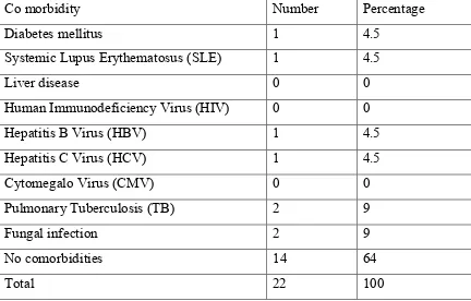
TABLE 1: PRETRANSPLANT COMORBIDITIES IN PATIENTS WITH
FUNGAL INFECTIONS
Co morbidity Number Percentage
Diabetes mellitus 1 4.5
Systemic Lupus Erythematosus (SLE) 1 4.5
Liver disease 0 0
Human Immunodeficiency Virus (HIV) 0 0
Hepatitis B Virus (HBV) 1 4.5
Hepatitis C Virus (HCV) 1 4.5
Cytomegalo Virus (CMV) 0 0
Pulmonary Tuberculosis (TB) 2 9
Fungal infection 2 9
No comorbidities 14 64
[image:42.612.104.536.404.680.2]37 No significant comorbidity was observed in 64% of the study population.
TABLE 2: TYPE OF RENAL DONATION IN THE STUDY POPULATION
Type of transplant Living donor Deceased donor Total
Number of infections 17 5 22
Percentage 77.5 22.5 100
77.5% of infections were noticed in living donor renal transplant
recipients compared to deceased donor renal transplant recipients (22.5%).
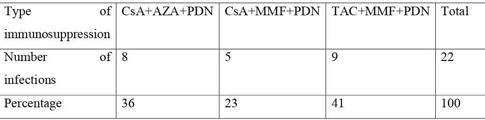
TABLE 3: TYPE OF IMMUNOSUPPRESSION AND NUMBER OF FUNGAL
INFECTIONS
Type of
immunosuppression
CsA+AZA+PDN CsA+MMF+PDN TAC+MMF+PDN Total
Number of
infections
8 5 9 22
Percentage 36 23 41 100
Among the twenty two patients with fungal infections, 41% received
tacrolimus (TAC), mycophenolate (MMF) and Prednisolone (PDN). Thirty
six percent received cyclosporine (CsA), azathioprine (AZA) and
Prednisolone. The rest received cyclosporine, mycophenolate and
[image:43.612.104.581.393.511.2]38
TABLE 4: TIME OF ONSET OF GRAFT DYSFUNCTION (GDF) AND
NUMBER OF INFECTIONS
Time of onset of
GDF
0 – 3 m 4 – 12 m 13 – 24 m No GDF Total
Number of
infections
12 4 4 2 22
Percentage 55 18 18 9 100
The average time to onset of GDF was 7.1 months. Fifty five percent
of patients with fungal infections developed GDF with in 3 months. Seventy
three percent of infections occurred within a year.
TABLE 5: TIME OF PRESENTATION OF FUNGAL INFECTIONS AFTER TRANSPLANTATION
Time of presentation 0 – 6 months 7 – 12 months > 13 months
Number of infections 12 2 8
Percentage 55 9 36
Mean 12
Range 0.5 – 83 months
Fifty five percent of patients developed fungal infections within 6
months. The mean time of presentation is 12 months (range 0.5 – 83
[image:44.612.107.576.126.242.2] [image:44.612.102.549.432.571.2]39
TABLE 6: FACTORS PREDISPOSING THE OCCURRENCE OF FUNGAL
INFECTIONS
Factors Present Absent Percentage P-value
GDF 20 2 91 0.000
Surgical procedure 3 19 14 0.001
Post Transplant DM 6 16 27 0.052
HBV 2 20 9 0.000
HCV 3 19 14 0.001
CMV 11 11 50 1.000
Bacterial infections 12 10 55 0.832
Anti Rejection Therapy 8 14 36 0.286
Leucopenia 11 11 50 1.000
Anemia (<11g/dl) 9 13 41 0.523
Thrombocytopenia 9 13 41 0.523
Binomial test has been used for analyzing the above table (p-value <
0.05 is significant)
Graft dysfunction alone seemed to be a risk factor for the occurrence
of fungal infection. Though many patients received anti rejection therapy
(ART, 36%) and cytomegalovirus (CMV, 50%) and bacterial infections
(55%), leucopenia (55%), anemia (41%) and thrombocytopenia (41%) were
present in many patients, they did not predispose to the occurrence of fungal
40
TABLE 7: FUNGAL INFECTIONS AND SITE OF OCCURRENCE
Site of fungal infection Number Percentage
Gastro intestinal tract 6 22
Lung 6 22
Upper respiratory tract 4 15
Urinary tract 6 22
Blood stream 3 11
Central nervous system 2 8
Fungal infections commonly occurred in gastrointestinal tract (GIT),
lung and urinary tract, each 22%. Other sites were upper respiratory tract
[image:46.612.104.503.464.636.2](15%), blood stream (11%) and central nervous system (CNS, 8%).
TABLE 8: TYPES OF RENAL HISTOPATHOLOGY
Histopathology Number Percentage
Acute cellular rejection 7 29
Chronic humoral rejection 2 8
Chronic allograft nephropathy 3 13
Acute tubular necrosis 4 17
Pyelonephritis 1 4
41 Fifteen of the twenty two patients underwent renal biopsy. Acute
cellular rejection (29%) was the commonest histopathology followed by
[image:47.612.101.536.217.374.2]acute tubular necrosis.
TABLE 9:
Organism Number Percentage
Candida species 14 62
Aspergillus 3 13
Mucor 4 17
Cryptococcus 1 4
Pneumocystis 1 4
TYPE OF FUNGAL PATHOGEN CAUSING INFECTION
Candida species (62%) is the commonest organism causing fungal
infection. The other common organisms are Mucor (17%) and Aspergillus
(13%).
TABLE 10: GRAFT AND PATIENT OUTCOME OF PATIENTS WITH
FUNGAL INFECTIONS
Outcome Number Percentage
Normal graft function 6 27
Stable graft dysfunction 7 32
Graft loss 9 41
Survived 11 50
[image:47.612.103.542.560.706.2]42
TABLE 11: FACTORS INFLUENCING PATIENT OUTCOME
Parameter Survived Expired P-Value
Deceased donor 4 1
0.000*
Living donor 7 10
TMP 4 5 0.682
CAP 4 4 1
CMP 3 2 0.631
DGF 4 3 0.666
ART 4 2 0.362
PTDM 4 2 0.362
HBV 1 1 1
HCV 2 1 0.557
CMV 5 6 0.687
Bacterial infection 7 5 0.416
Thrombocytopenia 3 6 0.211
Leucopenia 2 9 0.001*
Anemia 6 6 1
Alb < 3.5gm/dl 3 10 0.001*
GDF 9 11 0.152
Candida 7 7 1
Pneumocystis 1 0 -
Mucor 1 3 0.291
Aspergillus 2 1 0.557
Cryptococcus 0 1 -
Graft loss 3 6 0.211
Normal graft function 4 2 0.362
43 Student t test was used for analyzing the above data. p-value < 0.05
was considered as significant.
Fifty percent of patients with fungal infections expired. Graft loss
occurred in 41% of patients. Thirty two percent of patients continued to have
stable graft dysfunction.
Leucopenia and thrombocytopenia influenced patient outcome by
contributing to mortality (p value - 0.001). And also more significant
number of deaths occurred in patients who received renal allografts from
44
DISCUSSION
This study was conducted in the Department of Nephrology,
Government General Hospital, chennai during the period between Aug’ 08
and April’ 11. Twenty two patients were diagnosed with systemic fungal
infections during this period.
The mean age of the study patients was 35.55 yrs.
The male to female ratio was 1.75:1.
In a 10 year study done in Iran (from 1998 to 2008), the mean age of
patients was 49 yrs and the male to female ratio was 4.2:1.65 In a study by
Chugh et al, all patients were males with a mean age of 31.05 ±7.73 years
(range 21-42 years). 66
In the present study, living donor renal transplant recipients acquired
77.5% of infections compared to deceased donor recipients (22.5%).
In the present study, Fifty five percent of fungal infections occurred
with in 6 months of renal transplantation. Sixty four percent of infections
occurred within a year.
According to Abbott et al, majority of the fungal infections occurred
within 6 months67. In a study by Chugh et al, infection occurred within the
45
in the others.66 In a 10 year study done in Iran (from 1998 to 2008), 74% of
invasive fungal infections occurred with in 1 yr. In another retrospective
study (from 1987 to 1997) done in our department, out of 66 episodes of
fungal infection 4 episodes occurred within 1 month; 28 between 1 and 6
months; and 37 after 6 months i.e., nearly 50% of the infections occurred
within 6 months.68 This phenomenon may be because of the use of numerous
and higher doses of immunosuppressive agents.
In the present study, Candida species (62%) is the commonest
organism causing fungal infection. The other common organisms are Mucor
(17%) and Aspergillus (13%). Cryptococcus and pneumocystis constituted
4% each.
In a retrospective study done in our department, Candida was the
commonest pathogen, causing 50 of the fungal infection episodes (72.5%).
Aspergillus (11 episodes, 16%), Cryptococcus (3 episodes, 4.3%),
Pneumocystis (3 episodes, 4.3%), and Mucormycosis (2 episodes, 2.8%)
constituted the rest.68 In the Iranian study, mucormycosis (11/21) was the
commonest infection followed by Candidiasis (4/21) and Aspergillus
(3/21).65 In a study by fishman et al., candida and Aspergillus were the
common organisms.69 Infection with Cryptococcus neoformans was
46
species in two (11%), Aspergillus flavus in one (5.5%), and a mixed
infection with Aspergillus and Cryptococcus in one patient (5.5%).
In the present study, fungal infections commonly occurred in
gastrointestinal tract (GIT), lung and urinary tract, each 22%. Other sites
were upper respiratory tract (15%), blood stream (11%) and central nervous
system (CNS, 8%). In a retrospective study done in our department, sites of
infections were GI tract (35, 50.7%), respiratory tract (18, 26%), urinary
tract (8, 11.5%), CNS (3, 4.3%), and graft (2, 2.8%).The sites of infection
were almost similar to that in the present study.68
In the literature, the risk factors for developing fungal infection in the
post renal transplant setting were the following like deceased donor and
retransplantation, older age, high doses of immunosuppression for anti
rejection treatment, diabetes mellitus, CMV infection, bacterial infection
with prolonged antimicrobial therapy, surgical interventions, indwelling
catheters and anatomical abnormalities of the urinary tract.70
In the present study, graft dysfunction alone seemed to be a risk
factor for the occurrence of fungal infection. Though many patients received
anti rejection therapy (ART, 36%) and cytomegalovirus (CMV, 50%) and
47 thrombocytopenia (41%) were present in many patients, they did not
predispose to the occurrence of fungal infections. (No statistical
significance).
In a retrospective study done in our department, predisposing factors
were anti-rejection therapy in 24 cases, bacterial infections in 19, leukopenia
in 12, tuberculosis in 7, and CMV infection in 5.68 This difference may be
due to the small number of patients in the present study. In a study by Chugh
et al, graft function was normal at the time of diagnosis in 13 patients (68%)
while it was impaired (serum creatinine 160umol/l) in six patients (32%). 66
In the retrospective study done in our department, Six out of 10
diabetic recipients developed fungal infections.68 But in the present study,
only one patient had pre transplant diabetes mellitus. He developed fungal
infection in the post transplant period. In a study by Chugh et al, apart from
immunosuppressive drugs, predisposing factors included post-transplant
diabetes mellitus in two and leukopenia in two patients. Concomitant
bacterial infections were present in seven patients66.
In the present study 50% (11/22) of patients with fungal infection
died. In the retrospective study done in our department, a total of 36 out of
48 In the present study, high percentage of deaths occurred in patients
with mucormycosis (75%, 3 of 4); all of them had rhino cerebral Mucor with
GDF, post transplant diabetes mellitus and leucopenia. 50% of patients with
candida infections died. Three had blood stream and urinary tract infections,
the remaining 4 deaths were due to candida UTI and bacterial sepsis. All
those patients who had candida esophagitis survived. Thirty three percent of
patients with Aspergillus (1 out of 3) and 100% of cryptococcal meningitis
patients died (one patient).
In the Iranian study, 52.4% (11/21) of patients died due to fungal
infection, mostly due to Mucormycosis (74%) 65.
Leucopenia (9/11, 82%) and thrombocytopenia (6/9, 66%) influenced
patient outcome by contributing to mortality. And also more significant
number of deaths occurred in patients who received renal allografts from
living donor (10 out of 17, 59%). Their association was statistically
49
CONCLUSIONS
1. Candida species was the commonest fungal pathogen causing
infection in the renal transplant recipients (62%).
2. Gastrointestinal, lung and urinary tracts were the common sites of
fungal infection (22% each).
3. Majority of fungal infections occurred in the first year (64%).
4. Graft dysfunction predisposed to the occurrence of fungal infections.
5. The mortality rate was 50%.
6. Leucopenia (82%) and thrombocytopenia (66%) were associated with
50
BIBLIOGRAPHY
1. KL.Gupta: fungal infections and kidney. Indian J Nephrol 2001; 11: 147-154. 2. Singh et al: Clin Infect Dis2000:31; 545-53.
3. Rubin RH, Tolkoff-Rubin NE. Opportunistic infections in renal allograft recipients. Transplant Proc 1988; 6{Suppl 8]: 12-18.
4. Peterson PK, Balfour HH Jr, Fryd DS, Ferguson RM, Simmons RL. Fever in renal
transplant recipients: Causes, prognostic significance and changing patterns at the University of Minnesota Hospital. Am J Med 1981; 71: 345-351.
5. Tollemar J, Ringden O, Bostrom L, Nilsson B, Sundberg B. Variables predicting
deep fungal infectious in bone marrow transplant recipients. Bone Marrow Transplant 1989; 4- 635-641.
6. Hamacher J, Spiliopoulos A, Kurt AM, Nicod LP, and the Geneva Lung
Transplantation Group. Pre-emptive therapy with azoles in lung transplant patients. Eur Respir J 1999; 13:180–186.
7. George MJ, Snydman DR, Werner BG, et al. The independant role of
cytomegalovirus as a risk factor for invasive fungal disease in orthotopic liver transplant recipients. Am J Med 1997; 103:106–113.
8. Winston DJ, Emmanouilides C, Busuttil RW. Infections in liver transplant recipients. Clin Infect Dis 1995; 21:1077–1091.
9. Sia IG, Paya CV. Infectious complications following renal transplantation. Surg Clin North Am 1998; 78:95–112.
10.Wilczynski SW, Erasmus JJ, Petros WP, Vrdenburgh JJ, Folz RJ. Delayed pulmonary toxicity syndrome following high-dose chemotherapy and bone marrow transplantation for breast cancer. Am J Respir Crit Care Med 1998; 157:565–573.
51 12.Worthy SA, Flint JD, Muller NL. Pulmonary complications after bone marrow transplantation: high-resolution CT and pathologic findings. Radiographics 1997; 17:1359–1371.
13.Kaiser L, Huguenin T, Lew PD, Chapuis B, Pittet D. Invasive Aspergillosis clinical features of 35 proven cases at a single institution. Medicine 1998; 77:188–194.
14.Fitzsimmons EJ, Aris R, Patterson R. Recurrence of allergic bronchopulmonary aspergillosis in the posttransplant lungs of a cystic fibrosis patient. Chest 1997; 112:281–282.
15.Denning DW. Diagnosis and management of invasive aspergillosis. Curr Clin Top Infect Dis 1996; 16:277–299.
16.Berenguer J, Allende MC, Lee JW, et al. Pathogenesis of pulmonary aspergillosis
granulocytopenia versus cyclosporine and methylprednisone-induced
immunosuppression. Am J Respir Crit Care Med 1995; 152:1079–1086.
17.Saugier-Veber P, Devergie A, Sulahian A, et al. Epidemiology and diagnosis of invasive pulmonary aspergillosis in bone marrow transplant patients: results of a 5 year retrospective study. Bone Marrow Transplant 1993; 12:121–124.
18.Braun RS, Lake JR, Katzman BA, et al. Incidence and significance of Aspergillus cultures following liver and kidney transplantation. Transplantation 1996; 61:666–669.
19.Singh N, Arnow PM, Bonham A, et al. Invasive aspergillosis in liver transplant recipients in the 1990s. Transplantation 1997; 64:716–720.
20.Pomerance A, Madden B, Burke MM, Yacoub MH. Transbronchial biopsy in heart and lung transplantation: clinicopathologic correlations. J Heart Lung Transplant 1995; 14:761–773.
21.Ellis ME, Spence D, Bouchama A, et al. and the Fungal Study Group. Open lung
52 22.Caillot D, Casasnovas O, Bernard A, et al. Improved management of invasive pulmonary aspergillosis in neutropenic patients using early thoracic computed tomographic scan and surgery. J Clin Oncol 1997; 15:139–147.
23.Kanj SS, Welty-Wolf K, Madden J, et al. Fungal infections in lung and heart-lung transplant recipients. Report of 9 cases and review of the literature. Medicine 1996; 75:142–156.
24.Nunley DR, Ohori P, Grgurich WF, et al. Pulmonary aspergillosis in cystic fibrosis lung transplant recipients. Chest 1998; 114:1321–1329.
25.Klein DL, Gamsu G. Thoracic manifestations of aspergillosis. AJR Am J Roentgenol 1980; 134:543–552.
26.Kuhlman JE, Fishman EK, Siegelman SS. Invasive pulmonary aspergillosis and acute leukemia: characteristic findings on CT, the CT halo sign, and the role of CT in early diagnosis. Radiology 1985; 157:611–614.
27.Diederich S, Scadeng M, Dennis CH, Stewart S, Flower CH. Aspergillus infection of the respiratory tract after lung transplantation: chest radiographic and CT findings. Eur Radiol 1998;8:306–312.
28.Logan PM, Primack SL, Miller RR, Muller NL. Invasive aspergillosis of the airways: radiographic, CT, and pathologic findings. Radiology 1994; 193:383– 388.
29.Kemper CA, Hostetler JS, Follansbee SE, et al. Ulcerative and plaque-like tracheobronchitis due to infection with Aspergillus in patients with AIDS. Clin Infect Dis 1993; 17:344–352.
30.Mayer C, Appenzeller U, Seelbach H, et al. Humoral and cell-mediated autoimmune reactions to human acidic ribosomal P2 protein in individuals sensitized to Aspergillus fumigatus P2 protein. J Exp Med 1999; 189:1507–1512. 31.Castagnola E, Bucci B, Montinaro E, Viscoli C. Fungal infections in patients
53 32.Chu FE, Armstrong D. Candida Species Pneumonia. In: Sarosi GA, Davies SF, editors. Fungal Diseases of the Lung. 2nd Edn. New York, Raven Press, 1993; 10, 125–131.
33.Pennington JE. Opportunistic Fungal Pneumonias: Aspergillus, Mucor, Candida,
Torulopsis. In: Pennington JE, editor. Respiratory Infections: Diagnosis and Management. 3rd Edn. New York, Raven Press, 1994; 25: 533–549.
34.Buff SJ, McLelland R, Gallis HA. Candida albicans pneumonia: radiographic appearance. Am J Roentgenol 1982; 138:645–650.
35.Kassner EG, Kauffman SL, Yoon JJ. Pulmonary candidiasis in infants: clinical, radiologic, and pathologic features. Am J Roentgenol 1981;137:707–716
36.Watanakunakorn C. Acute pulmonary mycetoma due to Candida albicans with complete resolution. J Infect Dis 1983; 148:1131.
37.Leher RI, Howard DH, Syphred PS, et al. Mucormycosis. Ann Intern Med 1980;
93:93–108.
38.Aisner J, Sickles EA, Schimpff SC, Young VM, Greene WH, Wiernik PH. Torulopsis glabrata pneumonitis in patients with cancer: report of three cases. JAMA 1974; 230:584–585.
39.Perfect JR, Schell WA. The new fungal opportunists are coming. Clin Infect Dis 1996; 22:S112–S118.
40.Martino P, Gastaldi R, Raccah R, Girmenia C. Clinical patterns of Fusarium infections in immunocompromised patients. J Infect 1994; 28:7–15.
41.Khoury MB, Godwin JD, Ravin CE, Gallis HA, Halvorsen RA, Putman CE. Thoracic cryptococcosis: immunologic competence and radiographic appearance. Am J Roentgenol 1984; 141:893–896.
42.Patz EF, Goodman PC. Pulmonary cryptococcosis. J Thorac Imaging 1992; 4:51–
55.
43.Harari S. Current strategies in the treatment of invasive Aspergillus infections in immunocompromised patients. Drugs 1999; 58:621–631.
44.Braddy CM, Heilman RL, Blair JE. Coccidioidomycosis after renal
54
45.Mills W, Chopra R, Linch DC, Goldstone AH. Liposomal amphotericin B in the
treatment of fungal infections in neutropenic patients: a single-centre experience of 133 episodes in 116 patients. Br J Haematol 1994; 86:754–760.
46.Adler-Moore J. AmBisome targeting to fungal infections. Bone Marrow
Transplant 1994; 14:Suppl. 5S3–S7.
47.Anaissie E, Bodey GP, Kantarjian H, et al. Fluconazole therapy for chronic disseminated candidiasis in patients with leukemia and prior amphotericin B therapy. Am J Med 1991; 91:142–150.
48.Kauffman CA, Bradley SF, Ross SC, Weber DR. Hepatosplenic candidiasis: successful treatment with fluconazole. Am J Med 1991; 91:137–141.
49.Murphy M, Bernard EM, Ishimaru T, Armstrong D. Activity of voriconazole against clinical isolates of Aspergillus species and its effectiveness in an experimental model of invasive pulmonary aspergillosis. Antimicrob Agents Chemother 1997; 41:696–698.
50.Van't Wout JW, Novakova I, Verhagen CAH, Fibbe WE, de Pauw BE, van der Meer JWM. The efficacy of itraconazole against systemic fungal infections in neutropenic patients: a randomised comparative study with amphotericin B. J Infect 1992;22:45–52.
51.Denning DW, Tucker RM, Hanson LH, Stevens DA. Treatment of invasive aspergillosis with itraconazole. Am J Med 1989;8:791–800.
52.Viviani MA, Tortorano AM, Pagano A, et al. European experience with itraconazole in systemic mycoses. J Am Acad Dermatol 1990;23:587–593.
53.Nanas JN, Saroglou G, Anastasiou-Nana MI, et al. Itraconazole for the treatment of pulmonary aspergillosis in heart transplant recipients. Clin Transplant 1998;12:30–34.
55 55.Paradowski LJ. Saprophytic fungal infections and lung transplantation – revisited.
J Heart Lung Transplant 1997;16:524–531.
56.Radisic M, Lattes R, Chapman JF, et al. Risk factors for Pneumocystis carinii pneumonia in kidney transplant recipients: a case-control study. Transpl Infect Dis 2003; 5: 84–93.
57.Singh & Paterson: Review of fungal infections. Clin. Microbiol. Rev 2005; 8: 132-36
58.Microbiology Newsletter, Sir Gangaram Hospital, July 2007, Vol 12, No 2
59.Malini et al: Emergence of Non-Albicans Candida Species and Antifungal in a Tertiary Care Hospital, Jpn. J .Infect Dis 2005; 58: 344-348,
60.Verma et al: changing trends in candida infection. Indian J Med Res 117, March 2003; 122-128
61.Radisic M, Lattes R, Chapman JF, et al: Risk factors for Pneumocystis carinii pneumonia in kidney transplant recipients: a case-control study. Transpl Infect Dis 2003; 5: 84–93.
62.Braddy CM, Heilman RL, Blair JE: Coccidioidomycosis after renal
transplantation in an endemic area. Am J Transplant 2006; 6: 340–345.
63.Yamada H, Kotaki H, Takahashi T: Recommendations for the treatment of fungal
pneumonias. Expert Opin Pharmacother 2003; 4: 1241–1258.
64.Cordeiro RA, Brilhante RS, Rocha MF, et al: In vitro activities of caspofungin, amphotericin B and azoles against Coccidioides posadasii strains from Northeast Brazil. Mycopathologia 2006; 161: 21–26.
65.Behzad Einollahi et al: Invasive fungal infections following renal transplantation: A review of 2410 recipients. Ann Transplant, 2008; 13(4): 55-58.
66.K. S. Chugh, V. Sakhuja et al: High mortality in systemic fungal infections following renal transplantation in third-world countries. Nephrol Dial Transplant, 1993; 8: 168-172.
56 68.M.Jayakumar et al: Systemic fungal infections in renal transplant recipients at
Chennai, India. Transplantation Proceedings, 1998; 30:3135.
69.Fishman JA: Infection in Solid-Organ Transplant recipients. N Engl J Med, 2007; 357:2601-14.
70.Hardley S, Karchmer AW: Fungal infections in solid organ transplant recipients. Infect Dis Clin North Am, 1995; 9: 1045
71.Yamada H, Kotaki H, Takahashi T. Recommendations for the treatment of fungal