Dissertation Submitted to
THE TAMIL NADU DR. M.G.R. MEDICAL UNIVERSITY
in partial fulfillment of the regulations
for the award of the degree of
M.S. BRANCH – III
OPHTHALMOLOGY
GOVT. STANLEY MEDICAL COLLEGE & HOSPITAL
THE TAMIL NADU DR. M.G.R. MEDICAL UNIVERSITY
CHENNAI, INDIA.
This is to certify that the dissertation entitled “A CLINICAL
STUDY OF UVEITIS IN CHILDREN AND ADOLESCENTS” is
the bonafide original work of Dr. MAHARAJA DAVID GIDEON in
partial fulfillment of the requirements for M.S. Branch – III
(Ophthalmology) Examination of the Tamilnadu Dr. M.G.R. Medical
University to be held in September 2006.
DEAN
Govt. Stanley Medical College & Hospital,
Chennai-600 001.
PROF. A. PRIYA, M.S., D.O.,
Head of the Department Department of Ophthalmology Govt. Stanley Medical College &
I, Dr. MAHARAJA DAVID GIDEON, solemnly declare that
dissertation titled, “A CLINICAL STUDY OF UVEITIS IN
CHILDREN AND ADOLESCENTS” is a bonafide work done by me at Govt. Stanley Medical College & Hospital during 2003-2006
under the expert guidance and supervision of Prof. A. PRIYA, M.S.,
D.O. Head of the Department, Department of Ophthalmology.
The dissertation is submitted to The Tamilnadu, Dr. M.G.R.
Medical University, towards partial fulfillment of requirement for the
award of M.S. Degree (Branch – III) in Ophthalmology.
Place : Chennai.
Date :
Hospital, Dr. M. VASANTHA, M.D., for allowing me to avail the
facilities needed for my dissertation work.
I take great pleasure in expressing my deep sense of gratitude and respect for Prof. A. PRIYA, M.S.,D.O., Professor and Head of the Department of Ophthalmology and my unit Chief at Govt. Stanley Medical College and Hospital for her constant encouragement and permitting me to do the study.
I am extremely grateful to Prof. P. KUMARAVEL, M.S., D.O.,
Additional Professor of Ophthalmology for his guidance and encouragement and also for his constructive criticism which helped my work to take its present shape.
I am indebted to Dr. K. VINAYAGA MURTHY, M.S., DCH,
Registrar, Department of Ophthalmology for giving me guidance to conduct my study.
Last but not the least, my sincere thanks to all the patients who
co-operated for this study, without whom this study could not have been
AS : Ankylosing Spondylitis AS : Anterior Synechiae ATT : Antituberculous Treatment AU : Anterior uveitis
AV : Anterior vitreous BE : Both Eyes
BK : Band keratopathy C : Cells
CC : Complicated cataract CF : Counting fingers Ch : Choroiditis Patch Ch-C : Chest Clinic
CxR : Chest X-ray d : Days
DE : Divergent Eye De-C : Dental Consultation DV : Defective vision
ECCE : Extra Capsular Cataract Extraction ELISA: Enzyme Linked Immuno Sorbant Assay End : Endophthalmitis
Ex : Exudates F : Flare Fl : Floaters FL : Follows light HM : Hand Movements IU : Intermediate uveitis IV Ab : Intravitreal antibiotics
Jra : Juvenile Rheumatoid Arthritis KP : Keratic Precipitates
PA : Psoriatic Arthritis PaU : Pan Uveitis
PeS : Periocular Steroids PL : Perception of light PoU : Posterior Uveitis PPL : Pars Plana Lensectomy PPV : Pars Plana Vitrectomy PR : Projection of light
PRP : Pan Retinal Photocoagulation PS : Posterior Synechiae
R : Redness RE : Right Eye
Rh-C : Rheumatology Clinic SB : Snow banking
SICS : Small incision cataract surgery Sk-C : Skin Consultation
AFB : Acid Fast Bacilli
ANCA : Antinuclear Cytoplasmic Antibody
D : Dioptre
DC : Differential Count
ELISA : Enzyme Linked Immuno Sorbant Assay ENT : Ear, Nose and Throat
Hb : Haemoglobin
HLA : Human Leucocyte Antigen HM : Hand Movements
Ig : Immunoglobulin IUSG : International Uveitis Study Group
P : Partial
PCIOL : Posterior Chamber Intra Ocular Lens PL : Perception of Light
PPV : Pars Plana Vitrectomy PR : Projection of Light RA : Rheumatoid Arthritis RD : Retinal Detachment SLE : Systemic Lupus Erythematosis TB : Tuberculosis
TC : Total Count
1. INTRODUCTION 1
2. REVIEW OF LITERATURE 3
3. AIM 52
4. MATERIALS AND METHODS 53
5. ANALYSIS AND OBSERVATION 56
6. DISCUSSION 64
7. CONCLUSION 72
8. BIBLIOGRAPHY 76
9. PROFORMA 80
10. ABBREVIATIONS
11. MASTER CHART
INTRODUCTION
Uveitis refers to the inflammation of the middle vascular tunic of
the eye of called uvea.
The terminology uvea is derived from the latin word uva or grape
and consists of the iris, ciliary body and the choroid.
The frequency of uveitis in children under the age of 18, is
relatively low, the approximate incidence of 8-10% of the over all
uveitis cases from different world literature.
Unlike adults, the identification, history and symptoms, of uveitis
in a child is a diagnostic challenge, the complications are severe leading
to vision threatening problems, and the peculiar problem of development
of amblyopia in children.
SOME HISTORICAL FEATURES
1500 BC Hippocrates mentioned the typical finding of uveitis.
1801 AD – name Iritis introduced by Johann Adam Schmidt.
1853 – Vontolt classified uveitis aetiologically as scrofula 30%,
1931 – Kolmer (USA) put forward the view, that inflammation of
uveal tissue was due to bacterial toxins. The place of allergy as a potent
aetiological factor thus was established.
1950 – Toxoplasmic uveitis became a proven infection, thus
parasitic infection played a considerable role in etiology of posterior
REVIEW OF LITERATURE
ANATOMY OF UVEAL TRACT
The middle coat of the eye ball is between the sclera on the
outside and retina in the inside and is called the uveal tract.
It is divided into the choroid, ciliary body and the iris.
CHOROID
This extends from the ora serrata to the optic disc. Posteriorly it
merges with border tissue of elsching which surrounds the optic disc.
Anteriorly it continues as the ciliary body. The scleral surface of
choroids is shabby and irregular and the retinal surface is smooth and
shiny.
The choroid measures 0.22 mm in thickness, posteriorly and
0.1mm in thickness anteriorly. The choroid has a high blood flow with
pressure in the choroidal veins exceeding 20mm of Hg.
The following are the different layers of the choroid.
a) Suprachoroid lamina of FUSCA contains a delicate mesh work of
elastic fibres. It is lined by endothelium and contains potential
b) Layers of Haller and Sattler : These are layers of blood vessels.
Larger layer of blood vessels are called layer of Haller and the layer
of Sattler consists of medium sized vessels.
Stroma of these layers consist of collagen and elastic fibres –
Pigment granules are evenly distributed in the stroma. They are
responsible for absorbing excess light to prevent it from scattering.
LAYER OF CHORIOCAPILLARIES
The capillaries in this layer are of wide bore. The chorio
capillaries end in the ora serrata whereas other layers continue to the
ciliary body.
MEMBRANE OF BRUCH
It is a glossy homogenous membrane about 1.5µ thickness and is
also called lamina vitrea. It lies next to retina and lies closely adherent
to it.
It helps in passage of nutrients to retina and also helps in removal
of debris and metabolites.
CILIARY BODY
This is the part of the uvea which extends beyond the ora serrata
nasally. Extends more (8mm) temporally. The posterior part appears
smooth and is called pars plana. The anterior part is called pars plicata.
It presents as 70 radiating ridges with valleys in between.
CILIARIS
This is the muscle of the ciliary body. It is triangular in cross
section. It consists of bundle of muscle fibers running antero-posteriorly
in the outer layer and circular in the inner layer. Anterioposterior fibres
are called longitudinal fibres. They are attached to the scleral spur and
posteriorly they extend upto the equator.
Circular fibres occupy anterior and posterior parts of the ciliary
muscle. They are called Muller’s muscle. Muscle is supplied by short
ciliary nerve from the ciliary ganglion containing parasympathetic
fibres. Longitudinal fibres draw ciliary ring forward thus reducing
circumference. Circular fibres act as a sphincter and reduce
circumference of ciliary ring. They both slacken suspensory ligament of
lens and decrease the capsular tension in the lens.
CILIARY PROCESSES
Each process is a ridge about 2mm long and 0.5mm high.
IRIS
This is the anterior most part of the choroid. This forms a circular
disc in front of the lens with a central circular aperture called the pupil.
Peripherally this is attached to the anterior surface of ciliary
body. The anterior surface is divided into inner pupillary zone and outer
ciliary zone divided by the collarette. There are crypts adjoining the
collarette the epithelium is defective here and hence fluid can easily
leave the iris.
It consists of anterior endothelium, anterior limiting layer,
stroma, posterior membrane, epithelium. Anterior surface of iris is
smooth and velvety. Anterior surface presents with depression
adjoining the collarette called crypts of Fuchs. Minor arterial circle is
found in the region of collarette.
Posterior membrane consists of smooth muscle fibres called
dilator pupillae supplied by sympathetic fibres.
Sphincter pupillae is present in the stroma along with the blood
PATHOGENESIS OF UVEITIS
The etiopathogenesis of uveitis are several, and many theories
have been put forward.
INFECTIVE EXOGENOUS INFECTIONS
Can be due to introduction into the eye of organisms through a
perforating wound or ulcer. This results in suppurative iridocyclitis and
may progress to endophthalmitis and panophthalmitis.
Secondary Infections
This inflammation of the uveal tract is due to its spread from one
of the other ocular tissues (Cornea, sclera, or retina).
ENDOGENOUS INFECTIONS
This is a common cause of uveitis in which an infection lodged
any where else in the body can be transmitted to the uvea by the blood
stream.
Common infections are tuberculosis, syphilis, brucellosis, urinary
infection, osteomyelitis, toxoplasmosis, etc.
IMMUNE RELATED INFLAMMATION
They are very common. Primary source of infection, exists
infection. If it occurs again an immune process is set on the uveal
tissues, with deposition of immune complexes. Commonly they are due
to sinusitis, dental infections, pharyngotonsillitis, etc.
Acute iridocyclitis may be comparable to the antibody formation
in a lymph node wherein they are sensitized with thymus dependent
memory cells which may be implanted in the uvea. These cells
proliferate on contact with the antigen, should it reappear in the blood
stream and differentiate to form cytotoxic lymphocytes. This may
explain recurrence of uveitis in chronic inflammatory disease after the
infective form is treated.
HLA (Human Leucocyte Antigen) Associated : They occur more
frequently in persons with specific HLA antigens. Thus patients with
ankylosing spondylitis, juvenile chronic arthritis belonging to HLA B27
group present with iridocyclitis.
SYSTEMIC DISEASES
Uveitis are also associated with systemic etiologies like Stills’
disease, Wegener’s granulomatosis, sarcoidosis, Reiter’s disease. They
TRAUMATIC
Blunt or penetrating ocular trauma can produce features of
iridocyclitis, surgical procedures involving iris and rest of uvea can also
produce iritis.
IDIOPATHIC
A large percentage of uveitis can present without any identifiable
aetiological factor and are hence termed idiopathic.
SPECIFIC UVEITIS
Fuch’s uveitis syndrome, sympathetic ophthalmitis. VKH
CLASSIFICATION
Anatomical Classification (IUSG)
Anterior uveitis : Iritis
Anterior cyclitis
Iridocyclitis
Intermediate Uveitis : Posterior cyclitis
Hyalitis
Basal retinochoroiditis
Posterior Uveitis : Choroiditis
Retinochoroiditis
Neuroretinitis Pan Uveitis
ETIOLOGICAL CLASSIFICATION
Idiopathic, infective, immune related, neoplastic, traumatic.
PATHOLOGY
Granulomatous
Non-Granulomatous
Chronicity Acute
Recurrent-acute Chronic
Pattern Focal
Multifocal
DIAGNOSIS OF UVEITIS IN CHILDREN AND
ADOLESCENTS
HISTORY
PRESENT HISTORY
In the case of uveitis, the chief complaint is usually unilateral or
bilateral, photophobia, redness, floaters, blurred or decreased vision,
either alone or in combination.
Although blurred or diminished vision is the most common and
least localizing symptom, other symptoms can suggest a primary site of
intraocular inflammation. Juvenile idiopathic arthritis is a notable
exception in which pain, photophobia and redness and frequently absent.
These conditions can be very taxing for the ophthalmologist,
since the patient is asymptomatic and the child may not be able to come
forward with complaints of floaters or blurring.
Hence only periodic examination of these patients can help to
identify the disease.
PAST HISTORY
Previous attacks of uveitis, trauma, previous treatment history,
history of defective vision, amblyopia or strabismus since childhood that
herpes simplex or previous attacks of varicella zoster give an important
clue to the diagnosis.
Previous attacks of sinusitis, tonsillitis, systemic infections,
parasitic infections, attacks of arthritis and skin lesions are important in
further management.
PERSONAL HISTORY
A thorough dietary history, drug use, exposure history should be
elicited. History of contact with pets in lieu of toxoplasmosis and
toxocariasis and also pica habit in children which might lead to parasitic
infections are very pertinent.
FAMILY HISTORY
Genetics does appear to play a role in some forms of uveitis as
evidenced by their increased association with HLA. HLA B27 with
anterior uveitis often occur together with ankylosing spondylitis.
Reiter’s syndrome or inflammatory bowel disease. Other HLA
associations are HLA B51 with Bechet’s disease and HLA B53 and
HLA DR4 with VKH syndrome.
CLINICAL FEATURES AND EXAMINATION OF A CASE OF UVEITIS
The examination should start with the general condition of the
The child should be examined for anemia, lymphadenopathy.
The skin of the child should be thoroughly examined. The
presence of rashes, nodules or vitiligo can be diagnostic in children.
Hair loss, poliosis, granulomas and nodules of the lid has to be
noted.
VISUAL ACUITY
In pre verbal children,
If the child objects to occlusion of one eye it indicates poor visual
acuity in the other eye. Fixation test with prisms, covering one eye and
keeping a prism over the other eye is done. Now the cover is removed
from the eye and ability to maintain fixation is noted.
Preferential looking, rotation test, optokinetic nystagmus are
other methods of assessing visual acuity. Verbal children can undertake
picture naming test such as Kay pictures. Older children can be assessed
with a Sheridan-Gardner test and a E Snellen chart.
OCULAR EXAMINATION
After examining the lids and eye brows for hair loss, nodules,
dilated vessels, ulceration, nodules, phylcten. The congestion is divided
into ciliary and conjunctival congestion.
Ciliary congestion is pathognomonic of corneal lesion or
inflammation of the iris and ciliary body and also acute rise in intra
ocular pressure.
Cornea is examined for edema, band keratopathy, degeneration
and ulcers. Keratic precipitates are accumulations of inflammatory cells
over the desquamated sticky endothelium. Identification is important as
they some times may be the only signs of inflammation. They are
mostly found in the form of a triangle between the 4 – 8 o’ clock
position. This triangle is formed with the base towards the limbus. It is
called the Arlt’s triangle.
Keratic precipitates are classified as
Pigmented and crenated - old and inactive
White or yellow and round - Recent or currently active non granulomatous inflammations.
Larger and frequently greasy - Granulomatous appearance.
ANTERIOR CHAMBER
It is examined for depth and the clarity. Presence of aqueous
flare in graded by its intensity. Flare is due to disruption of the blood
aqueous barrier causing leakage of the proteins from the iris and ciliary
vessels. The aqueous cells are due to the extravasations of the
inflammatory cells from the vessels. They can be graded as follows (by
HOGAN’S Method)1.
AC cells –
0 - 0
1+ - 5-10 Cells
2+ - 11-20 cells
3+ - 21-50 cells
4+ - > 50 cells
AC Flare
0 - Normal
1+ - Faint (Just detectable)
2+ - Moderate (Iris details clear)
3+ - Marked (iris details hazy)
4+ - Severe (Fibrin, coagulated aqueous)
In addition to the above finding there may be collection of macro
the base of the anterior chamber. This is called hypopyon. Usually this
hypopyon is sterile and hence once the inflammation is brought under
control they get spontaneously absorbed. Some times in viral iritis there
may be accumulation of blood in the anterior chamber. This is called
hyphaema.
IRIS AND PUPIL
The iris colour and pattern may be lost due to inflammation and
leakage of the blood vessels. The whole iris becomes boggy and
edematous and thus they loose their architecture. The colour might be
lost in atrophic patches and also in heterochromia iridis as in Fuch’s
iritis.
Nodular deposits in the iris may be due to granulomatous
inflammation (ex. Bussaca nodules, Berlin’s nodules) and due to both
granulomatous and non-granulomatous uveitis (Koeppe’s nodules).
There may be synechiae (anterior and posterior) secondary to the
inflammation. Anterior synechiae is due to attachment between the iris
and the cornea where as posterior synechiae is due to attachment
between the iris and the lens. This posterior synechiae is very dangerous
because this blocks the flow of aqueous humour from the posterior
resulting in shallow anterior chamber and formation of peripheral
anterior synechiae. It produces severe secondary angle closure
glaucoma. The pupil is irregular in appearance in these cases producing
a festooned pupil when they are being dilated.
LENS
It may contain pigment deposits in its anterior capsule due to
deposition of iris pigments and also due to broken posterior synechiae.
Previous attacks of raised intra ocular pressure produces a grayish white
discolouration in the lens surface. This is called glaucomflecken.
Cataractous changes especially anterior sub capsular cataract and
posterior sub capsular cataract are common following chronic iritis and
due to prolonged steroid therapy.
VITREOUS
Vitreous inflammation may be diffuse or may be anterior or
posterior. The vitreous opacities are either floaters or cells. These
vitreous opacities can be graded by Hogan’s method.
The patient is examined using indirect ophthalmoscopy or slit
0 – Normal
1+ - Few Scattered fine and coarse opacities. Clear fundus details seen.
2+ - Scattered fine and coarse opacities fundus details can still be seen.
3+ - Many opacities and marked blurring of fundus details.
4+ - Dense opacities no view of the fundus.
Apart from this vitreous bands fibrous adhesions are also noted.
Fungal uveitis produces cotton ball like exudates floating in the
vitreous.
Vitreous hemorrhage has to be identified if present.
RETINA AND CHOROID
Retinal periphlebitis, vascular sheathing, neovascularizations,
occluded vessels are common things that has to be searched for in the
retinal vasculature. Both the posterior and the peripheral vessels have to
be scrutinized for new vessels.
Retinal lesions are manifested as yellow lesions with irregular
margins obscuring the vessels. They have associated superficial
hemorrhages.
Retinal hemorrhages, retinal necrosis and cystoid macular edema
there may be retinal folds and epiretinal membrane formation due to
chronic uveitis.
Fibrous bands originating from one part of the retinal surface to
the other and also from the retina into the vitreous can also be elicited
using an indirect ophthalmoscope. Choroidal inflammation are yellow
or grayish white lesions lying deeper to the retina with normal over
lying retinal vessels. Active inflammation is characterized by hazy
margins and surrounding hemorrhages. Old lesions are punched out,
well demarcated and are surrounded by pigments. The background
sclera is usually visible as the retina and choroid are usually destroyed
by the inflammation.
Choroidal inflammation can be focal, multifocal or diffuse. They
are caused by a wide variety of parasitic, bacterial and fungal infections.
The choroid is the vascular coat of the eye ball and hence blood from the
rest of the body pass through the choroid. Hence they are easily liable to
get infected by organisms from various parts of the body. Thus
choroidal inflammation is usually infective in origin whereas an anterior
uveitis is usually immunologically mediated and not usually due to a
OPTIC NERVE
It can be secondarily inflamed after severe retinits. Most of the
chorioretinitis are associated with subsequent papillitis. This results in
severe loss of vision and associated scotomas. Colour vision is also
severely affected as is contrast sensitivity. Papillitis has to be identified
early and treated vigorously with steroids. Failure to do so will lead to
secondary optic atrophy. This is the ultimate course of events in severe
fulminent chorioretinits due to cytomegolovirus, sarcoidosis, herpetic
retinal necrosis etc.
GONIOSCOPY
This is done to look for peripheral anterior synechiae, Berlin’s
nodules, new vessels in the angle and also membranes. Both the eyes
are compared with each other and the angle width is documented.
Associated pigmentation of the trabeculum is also documented in four
grades.
INTRA OCULAR PRESSURE
Intra ocular pressure is measured using Shiotz, indentation
tonometry. It can also be measured using an applanation tonometer.
Usually the intra ocular pressure is elevated in episodes of acute iritis.
angle. It may also be due to formation of posterior synechiae and also
peripheral anterior synechiae. This has to be identified and vigorously
treated with tension reducing measures like beta blockers. The intra
ocular pressure is also elevated in patients who are undergoing long term
treatment with steroid drops and they have to be regularly screened for
raised pressure.
These examination are usually a great challenge in young
children and pre verbal children and they have to be sedated or even
anesthetized to perform these intricate examinations.
DIAGNOSTIC TESTS
Complete blood cell count with differential count
It is necessary to find out the presence of infection in the blood
and also to find out the presence of acute or chronic inflammatory cells.
Also it is needed to find out the response to treatment as the cell count
comes back to normal if the treatment is successful. In patients with
long term steroid therapy or immunosuppressive therapy regular
monitoring of the blood cells has to be performed as they are likely to
Erythrocyte sedimentation rate
It is a non specific indicator of plasma fibrinogen in the blood
and also the globulin levels in the blood. This is elevated in chronic
infections and inflammations especially granulomatous inflammations
and systemic malignancies. Systemic malignancies can cause
masquerade syndromes in the eye where by they mimic regular
inflammatory conditions and can be confused with simple iritis. Using
the Westergren method normal ESR for children should be less than 10
mm per hour.
SPECIFIC TESTS
Syphilis serology. As syphilis forms a major part of both anterior
and posterior uveitis serological examination of this disease place a
major part in management of uveitis. VDRL test forms the corner stone
of serological examination of syphilis. The VDRL titre can be tested by
taking the patient’s, blood and agglutinating with a known antigen like
cardiolipin. This test however gives false positive and false negative
results. Antitreponemal antibody test such as FTA-ABS, MHATP has
100% sensitivity and specificity for syphilis regardless of the stage of
the disease. Unfortunately these tests remain positive for life and hence
contrast do reflect disease activity and therefore are used primarily to
gauge disease activity and response to treatment.
RHEUMATOID FACTOR (RF) TEST
Rheumatoid factor is an auto antibody directed against the Fc
fragment of the human IgG. About 80% of patients with rheumatoid
arthritis are RF positive defined as a titre of more than 1:80.
Rheumatoid factor sero positivity is non specific and best used to
support a clinical diagnosis of rheumatoid arthritis.
ANTINUCLEAR ANTIBODIES (ANA) :
The ANA test is typically performed by applying serial dilutions
of the patients serum to cultured tumour cells and then titrating for the
presence and pattern of nuclear auto antibody staining. The ANA test is
generally used to confirm the collagen vascular diseases particularly
systemic lupus erythematosus or Juvenile rheumatoid arthritis. Along
with ANA other tests like ANCA, Anti-double stranded DNA are more
specific tests for the diseases like Poly arteritis, Wegener’s
Granulomatosis.
HERPES VIRUS ANTIBODIES :
The prevalence of herpes virus antibodies is so high in the
meaningless. A negative test, however eliminates herpes virus infections
and therefore can be useful in selected instances. Herpes virus serology
remains positive for life. Hence in cases where the clinical diagnosis is
not very obvious herpes virus inclusion bodies can be seen by using
Giemsa Stains for the affected cells. The inclusion bodies are usually
seen in the nucleus of the affected cells.
HIV ANTIBODIES :
Most commonly detected by using an enzyme linked immuno
sorbant assay (ELISA), positive results are confirmed by a Western blot
test. HIV testing in uveitis is usually ordered in patients with known
HIV risk factors, severe or bilateral retinitis or choroiditis and suspected
Herpes Zoster Uveitis in children. HIV testing requires patient consent.
TOXOPLASMA ANTIBODIES :
Tests available to detect and quantify antitoxoplasma gondii
antibodies are Sabin Feldman (SF) dye test, immuno fluorescence
antibody (IFA) test and ELISA. Of these SF dye test remains the most
sensitive and specific test but it is technically difficult and of limited
availability, whereas IFA and ELISA are relatively easy and economical
and can be used to distinguish IgG and IgM anti toxoplasma gondii
remember that IgM anti toxoplasma antibodies may be elevated for upto
1 year after infection, limiting the accuracy with which they can date
acute infection and that antibody titres are generally less reliable in
patients with AIDS.
TOXOCARA ANTIBODIES :
Detected by haemoagglutination, complement fixation and
immunofluorescent antibody test. But the ELISA has the sensitivity and
specificity of 90%. Although a titre of more than 1:8 is considered
diagnostic for ocular toxocariasis, it is important to remember that
ocular toxocariasis is primarily a clinical diagnosis and negative titre
does not rule out the disease.
ANGIOTENSIN CONVERTING ENZYME (ACE LEVEL) :
ACE is produced primarily by capillary endothelial cells,
abundant in both lungs and liver and by macrophages. Clinically ACE
levels are elevated in more than two thirds of patients with active
disease of sarcoidosis.
Uveitis and a negative purified protein derivative with increased
ACE is fairly specific for sarcoidosis. Normal serum ACE level is
HUMAN LEUCOCYTE ANTIGEN (HLA) STUDY :
The surface membrane of human leucocyte contain HLA. These
are regulated by gene loci on chromosome 6. The reaction of these
antigens with specific antisera causes lysis of the cell membrane and
this is the basis of cell typing. HLAB-27 has been associated with
Ankylosing spondylitis, Reiter’s syndrome, Psoriatic arthritis and
arthritis associated with inflammatory bowel disease. HLAB-5 is
associated with Bechet’s disease.
DIAGNOSTIC PARACENTESIS :
Aqueous and vitreous samples are useful for polymerase chain
reaction (PCR) testing for toxoplasmosis and herpes virus particularly
in AIDS patients in whom diagnosis can be difficult.
To demonstrate local production of antibody the specific
antibody in the eye is measured relative to the total amount of globulin
in the eye. The formula for determining the value is
C = Antibody titre Aqueous humor x Ig Serum
Serum Aqueous humor
CONJUNCTIVAL AND LACRIMAL GLAND BIOPSY :
Reserved for those patients with visible conjunctival masses or
lacrimal gland enlargement as can occur with sarcoidosis, tuberculosis
or coccidio domycosis.
MUCOSAL BIOPSY :
The greatest use of oral mucosal biopsy is for the diagnosis of
Bechet’s syndrome, evidence of an occlusive vasculitis can greatly
support the diagnosis.
SKIN TEST :
Kveim’s Test :
Suspension of antigenic preparation from human sarcoid tissue is
injected intradermally and read at the end of 6 weeks when a papule
develops. The papules is biopsied for evidence of granuloma and giant
cells and epitheloid cells, with no caseation. This test is not easily
available.
Mantoux Test :
Purified protein derivative of tuberculin is injected intradermally.
It is a non-specific test. Because of prior exposure to tuberculosis a
representing inactive infection. Therefore it is disadvantageous as it
yields more false positive than true positive results.
Pathergy Test :
This is a scratch test done for Bechet’s syndrome. In this scratch
or a line drawn on the skin produces a wheal, the phenomenon is called
dermographism.
RAY CHEST, RAY SACROILIAC JOINTS AND SKULL X-RAYS :
Chest X-ray taken in patients suspected of having sarcoidosis or
tuberculosis.
Skull x-ray to evaluate patients with suspected congenital
toxoplasmosis and rubella for calcification.
X-ray sarcoiliac joints and x-rays of the peripheral joints are
taken in patients with suspected ankylosing spondylitis and juvenile
rheumatoid arthritis.
B – SCAN :
Echoes are plotted as dots instead of spikes and the brightness of
the dot indicates the sides of the received echo. The transducer is
moved in an arc above the eye and whole series of intensity registration
planes. B-scan produces a real time two dimensional gray scale display
of the eye and orbit. It is useful in delineating intraocular structures
with opaque media such as severe cataract and vitreous haemorrhage
and in evaluating vitreoretinal and orbital mass lesions. Dynamic
ultrasound allows differentiation of detachments of the retina from those
of the vitreous as well as identification of vascular abnormalities.
ULTRASOUND BIO-MICROSCOPY3:
This is another method of ultrasound examination of eye in
which high frequency ultrasound waves of 50-80Mhz are used that
allows histological evaluation of the anterior segment structures. It is
used specifically for defining abnormalities of anterior chamber angle,
limbus, ciliary body and anterior part of the retina. Snow banking and
organized exudates in the peripheral retina and also peripheral retinal
detachment can be clearly made out with ultrasound bio-microscope.
FLUORESCEIN ANGIOGRAPHY :
It is an invaluable aid in evaluating numerous pathological
changes in the eyes.
Fluorescein used are usually in 10% or 25% solutions. Major
uses of fluorescein angiography are :
b) Subretinal neovascular membranes
c) Disc leakage
d) Staining of retinal vessels
e) Neovascularisation of retinal vessels
f) Retinal pigment epithelium (window defects)
Fluorescein angiogram can cause anaphylactic shock due to
allergy to the dye. Hence all precautions to revive the patients in case of
shock should be kept ready before the test is performed.
INDOCYANINE GREEN (ICG):
It is used to mainly identify choroidal neovascular membranes. It
is well bound to plasma proteins and hence does not leak from the
normal choroidal vessels. The retinal pigment epithelium does not block
the choroidal fluorescence when ICG dye is illuminated with infrared
light. ICG has a longer wave length when compared to fluorescein.
Hence it is not blocked by the pigment layer. The disc is usually seen
as a dark shadow in the ICG angiogram. ICG is also used to study
patients with serpiginous choroiditis, bird shot choroiditis.
Laser Flare Photometry, optical coherence tomography are now
COMMON CAUSES OF SPECIFIC UVEITIS IN
CHILDREN AND ADOLESCENTS
ANTERIOR UVEITIS
Juvenile Rheumatoid Arthritis (JRA)
JRA is the most frequently identifiable etiology of pediatric
anterior uveitis. It is usually divided into systemic, polyarticular and
pauciarticular. Pauciarticular arthritis is defined as the involvement of
four or fewer joints in the first 6 months after the onset of the disease.
Uveitis is more common in pauciarticular type of JRA. JRA and
juvenile chronic arthritis together form the clinical spectrum of juvenile
idiopathic arthritis. It produces chronic inflammation of the synovium.
B-lymphocytes infiltration and expansion occurs in the joint space. This
produces a thick pannus in the joints leading to destruction of the
articular cartilage and leading to fusion of the adjoining bones.
Pauciarticular and polyarticular JRA affects girls more than the boys.
Systemic JRA is equal in both boys and girls. Patients with chronic
uveitis develop joints symptoms early in life, some by about 6 years4.
Laboratory diagnosis :
There is an elevated erythrocyte sedimentation rate and
This is more common in children with pauci artricular JRA. Titres are
usually in the range of 1:40 or 1:80. RA factor is usually rare and its
presence indicates persistent disease into adulthood. There is also
increased fibrinogen and D-dimer levels in the blood. Clinical
diagnosed is based on presence of arthritis in a child with negative RA
factor5.
JRA uveitis is most frequently a chronic non-granulomatous
iridocyclitis, bilateral in 70% of patients, common in female children.
Ocular inflammation is usually asymptomatic and eyes are
non-congested. Complications include cataract, glaucoma, band keratopathy,
posterior synechiae and phthisis bulbi.
The arthritis usually precedes the uveitis. Iridocyclitis occur in
approximately half of the affected patients within 2 years of the onset of
the arthritis. There is no correlation between the degree of arthritis
activity and ocular inflammation.
According to American Association of Paediatrics pauciarticular
individuals should be checked every 3 months6. Polyarticular patients
may be evaluated at 6 months intervals for the development of uveitis,
Management is by use of topical steroids and also periocular
steroids7. If the patients do not respond to the above methods systemic
steroids has to be started. In severe cases where intraocular damage is
rampant immunosuppressants and antimetabolites like methotrexate and
etanercept has to be used.
Ankylosing Spondylitis :
The onset of juvenile ankylosing spondylitis is usually at 8-10
years of age and males are more affected, 95% associated with HLA
B27. Frequently presents as a peripheral arthropathy before physical or
radiographic findings of sacroiliac joint involvement.
An acute, recurrent non-granulomatous iridocyclitis affects
10-20% of patients with juvenile ankylosing spondylitis. Typically
unilateral and may precede or follow the arthritis.
The patient might have severe iridocyclitis resulting in formation
of a hypopyon.
Management is by topical or systemic steroids or in resistant
cases by immunosuppressants.
Reiter’s Syndrome :
Triad of Reiter’s syndrome is non-infectious urethritis, arthritis
Although patients with Reiter’s syndrome may have sacroilitis and HLA
B 27 positivity, the presence of uveitis, keratoderma blenorrhagica of
palms and soles and ulceration of the mouth and genitals differentiates
this disorder from ankylosing spondylitis. Unlike Bechet’s disease the
mucosal lesions are usually not painful. A non-granulomatous anterior
uveitis affects 3%-12% of patients.
Bechet’s Disease :
In this condition the patient usually manifests with recurrent oral
ulceration, genital ulceration and severe uveitis. Patients might have
hypersensitivity and dermographism of the skin which is demonstrated
by the Pathergy test. The uveitis might manifest as a simple anterior
uveitis or may produce a severe vasculitis of the retina. This may lead
to vascular occlusion of the retina resulting in retinal infarcts and finally
leading to consecutive optic atrophy. In cases with vasculitis,
management has to be with immunosuppressants and antimetabolites as
only these can be vision saving.
Fuch’s Heterochromic Iridocyclitis :
Frequently asymptomatic, with chronic low grade iridocyclitis
atrophy is typical but not invariable. The keratic precipitates have a
characteristic small, stellate appearance.
Treatment with topical corticosteroids is usually ineffective.
Mydriatics are often not required because posterior synechiae are
uncommon. This commonly causes anterior subcapsular cataract which
may be the only symptom the patient might have as the eye is usually
quiet. These patients might have new vessels in the iris and anterior
chamber angle and when a paracentesis is done a typical type of
haemorrhage (Filiform) called Amslers sign is present. This is
pathognomonic of this disease.
Lens Induced Uveitis :
The phacolytic reaction usually occurs in the presence of a
hypermature cataract. The lens material acts as a chemical irritant,
probably acting directly on the iris and ciliary body. Macrophages enter
to engulf the liberated material. No polymorphonuclear cells are seen.
No keratic precipitates are usually seen in this condition. Management
is usually by means of steroids to control the inflammation and removal
of the cataractous lens with thorough washing of the anterior chamber.
In phacoanaphylatic uveitis, typically a break in the lens capsule
granulomatous uveitis after surgery or trauma. Poly Morpho Nuclear
cells and macrophages are found in the aqueous, iris and lens. There
may be also be white fluffy lens material seen in the anterior chamber
and also over the surface of the lens and in the angle of anterior
chamber. Patients have to be intensively treated with steroids and the
offending lens material has to be removed.
Acute Interstitial Nephritis :
It is an uncommon renal disorder, as a result of an immune
reaction to antibiotics, non-steroidal anti-inflammatory drugs or
infection. It is characterized by non-specific systemic complaints of
low-grade fever, pallor, fatigue and weight loss and may be associated
with increased Erythrocyte sedimentation rate, increased serum
creatinine level, proteinuria, glycosuria, microhematuria, leucocyturia,
and excretion of casts. A bilateral anterior uveitis may precede, follow
or occur concomitantly with this renal disorder. This diagnosis is
established by renal biopsy. The prognosis in childhood is good.
Tuberculosis :
This may cause severe granulomatous and also
non-granulomatous iridocyclitis usually a result of systemic tuberculosis.
the conglomerate form. These patients along with treatment by topical
steroids also need to be treated with antituberculous drugs for control of
the disease process.
Sarcoidosis :
This causes both non-granulomatous and granulomatous
iridocyclitis. Lesion may be present all throughout the body. Biopsy of
the sarcoid nodules may reveal non-caseating granulomas. These cases
respond well to systemic and topical steroids.
Leprosy :
It causes severe anterior uveitis both in the multi bacillary and
pauci bacillary forms. In multi bacillary form there is direct invation of
the anterior chamber by the bacilli. It forms an iris pearl in the anterior
surface of the iris. Treatment is both with steroids and anti-leprosy
drugs.
Herpes :
Herpes iridocyclitis can be caused by both herpes zoster and
herpes simplex viruses. Sometimes they follow severe corneal dendritic
ulcerations. They also may accompany interstitial and disciform
keratitis. The anterior chamber reaction may be due to immunologically
viruses in to the anterior chamber. Haemorrhagic uveitis is a common
manifestation of herpetic iritis.
INTERMEDIATE UVEITIS
Early in the course of the disease, the presence of anterior
vitreous cells is the only sign. Initially there are fine and separate, but as
the disorder progresses they aggregate and may acquire a fibrillar state
in the anterior 1/3rd of the vitreous. There is usually posterior
subcapsular cataract formation at the time of presentation. The patient
usually has no acute symptoms of redness or pain and the only
symptoms may be floaters in front of the eye while the patient is
viewing a light background. Progressively the patient may slowly
develop a decrease in visual acuity as the vitreous cells and floaters
become more organized.
Scleral depression early in the course of illness will reveal a
graywhite exudates in the pars plana. This is done by using either an
indirect ophthalmoscope or a three mirror lens using a slit lamp. Other
signs are peripheral retinal vasculitis with sheathing of both the venules
and the arterioles. When vision is reduced below a level of 6/18,
Local corticosteroid are used only if the patients vision is 6/12 or
better and significant anterior chamber reaction is present. Patients with
minimal anterior chamber reaction and vision of 6/12 or better receive
no treatment. If vision is less than 6/12 and subretinal exudation, optic
nerve papillitis or disturbing floaters are found, periocular steroids are
given weekly (upto 6 doses). The commonly used periocular steroids is
triamcinolone acetonide in doses of 20-40mg/dose. This can be given
until visual improvement occurs. If these fails to achieve the desired
result, retinal cryopexy should be applied, if cryopexy also fails and the
disease is bilateral, cyclosporine should be employed. The common
causes of intermediate uveitis are usually idiopathic in nature where the
etiology is not usually found. Other specific etiologies are sarcoidosis,
sympathetic ophthalmitis, multiple sclerosis and also secondary to septic
foci else where in the body.
POSTERIOR UVEITIS
Toxoplasmosis :
It is the most common identifiable cause of pediatric uveitis.
Infection during the first trimester is associated with neonatal
convulsions, intracranial calcifications and chorioretinitis. Infants
Majority of the cases in children are recurrences of a congenital
infection8.
This protozoa is usually found in cats and may be acquired by
close contact with the animals or with contamination of the cat feces. It
is present in three forms, the sporozoites, bradyzoites and tachyzoites.
Tachyzoites are the most virulent form of the protozoa. Tachyzoites are
responsible for causing severe tissue damage. Bradyzoites are usually
the dormant stage of the protozoa.
Inactive atrophic scars usually affecting the posterior pole, occur
in approximately 80% of infected new borns. Ocular toxoplasmosis in
older children and adults represents a reactivation of the subclinical
congenital infection of the retina. The posterior uveitis may be
associated with a granulomatous or non-granulomatous iridocyclitis.
Congenital toxoplasmosis usually occurs in and around the macula. The
patients usually develops a divergent squint as they usually have the
disease from their intrauterine life and are not able to use the eye to fix
light. This divergence may be the only sign of the macular choroiditis in
these children. Also children might get affected by close contact with
infected cats and may have the same manifestation as the adult form of
the disease. The disease might present with focal, multifocal and diffuse
Treatment consists of the combined use of pyrimethamine,
folinic acid supplement and sulfadiazine. Clindamycin may be used in
addition to these medications or in substitutions for pyrimethamine. The
ELISA is the most sensitive and specific test. Any positive titer is
significant in evaluating the diagnosis of toxoplasmosis. Systemic
corticosteroids used under antimicrobial coverage. Not all cases are
treated in immunocompetent children. The cases that are treated either
have a lesion in the papulo macular bundle or near a major vessel or
close to the optic disc. In immunosupressed children all the lesions
irrespective of locations has to be treated vigorously.
Toxocariasis :
It is primarily a disease of children, acquired by the ingestion of
soil containing the eggs of the canine intestinal round worm, toxocara
canis. It is unusual for visceral larva migrans and ocular toxocariasis to
affect the same person.
Ocular form is typically found in patients approximately 7 – 10
years old. Usually unilateral and presents as strabismus, leucocoria, or
decreased vision. May be present as a chronic endophthalmitis,
peripheral chorioretinal granuloma or posterior pole chorioretinal
formation of fibres bands which leads to tractional retinal detachment
and dragging of the macula and the disc.
Lab diagnosis is by the light sensitivity and specificity (90%) of
an ELISA titre for toxocara. Oral or periocular corticosteroid are used
to treat toxocara endophthalmitis. Antihelminthics are not effective for
ocular toxocariasis9. In cases with tractional bands and macular traction
viterectomy with release of the traction bands has to be performed to
prevent a tractional retinal detachment.
Congenital Syphilis :
Infants with congenital syphilis have been born with active
disease, in some active choroiditis may be seen, but in most the only
evidence of choroiditis, is the presence of segmental pigmentation in
the posterior pole. Cataract may begin earlier, than would be expected.
Glaucoma is a late sequelae. Decades later, recurrence of iritis and
interstitial keratitis are puzzling but they respond to topical
corticosteroid and cycloplegics.
Fungal Disease
Candidiasis and other fungal entities are rare causes of posterior
uveitis in children. Vitrectomy and systemic and intravitreal antifungal
Viral Disease :
Rubella retinitis is the most common ocular features of the
maternal rubella syndrome. Unilateral or bilateral pigment deposits,
usually limited to the posterior pole, vary from powdery or granular to
more discrete shapes. Cytomegalo virus retinitis is a common cause of
blindness in individuals with immunosuppression. They may occur in
the indolent form as well as the fulminating form. Early antiviral
treatment is vision saving. Herpes virus can produce progressive outer
retinopathy in immunosuppressed children and acute retinal necrosis in
immuno competent healthy children. This leads to severe irreversible
damage to the retina with subsequent consecutive optic atrophy. Early
treatment with systemic acyclovir is useful to reduce the damage caused
by the virus.
PAN UVEITIS
Endophthalmitis :
Endophthalmitis is intraocular inflammation predominantly
involving the vitreous cavity and anterior chamber of the eye.
Contiguous ocular structures such as the retina or choroid may also be
Endogenous endophthalmitis results from blood borne spread of
bacteria or fungi during generalized septicemia, characterized by an
acute onset with pain, decreased vision, hypopyon and vitritis. Most
common organisms are streptococcus pyogenes, staphylococcus aureus,
neisseria meningitides, H. influenza and E. coli, Candida is the most
common fungal aetiology.
Infectious endophthalmitis is an exogenous infection in which
organisms enters through a surgical incision, a traumatic laceration or a
conjunctival filtering bleb. The incidence of post traumatic
endophthalmitis ranges between 4% - 8%. Most common organisms
found are S. epidermidis, bacillus species, streptococcus species, S.
Aureus and various fungi.
Most common signs of endophthalmitis are decreased vision,
anterior chamber reaction (hypopyon) and vitritis. Conjunctival
chemosis, hyperemia, eyelid edema and corneal edema may also be
observed. There will be no fundal red glow. Patients will have
symptoms of severe pain.
Diagnosis is by culture and sensitivity of aqueous tap and
vitreous tap. Drops of the sample should be placed into blood agar,
of the aqueous and vitreous specimens should be placed in clean slides
for Gram and Giemsa stains for bacteria and fungi. Management should
be instantaneous and one should not wait for the culture reports. If the
patient has visual acuity of hand movements or better they should be
subjected to intravitreal antibiotics usually a cephalosporin (third
generation) and an aminoglycoside (amikacin). If the visual acuity is
less than hand movements then the patient should be subjected to
immediate core vitrectomy.
Sarcoidosis :
Sarcoidosis is a chronic multisystemic disorders of unknown
etiology. Paediatric sarcoidosis has two peaks of incidence, occurring
between ages 10 and 15 and in children younger than 5 years old.
It is a common cause of panuveitis consisting of granulomatous
and non-granulomatous anterior uveitis, intermediate uveitis and also
posterior uveitis. Complications are band keratopathy, cataract and
retinal periphlebitis and optic neuritis. Management is with systemic,
topical and periocular steroids.
Sympathetic Ophthalmia :
It is a bilateral granulomatous panuveitis that develops after a
develop within 3 months after trauma in 70% of the cases and within
one year in 90% of the cases.
Initially, photophobia and blurred vision secondary to loss of
accommodation and later a bilateral granulomatous uveitis develops.
Multiple signs of posterior segment inflammation including cells and
haze in the vitreous, choroidal thickening and optic nerve edema also
develops.
The first sign of sympathetic ophthalmia is retrolental flare. The
pathogenesis is usually the exposure of the uveal tissue to the superficial
conjunctival and episcleral vessels which sensitizes the blood to the
uveal antigen and triggers an inflammatory reaction in the other eye.
Dalen – Fuchs nodules which are small yellow-white infiltrates in
the retinal periphery at the level of retinal pigment epithelium are
characteristic, but not pathognomonic for sympathetic ophthalmia. The
Dalen – Fuchs nodules contain chronic inflammatory cells like
macrophages plasma cells and epitheloid giant cells.
Vogt – Koyanagi – Harada Syndrome
Vogt-Koyanagi – Harada syndrome is a multisystemic disorder of
unknown etiology. Although it usually affects young and middle aged
and heavily pigmented persons are more often affected than whites. The
Vogt-Koyanagi syndrome consists of skin lesions like hypopigmented
patches and poliosis of the lashes along with anterior uveitis. Whereas
the Harada syndrome usually consists of posterior uveitis along with
TREATMENT
Mydriatics and Cycloplegics:
Topical cycloplegics are beneficial for breaking or preventing the
formation of posterior synechiae and for providing relief of photophobia
secondary to ciliary spasms. It also relieves the pain associated with
spasm of the ciliary body. It also reduces the leakage of the vessels thus
reducing the inflammation in the aqueous. The stronger the
inflammatory reaction the more frequent is the dosage of cycloplegics.
Short acting drop such as cyclopentolate or long acting drops such as
atropine may be used.
Corticosteroid :
Corticosteroid are the mainstay of uveitis therapy. The amount
and duration of corticosteroid therapy must be determined on individual
basis. The minimum amount needed to control inflammation should be
prescribed to reduce complications of the treatment. If steroid therapy is
used longer than 2-3 weeks, the dosage should be tapered before
discontinuation. The dosage should be increased when surgical
intervention is required to prevent exacerbation of uveitis
Topical steroids are used only for anterior uveitis because they do
not reach a therapeutic level in tissues behind the lens. Strong steroids
such as dexamethasone, betamethasone and prednisolone must be used.
Periocular steroids are able to reach therapeutic concentrations
behind the lens. A long-lasting effect can be achieved if a depot
preparation such as triamcinolone acetonide or methylprednisolone
acetate is used.
Periocular sub-tenon injections can be given either anteriorly or
posteriorly. Anterior subtenon injections are usually given for severe
anterior uveitis, whereas the main indication for posterior subtenon
injection is intermediate and posterior uveitis.
Indications for systemic corticosteroid are intractable anterior
uveitis which has failed to respond to both topical therapy and anterior
subtenon injection, intermediate uveitis which has failed to respond to
posterior subtenon injections, and in certain types of posterior or pan
uveitis with severe bilateral involvement.
They are usually started with a large dose and then reduced. The
initial dose of prednisolone is 1-1.5mg/kg body weight. The total dose
should be taken before eating breakfast. Once the inflammation is
steroids are given for less than 2 weeks there is no need for gradual
reduction of the dose.
Ocular complications of prolonged corticosteroid therapy are
cataract formation glaucoma, corneal / scleral thinning or perforation,
ptosis, scarring of conjunctiva / Tenon’s capsule.
Systemic complications of prolonged corticosteroid therapy are
weight gain, sodium and fluid retention, peptic ulcers, diabetes mellitus,
hypertension, osteoporosis, mental status changes, acne and
exacerbation of systemic infections.
Methotrexate :
Commonly used drug. It is an anti-metabolite. It inhibits
dihydrofolate reductase enzyme. Toxicity is specifically on the bone
marrow and the epithelial cells on the body. There may be suppression
of the normal function of the bone marrow leading to aplastic anaemia
and also toxicity involving the gastric and intestinal mucosa which
results in ulcerations and gastrointestinal bleeding. It is used in severe
cases of juvenile rheumatoid arthritis, other types of spondylo
arthropathies, Bechet’s disease. Methotrexate is given along with folinic
Dosage is 10-25mg/m2 per week either in a single weekly dose or
divided daily dosage with a break of 2 days for every 5 days.
Etanercept :
This drug inhibits the tumour necrosis factor- α receptor. Thus it
prevents the binding of the factor to its receptor and thus prevents the
severe inflammatory effects of the TNF α. Dosage is 0.8 to 1mg/ kg
body weight / week. It is used in steroid resistant cases and also in
severe vision threatening uveitis. It is very useful to prevent joint
damage in children with juvenile rheumatoid arthritis as this factor is
responsible for the irreversible damage of the joint articular surface.
Cyclosporin, Tacrolimus, Azathioprine and Cyclophosphamide
are useful in modulating and controlling the devastating complications
of immunologically mediated uveitis and also in children who unable to
AIM
The aim is to analyse uveitis in children and adolescents with
reference to age, sex, laterality, chronicity, severity, etiology, clinical
presentation, complications and the various treatment modalities
MATERIALS AND METHODS
The study design was a prospective case study of 100 children
and adolescents (<18 years) with uveitis presenting themselves in
Stanley Medical College during the period of October 2003 to
December 2005 for a period of 26 months.
These patients were questioned for presenting complaints (if they
were quite small and not reliable) history was taken from their parents.
The duration of illness, the symptoms, (causative factor if any)
associated symptoms esp. joint pain, loss of weight, appetite evidence of
focal sepsis were enquired in detail.
Contact history among the children were looked into. Any
exposure history in parents were elicited.
A thorough, systemic examination with special attention to
musculo cutaneous, musculo skeletal, gastrointestinal, cardio pulmonary
and neurological systems was made.
Complete ocular examination was done with special attention to
visual acuity, slit lamp examination, intraocular tension, direct and
All these patients were subjected to a battery of investigations
including total blood count, differential blood count, erythrocyte
sedimentation rate, blood sugar, motion for ova and cysts.
Suspected cases with history of contact and systemic signs were
further subjected to, mantoux test, chest x-ray, x-ray sacro-iliac joints
and x-ray of peripheral joints. Investigations in the order of rheumatoid
factor, antinuclear antibody, C-reactive protein, anti-cytoplasmic nuclear
antibody were done.
ELISA tests (IgM) for toxoplasma and toxocara were done for
necessary cases to confirm the clinical diagnosis.
Opinions of consultants in fields like, rheumatology, skin,
neurology were obtained, for the patients.
The patients were sent to ENT, skin, dental and chest clinics to
rule out any septic foci that might be the cause for the uveitis.
In patients with endophthalmitis, vitreous tapping was done and
sent for culture and sensitivity.
Some patients were referred for ultrasound B-Scan of the eye
especially cases with opaque media to rule out posterior segment
Regular screening of patients from the rheumatology clinic was
done to identify cases of uveitis even though their eyes were quiet.
The patients were treated symptomatically, while they were
investigated for any underlying systemic disorder.
The patients with anterior uveitis were treated with cycloplegics
and topical steroids. Patients with severe grade of iritis were treated
with systemic and periocular steroids along with the topical medications.
Cases with endophthalmitis were managed with cycloplegics,
topical antibiotics, systemic antibiotic and intravitreal antibiotics.
Unresponsive cases were subjected to core vitrectomy.
Cases with band keratopathy underwent chelation with sodium
versenate for their removal.
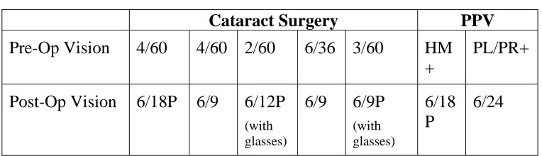
Surgery in the form of cataract removal with intraocular lens
implantation was done in cases with complicated cataract.
Post inflammatory glaucoma was managed with antiglaucoma
measures along with steroids and followed up, regularly. Chorioditis
that were active were followed up after management of active phase.
Cases needing vitrectomy for traction bands, were referred for surgery
ANALYSIS AND OBSERVATION
[image:65.612.121.493.189.314.2]These were the observations of our study.
Table 1 Age Incidence
Age in Years No. of Cases Percentage
0-4 5 5%
5-9 35 35%
10-14 44 44%
15-18 16 16%
In our study the maximum number of cases of uveitis in children
were found to be within the age groups of 10-14 years and 5-9 years,
comprising 44% and 35% of the cases respectively. Least number of
cases were found to be in the age group of 0-4 years.
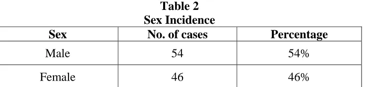
Table 2 Sex Incidence
Sex No. of cases Percentage
Male 54 54%
Female 46 46%
Out of the 100 cases in this study. 54 cases were males and 46
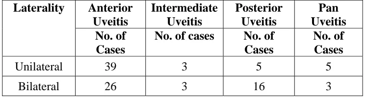
[image:65.612.125.494.442.530.2]Table 3 Laterality
Laterality No. of Cases Percentage
Unilateral 52 52%
Bilateral 48 48%
Regarding the laterality out of 100 cases in our study, 52 cases
were unilateral and 48 cases were bilateral.
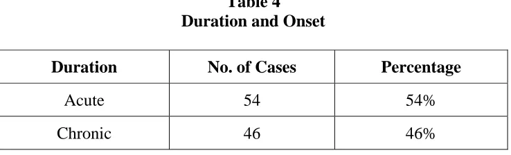
Table 4 Duration and Onset
Duration No. of Cases Percentage
Acute 54 54%
Chronic 46 46%
Out of our 100 cases 54% presented with acute onset of less than
6 weeks and 46% with chronic duration. Acute on chronic cases were
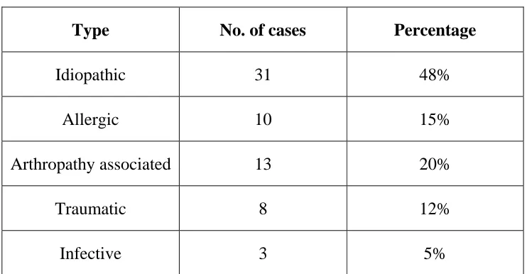
[image:66.612.123.492.348.460.2]Table 5
Aetiological Analysis (Anterior Uveitis)
Type No. of cases Percentage
Idiopathic 31 48%
Allergic 10 15%
Arthropathy associated 13 20%
Traumatic 8 12%
Infective 3 5%
In our study out of the 65 cases of anterior uveitis (31 cases).
48% were formed by idiopathic causes. Iritis secondary to septic foci in
the body formed 15% of cases. Anterior uveitis associated with juvenile
rheumatoid arthritis and sero negative arthropathies amounted to 20% of
cases (13 cases). Trauma was the cause for 12% of cases. Infective
causes amounted to 5% of cases (3 cases). They belonged to children
with herpes simplex uveitis (two) and one with fungal corneal ulcer with
Table 6
Aetiological analysis of posterior uveitis
Aetiological Type No. of cases Percentage
Tuberculosis 4 19%
Toxoplasmosis 12 57%
Toxocara 1 5%
Others 5 19%
In our study out of the 21 cases of posterior uveitis. Tubercular
etiology was found in 4cases, 12cases of toxoplasmosis involving both
old and fresh cases and one case of Toxocara endophthalmitis, four
cases of chorio retinal vasculitis namely one each of Eale’s disease,
SLE, cytomegalovirus retinitis in immunosuppressed child and one due
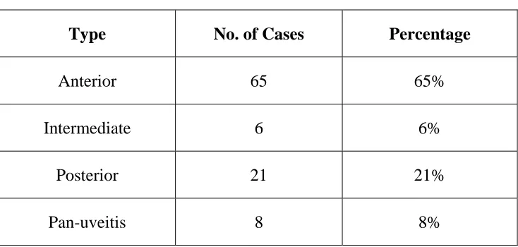
Table 7
Anatomical Classifications
Type No. of Cases Percentage
Anterior 65 65%
Intermediate 6 6%
Posterior 21 21%
Pan-uveitis 8 8%
Based on anatomical classification (IUSG) in our study 65 cases
(65%)were anterior uveitis, 6 cases (6%) were intermediate uveitis, 21
cases (21%) were posterior uveitis and 8 cases (8%) were pan uveitis.
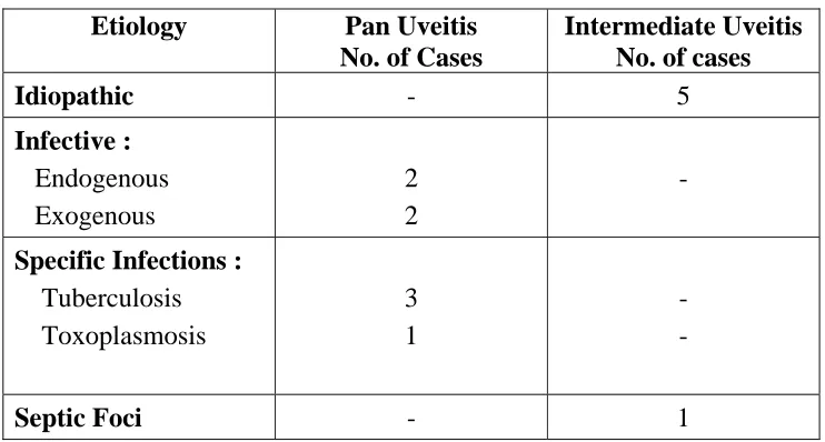
In intermediate uveitis 5 cases were idiopathic and one case was
due to focal sepsis.
In pan uveitis four cases were due to infection with bacterial
seeding, ultimately leading on to endophthalmitis. 3 cases were due to