REAL TIME THREEDIMENSIONAL ECHOCARDIOGRAPHY IN
TRICUSPID VALVE DISEASES
Dissertation submitted for
D.M. DEGREE EXAMINATION
BRANCH II – CARDIOLOGY
MADRAS MEDICAL COLLEGE
AND
GOVERNMENT GENERAL HOSPITAL
CHENNAI – 600 003
THE TAMIL NADU DR.M.G.R MEDICAL UNIVERSITY
CHENNAI – 600 032
AUGUST 2009
“learn to heal”
CERTIFICATE
This is to certify that the dissertation entitled “REAL TIME THREE
DIMENSIONAL ECHOCARDIOGRAPHY IN TRICUSPID VALVE DISEASES” is the
bonafide original work of DR.J.JACOB JUSTIN in partial fulfillment of the requirements for D.M. Branch-II (CARDIOLOGY) examination of THE TAMILNADU DR.M.G.R.
MEDICAL UNIVERSITY to be held in August 2009.The period of post-graduate study and
training was from August 2006 to July 2009.
THE DEAN PROFESSOR R. SUBRAMANIAN. M.D., D.M. MADRAS MEDICAL COLLEGE & PROFESSOR AND HEAD OF THE
GOVERNMENT GENERAL HOSPITAL DEPARTMENT OF CARDIOLOGY CHENNAI - 600 003. MADRAS MEDICAL COLLEGE &
GOVERNMENT GENERAL HOSPITAL
CHENNAI - 600 003.
DECLARATION
I Dr.J.JACOB JUSTIN, solemnly declare that this dissertation entitled, “REAL TIME
THREEDIMENSIONAL ECHOCARDIOGRAPHY IN TRICUSPID VALVE
DISEASES”is a bonafide work done by me at the department of Cardiology, Madras Medical
College and Government General Hospital during the period 2006 – 2009 under the guidance
and supervision of the Professor and Head of the department of Cardiology of Madras Medical
College and Government General Hospital, Professor R. SUBRAMANIAN .MD.DM . This dissertation is submitted to The Tamil Nadu Dr.M.G.R Medical University, towards partial
fulfillment of requirement for the award of D.M. Degree (Branch-II) in Cardiology.
Place : Chennai
ACKNOWLEDGEMENTS
A great many people made this work possible. I thank my Dean for allowing me to conduct this study.
My warmest respects and sincere gratitude to our beloved Professor R. SUBRAMANIAN
MD. DM Professor and Head of the Department of Cardiology, Government General
Hospital, Chennai who was the driving force behind this study. But for his constant guidance this study would not have been possible.
My respectful thanks to Prof R.Alagesan[Rtd] for his constructive ideas, personal guidance and involvement in this study.
I am indebted to Prof. Geetha Subramanian, Prof. B.Ramamurthy Prof. P.Arunachalam Prof M. Somasundaram , and Prof.V.Dhandapani without whom, much of this work would not have been possible.
I acknowledge Dr M.A.Rajasekar for the many useful comments he made during this project .In addition,I am grateful to Dr.G.Gnanavelu, Dr.S.Venkatesan Dr.P.S.Mohanamurugan, Dr.G.Ravishankar, Dr.JustinPaul, Dr. C.Elangovan and Dr.G.Pradap kumar for all guidance.
CONTENTS
Page
1. Introduction 1
2. Aim of the study 4
3. Review of literature 5
4. Material and methods 48
5. Results and Data analysis 56
6. Discussion 83
7. Conclusion 93
8. Bibliography
9. Glossary & Acronyms
INTRODUCTION
The tricuspid valve is a forgotten valve It has multi-component complex structure . The
tricuspid valve is composed of three leaflets (anterior, posterior and septal) attached to a fibrous
annulus.The three-dimensional shape of the tricuspid annulus is complex and does not
conform to a flat ring .Tricuspid regurgitation is the most common pathology affecting the
tricuspid valve. An understanding of the pathological process underlying tricuspid regurgitation
is necessary to determine the optimal management strategy. Usually tricuspid regurgitation is
secondary to left-sided valvular pathology (mostly mitral valve disease) with pulmonary
hypertension and right ventricular dilatation. Because the tricuspid annulus is a component of
the right ventricle it will dilate also. Unfortunatelytricuspid valve evaluation continues to be a
major problem in the surgical decision making process.
Unlike the aortic and mitral valve it is not possible to visualize all tricuspid valve cusps
simultaneously in one cross-sectional view by standard transthoracic two- dimensional
echocardiography During transesophageal 2D Echo small changes in transducer angle, probe
position and rotation may bring to light some additional tricuspid valve details .However,
because of the position of the tricuspid valve in the far field in relation to probe,
transesophageal 2DEcho can still only provide limited information and can also not visualize
decision to repair the tricuspid valve may be changed due to discrepant tricuspid annular
diameter findings between pre-operative two-dimensional transthoracic or transesophageal
echocardiography and direct surgical visualization . These unsatisfactory results call for a
reappraisal of surgical techniques for TR that in the past might have been based on an
incomplete knowledge of tricuspid annulus geometry.
Tricuspid vavle is affected by a variety of congenital and acquired diseases
Rheumatic heart disease causes tricuspid valve stenosis in up to 8% of patients. Unfortunately,
tricuspid valve stenosis is easily missed at clinical examina tion except in advanced cases
when a high degree of clinical suspicion exists. Undetected and thus uncorrected tricuspid
valve stenosis may lead to postoperative low cardiac output despite successful relief of
left-sided valve disease and carries a high mortality and morbidity.
Two-dimensional echocardiography (2DE) can detect thickened tricuspid valve leaflets
and a reduced tricuspid valve orifice diameter, and continuous-wave Doppler allows
estimation of the tricuspid transvalvular pressure gradient. However, in most patients, it is not
possible to visualise all three tricuspid valve leaflets simultaneously with 2DE and measure the
tricuspid valve area .
Transthoracic real-time three-dimensional echocardiography (RT3DE) may be a
valuable imaging modality for the examination of stenotic tricuspid
valves because all leaflets can be seen simultaneously and studied from both atrial and
ventricular aspects. Ebsteins anomaly is characterized by by tethering of the tricuspid valve to
value in making the diagnosis of this entity. It does not visualize the posterior leaflet involved
in tethering .
Also it does not provide a comprehensive assessment of the tethered and non tethered
areas of tricuspid leaflets when repair is considered Obviously, 2D TTE is limited in
visualizing the complete tricuspid anatomy . Since real-time 3D echocardiography (RT3DE),
has become available for clinical practice, it is now possible to examine the tricupid valve
more completely. In three-dimensional (3D) echocardiography studies this goal could be
achieved but at the cost of an increase in procedural duration . Real-time three-dimensional
echocardiography (RT3DE) can visualize the tricuspid valve from both the ventricular and
atrial side in detail .The real-time, 3- dimensional echocardiography (RT3DE) can be used to
provide fast and noninvasive estimates with high image resolution that is more accurate and
AIM OF THE STUDY
The study aims at utilizing Real time three-dimensional transthoracic echocardiography
(RT3DE) technique for
a. qualitative and quantitative analysis of tricuspid valve in both normal and
dieased states
b. comprehensive assessment of individual leaflets of tricuspid valve commisures
,leaflets ,valve area and annulus in normal individuals
c. detailed analysis of tricuspid annulus, commisures, valve area and leaflets in
patients with functional regurgitation.
d. assessment with descriptive tricuspid valve morphology for each tricuspid valve
leaflets and each tricuspid valve commissure and tricuspid valve area in
patients with rheumatic tricuspid valve stenosis
e. comprehensive assessment of individual leaflets of tricuspid valve and annulus
in patients with organic tricuspid valve diseases like ebsteins anomaly and
infective endocarditis
The study aims to compare the findings between 2D TTE and RT3D echo.
REVIEW OF LITERATURE
ANATOMY Leaflets
The tricuspid valve has three distinct leaflets described as septal, anterior, and posterior.
The tricuspid orifice is placed in the medial and anterior part of the floor of the right atrium and
with the heart in its natural position, the orifice is almost vertical. . Of the valve cusps the
anterior is usually the largest and extends from close to the infundibulum to nearly the lateral
margin of the orifice .The posterior leaflet is smaller and appears to be of lesser functional
significance. The septal leaflet is in immediate proximity of the membranous ventricular
septum, and its extension provides a basis for spontaneous closure of the perimembranous
ventricular septal defect.
Papillary Muscles & Chordae
There are three sets of small papillary muscles, each set being composed of up to three
muscles. Of the main papillary muscles the large anterior muscle is inserted into the adjacent
margins of the anterior and posterior cusps, the smaller posterior into the posterior and septal
cusps, and the small septal muscle into the septal and anterior cusps. The chorde tendine and
papillary muscles are thus attached almost in a ring around the tricuspid valve orifice and tether
chordae tendinae arising from each set are inserted into two adjacent leaflets. The anterior set
chordae insert into half of the septal and half of the anterior leaflets. The medial and posterior
sets are similarly related to adjacent valve leaflets
Tricuspid annulus
The tricuspid valve is the most apically placed valve with the largest orifice among the
four valves. The tricuspid annulus is oval-shaped and when dilated becomes more circular. It is
20% larger than mitral valve annulus . Normal Tricuspid valve annulus diameter is 3.0 to 3.5
cm The cavity of the right ventricle has a roughly three-sided pyramidal shape with the
tricuspid orifice set in the right posterior wall. In the roof of the ventricle a thick muscular
ridge, the crista supraventricularis separates the tricuspid orifice from the infundibulum and
thus helps to divide the ventricular cavity into a posterior inflow tract and an anterior outflow
tract.51 The tricuspid orifice projects into the cavity of the right ventricle rather like a funnel
and is set an angle of roughly 60° to the outflow tract The function of the valve may be studied
in the dead heart by distending the ventricular cavity with water (Brunton, 1906). Viewed from
the right atrium, valve closure is seen to be effected by apposition of the atrial surfaces of the
cusps almost at their margins The cusps bulge upwards like sails but are prevented from
Tricuspid valve by 3D echo
ò
The mitral valve, in contrast to the tricuspid, does not project forwards into the
ventricular cavity but is set more or less flush with the wall. In other words the inflow and
outflow tracts of the left ventricle are parallel to each other, whereas on the right they are at an
angle of about 60 degrees .
Therefore, by moving laterally in systole, the anterior cusp of the mitral valve can help
to obliterate its inflow tract whilst no such action is possible with the tricuspid valve. In
addition, according to Walmsley (1929), the two large mitral papillary muscles actually
obliterate part of the inflow tract in systole: there is no comparable muscular action in the right
ventricle.21 It is therefore apparent that the tricuspid valve is anatomically much weaker than
the mitral as regards the prevention of incompetence. The tricuspid valve may malfunction due
to structural malformationor secondary to other cardiac pathology
ETIOLOGY
The most common presentation of Functional Tricuspid valve disease is of tricuspid
regurgitation due to
• Right ventricular hypertension due to pulmonary hypertension either primary or
secondary to left sided diseases
As pulmonary hypertension developsleading to right ventricular dilatation, the tricuspid
valveannulus will dilate. The circumference of the
annulus lengthensprimarily along the attachments of the anterior and posteriorleaflets. The
ventricular dilationprogresses, the chordal papillary muscle complex becomes
functionallyshortened. This combination prevents leaflet apposition, resultingin valvular
incompetence.
Eisenmenger's syndrome and primary pulmonary hypertension leadto the same
pathophysiology of progressive right ventriculardilation, tricuspid annular enlargement, and
valvular incompetence.
Etiology of Primary Tricuspid Valve Disease
• Congenital
-Ebstein’s anomaly
-Congenital tricuspid stenosis
-Tricuspid atresia
• Rheumatic valve disease, generally in association with rheumatic mitral valve disease
• Infective endocarditis
• Carcinoid heart disease
• Toxic (eg, Phen-Fen valvulopathy or methysergide valvulopathy)
• Tumors (eg, myxoma)
•
•
• Iatrogenic—pacemaker lead trauma
• right ventricular infarction.
• Marfan syndrome can lead to prolapsing leaflets, elongationof chordae, or chordal
rupture producing valvular incompetence.
CLINICAL PRESENTATION
Tricuspid insufficiency has been recognized clinically since 1836 when Benson
described the jugular venous wave abnormalities in a case proven at autopsy. In the following
year King stated that "on occasion of most copious influx (the right ventricle) becomes dilated:
upon which the curtains of the tricuspid valve are drawn aside, an aperture of reflux is
produced, and the force of the ventricle is diverted from the pulmonary circulation." The
clinical observation that patients with pulmonary hypertension had less orthopnea and
paroxysmal nocturnal dyspnea once they developed tricuspid insufficiency would seem to
support King's concept. This is consistent with the hypothesis that rising pulmonary arteriolar
resistance by producing pulmonary hypertension, right atrioventricular distention, and tricuspid
insufficiency, diminishes forward flow and progressive overloading of the left atrium and
pulmonary capillary bed. Patients with tricuspid regurgitation have the presenting symptomsof
fatigue and weakness related to a reduction in cardiac output. f
Right heart failure leads to ascites, congestive hepatosplenomegaly,pulsatile liver,
pleural effusions, and peripheral edema. Inthe late stages these patients are wasted with
cachexia, cyanosis,and jaundice.
stages of severity of tricuspid regurgitation It is generally agreed that the initial change in right
atrial curves is a decrease in the depth of the X descent which eventually almost disappears as
the V wave becomes larger, leading to "ventricularization" of the curve with severe tricuspid
insufficiency . Impressive jugularvenous distention with an s wave or fused c and v waves, followedby a prominent y descent, is present. During inspiration thisfinding is accentuated because of the physiologic increase invenous return
Duroziez is credited with the description of the physical findings of tricuspid
insufficiency. Included were a xiphoid systolic murmur, enlarged right atrium, distended neck
veins with systolic pulsation, hepatic enlargement and pulsation, and peripheral cyanosis. Atrial
fibrillation is common. The cardiac exam is notable for anS3 that increases with inspiration and
decreases with a Valsalvamaneuver, increased P2 if pulmonary hypertension has
developed,and a parasternal pansystolic murmur increasing with inspiration. The characteristic
murmur with its inspiratory accentuation may be missed
easily in the presence of other systolic murmurs or in mild tricuspid insufficiency.The hepatic
pulsation is readily detected only with severe tricuspid insufficiency . Furthermore, the
presence and severity of tricuspid insufficiency are remarkably labile, being greatly influenced
by exercise, deep breathing, emotions, and the degree of cardiac compensation .
PATHOPHYSIOLOGY
The tricuspid annulus dilatation and leaflet tethering were importantmechanisms in the
development of functional TR. Annulusdilatation compromised leaflet closure or coaptation by
limiting the amount of leaflet overlap. Changes in RV geometry presumably caused
The chest x-ray demonstrates cardiomegaly, increased right atrialand ventricular size, a
prominent azygous vein, pleuraleffusion, and upward diaphragmatic displacement due to
ascites.Echocardiography best assesses the degree of regurgitation,structural abnormalities of
the valve, pulmonary artery pressures,and right ventricular function both preoperatively and
intraoperatively.A shift in the atrial septum to the left and paradoxical septalmotion are
consistent with right ventricular systolic overload.Pulsed Doppler and color flow help identify
systolic right ventricularto right atrial flow with inferior vena cava and hepatic veinflow
reversal. Contrast echocardiography can be useful witha rapid saline bolus injection producing
microcavities thatare visible on echo, demonstrating to-and-fro motion acrossthe valve orifice
and reversal into the inferior vena cava andhepatic veins.
Angiocardiography has been unreliable in demonstrating tricuspid insufficiency.27
Thehigh-pressure delivery of contrast material into the right ventricle may cause the catheterto
regurgitate into the right atrium or stimulate premature ventricular contractions which produce
insufficiency. Only if adequate opacification is achieved and no regurgitation is seen can one
state confidently that tricuspid insufficiency is absent. Cardiac catheterization will document
increased right atrialand right ventricular end-diastolic pressure. The right atrialpressure
tracing has an absent X descent, prominent V wave,and "ventricularization" of the right atrial
tracing, and the degree of pulmonary artery hypertension is documented. Pulmonaryartery
pressures of over 60 mm Hg are usually due to left-sidedlesions leading to secondary tricuspid
regurgitation. A rightventriculogram has been used but is unnecessary with
It is well known that after mitralvalve surgery patients may clinically deteriorate due to
underestimated tricuspid pathology and significant residual or developing TR . Tricuspid
regurgitation in patients with mitral valve disease is associated with poor outcome and predicts
poor survival,heart failure, and reduced functional capacity2. It might appear many years after
surgery
andmight not resolve after correcting the mitral valve lesion. Late TR mightbe caused by, left
heart disease, right ventricular dysfunction and dilation, persistentpulmonary hypertension,
chronic atrial fibrillation, or by organic(mainly rheumatic) tricuspid valve disease. Most
commonly, lateTR is functional and isolated, secondary to tricuspid annulardilation. Outcome
of isolated tricuspid valve surgery is poor,because RV dysfunction has already occurred at that
point inmany patients. Mitral valve surgery or balloon valvotomy should be performedbefore
RV dysfunction, severe TR, or advanced heart failurehas occurred.
Tricuspid annuloplasty with a ring should be performedat the initial mitral valve
surgery, and the tricuspid annulus diameter>3.5 cm is the best criterion for performing the
annuloplasty4
The ideal tricuspid valve annuloplasty would resolve the deficiency in tricuspid valve
coaptation caused by both tricuspid annular dilatation and leaflet tethering. However, the
concept of current TV annuloplasty is to stabilize the area of the tricuspid annulus that is
primarily responsible for annular dilatation. Therefore, annuloplasty performed to reduce the
tricuspid annulus might not be sufficient tocorrect the tethering of tricuspid vave . Tricuspid
annuloplasty28. A similar finding of echocardiographic
predictors has been described for mitral valve annuloplasty in patients with functional mitral
regurgitation.
Calafioreet al showed that mitral valve coaptation heightwas a preoperative predictor
for the failure of mitral annuloplasty in patients with functional mitral regurgitation3. In
contrast, the degree of tricuspid annulus dilatation was also a preoperative risk factor for
postoperative negative results. 3In, a future tricuspid valve surgery addressing both leaflet
tethering and annular dilation should be developed to overcome the limitations of annular
reduction alone in functional TR. Furthermore, assessment of preoperative tricuspid valve
tethering might be essential to define thesurgical indication for tricuspid valve replacement in
patients with severefunctional TR .Most surgeons consider the use of an annuloplasty ring an
essential part of basic repair techniques for obtaining good results. However, the rings currently
being used for tricuspid annuloplasty were originally released for the mitral valve, and
moreover, most rings are formed in a single plane with variation, whereas the actual tricuspid
annulus may have a nonplanar, or 3D, structure5,6.
Although 2D TTE is helpful to assess tricuspid valve function and to detectTR severity
it has important limitations in describing tricuspid valve morphological details, such as
tricuspid annular diameter and annular area. The impact of echocardiographic-guided treatment
on outcome after tricuspid valve surgery is not well defined. A limitation of the 2D
echocardiography was the
In a study in Int J Cardiovasc Imaging (2007) “Assessment of normal tricuspid valve
anatomy in adults by real-time three-dimensional echocardiography” by Ashraf M. Anwar et al
41 reports that it was possible to analyse tricupid valve anatomy by RT3DE and a detailed
anatomical structure including unique description and measurement of tricuspid annulus shape
and size, tricuspid leaflets shape, and mobility, and tricuspid commissural width in majority of
patients. Moreover, RV size and geometryare technically difficult to determine accurately with
2D echocardiography because of its anatomic complexity can be measured with 3D echo.
Changes of ventricular geometry may cause the tethering of tricuspid leaflet through the
displacementof the papillary muscles, determining the outcome of tricuspidannuloplasty.
Three-dimensional imaging techniques may havethe potential to provide more accurate
information for tricuspid valve deformation ,RV function and geometry, RT3DE combined
with software is useful for assessing the unique 3D geometry of the tricuspid annulus in
healthy subjects and in patients with functional TR. In a study by Michael Y. Henein, Christine
A. O'Sullivan, Wei Li, Mary Sheppard, Yen Ho, John Pepper, Derek G. Gibson Departments of
Echocardiography, Cardiac Morphology, Cardiac Pathology and Cardiac Surgery, Royal
Brompton Hospital, London, UK “Evidence for rheumatic
valve disease in patients with severe tricuspid regurgitation long after mitral valve surgery”
(The Journal of Heart Valve Disease 2003)30 states that severe tricuspid regurgitation is a well-recognized, long-term complication of rheumatic mitral valve replacement that impairs the
functional results of surgery, its exact basis remains unclear and its management is
unsatisfactory. A detailed assessment of tricuspid valve morphology and function using 2D
of this condition may thus improve its long-term management, possibly with early tricuspid
valve repair.
In patients with predominanttricuspid regurgitation, the negative consequences of
rightventricular volume overload develop slowly. Acute tricuspidregurgitation due to
traumatic rupture or complete excisionof the tricuspid valve as the treatment for infective
endocarditiscan be well tolerated for years if the pulmonary artery pressureis not elevated45
TRICUSPID STENOSIS;
It has become almost traditional to state that tricuspid stenosis is rare and that the
diagnosis is difficult . Mackenzie (1908) wrote that he had heard a tricuspid murmur only three
times in his life46. The stress that has been laid on the rarity and difficulty of diagnosis of the
affection combined with statements such as those above, have tended to make the average
practitioner forget that the tricuspid valve is not infrequently affected. . Tricuspid stenosis is
most
commonly rheumatic. It is extremelyrare to have isolated tricuspid stenosis, as some degree
oftricuspid regurgitation will be present. Mitral valvedisease coexists with occasional
involvement of the aortic valve.The third world and especially the Indian subcontinent
stillhave a significant prevalence of rheumatic tricuspid valvulardisease
INCIDENCE
There exists a good deal of discrepancy between several of the analyses that have been
following figures of the incidence of valve injury in 97 cases of rheumatic heart disease,
including the lesser as well as the greater degrees of involvement.
These were combined in the following ways:
Mitral alone .. .. .. .. .. .. .. 27 cases
Mitral and aortic .. .. .. .. .. 35
Mitral and tricuspid .. .. .. .. .. 12
Mitral, aortic, and tricuspid .. .. .. .. .. 21
Mitral, aortic, tricuspid, and pulmonary .. .. .. 2
The incidence at Bristol of clinical tricuspid stenosis in rheumatic heart disease was 14 per
cent. Cabot (1926) gives the incidence at 15 per cent while Dressler
and Fischer put the incidence at 24 per cent. Bland, Jones, and White (1935) analyzed the
pathological findings in 100 cases of fatal rheumatic disease below the age of 21 in whom the
diagnosis of tricuspid valve disease had not been made during life. Out of 100 cases, the mitral
valve was affected in 98 instances, the aortic in 71, the tricuspid in 30, and the pulmonary valve
in 5,
PATHOLOGY
According to the pathologist, the tricuspid valve becomes stenosed when the ostium is
reduced to between 6 and 8 cmsq., the normal being between 12 and 14 cm sq. (Cabot,
The valve becomes incompetent for three reasons: (1) extensive scarring in which
regurgitation accompanies stenosis; (2) slight shortening of the chorda tendinee or fibrosis of
the valve edge resulting in regurgitation, without necessarily causing stenosis; The anatomic
features of stenosis are similar to mitral stenosiswith fusion and shortening of the chordae
commissural fusion and leaflet thickening.Fusion along the free edges and calcific deposits on
the valveare found late in the disease. The preponderance of cases arein young women.
CLINICAL DIAGNOSIS
Duroziez (1868) reported 10 cases of tricuspid stenosis with ages varying from 22 to 64,
and asserted that tricuspid disease was more common
than was usually accepted. He was impressed by the fact that many could lie flat in bed in spite
of gross cardiac deficiencies: He drew attention to the diastolic murmur at the lower end of the
sternum that pointed to the diagnosis, and wrote: " The disease should be diagnosed when the
patient is a female, has a history of rheumatism, and of dvspnea, palpitation, edema, often with
remissions and exacerbations, is cyanosed, has mitral stenosis with an enlarged right heart,
particularly if the atrial enlargement can be made out . If in addition to this the patient has a
separate murmur best heard over the ensiform or the fifth or sixth right costal cartilage, then the
diagnosis becomes reasonably certain. There is no one infallible diagnostic sign but there are
numerous signs, any one of which should initiate a search for other clues. The diagnosis may
be suspected from the history alone as Levine and Thompson (1937) have pointed out.
Any adult patient who has repeated attacks of edema and ascites and yet is able to lead a
the early demise of the patient, probably has tricuspid stenosis. Several observers, have
commented on the peculiar colour, a mixture of jaundice and cyanosis, that is presented by
these patients, pointing out that this should always suggest the possible diagnosis of tricuspid
stenosis. Much reliance cannot, however, be placed on this sign in as much as its commonest
pathogenesis lies in the inability of an engorged or diseased liver to excrete rapidly enough the
blood pigment from a pulmonary infarct .A
patient with a rheumatic heart and ascites who is able to sleep without extra pillows probably
has tricuspid stenosis. This is due to the lack of pulmonary vascular engorgement, as confirmed
by X-ray study, resulting from the obstruction to the free flow of blood through the right heart.
Clinical features are consistent with reduced cardiac outputproducing the symptoms of
fatigue and malaise. Significant liverengorgement produces right upper quadrant tenderness
with apalpable liver with a presystolic pulse. Ascites produces increasedabdominal girth.
Significant peripheral edema or anasarca candevelop. Severe tricuspid stenosis may mask or
reduce the pulmonarycongestion of mitral stenosis due to reduced blood flow to theleft side of
the heart. The low output state of the patientis prominent Pulsation in the neck veins has for
over one hundred years excited comment, whether the jugular polygram is diagnostic. The
elevated venous blood pressures between 19 and 27 cm. of water noted in the absence of
(edema, constrictive pericarditis, and mediastinal obstruction should be taken as a strong
diagnostic point in favour of tricuspid stenosis.
If the patient remains in normal sinus rhythm, the right atrialtracing and jugular venous
Tschilikin (1930) stated that the only pathognomonic sign of tricuspid stenosis is a
localized diastolic murmur at the lower end or to the right of the sternum. However, this sign is
often, probably usually, absent, The cardiac
murmur is mid-diastolic,increases with inspiration, is heard maximally along the leftsternal
border, and may have an opening snap.
Atrial fibrillation occur just as frequently in cases of pure mitral valve disease,
especially the so-called " tight type," as in tricuspid stenosis. The circulation times were all
markedly prolonged, the right heart times averaging 20 seconds (ether method) against a
normal average of 6 seconds.As the right atrial pressure increases, venous congestion leadsto
distention of the jugular veins, ascites, pleural effusion,and peripheral edema. The right atrial
wall thickens and theatrial chamber dilates.
The chest x-ray demonstrates cardiomegaly with an increase inthe right atria and
pulmonary artery size. Dressler and Fischer emphasized as an important diagnostic pointer the
presence of marked enlargement of the heart to the right by X-ray especially when associated.
with absence of pulmonary congestion at the hilus of the lung.. Lian and Marchel (1936) drew
attention to the deviation of the esophagus to the left in the presence of marked enlargement of
the heart to the right.The ECG will demonstrateincreased P-wave amplitude if the patient is in
normal sinusrhythm.
Echocardiography reveals the diagnostic features ofdiastolic doming of the thickened
tricuspid valve leaflets,reduced leaflet mobility, and a reduced orifice of flow. TheDoppler
gradient between the right
atrium and right ventricleis significantly elevated at 2 to 5 mm Hg mean pressure17 .The
patient's ability to tolerate stenotic lesions of the tricuspidvalve is dictated to a large degree by
the natural history ofthe mitral or aortic valve disease.
Although thetricuspid valve leaflets can sometimes be visualised simultaneouslyby 2DE
from an angulated view, measurement of Tricuspid valve area is only rarely possible because
even when all leaflets are visualised,simultaneously the image cross section will not be at the
correcttricuspid valve area level. Unfortunately, from all other 2DE views, includingthe
atypical parasternal projection, only two tricuspid valve leaflets can be visualised
simultaneously. Estimation of the transtricuspid pressure gradient is usually only performed
when a morphologicallyabnormal tricuspid valve is seen. Therefore, good
morphologicalimaging and description of the tricuspid valve are essentialin identifying
tricuspid valve.
With RT3DE, each separatetricuspid valve leaflet can be assessed with regard to
thickness,mobility, calcification and its relationship to other tricuspidvalve leaflets.29 In
addition, RT3DE provides unique tricuspidvalve measurements such as Tricuspid valve area
3D and commissural width at thetime of maximal tricuspid valve opening.
In a study by Ashraf M Anwar et tal in Heart 2007 Evaluation of rheumatic tricuspid
valve stenosis by real-time three-dimensional
than tricuspid gradient for the separation of patients with rheumatic heart disease with
tricuspid valve involvement from those with rheumatic heart disease without tricuspid valve
involvement or from normal controls, tricuspid valve area 3D correlated with the mean
transtricuspid pressure gradient
Ebstein's anomaly of the tricuspid valve
Ebstein anomaly is characterized by heterogenous and variable tethering of the Tricuspid valve to the underlying right ventricle and ventricular septum .with the septal and the posterior leaflets more commonly involved in tethering and dysplasia of the leaflets. The variable degrees of redundancy and dysplasia determine the displacement of the functional tricuspid opening toward the trabecular portion and outflow tract of the right ventricle . The
histopathology of the right ventricular wall reveals a decrease or total absence of myocardial fibers in the inlet portion . The apical trabecular portion of the right ventricular free wall shows a characteristic pattern of anomalous muscular bands that connect the ventricular septum to the free wall . Moreover, in the infundibular portion of the ventricular outflow tract, myocardial fibers are diminished in number, making it very thin. Subpulmonary and pulmonary
obstructions can also be present In addition, 25% of patients with Ebstein's anomaly also present with a Wolf-Parkinson-White pre-excitation syndrome
The hemodynamic effects are related to the tricuspid regurgitation, the existence of an atrial septal defect and the degree of dysfunction of the atrialized portion of the right ventricle. Some cases have tricuspid stenosis owing to reduction of the functional valve opening
secondary to fusion of the leaflets. Echocardiography is the non-invasive method of choice in the diagnosis of this condition, as it makes it possible to evaluate the degree of leaflet tethering, the characteristics of the leaflets and the subvalvular apparatus, the degree of regurgitation or tricuspid stenosis, the morphological and functional alterations in the atrialized right ventricle and associated anomalies, all of which aid in planning the type of surgery to perform .54
Tethering of the septal leaflet results in apparent displacement of of its orgin toward the
apex of RV which is easily diagnosed on 2D echo For definition, the distance between mitral or
tricuspid annulus plane and the insertion of tricuspid pathologic leaflet is greater than 1 cm (or
more than 0.8 mm/m²). Frequently a tethering of posterior tricuspid leaflet to RV wall is
present. These modifications lead to atrialization of a RV The atrialized RV/total RV ratio
more than 30% indicates severe disease with poor prognosis. Commonly the tricuspid
of limited value in not delineating the morphology of the valves it also doesn’t visualize the
posterior
leaflet involved in tethering .it does not provide comprehensive assessment of the tethered and
non tethered areas of tricuspid leaflets which have an implication in the management of
tricuspid valve repair. 3D echo visualizes the posterior leaflet involved in tethering .and
provide comprehensive assessment of the tethered and non tethered areas of tricuspid leaflets .
In addition 3D echo demonstrates characteristic bubble like appearance produced by bulging of
the non tethered areas of tricuspid leaflets.
In a study “live real time three-dimensional transthoracic echocardiographic assessment
of ebstein's anomaly by Vinod patel, Navin c nanda. echocardiography, volume 22, november
2005” 56 states that the technique was found useful in assessing the distribution and extent of
tethering of each of the three leaflets of the tricuspid valve to the underlying right ventricular
walls and the ventricular septum. The characteristic bubble-like appearance resulting from
bulging of the non-tethered areas of the tricuspid leaflets was also well visualized in three
dimensions and their size measured. Thus, an estimate of the nontethered or free segments of
all three leaflets of the tricuspid valve could be obtained using this technique. This has
important implications when considering these patients for surgical repair of the tricuspid valve
. Visualization of all three leaflets of the tricuspid valve and their extent of tethering by 3D
TTE also made it easier to identify the boundaries of the functioning right ventricular chamber
Endocarditic lesions and vegetations are clearly visibleby echo; the valve may be
destroyed and septic pulmonary emboliare a common feature. The tricuspid valve in carcinoid
syndromeis thickened with retracted leaflets fixed in a semi open postionthroughout the
cardiac cycle.
Echocardiographic assessment of "tricuspid valve"
Tricuspid valvular apparatus can be evaluated by transthoracic and transesophageal
echocardiography10., The three-dimensional echocardiography will offer the integral image of
tricuspid valve .The TV can be well visualized from multiple transthoracic echocardiographic
(TTE) views
Parasternal short-axis view; septal and posterior leaflets are seen
Apical four chambers view;
Subcostal view.
Right ventricular inflow tract view, - In the these three views the septal and anterior
leaflets can be visualized.
Transesophageal echocardiography (TEE) offers multiple views for the tricuspid valve
evaluation planes23.24
- 4C view, similar to TTE 4C view;
- At the base of the heart in a 60° view.
- Bi-caval view;
- Gastric longitudinal view of RV.)
The last two allow the visualization of the anterior and posterior leaflets
The tricuspid inflow and tricuspid regurgitation can be evaluated from multiple
ecocardiographic windows by spectral and color Doppler31. The effective orifice area of the tricuspid valve is greater than that of the mitral valve and the right cavities pressure is smaller than on the left. Because of that the inflow velocities are lower for TV than for the mitral valve. Normally, the tricuspid diastolic flow E/A ratio exceeds 1.0.1 Color flow imaging can be used to detect the presence of tricuspid regurgitation (TR). This is present physiologically in 80-90% of healthy subjects. The physiological TR has low velocities and right ventricular systolic pressure is in the normal range.
The physiological TR with the following criteria44:
- TR jet in RA < 1 cm long;
- TR jet area < 2.5 cm2;
- TR jet area / RA area < 18%.
Tricu spid st nosis (e TS )
The echocardiography offers a etiologic, severity and functional diagnosis of this
valvulopathy.The diagnosis uses M mode and two
dimensional (2D) data, which described the morphology and mobility of tricuspid valve. The
continuous wave and color Doppler registered the diastolic tricuspid turbulent flow. The
normal tricuspid inflow velocity is less than 0.5 to 1 m/s, with a mean gradient less than 2 mm
Hg
stenosis:
- Continuous wave Doppler measurement of maximum and medium velocities and
pressures on tricuspid inflow wave (Vmax, Vmean, Pmax, Pmean)
- Pressure half time (PHT) method with the constant of 190:
TV orifice area (S) = PHT/190
- The continuity equation.
The TS is considered severe when P mean ≥ 7 mm Hg, S ≤ 1 cm2, PHT > 190 ms.
The echocardiographic evaluation must include the measurements of the right atrium
enlargement.
Tricuspid stenosis Severity
Mild moderate severe
Mean gradient <2mm Hg 2-7 mm Hg >7mm Hg
17 1
T
ric u spid r e g u rgitation ( TR )
Doppler echocardiography represents the gold standard for diagnosis of TR. Continuous
wave and color Doppler are the main methods of detection and quantification of severity of TR.
Echocardiographic diagnosis uses M mode and 2D indirect data such as annulus dilation,
The underlying cause of TR can be detected by 2D and M-mode echocardiography. TR
can be due to primary disease of TV or secondary to annulus dilation (functional TR).
Mild TR Moderate TR Severe TR
Valve morphology normal Normal/abnormal abnormal
RA/RV/IVC size normal Normal/dilated Dilated unless acute
TR
CW jet Less intense Dense Dense with late
delay
Jet area <5cm2 5-10cm2 >10cm2
Jet/RArea <20% 20-40% >40%
Venacontracta small <7mm >7mm
Hepatic vein flow Systolicdominance Systolic blunting Systolic reversal
18The severity of TR can range from mild to severe. It is important to remember that
maximum velocity of tricuspid regurgitant flow represents the
pressure difference between RV and RA, and has nothing to do with TR severity grade. More
than that, the intensity of continuous wave signal is directly proportional with TR severity and
the shape of flow can suggest the acute TR (flow with early peak velocity and cut-off of
descendent part of the slope).
Quantification of TR can be done in a manner analogous to that for the mitral valve but
the publishing data are less well established. The intensity and the shape of continuous wave
signal is useful to evaluate the TR severity of to differentiate an acute TR from a chronic one.
respiratory variation in size can be detected. Additionally retrograde systolic flow in hepatic
veins can be seen.
SURGICAL DECISIONS
The cardiologist and the cardiac surgeon faces the decision ofwhen to intervene and
when to surgically repair or replace thetricuspid valve.7 The choice of the reparative technique
to usefor a durable result must be evaluated as well as evaluationof which type of valve,
mechanical or bioprosthesis, to employto maximize durability and minimize complications
(i.e., thrombosisand thromboembolism). The surgical literature can be misleadingdue to case
selection bias and the various time frames of retrospectivereviews.
r
ANNULOPLASTY TECHNIQUES
Techniques to deal with a dilated tricuspid valve annulus withnormal leaflets and
chordal structures include plication ofthe posterior leaflet's annulus (bicuspidization), partial
purse-stringreduction of the anterior and posterior leaflet annulus (DeVegatechnique), and
rigid or flexible rings or bands placed to reducethe annular size and achieve leaflet coaptation.
The preoperativeand intraoperative echocardiograms are valuable assessment toolsto help the
surgeon understand the structure and function ofthe valve14.
The degree of pulmonary hypertension, right ventricular dilatation,and systolic function
coupled with the size of the right atriummust be factored into the surgical decision making.
palpate the tricuspid valve and withdrawingthe finger tip 2 to 3 cm from the valve orifice
trying to accessthe force of the regurgitant jet is of less importance in thecurrent era of cardiac
surgery. Minimal right atrial enlargement and +1 to +2 regurgitationwill usually resolve after
surgery on left-sided valve lesions,especially if the pulmonary hypertension resolves.
Otherwise,tricuspid annuloplasty should be performed to help improve theearly postoperative
course and prevent residual or progressivetricuspid regurgitation. Special note should be taken
in assessingthe foramen ovale for patency. These lesions should always
besutured closed, reducing the possibility of arterial desaturationfrom right to left shunting or
paradoxical embolization.
Bicuspidization
Suture plication todeal with mild dilation of the annulus is accomplished by
placingpledgeted mattress sutures from the center of the posteriorleaflet to the commissure
between the septal and posterior leaflets.A second suture is often necessary to further reduce
the annulus,ensuring proper leaflet coaptation while providing an adequateorifice for flow. An
annuloplasty ring can be inserted to furthersupport the annular reduction
DeVega Technique
The DeVega technique can also be employed for mild to moderateannular dilation. The
technique employs a 2-0 Prolene or Dacronpolyester suture placed at the junction of the
annulus and rightventricular free wall, running from the anterior septal commissureto the
parallel and close to thefirst suture line in the same clockwise direction, placing itthrough a
second pledget at the posterioseptal commissure. Thesuture is tightened, producing a
purse-string effect and reducingthe length of the anterior and posterior annulus to provideadequate
leaflet coaptation and orifice of flow. The judgment regarding the degree of annular reduction
has
variedfrom the guideline of being able to insert 2-1/2 to 3 finger breadthssnugly through the
valve orifice to using the ring annuloplastysizers designed for the tricuspid valve. An
annuloplasty sizer,chosen by measuring of the intertrigonal distance, can be usedas a template
while tying the purse-string suture to achievethe proper degree of reduction. The DeVega and
suture plicationtechniques should be reserved for mild annular reductions andsituations in
which the structural integrity of the annulusis not absolutely necessary for long-term success
(i.e., functionaltricuspid regurgitation expected to resolve over time)13. In thesesituations the
annuloplasty provides a competent tricuspid valveduring the early postoperative course while
the heart remodelsafter surgical treatment of the left-sided valvular lesions.
Rings and Bands
Significant degrees of annular reduction requiring durabilityare best accomplished with
rigid rings (Carpentier-Edwards),flexible rings (i.e., Duran), or flexible bands (i.e.,
Cosgroveannuloplasty system). The length of the base of the septal leaflet(intertrigonal
distance) determines the size of the ring orband. These devices avoid suture placement in the
region ofthe AV node to avoid postoperativeconduction problems.It allows the tricuspid
leaflets.Overly aggressive annular reduction can lead to ring dehiscencedue to excessive
tension on the tenuous tricuspid valve tissue t
Intraoperative assessment of the repair
Assessment of tricuspid valve competence after the annuloplastyrequires filling the right
ventricle with saline and observingthe leaflet coaptation.. If the result appearsinadequate,
replacement should be performed. Final assessment is by TEE examination after completely
weaning from cardiopulmonarybypass with appropriate volume and afterload
adjustment12,13.
Tricuspid valve replacement
Tricuspid valve replacement with a homograft is complicated.The homograft tissue is a
mitral valve. Sizing is performed by measuring the intratrigonal distances13. Fixationof the
papillary muscles is either intracavity (right ventricle)or through the wall of the right
ventricle.. An annuloplasty ring isinserted to prevent dilatation and ensure adequate leaflet
coaptation.Special care is necessary in suture placement to avoid conductiondisturbances..
Similarto mitral valve replacement, leaflet and chordal preservationshould be performed to
maintain annular papillary muscle continuity.
Carpentier techniques for mitral valve repair can be appliedto the tricuspid valve.
Traumatic disruptions, occasionally endocarditis with healed lesions and perforations, or the
resections of the anterior
(limited) or posterior(extensive) leaflets, and ring annuloplasty are standard techniquesto
produce competent valves and avoid replacement. In infective endocarditis Tricuspid valve
excision is possible if pulmonary pressuresare not elevated and the degree of infection is
extensive.Blood flows passively through the right heart to the lungs.After eradication of the
infection, a second-stage procedurewith valve replacement can be performed months to years
laterwhen all infection is eradicated. 55 .Homograft tissue is often versatile for partial or total
tricuspidvalve repair or replacement.
PROSTHETIC VALVE CHOICE
The choice of prosthesis follows an algorithm similar to thatused for valve replacement
in other cardiac valve positions.The patient's age, anticoagulation considerations, whether
thepatient is a young woman during her childbearing years, andsocial issues must be
considered. The previously reported poorresults with mechanical valves in the tricuspid
position weredue to valve thrombosis. Most of these reports were during theera of cage-ball
and tilting-disc prosthesis. More recentreports with the St. Jude bileaflet valve have provided
encouraging data, allowing the surgeon to recommend a mechanical valve withconfidence to
younger patients who do not have a contraindicationto anticoagulation. . Bioprostheses, both
porcine and of pericardialtissue, have functioned well in the tricuspid position.The data
demonstrate a longer
duration of freedom from structuralvalve dysfunction or re-replacement for a bioprosthetic
In the tricuspid position it is always possible to place largebioprosthetic or mechanical
valves. Prostheses with more thana 27-mm internal diameter do not have clinically
significantgradients. Therefore hemodynamic performance is rarely an issuefor tricuspid valve
replacement. The data demonstrate excellentresults with modern bileaflet mechanical valves.
Series comparingbioprosthetic and mechanical valves have been consistent indemonstrating
equality during the period of follow-up.
Some patients with mitral valve disease undergoing surgery withtricuspid regurgitation
do not require surgical treatment ofthe tricuspid valve. Guidelines to identify these patients
arepoorly developed35. Experience has shown that careful observationof the patient
preoperatively is quite valuable. Absence oftricuspid valve regurgitation during periods of
good medicalcontrol, absence of tricuspid regurgitation by transesophagealecho at the time of
operation, minimal elevation of pulmonaryvascular resistance, and absence of right atrial
enlargementare helpful findings permitting the surgeon to confidently replacethe mitral valve
without performing an annuloplasty or replacementof the tricuspid valve.37,38
Clinical experience has demonstrated that up to 20% of patientsundergoing mitral valve
replacement receive a tricuspid
annuloplastybut less than 2% require replacement. The surgeon's clinicaljudgment and
experience guide the approach to tricuspid valvesurgery and ultimately lead to variability in
reported clinicaldata. The accuracy of the judgments can be guided by assessmentof the risk
factors for persistent or progressive tricuspidvalve regurgitation.26 They are related to
regurgitation or the misjudgment of its severity, failure ofthe pulmonary hypertension and
pulmonary vascular resistance to resolve, resulting in persistent impairment in right
ventricularfunction, and failureto recognize organic tricuspid valve disease.
The durability of simple annuloplasty techniques such as bicuspidizationand the
DeVega has been good when employed only for mild tomoderate degrees of functional
tricuspid regurgitation withsuccessful resolution of pulmonary hypertension after the
mitralvalve operation. Extensive experience with the tricuspid annuloplastyusing the Duran,
Carpentier-Edwards, or Cosgrove rings or bandsresulted in an 85% freedom from moderate to
severe tricuspid regurgitation at 6 years. The subsequent requirement for tricuspidreoperation
is very low. Inadequate resolution of the mitraldisease and persistent pulmonary hypertension
with right ventriculardilation and dysfunction are the major predictors of poor late results. r
The display of cardiac anatomy in three dimensions from any perspective would have
clear advantages over conventional 2D imaging and provide an insight into the functional and
anatomic properties of cardiac structures. One of the main advantages of live 3D TTE is its
“surgeon’s eye” view of the cardiac anatomy36. The technique is finding a niche in
preoperative planning, particularly for tricuspid valve repair A volume rendered 3D image of
the tricuspid valve can be reconstructed from any perspective. So, preoperative detection of
tricuspid valve by 3D ECHO will be helpful for the surgeon in better planning
THREE DIMENSIONAL ECHOCARDIOGRAM
diagnostic capabilities of cardiac ultrasound.
BACKGROUND
Attempts to record and display ultrasound images in 3D format were first reported in the
1960s. More than a decade later, investigators began to obtain 3D ultrasound images of the
heart. Through the careful tracking of a transducer, a sequence of 2-dimensional (2D)
echocardiograms could be recorded, aligned, and reconstructed into a 3D data set. This
methodology was limited by the need for offline data processing to create and display the 3D
images. In the early 1990s, von Ramm and colleagues developed the first real-time 3D (RT3D)
echocardiographic scanner capable of acquiring volumetric data at frame rates sufficient to
depict cardiac motion25.
d
METHODOLOGY
Transducer design
Breakthrough in 3D technology is possible by piezoelectric crystals development.
Conventional transducer- consist of 64 to 128 elements . These elements are arranged along a
single row. 3D transducer has more than 3000 elements arranged in rows and columns.. These
elements are electrically independent. 3D transducer has 150-boards. In these dense array
real-time 3D transducer, each square represents an element and Entire crystal of the transducer
head is sampled or covered with elements33. The micro beam former is required for this
So, beem steering is very powerful.
Reconstruction Techniques
Early approaches to 3D echocardiography were based on the principle that a 3D data set
could be reconstructed from a series of 2D images. In this method, serial 2D images are
obtained using either freehand scanning or a mechanically driven transducer that sequentially
recorded images at predefined intervals. More recently, the use of a transesophageal or
transthoracic multiplane probe has emerged as a readily available method to obtain rotational
images at defined interval angles around a fixed axis. Typically, images are
collected over a 180-degree rotation at set intervals. To minimize reconstruction artifacts,
sequential images are gated to both electrocardiography and respiration. Acquisition of a
complete data set typically takes 1 to 5 minutes, depending on respiratory and heart rates and
the predefined spatial intervals.The quality of 3D reconstructions from 2D images depends on a
number of factors, including the intrinsic quality of the ultrasound images, the number of the
2D images used to reconstruct the 3D image, the ability to limit motion artifact, and adequate
ECG and respiratory gating. Once the 2D images have been obtained, they are processed
offline with customized or commercially available software. The cardiac structures are
manually or semiautomatically traced to the 3D spatial coordinates to reconstruct a 3D image
Real-Time 3D Acquisition Methods
The development of RT3D echocardiographic systems circumvents many of the
ultrasound elements arranged in a grid fashion transmitting at a frequency of 2.5 or 3.5 MHz.
Current RT3D systems use matrix-array transducer technology with a greater number of
imaging elements, typically containing more than 3000 imaging elements, compared with the
256 in the sparse-array transducer. These current matrix-array transducers offer
improved resolution and are rapidly becoming the primary technique for 3D data acquisition in
clinical and research practice.
In addition, these matrix-array transducers display either online 3D volume-rendered
images or 2 to 3 simultaneous orthogonal 2D imaging planes. To minimize reconstruction
artifacts, data should be acquired during suspended respiration if possible.39
3D spatial modes consists of
1-live 3D mode –instantaneous 2- live 3D mode zoom-instantaneous
3-full voume –gated. 4-3D colour doppler- gated.
Live 3D mode/zoom –instantaneous
The system scans in real time 3 dimensions.If the transducer comes off the chest –the
image disappears.The volume pyramid may be reduced to zoom in three dimensions- Live 3D
zoom-instantaneus RT3D is the only on-line 3D method based on real time volumetric
scanning, as compared with other 3D imaging techniques- MRI, CT.
Full voume –gated
Gating is done by stitching 4 (or more) gates together in full-
volume mode. This can generate > 90 degree scanning volumes at frame rates >30 Hz.
Once a 3D data set is acquired, it must be sliced or “cropped” to visualize the cardiac
structures within the pyramid Multiple cropping methods are available, but a common method
displays 2 or 3 imaging planes simultaneously. Each of these imaging planes can be
manipulated separately to appropriately align the cardiac structures. Another cropping method
involves a single-slice plane that can be manually adjusted to expose and display the cardiac
structures of interest
Technical Factors
The technical aspects of acquiring a high-quality, diagnostic 3D echocardiogram are
similar to those for 2D echocardiography . Where as in 2D display which has rows and blocks of pixels( picture elements) in 3D data set it consists of bricks and pixels called volume
elements or voxels. 3D Data set (collection of voxels) can be rotated with respect to the
computer screen. . The use of optimal gain settings before acquisition is essential for accurate
diagnosis.. Most 3D echocardiographic systems use some form of gating to obtain volumetric
data. Gated data sets are most challenging in patients with arrhythmias or respiratory
Protocols
A complete 3D echocardiographic study includes an assessment of ventricular function,
valvular morphology, and hemodynamic status. Unlike 2D echocardiography, in which
standard views are described based on the plane through which they pass, 3D echocardiography
is inherently volumetric. As such, it permits both an external view of the heart and multiple
internal perspectives (through cropping). A general approach is to describe cardiac structures
using both the ultrasound plane and the viewing perspective. Three orthogonal planes are
recommended: (1) the sagittal plane, which corresponds to a vertical, long axis view of the
heart; (2) the coronal plane, which corresponds to a 4-chamber view; and (3) the transverse
plane, which corresponds to a short-axis view. Each plane can be viewed from 2 sides, which
represent opposite perspectives; for example, the transverse plane, which represents the
short-axis view, can be visualized from the perspective of the apex or base; the coronal plane can be
viewed from above or below; and the sagittal plane can be viewed from the left or right. The
choice of narrow-angle or wide-angle imaging acquisition modes depends on the cardiac
structure to be examined. For imaging of the ventricles, it is best to use a wide-angle
acquisition in the apical window. Instead of a complete 3D echocardiogram, a more focused 3D
imaging study may be appropriate in some cases. For example, in a patient with tricuspid
stenosis, the 3D portion of the study may be limited to visualization and quantification of the
tricuspid orifice. The ability
to extract hemodynamic information derived from 3D color Doppler ultrasonography is
currently being investigated. To capture and analyze color flow imaging in 3 dimensions, the
beam aligned as parallel as possible to the direction of blood flow. The color Doppler flow
patterns can be analyzed in multiple views to provide a complete assessment of the color
Doppler data
The benefits of 3DE are particularly well suited to the study of the tricuspid valve . The
assessment of patients with functional tricuspid regurgitation and tricuspid stenosis is one of
the most promising clinical applications of this technology. For reconstruction of the tricuspid
valve in adult patients, transthoracic echocardiography is the routine approach for 2D image
acquisition. Images from transthoracic echocardiography are interfaced with a 3D computer
system which incorporates the steering logic for acquisition of a rotational dataset and software
for 3D reconstruction and display. From the resultant dataset, novel 2D cut planes in any
orientation can be selected (any plane echo) and multiple parallel cross sectional 2D slices can
be generated in any desired plane (para plane echo).
However, as with any emerging technology, the enthusiasm to embrace a new technique
must be tempered by a critical appraisal of the evidence supporting its use. It is essential to
assess the limitations as well as the unique
capabilities it provides. To justify the use of a new 3D modality, its unique contribution to
clinical practice must be critically analyzed.
In a study done by “Renate schnabel,.second johannes gutenberg-university, germany,
(echocardiography, volume 22, january 2005)” 57 to demonstrate the feasibility of
transthoracic three-dimensional real-time echocardiography (3D TTE) supplemental to routine
TTE of the tricuspid valve can be performed in addition to routine 2D echocardiography
within a reasonable time and with high assessability of important features in patients with right
ventricular failure. Thus, its feasibility may encourage prospective studies on its potential for
more detailed noninvasive diagnosis and preoperative planning. In a study by “koteswara
Pothineni, et al university of alabama at birmingham in echocardiography, volume 24, may
2007 , “live/real time three-dimensional transthoracic echocardiographic assessment of tricuspid
valve pathology: incremental value over the two-dimensional technique”42 ,states that our preliminary experience with 3D TTE has demonstrated substantial incremental value over 2D
TTE in the assessment of various tricuspid pathologies. In addition, color doppler 3D TTE
provided an estimate of quantitative evaluation of TR severity, since the exact shape and size of
the vena contracta could be accurately assessed t
In a study by “Thanh-Thao Ton-Nu, MD Geometric Determinants of Functional
Tricuspid Regurgitation Insights From 3-Dimensional Echocardiography [Circulation. 2006;]”58 states that Little is known about the normal 3D shape of the TV and annulus and
changes that occur with functional TR. Their study was to examine the 3D geometry of the TV
annulus (TVA) under normal circumstances and in patients with functional TR, relating the
geometric changes in the tricuspid annulus between normal controls and functional TR. They
concluded that the normal Tricuspid Annulus by 3D echo is a bimodal non planar structure.
With development of functional TR,the annulus becomes larger, more planar, and circular. The
changes in annular geometry that occur with functional TR can be visualized by 3D Echo and
MATERIAL AND METHODS
This study was performed in the Department of Cardiology, Government General
Hospital, Chennai, during the year 2006– 2009.
STUDY INDICATION
Study indication was for the comprehensive assessment of qualitative and quantitative
analysis of tricuspid valve in both normal and diseased states and its correlation with 3D
echocardiography and 2D echocardiography
STUDY GROUP SELECTION
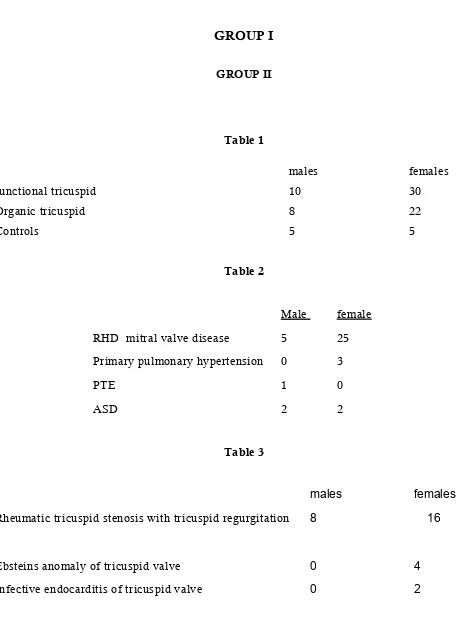
Seventy patients and ten controls attending government general hospital were
selected for the study. Patients suspected of tricuspid valve disease both organic and functional
were carefully evaluated by history taking, physical examination and laboratory tests, including
electrocardiography chest radiographs, and echocardiography. Color Doppler 2D echo was
done in all cases They were divided into three groups
Group 1
Patients with anatomical normal tricuspid leaflets with right ventricular pressure or
volume over load. It includes
1. Rheumatic heart disease with mitral heart disease with pulmonary hypertension