A STUDY TO DETERMINE
THE FREQUENCY OF OCULAR
MANIFESTATIONS IN HIV/AIDS PATIENTS
DISSERTATION SUBMITTED FOR
MASTER OF SURGERY DEGREE
BRANCH – III - OPHTHALMOLOGY
MARCH 2010
THE TAMILNADU
DR.M.G.R. MEDICAL UNIVERSITY
CERTIFICATE
This is to certify that this dissertation entitled "A
STUDY TO DETERMINE THE FREQUENCY OF OCULAR MANIFESTATIONS IN HIV/AIDS PATIENTS" is the bonafide
original work of Dr.VENI PRIYA.S., in partial fulfillment of the
requirement for M.S., (Branch III) Ophthalmology examination of
the Tamil Nadu Dr.MGR Medical University to be held in March
2010.
Dr.A. SULAIMAAN, M.S.,D.O.,
Head of the Department Department of Ophthamology
DECLARATION
I, Dr.VENI PRIYA.S., solemnly declare that this dissertation
"A STUDY TO DETERMINE THE FREQUENCY OF OCULAR MANIFESTATIONS IN HIV/AIDS PATIENTS "is a
bonafide record of work done by me in the Department of Ophthalmology, Madurai Medical College, Madurai under the
guidance of Dr.A. SULAIMAAN, M.S.,D.O., Head of the
Department, Department of Ophthalmology, Madurai Medical
College, Madurai.
This dissertation is submitted to the Tamilnadu
Dr.M.G.R.Medical University, Chennai in partial fulfillment of the
University regulations for the award of M.S Degree (Ophthalmology) Branch-III, Examination to be held in March 2010.
Place: Madurai
ACKNOWLEDGEMENT
I wish to thank THE DEAN, Madurai Medical College for permitting me to carryout this study.
With sincere gratitude, I am deeply indebted to
Prof.Dr.A.SULAIMAAN, M.S.,D.O., Professor and Head of
Department of Ophthalmolgy for his valuable guidance, help, expert
opinion, encouragement and kindness throughout this study.
I am extremely thankful to Prof. Dr.P.THYAGARAJAN,
M.S.,D.O., Additional Professor for his immense support , kindness
, guidance and encouragement during the course of my study.
I record my heartfelt gratitude to my Assistant Professors, Dr.
G.S. SRINIVASAN, M.S., D.O., Dr. T.BADRINARAYANAN, M.S.,
D.O., Dr.A.R.ANBARASI, M.S.,D.O., Dr.N.PARVATHASUNDARI,
M.S.,D.O., Dr.S.V.CHANDRAKUMAR, M.S.,D.O., Dr.
V.ANBUKANI, D.O. & Dr. SELVAKADUNGOVAZHIATHAN,
D.O. for their whole hearted support, valuable suggestion and
I am particularly thankful to my fellow post graduate
colleagues and friends for their valuable support and to all the staff
members who have made this study possible.
Above all I powerfully thank all the patients who have
submitted themselves for this study and made it possible and
CONTENTS
Sl.No. Title Page No.
1. INTRODUCTION 1
2. OCULAR MANIFESTATIONS 8
3. REVIEW OF LITERATURE 21
4. AIM OF THE STUDY 25
5. MATERIALS AND METHODS 26
6. OBSERVATION AND RESULTS 29
7. DISCUSSION 43
8. SUMMARY 53
9. CONCLUSION 55
10. BIBLIOGRAPHY
CONSENT FORM
PROFORMA
MASTER CHART
INTRODUCTION
AIDS is a potentially fatal multisystem disease characterized
by Profound disruption of the Immune system & a tendency for
various opportunistic infections & neoplasms.
AIDS was first recognized in United States of America in the
summer of 1981. Since then it has emerged as a global pandemic health problem of extra ordinary proportions & unprecendented
emergency.
At the end of 2007, 33.2 million people were living with HIV as per Joint United Nations Programme on HIV/AIDS. There are
nearly 5.7 million people infected with HIV in India.
Ocular manifestations are seen in 40 – 70 % of HIV patients. It can be caused by opportunistic infections, immunological reactions,
neoplasms & by HIV infection per se. It can affect almost all the
structures of the eye.
Life time cumulative risk of atleast one abnormal ocular lesion
developing in HIV patient ranges from 52 – 100 % in various
CD4 lymphocyte cells are proved to be a reliable predictor of
ocular manifestations of HIV infection. Ocular lesions are usually
seen in the end stages of the disease when the immunity is the lowest
(low CD4 count). With the introduction of HAART in 1990s, the incidence of ocular manifestations has come down.
Many a times ocular lesions may be the first clinical presentation
& can help the clinician to suspect the underlying HIV infection. Ocular lesions can be categorized by the ocular structures
involved as below
• Anterior segment manifestations – seen in upto 50% of HIV
patients
• Posterior segment manifestations – the most common & the
most severe vision threatening manifestations seen in > 50% of HIV infected patients. HIV microangiopathy is the most
common lesion followed by CMV retinitis. • Ocular adnexal manifestations.
• Neuro ophthalmic manifestations – seen in 10 – 15 % of HIV
Early recognition of ocular lesions will help early institution of
therapy & there by preventing visual loss. This is of even greater
significance nowadays, following the introduction of HAART &
HUMAN IMMUNODEFICIENCY VIRUS
HIV is a RNA virus belonging to human Retro virus,
Lentivirus subfamily. It has a unique reverse transcriptase enzyme.
HIV 1 & HIV 2 are currently known to infect human beings. In India, HIV Type 1c is the commonest type reported, though both
types exist.
MORPHOLOGY OF VIRUS:
Virus is 120 nm in diameter consisting of an outer envelope, a core shell of protein & a cone shaped inner core containing RNA
genome, reverse transcriptase enzyme & core polypeptides.
There are 3 structural genes 1. ‘gag’ gene ( group antigen)
2. ‘pol’ gene ( polymerase)
3. ‘env’ gene ( envelope antigen)
Apart from these genes, the virus also contains additional
regulatory genes like tat, rev, ref, vif, vpr & vpu.
STAGES OF VIRAL REPLICATION:
• HIV glycoprotein 120 attaches to the CD 4 receptors on CD4
• RNA genome of HIV is converted to DNA by reverse
transcriptase enzyme.
• Viral DNA then enters the host nucleus & gets incorporated
into the host cell DNA with the help of endonuclease.
• Virus components are produced by the host cell. Mature
viruses burst out of the host cell, lysing them, to infect new
cells Viral turn over is very high upto 10^9 particles every
1.5 – 2 days.
ROUTES OF TRANSMISSION:
The major routes of transmission are sexual contact,
parenteral exposure to blood & blood products & vertical
transmission.
WHO CLINICAL STAGING OF HIV/AIDS FOR ADULTS &
ADOLSCENTS, 2006.
CLINICAL STAGE 1:
• Asymptomatic
• Persistent generalized lymphadenopathy
CLINICAL STAGE 2:
• Unexplained weight loss
• Herpes zoster
• Minor mucocutaneous manifestations
CLINICAL STAGE 3:
• Unexplained severe weight loss
• Chronic diarrhea > 1 month duration
• Persistent fever > 1 month duration
• Oral candidiasis
• Oral hairy leukoplakia
• Pulmonary tuberculosis
• Severe bacterial infections
• Unexplained anemia
CLINICAL STAGE 4:
• HIV wasting syndrome
• Pneumocystis pneumonia
• Esophageal candidiasis
• Cytomegalovirus infection
• CNS toxoplasmosis
• HIV encephalopathy
• Kaposi sarcoma
• Lymphoma
CDC REVISED CLASSIFICATION OF HIV DISEASE – 1993
Absolute CD4 count cells/cu mm A Asymptomatic/ PGL/ acute seroconversion illness B HIV related conditions not included in A or
C
C Clinical conditions listed in AIDS
surveillance case definition >500
OCULAR MANIFESTATIONS OF HIV
Ocular involvement in AIDS is very common & includes various clinical presentations.
The first report of the ocular manifestation in AIDS was
done by Holland et al in 1982. In India, it was reported by J.Biswas
et al in 1995.
In general CD4 count has been used to predict the onset of
certain ocular infections in HIV patients.
CD4 COUNT cells/ cu mm
OCULAR MANIFESTATIONS
<500 <250 <100
Kaposi sarcoma,lymphoma, TB Pneumocystis, toxoplasmosis
Retinal/conjuctival microvasculopathy, VZ retinitis, CMV retinitis, MAC infections, cryptococcosis, microsporidiosis, HIV encephalopathy, progressive multifocal leukoencephalopathy
OCULAR ADNEXAL MANIFESTATIONS
Adnexal manifestations are seen in 25% of HIV patients & it
Conjunctival microvasculopathy
Conjunctival OSSN
Kaposi sarcoma
HERPES ZOSTER OPHTHALMICUS:
It is a painful vesiculo bullous dermatitis resulting from the
reactivation of the latent varicella zoster virus in the ophthalmic
division of the trigeminal nerve.
It is seen in 5-15% of HIV patients. Any patient younger than
50 years of age presenting with HZO should be suspected to have
underlying immunosuppression like HIV.
In HIV patients, HZO have prolonged & severe course with a
higher rate of painful sight threatening complications & increased
incidence of post herpetic neuralgia. It may also occur bilaterally & without associated skin lesions.
HZO is associated with conjunctivitis, keratitis, scleritis,
blepharitis, uveitis, retinitis , encephalitis, hemorrhagic hypopyon. Tissue damage is mediated through occlusive vasculitis.
TREATMENT:
INDUCTION DOSE:
I.V. Acyclovir 10mg/kg body weight TID – 7 days MAINTENANCE DOSE:
oral acyclovir 800mg 5 times a day for atleast 3-5 weeks.
Other drugs : famciclovir, I.V. foscarnet
LID INFECTIONS
Often severe & recurrent blepharitis, stye & lid ulceration can
occur.
TREATMENT: lid hygiene, antibiotics
MOLLUSCUM CONTAGIOSUM
Highly contagious infection caused by DNA pox virus.
The eyelid is involved in 5% o HIV infected patients. In these
patients, they are multiple, rapidly growing, bilateral, confluent &
more prone for secondary infections. They tend to recur after removal.
The virus particles may release into tears & cause toxic
keratoconjunctivitis. TREATMENT:
Cryotherapy with podophyllotoxin cream as an adjunct &
CONJUNCTIVAL MICROVASCULOPATHY
An asymptomatic vascular changes including segmental
vascular dilatation & narrowing, microaneurysm, comma shaped
vascular fragments & sludging of blood columns. It is seen in 70 – 80% of HIV patients.
Specific etiology is not known. However increased plasma
viscosity & immune complex deposition related to HIV or the direct infection of HIV in the endothelium may be the cause.
No treatment required.
CONJUNCTIVAL OSSN
It is usually seen in old age > 70 years and rarely seen in
normal persons.
In HIV patients, the risk of developing OSSN increases by 10-13 fold & occurs in young age. It constitutes the third most
common neoplasm associated with HIV. It bears no correlation with
CD4 count.
Hypothesis proposed for etiology:
1. generalized depression of immune surveillance with
Usually presents as pink papillomatous mass with feeder
vessels in the limbus.
TREATMENT:
Local excision with 2-3 mm safety margin & underlying scleral resection
Adjunctive: cryotherapy to the conjunctival margins,
brachytherapy, topical chemotherapy ( mitimycin c, 5 – fluorouracil, IFN α 2B)
Enucleation , exenteration can be done if the tumour is advanced.
KAPOSI SARCOMA
It is a highly vascularised, painless mesenchymal tumour that
affect skin & mucous membrane. Propable etiology may be due to
infection of human herpes virus – 8.
Conjunctival KS is seen in 10 – 20% in HIV patients . But in
India it is very rare, because of low incidence of HHV – 8.
ANTERIOR SEGMENT MANIFESTATIONS:
>50% of HIV patients have anterior segment manifestations.
It includes
1. Keratitis
2. Keratoconjunctivitis sicca
3. Iridocyclitis
KERATOCONJUNCTIVITIS SICCA:
Results from deficiency of any tear film layer; usually seen in the
late stage.
It is seen in 10 -20 % of HIV infected patients. Etiology is related
to HIV mediated inflammation & damage to the accessory glands &
to the direct effect of HIV virus itself on conjunctiva.
TREATMENT:
Lubricants & eye ointments
KERATITIS:
VIRAL:
Herpes is the most common cause of infectious keratitis. In HIV patients, they tend to have a longer, more severe course. They
recur more often & resistant to treatment.
HSV stromal disease occur infrequently. HZV – usually associated
with HZO iritis & increase in IOP may occur.
TREATMENT: Topical & systemic antiviral agents.
BACTERIAL:
Frequency not increased but generally more severe in HIV
patients. It may occur bilaterally, involve multiple pathogens &
carry a higher risk of perforation.
Ocular flora in HIV patients is not different from that of
general population, but the risk of infection with the normal flora is
high in severely immuno suppressed individuals.
MICROSPORIDIA:
Cause multiple diffuse punctuate epithelial keratopathy &
follicular conjunctivitis. Massom trichrome or giemsa stain can be used for diagnosis.
TREATMENT: topical fumagilin, oral albendazole, oral
itraconazole.
IRIDOCYCLITIS
It is rarely associated with HIV disease. It is usually associated
Rifabutin & cidofovir may induce iritis.
Immune recovery uveitis in CMV infected patients on HAART
therapy.
POSTERIOR SEGMENT MANIFESTATIONS
Seen in more than 50% of HIV infected patients.
It includes
1. non infectious – HIV microangiopathy & 2. infectious
These lesions can potentially affect the vision, if not treated
early.
HIV MICROANGIOPATHY:
The most common ocular lesion seen in 50-70% of HIV
infected patients. The lesion includes cotton wool spots,
hemorrhages & microaneurysm, which are generally asymptomatic
& transient. BRVO, BRAO & ischemic maculopathy may occur. Etiology may be due to
Increased plasma viscosity & fibrinogen levels
Circulating immune complexes
Infectious damage to the endothelium
No treatment required. HAART decreases the prevalence of
CMV RETINITIS:
The most common opportunistic intra ocular infection in HIV
patients seen in 15 – 40% .It occurs when the CD4 count is <100
cells / cu mm.
Clinically present as three forms:
1. perivascular fluffy white lesion with many scattered
hemorrhages.
2. more granular lesion with few hemorrhages with central area
of atrophic retina.
3. frosted branch angitis – rare
These lesions expand in brush fire pattern. Anterior spill over
uveitis may occur with fine stellate KPs.
Complications include retinal detachment & optic nerve involvement.
CMV IN HAART ERA:
After the introduction of HAART,
¾ the incidence of CMV retinitis has decreased by 75%,
¾ the risk of vision loss is lower,
Patients treated with HAART with sustained CD4 count
elevation of >100 cells/cu mm for atleast 3-6 months & with well
healed CMV retinitis inactive for 3-4 months can be given a trial of
withdrawal of CMV therapy.
IMMUNE RECOVERY UVEITIS:
It is an intra ocular inflammation caused by the reconstituted
immune system with HAART therapy in response to the persistence of CMV antigen.
It may present as anterior uveitis, vitritis, papillitis & macular
edema. Complications include cataract, CME & epiretinal membrane formation.
TREATMENT OF CMV RETINITIS: ANTI CMV DRUG INDUCTION MAINTENANC E SIDE EFFECTS Ganciclovir Oral Intravenous intravitreal Not used 5mg/kgBDx2wks 200-2000 mcg /0.1 ml 1gm TID 5mg/ kg daily
Weekly once Neutropenia, thrombocytopenia , anemia, nephrotoxicity Valganciclovir
oral 900mgX2wks 900mg OD Same as above
Foscarnet Intravenous
intravitreal
60mg TID X 3 wks
2.4 mg /0.1 ml ; twice weekly
90 mg/kg OD
Once a week
Nephroyoxicity, electrolyte imbalance Cidofovir: Intravenous intravitreal
5mg/kg once a wk X 2wks 15 – 20 mcg/0.1
ml
5mg/kg every 2 weeks Once in 6 wks
Proteinuria, hypotony,iritis,
neutropenia, peripheral neuropathy fomiversin 330 mcg; 2 doses
for every 2 weeks
2 doses for every month
GIOD - Ganciclovir Intraocular Device
It is a sustained release drug delivery device that contains about 4.5 – 6mg of ganciclovir. It is surgically placed into the
Investigational drugs: maribavir, tomeglovir
RD in CMV retinitis is characterized by multiple breaks, so
vitrectomy with silicon oil injection is the preferred modality of treatment.
TOXOPLASMA RETINOCHOROIDITIS
It accounts for 1% of the AIDS related retinal infections. The typical ocular presentation is
A focus of retinitis adjacent to a pigmented retinochoroiditic scar.
Vitritis , spill over anterior uveitis, focal retinal vasculitis are associated factors.
TREATMENT:
DRUGS DOSAGE SCHEDULE
SIDE EFFECTS
pyrimethamine 75mg/day X2 days; 25mg/day X 4 wks
Bone marrow depression, GI
intolerance, teratogenic sulfadiazine 2g orally followed by
1 gm QID X 4 wks
Renal crystallization, skin rash, SJ
OCULAR SYPHILIS:
It is seen in 1-2 % of HIV patients. In HIV patients, ocular
syphilis is often associated with neurosyphilis. The most common
presentation is uveitis. Others include retinitis, vasculitis, optic neuritis & papilloedema. Treatment: 12-14 million units /day of IV
penicillin G – 14 days.
FUNGAL INFECTIONS:
Candida, Cryptococcus, pneumocystis carinii infection can
occur.
NEURO OPHTHALMIC MANIFESTATIONS:
Seen in 10-15% of HIV patients. It may be due to direct effect
of HIV or secondary to opportunistic infection of CNS.
Cryptococcal meningitis & intracerebral toxoplasma may cause papilloedema, optic atrophy & cranial nerve palsies.
Neurosyphilis, progressive multifocal leukoencephalopathy, CNS
lymphoma & intra cerebral infection with herpes may also be related to neuro phthalmic manifestations.
ORBITAL MANIFESTATIONS:
REVIEW OF LITERATURE
Kuppermann BD et al (1993) studied the prevalence of CMV retinitis & HIV retinopathy in HIV patients. In his study, CMV
retinitis was seen in 20% patients& HIV retinopathy in 35% . In pts
with CD4 count < 50cells /cumm, 30% had CMV retinitis & 45% had HIV retinopathy. In patients with CD4 count > 50cells /cu mm,
none of the patients had CMV but 16 % had HIV retinopathy.
DA Jabs et al (1995) studied the frequency of ocular complications in HIV patients . He reported CMV retinitis in 37% of
HIV patients & HIV retinopathy in 50%. toxoplasma, VZ retinitis,
pneumocystis choroidopathy seen in < 1%. HZO was seen in 3% of
patients & in all stages of the disease. NO manifestations were seen in 6% of patients secondary to cryptococcal meningitis.
J Biswas et al (1998) reported CMV retinitis in 24% & HIV retinopathy in 19% of cases with ocular manifestations. Optic
atrophy & RD were attributed to CMV retinitis. Endogenous
endophthalmitis was seen in 6% of cases.
DK sahu et al (1999) evaluated the prevalence of ocular manifestations in a group of 19 patients with HIV from South India.
involvement. All 19 patients had posterior segment involvement
primarily. HIV retinopathy was present in 34%, CMV retinitis in
39%, HS related ARN & retinitis in 11%, TB choroiditis in 11%,
while HZ retinitis & presumed P.carinii choroidopathy each were observed in 2.5% of eyes.
Macdonald JC et al (2000) studied the effect of immune
recovery in HIV patients treated with HAART on CMV retinitis. They have reported that in patients treated with HAART, with
sustained elevation of CD4 count & healed inactive CMV retinitis >
4 months are likely to remain healed if anti CMV therapy is withdrawn.
J. Biswas et al (2000) reported that the spectrum of ocular
lesions associated with HIV infection in India is different. The prevalence of CMV retinitis & HIV retinopathy is lower in India.
They reported CMV retinitis in 17% & HIV retinopathy in 15 %.
Furrer et al (2003) evaluated the prevalence of HIV retinopathy. HIV retinopathy was present in 24% & opportunistic
viral retinitis in 7% of HIV patients. In this study, the
Goldberg et al (2005) studied the effectiveness of HAART on
HIV associated retinopathies. They reported that CMV retinitis
declined by 80% in the HAART era. The survival also increased
from 6-10 months to 1 year. Immune recovery uveitis may occur in upto 63% of patients with regressed CMV retinitis on HAART.
Pradyot Biswas et al (2008) studied the ophthalmic
manifestations in HIV patients in Eastern India. Ophthalmic manifestations were found in 29.14%. In that, 64.7% had posterior
segment manifestations, 23.52% had neuro ophthalmic
manifestations, 19.60% had anterior segment lesions 15.69 % had adnexal lesions. HIV retinopathy was the commonest involving 23
eyes. CMV retinitis was seen in only 10 eyes. Ophthalmic
manifestations were less in this study than reported in earlier literature in India & abroad.
Venkatesh et al (2008) studied the ophthalmic manifestations
of HIV in India in the HAART era in this study, 45% had ophthalmic manifestations, the most common being the CMV
retinitis. HIV retinopathy was seen in 11%, IRU in 5% , ARN in 3%,
with ophthalmic manifestations as the only presenting sign of HIV
infection. Among those who had ocular manifestations, 50% had
CD4 count < 100cells /cu mm & 70% had CD4count < 200 cells
AIM OF THE STUDY
The aim of this study is to determine the frequency of various
ocular manifestations in HIV infected patients.
Objectives :
¾ To correlate the various ocular manifestations with the CD4
count
¾ To study the frequency of various ocular manifestations in
different age groups
¾ To study the sex distribution of various manifestations
¾ To analyse the frequency of occurrence of other systemic
comorbid conditions.
¾ To compare and analyse the present study with inference to
other studies in literature
MATERIALS AND METHODS
SITE OF THE STUDY:
The study was carried out in the Department of
Ophthalmology, Government Rajaji Hospital, Madurai.
PERIOD OF THE STUDY :
The study was conducted from November 2008 to October
2009.
INCLUSION CRITERIA :
1. Patients diagnosed as HIV infected (confirmed by ELISA /
TRIDOT test)
2. Patients of all age groups are included.
EXCLUSION CRITERIA :
1. Patients not willing to participate in the study
2. Seriously ill patients who cannot cooperate for ophthalmological examination.
ETHICAL COMMITTEE APPROVAL :
The study was submitted for approval of ethical committee meeting at Dean’s chamber at Government Rajaji Hospital and
SAMPLE SIZE:
100 patients from both sexes
DESIGN OF THE STUDY:
Cross sectional study (also known as Prevalence study)
SELECTION OF THE STUDY SUBJECTS :
Patients diagnosed as HIV infected attending ophthalmology
out patient department GRH and those referred from ART centre and other departments, fulfilling the inclusion criteria are included in the
study.
PROCEDURE OF THE STUDY :
Following details were collected from each patient.
General symptoms ocular symptoms, past history of
tuberculosis, treatment history (HAART, prophylaxis treatment). Risk factors like sexual promiscuity, sexually transmitted diseases,
blood transfusion and IV drug abuse are recorded using a master
chart for each patient.
Examination to rule out any systemic illness which includes
examination of
¾ Skin and mucosa
¾ Respiratory system
¾ Cardiovascular system
¾ Respiratory system
¾ Gastrointestinal tract
Detailed ophthalmic evaluation :
¾ Preliminary oblique examination done
¾ Visual acuity checked with Snellen’s chart
¾ Anterior segment was examined using slit lamp
biomicroscopy.
¾ Dilated fundus examination was done with Direct and indirect
ophthalmoscopy.
¾ Findings were noted. Appropriate photographs were also
OBSERVATION AND RESULTS
Total number of patients - 100
No. %
No. of patients with ocular manifestations 54 54% No. of patients with anterior segment
manifestation
7 12.9 %
No. of patients with posterior segment manifestations
17 31.5%
No. of patients with neuroophthalmic manifestations
4 7.4%
No. of patients with adnexal manifestations 43 79.62%
Table 1 AGE DISTRIBUTION Age in years No.of cases % Ocular manifes tations AS manifes tations PS manifes tations Adnexal Manifes tations
0 - 10 3 3 0 0 0 0
11-20 3 3 0 0 0 0
21-30 32 32 20 (62.5%) 1 (3.1%) 6 (18.75%) 18 (56.25%) 31-40 41 41 22
(53.65%) 5 (12.2%) 6 (14.63%) 16 (39%) 41-50 18 18 11
(61%) 1 (5.5%) 5 (27.7%) 8 (44.4%) 51-60 3 3 33
(1%)
0 0 1 (33.3%)
>60 0 0 0 0 0 0
Majority of the patients in this study were in the age group of
31-40 (40%) and 21-30 (35.5%) corresponding to sexually active
Table 2 SEX DISTRIBUTION Sex No.of cases % Ocular manifestations AS mani festations PS manifestations Adnexal mani festations
Male 60 60 32
(53.3%) 5 (8.3%) 9 (15%) 24 (40%)
Female 40 40 22
(55%) 2 (5%) 8 (20%) 19 (31.6%)
100 100 54 7 17 43
Table 3
ASSOCIATED SYSTEMIC INFECTIONS
Systemic infections
No.of cases % Ophthalmic manifestations TB 24 24% 14
Herpes (Oral and genital)
7 7% 3
Oral candidiasis 10 10% 5
Cryptococcal
meningitis
1 1% 1
Tuberculosis is the most common systemic disease associated
with HIV infected patients.
Table 4
CD4 COUNT & OCULAR MANIFESTATIONS
CD4
count
No.of patients /
%
No.of pts with ocular manifestations
No.of pts with AS manifestations
No.of pts with PS manifestations
< 100 23(23%) 14 (60.87%) 1 (4.34%) 7 (30.4%)
100 - 200 30(30%) 16 (53.3%) 1 (3.3%) 4(13.33%)
200 – 300 19(19%) 11(57.9%) 0 2(10.52%)
300 – 400 13(13%) 6(46%) 2(15.4%) 1(7.7%)
≥ 400 15(15%) 7(46.67%) 3(20%) 3(20%)
Total 100 54 7 17
Table 5
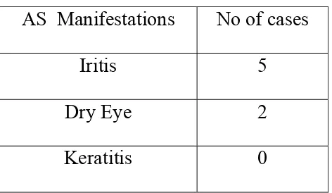
ANTERIOR SEGMENT
AS Manifestations No of cases
Iritis 5
Dry Eye 2
Keratitis 0
IRITIS
1 case associated with CMV retinitis 1 case associated with TB Panuvetis
1 case associated with toxoplasma
Table 6
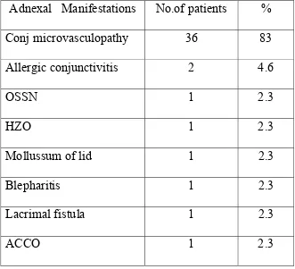
ADNEXAL MANIFESTATIONS
Adnexal Manifestations No.of patients %
Conj microvasculopathy 36 83
Allergic conjunctivitis 2 4.6
OSSN 1 2.3
HZO 1 2.3
Mollussum of lid 1 2.3
Blepharitis 1 2.3
Lacrimal fistula 1 2.3
ACCO 1 2.3
CONJUNCTIVAL MICROVASCULOPATHY
Seen in 36 patients, accounting for 83% of adnexal manifestations.
19.4% of patients with conjunctival microvasculopathy
[image:42.612.125.530.378.638.2]have been associated with retinal microvasculopathy.
Table - 7
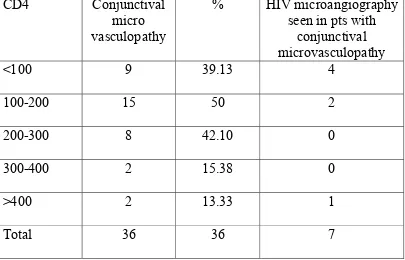
CD4 COUNT & CONJUCTIVAL MICROVASCULOPATHY
CD4 Conjunctival micro
vasculopathy
% HIV microangiography seen in pts with
conjunctival microvasculopathy
<100 9 39.13 4
100-200 15 50 2
200-300 8 42.10 0
300-400 2 15.38 0
>400 2 13.33 1
Table 8
POSTERIOR SEGMENT MANIFESTATIONS
Seen in 17(17%) patients
POSTERIOR SECALENT MANIFKCTATIONS
No of patients %
HIV microangiography 10 58
CMV retinitis 4 23.5
Toxoplasma retinitis 1 5.9
TB Panuveitis 1 5.9
Old choroiditis scar 1 5.9
Table 9
HIV MICROANGIOPATHY
Comprises 58% of patients having PS manifestations and is
the most common PS manifestation.
CD 4 count and HIV microangiopathy
CD4 HIV micro
angiography
% Bilateral
<100 5 21.7 3
100-200 2 6.7 0
200-300 1 5.26 0
300-400 0 0 0
>400 2 13.33 1
Total 10 4
With the decrease in the CD4 count, the incidence of HIV
Table 10
AGE DISTRIBUTION OF HIV MICROANGIOPATHY
Age HIV micro
angiography
% Bilateral
0-10 0 0 0
11-20 0 0 0
21-30 2 6.25 1
31-40 4 9.75 0
41-50 4 22.2 3
51-60 0 0 0
>60 0 0 0
CMV RETINITIS
[image:46.612.109.512.306.524.2]Seen in 4 (23.5%), patients with posterior segment manifestations.
Table 11
CD4 Count and CMV retinitis
CD4 CMV retinitis
% Bilateral ART taking
< 100 1 4.3 1 Yes
100-200 2 6.7 1 Yes
200-300 1 5.3 0 Yes
300-400 0 0 0 0
> 400 0 0 0 0
All the patients were on ART and 3 patients had CD4 count
> 100 cells / cumm.
NEURO OPHTHALMIC MANIFESTATIONS
[image:47.612.109.508.342.552.2]Seen in 4 patients. Majority were sequelae of CNS lesions.
Table 12
CD4 Count & Neuro ophthalmic manifestations
CD4 count No.of
patients / %
No.of patients with ocular manifestations
No.of patients with neuro ophthalmic
manifestations
< 100 23(23%) 14 (60.87%) 2 (8.7%)
100 - 200 30(30%) 16 (53.3%) 0
200 – 300 19(19%) 11(57.9%) 0
300 – 400 13(13%) 6(46%) 1 (7.7%)
≥ 400 15(15%) 7(46.67%) 1 (6.67%)
Table 13
HAART & OCULAR MANIFESTATIONS
No of Patients
No of patients with ocular manifestations AS manifes tations PS manifes tations Neuro ophthal manifestations On HAART 18 (19%) 13 (72%) 12 (66.67%) 5 (27.78%) 3 (16.675) Not on HAART 82 (82%) 41 (50%) 36 (43.9%) 12 (14.63%) 1 (1.22%)
Patients on HAART had more ocular manifestations than those
patients not on HAART. HAART taking patients would be in the late
stage of the disease with low CD4 count and so they are more prone for
DISCUSSION
The incidence of ocular manifestations in HIV infection has
been reported on various studies to occur upto 100% (Ryan).
J Biswas et al reported an incidence of 40-70%. In this study,
Ocular manifestations are seen in 49% of patients.
Adrenal manifestations are most commonly seen in 79.6% of
patients with ocular manifestations followed by posterior segment
manifestation seen in 31.5% of patients. Anterior segment manifestations are seen in 13% and neuro ophthalmic manifestations
in 7.4%.
Pradyot Narayan Biswas et al (2008) reported 29.14% of
ophthalmic manifestations. Of them 64.7% had posterior segment lesion, 23.52% had neuroophthalmic manifestations, 19.6% had
anterior segment lesion 15.69% had adnexal lesion.
Venkatesh et al (2008), reported ocular manifestations in 45%
of patients, CMV retinitis being the most common.
The ocular manifestations in HIV infected patients are less
common in this study than reported in earlier literature in India and abroad. This is supported by the studies of J Biswas et al (2000) &
among patients, lack of adequate knowledge among health
professionals and lack of facilities to look for ophthalmic
manifestations in HIV infected individuals in India.
Majority of patients in this study are in the age group of 20 – 40 (73%), which is comparable to the WHO estimates (62.39%).
This is attributed to the fact that this being the sexually active age
group, the risk of exposure is very high. The increased awareness of the disease and early reporting in this age group can also be a
contributing factor.
The gender incidence in this study is 1.5 : 1 (male : female). This male predominance is explained by their high risk of exposure
due to their nature of work and economic freedom. According to
WHO estimates among the adult living with AIDS/HIV 59% are males and 41% are females. In this study, it is 60% and 40%
respectively which correlates well with the WHO reported incidence.
In this study, the most common systemic disease associated with HIV / AIDS patients is tuberculosis seen in 24% of patients oral
candidiasis was seen in 10% and Herpes in 7% of patients.
seen. Among tuberculosis patients, ocular manifestations are seen in
58.3%.
CD4 T cell count is an important predictor of immune
suppression in HIV / AIDS patients. In this study, 11% of patients had CD4 < 50 cells / mm3, 23% had CD4 count < 100 cells/mm3
and 53% had CD4 count < 200 cells / mm3.
In this study, 72% of patients show ocular manifestations in HIV patients with CD4 count < 50 cells / mm3. In patients with CD4
count < 100 cells per cumm, 60.87% had ocular manifestations &
between 100-200, 53.3% had ocular manifestations. Ocular manifestations decrease in prevalence with increase in CD4 count.
The observation concludes that the CD4 count is inversely
related to the ocular manifestations in HIV patients with the decrease in CD4 count, the immunity decreases and the increase in occurrence
of opportunistic infections, malignancies and other manifestations in
HIV patients.
In terms of ocular symptoms, majority of patients are
asymptomatic and only 12 patients (12%) are symptomatic.
of defective vision and flashes/floaters. HIV microangiography and
confunctional microvasculopathy patients are asymptomatic.
Anterior segment manifestations are seen in 12.96% of
patients with ocular manifestations. Out of 7, 5 (71.4%) had iritis and 2 (28.5%) had Dry eye.
Among 5 Iritis affected patients, 3 had posterior segment
inflammation. One patient had CMV retinitis, one patient had Toxoplasmo retinitis and one patient had pan uveitis 2 patients had
old iritis with festooned pupil. Thus implies that most of the iritis in
HIV patients are associated with or “spill over” from posterior segment inflammation. Most of these patients were treated with
HAART.
J. Biswas et al (2008) reported that iritis is rarely associated with HIV diseases. Majority of cases are associated with retinal or
choroidal infection such as CMV, tomoplasma, TB, syphilis etc.
No cases of keratitis are reported in this study.
Posterior segment manifestations are reported in 31.48% of
patients with ocular manifestations. It is the 2nd most common
41.17% patients with posterior segment manifestations had
CD4 count < 100 cells / mm3. With decrease in CD4 count, the
incidence of posterior segment manifestations has increased.
HIV microangiography is the most common retinal manifestation in this study affecting 10% of patients and 58% among
patients with ocular manifestations. Cotton wool spots are the most
common manifestation followed by hemorrhage.
Kuppermann BD et al (1993) reported HIV retinopathy in 35%
of patients. In patients with CD4 count < 50 cells / mm3, 45% had
HIV retinopathy where as in patients with CD4 count > 50 cells / mm3 only 16% had HIV retinopathy.
DA Jabs et al (1995) reported HIV retinopathy in 50% of HIV
patients.
But studies in India showed, low prevalence of HIV
microangiography which correlates well with this study.
J. Biswas et al (2000) reported HIV retinopathy in 15% of HIV patients.
Venkatesh et al (2008) reported the prevalence of HIV
In this study, in patients with CD4 count < 100, 21.7% had
HIV microangiography whereas only 6.7% had HIV micro
angiography in patients with CD4 count 100-200. This support the
literature that when CD4 count decreases the prevalence of HIV retinopathy increases.
The prevalence of HIV retinopathy has increased with increase
in age in this study. 6.25% affected in the age group between 20-30, 9.75% in 31-40 age group and 22.2% in 41-50 age group.
Furrer et al (2003) reported that HIV microangiography is
associated with higher age group and higher viral load of HIV.
J Biswas et al (2008) related that the occurrence of HIV micro
angiography correlates well with the conjunctional
microvasculopathy. In the present study, 7 (70%) patients out of 10 patients with retinal microangiography had conjunctival
microvasculopathy.
Clinical diagnosis of CMV retinitis is made in 4 patients (4%) in the total study group and in 23.5% of patients with posterior
segment manifestations. Majority of them complained of defective
Various literature gives incidence of CMV retinitis as high as
15-40%. But in this study group, CMV retinitis is reported in only
4% of total study group and 23.5% of those with posterior segment
manifestations. The higher incidence in literature may be because the studies elsewhere conducted dealt with full blown cases of
AIDS, while this study is a mixture of asymptomatic cases with
fewer cases of AIDS.
All the 4 patients with CMV retinitis were on HAART. Only
one patient had CD4 count < 100 cells / cu.mm. Other 3 had CD4
count > 100 cells / cu.mm.
Various literature states that with the introduction of HAART,
the incidence of CMV retinitis has come down approximately by
75%. But in this study, CMV retinitis is seen in patients treated with HAART. This is explained by Brian et al that CMV retinitis may
occur with CD4 count > 100 cells / cu mm, because there may be
incomplete restoration of the immune repertoire against CMV with HAART therapy.
Toxoplasma retino choroiditis is seen in 1 patient. It is a
“Spill over” anterior uveitis was also seen. Serological tests were
not done due to lack of facility.
The panuveitis is seen in 1 patient secondary to TB. The
patient is treated with ATT and steroids. ARN, PORN are not reported in this study.
Ocular adnexal manifestation is seen in 43% of HIV infected
patients, conjunctival microvasculopathy being the commonest manifestation.
Conjunctional microvasculopathy is seen in 36% of patients in
the study and in 83% of patients with adnexal manifestation. Literature evidence shows that 70-80% of HIV patients show
conjunctival microvasculopahty and it correlates with the retinal
microangiopathy. 19.4% of patients with conjunctival microvasculopathy are associated with retinal microangiopathy.
Ocular surface squamous neoplasia is seen in 1 patient with
CD4 count 50 cells / cu mm. Nodular vascularised mass is seen in temporal limbus. Excision biopsy was done. HPE report confirmed
HZO is seen in 1 patient. The CD4 count is 345 cells. J.
Biswas et al reported 1 case in 100 cases of HIV patients in 1999 &
7 cases per 100 HIV patients in 2008. HZO has been reported to
occur in all stages of HIV.
Other manifestations include ACCO, Allergic conjunctivitis,
molluscum of the lid, Blepharitis and lacrimal fistula. keratitis is not
reported in this study.
Neuro ophthalmic manifestations are seen in 4% of patients in
study group and 7.4% of patients with ocular manifestations. 1
patient had papilloedema with headache, vomiting, probably due to increased intracranial hypertension. 1 patient had secondary optic
atrophy with bilateral LR palsy secondary to crptococcal meningitis.
AIDS dementia complex was associated with 1 patient with primary optic atrophy. LMN type facial N palsy was seen in 1 patient.
Majority of neuro ophthalmatic manifestations are secondary to CNS
lesions which is in accordance with literature evidence.
Orbital cellulitis is seen in 1 patient (1%) secondary to abscess
Among 100 patients, 2 patients presented with ocular
manifestations as their first presentation which led to HIV diagnosis.
From the present study, we can conclude that anterior segment
manifestations occur as often as the posterior segment manifestations. Majority of ocular manifestations are seen in patient
with CD4 count < 100 cells / cu mm. But vision threatening lesions
SUMMARY
The present cross sectional study is an attempt to assess the
frequency of ocular manifestations in HIV infected patients and its
correlation with CD4 count.
The study included 100 patients with 60 males and 40 females
infected with HIV / AIDS attending ophthalmology outpatient ward
in Govt Rajaji Hospital or referred from ART centre and other departments.
¾ Ocular manifestations seen in 54 (54%).
Among patients with ocular manifestations.
¾ No. of patients with posterior segment manifestation is 17
(31.48%)
¾ No.of patients with anterior segment manifestations is 7 (12.96%)
¾ No.of patients with adnexal manifestations is 43 (79.62%)
¾ No. of patients with neuro ophthalmic manifestations is 4
(7.4%)
¾ Majority belong to the age group of 20-40 years (73%)
¾ 60.8% of patients and CD4 count < 100 cells / cu mm have
ocular manifestations. But vision threatening lesions like CMV
retinitis and toxoplasma retinitis have been reported in patients
with CD4 count > 100 cells / cu mm in this study.
¾ HIV microangiography is found to be the most common
retinal manifestations (10%). It correlates well with the
occurrence of conjunctival microvasculopathy.
¾ Comparing the prevalence of retinal manifestations on HIV /
AIDS with the international literature evidence, the prevalence
CONCLUSION
Ophthalmic manifestations of HIV disease are increasingly
being recognized in the present era due to increased longevity of
patients after HAART.
This emphasizes the necessity for early recognition of ocular
lesions and early treatment to provide good quality of vision, thereby
providing good quality of life to patients infected with HIV.
In this study, sight threatening lesions like CMV retinitis and
toxoplasma retinitis were reported in patients with CD4 count > 100
cells / cu mm.
Regular Ophthalmic examination must be done preferably once in 6 months in patients with CD4 count > 100 cells/ cumm and
once in 3 months in patients with CD4 count < 50 cells cu mm, since the sight threatening lesions can occur at any stage of HIV infection.
Screening for retinal disease can be done at home by patients
themselves using Amsler’s grid test once weekly by which they can
HIV patients should be educated about the ocular
manifestations and should be advised to undergo regular ophthalmic
examinations.
Health care professionals should be trained and educated to pick up early cases of ophthalmic manifestations of HIV/AIDS
thereby preventing vision threatening complications.
Timely referral for complete ophthalmic examination and early institution of therapy should be done to help the cause of people
BIBLIOGRAPHY
1. Sahu DK, Namperumalsamy P, Walimbe P, Rajalakshmi C. Ocular
manifestations of HIV infection/AIDS in South Indian patients.
Indian J Ophthalmol 1999;47:79-85
2. Furrer H, Barloggio A, Egger M, Garweg JG; Swiss HIV Cohort
Study. Retinal microangiopathy in human immunodeficiency virus
infection is related to higher human immunodeficiency virus-1 load
in plasma. Ophthalmology. 2003 Feb;110(2):432-6
3. Kuppermann BD, Petty JG, Richman DD, Mathews WC, Fullerton
SC, Rickman LS, Freeman WR. Correlation between CD4+ counts
and prevalence of cytomegalovirus retinitis and human
immunodeficiency virus-related noninfectious retinal vasculopathy
in patients with acquired immunodeficiency syndrome. Am J
Ophthalmol. 1993 May 15;115(5):575-82.
4. Goldberg DE, Smithen LM, Angelilli A, Freeman WR.
HIV-associated retinopathy in the HAART era. Retina. 2005
Jul-Aug;25(5):633-49; quiz 682-3
5. Whitcup SM. Cytomegalovirus retinitis in the era of highly active
antiretroviral therapy. JAMA. 2000 Feb 2;283(5):653-7
6. Whitcup SM, Fortin E, Lindblad AS, Griffiths P, Metcalf JA,
T, Davey RT Jr, Falloon J, Walker RE, Kovacs JA, Lane HC,
Nussenblatt RB, Smith J, Masur H, Polis MA. Discontinuation of
anticytomegalovirus therap.
7. Reed JB, Briggs JW, McDonald JC, Freeman WR, Morse LS.
Highly active antiretroviral therapy-associated regression of
cytomegalovirus retinitis: long-Term results in a small case series.
Retina. 2001;21(4):339-43.y in patients with HIV infection and
cytomegalovirus retinitis. JAMA. 1999 Nov 3;282(17):1633-7.
8. Macdonald JC, Karavellas MP, Torriani FJ, Morse LS, Smith IL,
Reed JB, Freeman WR. Highly active antiretroviral therapy-related
immune recovery in AIDS patients with cytomegalovirus retinitis.
Ophthalmology. 2000 May;107(5):877-81; discussion 881-3.
9. Reed JB, Briggs JW, McDonald JC, Freeman WR, Morse LS.
Highly active antiretroviral therapy-associated regression of
cytomegalovirus retinitis: long-Term results in a small case series.
Retina. 2001;21(4):339-43.
10.Lin DY, Warren JF, Lazzeroni LC, Wolitz RA, Mansour SE.
Cytomegalovirus retinitis after initiation of highly active
antiretroviral therapy in HIV infected patients: natural history and
11.Roels P. Ocular manifestations of AIDS: new considerations for
patients using highly active anti-retroviral therapy (HAART).
Optometry. 2004 Oct;75(10):624-8.
12.Arevalo JF, Mendoza AJ, Ferretti Y. Immune recovery uveitis in
AIDS patients with cytomegalovirus retinitis treated with highly
active antiretroviral therapy in Venezuela. Retina. 2003
Aug;23(4):495-502.
13.Deayton JR, Wilson P, Sabin CA, Davey CC, Johnson MA, Emery
VC, Griffiths PD. Changes in the natural history of
cytomegalovirus retinitis following the introduction of highly
active antiretroviral therapy. AIDS. 2000 Jun 16;14(9):1163-70.
14..Kempen JH, Min YI, Freeman WR, Holland GN, Friedberg DN,
Dieterich DT, Jabs DA; Studies of Ocular Complications of AIDS
Research Group. Risk of immune recovery uveitis in patients with
AIDS and cytomegalovirus retinitis. Ophthalmology. 2006
Apr;113(4):684-94.
15.Karavellas MP, Azen SP, MacDonald JC, Shufelt CL, Lowder CY,
Plummer DJ, Glasgow B, Torriani FJ, Freeman WR. Immune
recovery vitritis and uveitis in AIDS: clinical predictors, sequelae,
16.Jyotirmay Biswas, MS and S Sudharshan, DO. Anterior segment
manifestations of human immunodeficiency virus/acquired
immune deficiency syndrome. Indian J Ophthalmol. 2008 Sep–Oct;
56(5): 363–375.
17.Biswas J, Madhavan HN, George AE, Kumarasamy N, Solomon S.
Ocular lesions associated with HIV infection in India: A series of
100 consecutive patients evaluated at a referral center. Am J
Ophthalmol. 2000;129:9–15.
18.D A Jabs.Department of Ophthalmology, Johns Hopkins
University School of Medicine, USA. Ocular manifestations of
HIV infection. Trans Am Ophthalmol Soc. 1995; 93: 623–683.
19.Biswas J, Kumaraswamy N, Soloman S. Ophthalmic
manifestations of Human Immunodeficiency Virus (HIV) in India.
Indian J Ophthalmol. 1999;47:87–93.
20.Sudharshan Introduction and immunopathogenesis of AIDS. Indian
J Ophthalmol. 2008;56:357–62. Banker A. Posterior segment
manifestations of HIV/AIDS. Indian J Ophthalmol. 2008;56:377–
83
21.Kempen J. Medical management of human immunodeficiency
22.Venkatesh K. Impact of highly active antiretroviral therapy on
ophthalmic manifestations in HIV/AIDS. Indian J Ophthalmol.
2008;56:391–3. [PubMed]Ophthalmic Epidemiol. 2008
Jul-Aug;15(4):264-71.
23.Ophthalmic manifestations of HIV infections in India in the era of
HAART:analysis of 100 consecutive patients evaluated at a tertiary
eye care center in India.
24.Gharai S, Venkatesh P, Garg S, Sharma SK, Vohra R.Dr Rajendra
Prasad Center for Ophthalmic Sciences, All India Institute of
Medical Sciences, New Delhi, India. sujitgharai@gmail.com
Ocular lesions associated with HIV infection in India: a series of
100 consecutive patients evaluated at a referral center.
25. Biswas J, Madhavan HN, George AE, Kumarasamy N, Solomon
S.Medical and Vision Research Foundation, Chennai, India.
26.Arevalo JF, Russack V, Freeman WR, New ophthalmic
manifestations of presumed rifabutin associated uveitis.
Ophthalmic surg lasers. 1997 28: 321-324.
27.Aslanides 1M et al Oral valacyclovir in the treatment of ARN
syndrome. R Retina 2002; 22: 353-354.
28.Berger BB et al Miliary toxoplasmic retinitis in AIDS Arch Oph.
29.Cano-parra JL et al Milary toxoplasmic Retinitis in AIDS Ocul
immuno inflammation 2000; 8:127-130.
30.Causey DM, Rarick MD. Foscamet treatment for resistant HSV
disease. Stockholm 1988 abstract 3589.
31.Cochereau - Massin et al Ocular toxoplasmosis in AIDS AMJ Oph
1992; 114:130-135.
32.Cole EL, DM, Calabrese LH et al. HZO and AIDS. Arch Oph
1984.102: 1027-1029.
33.Conway MD, Tong P, Olk TJ BRAO/BRVO and optic disc
neovascularisation associated with H1V and CMV retinitis. Int
Oph 1995-96. 19: 249-252.
34.Crowe S.M., Carlin JB, Stewart K.I, Lucas C.R and Hoy JF.
Predictivevalue of CD4+ lymphocyte numbers for the development
ofopportunistic infections and malignancies in HIV infected
persons. J.Acquir. Immune Defic. Syndr. 4:770, 1991.
35.Culbertson WW, Atherton SS. Acute retinal necrosis and similar
retinitis syndrome. Int Oph Clin 1993; 33: 129-143.
36.Dunn JP, Martin DF. Treatment of CMV retinitis in era of
HAART.
37.Edwards JE et .al Ocular manifestations of candida septicemia.
38.Elkiness et al Ocular toxoplasmosis misdiagnosis as CMV retinitis
in AIDS. Ophthalmology 1994;10_:499-507.
39.Engstrom RJ et al PORN syndrom_. A varient of necrotizing
herpetic retinopathy in patients with AIDS Oph 1994;101:
1488-1502.
40.Fardeau C et al Diagnosis of toxoplasmic Retino choroiditis with
atypical clinical feature. AMJ oph 2002; 134:196-203.
41.Forster OJ et al. Rapidly progressive PORN in AIDS. Am J Oph
1990;110: 341-348.
42.Freeman WR et al. Prevalence and significance of AIDS. Related
microvasculopathy. Am J Oph 1989; 107: 229-235.
43.Freeman WR, Chen A, Henderly DE et al. prognostic and systemic
Hodge WG et al. Clinical risk factors for CMV retinitis in patients
with AIDS. Oph 2004; 111: 1326-1333.
44.Hollan GN et al AIDS, Ocular manifestations. Ophthalmology
1983 ;90 : 859-873.
45.Holland FM. Ocular toxoplasmosis in immuno compromised host.
Oph1989; 13:399-402.
46.Holland GR. et al Ocular infections associated with AIDS in
duanes clinical ophthalmology cleap 82.
CMV retinitis in patients with AIDS 1988-1994.
48.Holland GN et al. A controlled retrospective study of gancyclovir
treatment ofCMV retinitis. Arch Oph 1989; 107: ) 759-1766.
49.Rolland GN et al. Retinal CWS in patients with AIDS N. Engl J
Med 1982,307: 1704.
50.Holland GN, Tufail A, Jordan MC. CMV disease. Ocular
infections and immunity Mosby 1996, 1088-1129.
51.Irvine AR. Treatment of RD due to CMV retinitis in AIDS
PROFORMA - HIV RETINOPATHY
IDENTITY NO : AGE: SEX:
EDUCATION : OCCUPATION :
MARITAL STATUS : ROUTE OF TRANSMISSION:
HIV TIME OF DIAGNOSIS:
ART DRUGS: DATE OF STARTING: DURATUON:
IF YES, MISSED DOSES/DEFAULT: HOW MANY TIMES:
NO OF DAYS:
OPPORTUNISTIC INFECTIONS:
VDRL : TB: HERPES SIMPLEX:
HERPES ZOSTER: CMV: PCP: TOXOPLASMA :
OTHERS :
PREVIOUS VISIT: DATE : RETINAL FINDINGS:
INITIAL VISIT STUDY VISIT
CD4 COUNT WHO CLINICAL STAGING
KEY TO MASTER CHART
SEX:
Male – 1, female – 2
ART TAKING:
Yes – 1, no – 2
AS MANIFESTATIONS:
NAD – 1
Conjunctival microvasculopathy – 2
HZO – 3
Dry eye – 4
OSSN – 5
ACCO – 6
Vernal conjunctivitis – 8
Blepharitis – 9
Orbital cellulites – 10
Iritis – 11
Lacrimal fistula – 12
PS MANIFESTATIONS:
NAD – 1
HIV microangiopathy – 2
CMV retinitis – 3
Toxoplasma retinitis – 4
Old choroiditis scar – 5
Panuveitis – 6
NEURO OPHTHALMIC MANIFESTATIONS:
NAD – 1
Optic atrophy – 2
6th cranial nerne palsy – 3
7th cranial nerve palsy – 4
Papilloedema – 5
TB :
Yes – 1
ABBREVIATIONS
AIDS – Acquired immuno deficiency syndrome
ARN - Acute retinal necrosis
ART - Anti retroviral therapy CME - Cystoid macular edema
CMV - Cytomegalo virus
CWS - Cotton wool spots
ELISA - Enzyme linked immmunosorbent assay
HAART - Highly activated antiretroviral therapy
HHV - Human herpes virus
HIV - Human immunodeficiency virus
HSV - Herpes simplex virus
HPV - Human papilloma virus HZV - Herpes zoster virus
MAC - Mycobacterium avium complex
OSSN - Ocular surface squamous neoplasia PORN - Progressive outer retinal necrosis
RD - Retinal detachment
CONSENT FORM
I was informed & explained of the purpose & nature of the
study. I am willing to participate in this study. I hereby give my full
consent for the study.
Signature of the patient
HERPES ZOSTER OPHTHALMICUS BLEPHARITIS
CONJUNCTIVAL OSSN
PRE OPERATIVE
HIV MICRO ANGIOPATHY
CMV RETINITIS
ORBITAL CELLULITIS
S.No. op no age sex CD4 CD3 ALC ART RE AS LE AS RE PS LE PS NO TB ASS CONDITIONS
1 415645 27 1 75 2 2 2 1 1 1 1
2 415624 39 1 131 976 1243 2 1 1 1 1 1 2
3 415734 39 1 45 1 5 1 1 1 1 2
4 415827 35 2 136 1124 1584 2 1 1 1 1 1 2
5 415398 26 1 161 1932 2312 2 2 2 1 1 1 1
6 415625 32 1 375 2127 2335 2 1 1 1 1 1 2
7 415892 6 2 455 2824 3815 2 1 1 1 1 1 2
8 415439 21 2 593 3063 3576 2 1 1 1 1 1 2 genital warts
9 415289 25 2 58 1192 1398 2 1 1 1 1 1 2
10 415425 36 2 169 2310 2806 2 2 2 1 1 1 2
11 426749 35 2 155 2 2 2 1 1 1 2
12 416153 29 1 111 1423 1587 2 2 2 1 1 1 1
13 415804 36 1 187 2 1 1 1 1 1 1
14 416337 37 1 190 1380 1966 2 1 1 1 1 1 2
15 416451 39 1 5 183 1 1,4 1,4 1 1 2,3 1 cryptoccal meningitis
16 415382 42 1 816 2157 2605 2 1 11 1 4 1 2
17 527918 42 1 49 1 2 2 2 2 1 2
18 103833 35 2 419 2 1 1 1 1 1 2
19 416753 60 2 210 3723 3953 2 1 1 1 1 1 2
20 417221 35 1 245 2 1 1 1 1 1 2
21 417216 41 1 165 2 1 1 1 1 1 2
22 418142 21 2 316 2 11 11 6 6 1 2
23 419626 35 1 156 2 2 2 2 1 1 2
24 420402 35 1 312 1267 1924 2 2 2 1 1 1 1
25 419925 47 1 1303 3120 4333 2 1 1 2 2 1 2
26 424752 40 1 827 3308 3959 2 2 2,11 1 1 1 2
27 413289 32 1 75 1192 1525 2 1 1 5 1 1 2 oral candida
28 429864 40 1 52 543 715 2 1 1 1 2 1 2
29 428753 24 2 28 1 2 2 1 1 4 2
30 430130 38 1 345 2 10 1 1 1 1 2
31 413526 26 2 260 1269 1 8 8 3 5 1 2
32 413529 34 1 116 598 1 2 2,11 1 3 1 1
33 412356 25 2 442 6434 7061 2 2 2 2 1 1 1
34 432167 31 2 376 2306 2814 2 1 1 1 1 1 2
36 667/09 24 2 272 1717 2233 2 2 2 1 1 1 2 vulvovaginal candidiasis
37 563/08 45 1 354 2 1 1 1 1 2 1 AIDS dementia complex
38 464/09 38 1 97 1526 1795 1 1 1 1 1 1 1 oral candida
39 431837 37 1 314 2 1 1 1 1 1 2
40 639/09 27 2 195 623 850 2 8 8 1 1 1 2
41 706/09 23 2 1227 2596 3251 2 1 1 1 1 1 2
42 396/09 30 2 168 1398 1859 1 2 2 3 3 1 1
43 1760/08 34 2 249 1698 1895 2 2 2 1 1 1 1
44 01/07art 52 1 86 1019 1165 2 1 1 1 1 1 1
45 3374 37 1 145 2 2 2 1 2 1 2
46 2248/08 10 2 781 2718 3305 2 1 1 1 1 1 2
47 2247/08 7 1 696 2764 3656 2 1 1 1 1 1 2
48 029/09 31 2 37 1209 1 9,12 9,12 1 1 1 2
49 1742/07 29 1 165 2232 2558 2 1 1 1 1 1 1
50 762/09 42 1 116 822 1020 2 1 1 1 1 1 2
51 766/09 42 1 30 691 751 2 1 1 1 1 1 2
52 760/09 36 1 124 1535 1807 1 1 1 1 1 1 2
53 4059 35 1 129 2 1 1 1 1 1 2
54 758/09 47 1 44 855 1246 2 2 2 2 1 1 2 oral candida
55 753/09 37 2 141 1097 1376 2 2 2 1 1 1 2
56 752/09 16 2 167 1288 1559 2 1 1 1 1 1 1
57 770/09 50 2 359 1747 2374 2 1 1 1 1 1 2
58 779/09 45 1 234 2 2 2 1 1 1 2
59 774/09 37 1 191 1394 1613 2 2 2 1 1 1 2
60 752/09 47 2 205 2 2 2 1 1 1 2
61 720/09 37 1 31 741 1353 2 1 1 1 1 1 1 herpes zoster
62 145/08 27 2 52 1 2 2 3 3 1 1 esophagial candidiasis
63 1850/06 40 2 219 1291 1568 1 2 2 1 1 1 2 chicken pox
64 790/09 33 1 275 1112 1559 2 1 1 1 1 1 1
65 820/06 38 1 96 91 1092 2 1 1 1 1 1 1
66 148/08 45 2 159 997 1284 2 1 1 1 1 1 2
67 206/07 30 1 836 2568 33 1 1 1 1 1 5 2
68 483/06 35 2 549 1636 2123 1 1 1 1 1 1 2 oral candida
69 837/05 45 2 70 1022 1392 2 2 2 2 2 1 2 herpes zoster
70 829/08 40 2 288 2 1 1 2 1 1 1 enital molluscum, oral candid
72 2203/08 30 2 136 1124 1584 2 2 2 1 1 1 2
73 701/05 35 2 1551 3915 5272 1 11 11 1 1 1 1
74 729/09 25 1 29 1253 1513 2 2 2 1 1 1 2
75 621/09 43 1 166 1101 1268 2 2 2 1 1 1 2
76 734/09 30 1 212 1190 1691 2 1 1 1 1 1 2
77 2147/06 13 1 271 1001 1907 2 1 1 1 1 1 2
78 707/09 26 2 20 275 373 2 1 1 1 1 1 2
79 796/09 51 1 354 2 2 2 1 1 1 2
80 755/09 44 1 59 839 1115 2 2 2 1 1 1 1
81 1730/08 27 1 134 1220 1563 1 2 2 1 1 1 1 genital herpes
82 791/09 40 1 269 1639 1863 2 2 2 1 1 1 2
83 783/09 30 1 70 1604 2004 2 1 1 1 1 1 2
84 1153/07 24 1 433 980 1312 2 6 6 1 1 1 2
85 49192 31 2 256 2 1 1 1 1 1 2
86 826/09 43 1 299 2092 2865 2 2 2 1 1 1 2
87 2069/07 23 2 240 2 1 3 1 1 1 2
88 49773 23 2 142 14 189 2 1 1 1 1 1 2 pancytopenia
89 1306/07 35 1 322 1704 2497 2 4 4 1 1 1 2 genital herpes,oral candida
90 2345 24 1 604 2760 3486 2 1 1 1 1 1 2
91 857/09 28 1 124 1043 1547 2 1 1 1 1 1 2
92 767/09 28 1 205 943 120 1 1 1 1 1 1 2
93 873/09 30 2 47 473 600 2 2 2 2 2 1 2
94 2089/07 29 2 137 680 1157 2 2 2 1 1 1 2
95 884/08 13 2 299 3471 4357 2 1 1 1 1 1 2
96 885 42 2 62 565 762 2 1 1 1 1 1 1 herpes
97 652/09 40 1 303 1718 1917 2 1 1 1 1 1 2
98 876/09 33 1 199 1369 1802 2 2 2 1 1 1 2
99 878/09 43 1 303 1718 1917 2 1 1 1 1 1 2
0 0
20
22
11
33
0
0 5 10 15 20 25 30 35
NO.OF CASES
0 - 10 11 -20 21-30 31-40 41-50 51-60 >60
AGE IN YEARS AGE DISTRIBTION