Copyrightq1997, American Society for Microbiology
Binding of the E1 and E2 Proteins to the Origin of Replication
of Bovine Papillomavirus
TIINA SEDMAN, JUHAN SEDMAN,ANDARNE STENLUND*
Cold Spring Harbor Laboratory, Cold Spring Harbor, New York 11724
Received 13 September 1996/Accepted 11 December 1996
DNA replication of bovine papillomavirus (BPV) requires two viral proteins encoded from the E1 and E2 open reading frames. E1 and E2 are sequence-specific DNA binding proteins that bind to their cognate binding sites in the BPV origin of replication (ori). The E1 and E2 proteins can interact physically with each other, and this interaction results in cooperative binding when binding sites for both proteins are present. We have analyzed the binding of E1 to the ori in the absence and presence of E2, using DNase I footprint analysis, gel mobility shift assays, and interference analysis. We have also generated a large number of point mutations in the E1 binding site and tested them for binding of E1 as well as for activity in DNA replication. Our results demonstrate that E1 binds to the ori in different forms in the absence and presence of E2 and that E2 has both a quantitative and a qualitative effect on the binding of E1. Our results also suggest that the ori contains multiple overlapping individual E1 recognition sequences which together constitute the E1 binding site and that different subsets of these recognition sequences are used for binding of E1 in the presence and absence of E2.
The papillomaviruses constitute a large family of viruses that cause benign tumors (warts) in their hosts. In recent years, these viruses have been found to have substantial clinical im-portance (34, 42), and a more detailed understanding of the viral life cycle is of crucial importance for an understanding of the disease and its transmission and for the development of therapeutic measures. A coherent picture of the requirements for viral DNA replication has emerged only in recent years, and the understanding of papillomavirus replication is there-fore relatively primitive. DNA replication of bovine papillo-mavirus (BPV) in vivo requires two viral proteins encoded from the E1 and E2 open reading frames (36). The E1 protein has sequence-specific DNA binding activity (13, 14, 19, 30, 35, 37, 39, 40), DNA helicase activity, and DNA-dependent ATPase activity and can serve to unwind the origin of replica-tion (10, 28, 40). E1 is also the only viral protein required for initiation of DNA replication in an in vitro replication system (5, 21, 24, 28, 40, 41). In this respect, the E1 protein appears to be the counterpart of simian virus 40 large T antigen, capable of serving as an initiator.
The minimal origin of replication (ori) consists of a se-quence approximately 60 bp in length (37, 41). This sese-quence
contains three elements, an A1T-rich sequence, an 18-bp long
palindromic sequence that serves as a binding site for the E1 protein (13, 37, 41), and a 12-bp-long sequence that constitutes a binding site for the E2 protein (15). The function of the
A1T-rich sequence is not clear, but it has been suggested that
this sequence may serve as a site for initial melting of the DNA helix (10). A unique and interesting aspect of papillomavirus replication is the specific requirement for E2 for viral DNA replication in vivo (37, 38). The E2 protein is a sequence-specific transcriptional activator that regulates viral gene ex-pression (1, 11, 20, 31), but E2 also has a direct role in DNA replication. The requirement for E2 is highly specific; for ex-ample, a hybrid activator, VP16-E2 (16), that contains the
activation domain from the herpes simplex virus VP16 fused to the DNA binding domain of E2 fails to support replication in vivo (37).
Several possible functions have been proposed for E2 in viral DNA replication. It has been demonstrated that E2, as well as several other transcription factors, can interact with the single-stranded DNA binding protein replication protein A (RPA), and a role for E2 in recruiting RPA to the ori has been sug-gested (12, 16). It has also been shown that E2, as well as other transcription factors, can function to prevent nucleosomal re-pression of replication on chromatin templates assembled in vitro (7, 8, 17). However, neither of these activities can entirely explain the requirement for E2 for replication in vivo because replication in vivo shows a stricter specificity than that ob-served in either of these in vitro assays. For example, Gal4-VP16, which is capable of interacting with RPA (16) and also can serve to derepress chromatin templates for BPV replica-tion in vitro, lacks activity for replicareplica-tion in vivo and cannot replace E2 (17).
It has been well established that the E1 and E2 proteins can interact physically. This interaction, which has been detected by both coimmunoprecipitation and two-hybrid analysis (3, 4, 18, 23), results in cooperative binding of E1 and E2 to the ori (10, 26, 29, 30, 41). We have recently demonstrated that this ability of E2 to interact with the E1 protein is required for E2 activity in viral DNA replication (26). We have also demon-strated that E1 by itself binds with a low degree of sequence specificity: one of the consequences of the interaction with E2 is that E1 in the presence of E2 can bind to the ori with substantially increased sequence specificity. Based on these observations, we have proposed that a major function of E2 in replication is to serve as a specificity factor for binding of E1 to the ori (26).
In this study, we have analyzed the DNA binding properties of the E1 protein in the absence and in the presence of E2 by using footprinting and interference studies as well as muta-tional analysis of the E1 binding site at the ori. We find not only that the effect of E2 on binding of E1 to the ori is a quantitative effect but that also the mode of E1 binding in the
* Corresponding author. Mailing address: Cold Spring Harbor Lab-oratory, 1 Bungtown Rd., Cold Spring Harbor, NY 11724. Phone: (516) 367-8407. Fax: (516) 367-8454. E-mail: [email protected].
2887
on November 9, 2019 by guest
http://jvi.asm.org/
presence of E2 is substantially different than in the absence of E2, consistent with the finding that different numbers of E1 molecules bind to the ori in the two complexes (27). Thus, in the presence of E2, E1 can bind to the ori in a form that is incapable of binding DNA in the absence of E2. These results suggest that the assembly of an initiator complex may require a stepwise addition of E1 molecules onto the ori.
MATERIALS AND METHODS
Expression and purification of E1 and E2 proteins. (i) E2 protein.Escherichia coliBL21(DE3) was transformed with the E2 expression plasmid pET E2. Liquid cultures were inoculated and grown at strictly controlled temperature (19 to 208C) until the optical density at 600 nm reached 0.6. The cultures were induced by the addition of isothiogalactopyranoside (IPTG) to 0.3 mM and grown for an additional 8 h. The bacteria were lysed in 40 ml of buffer A (50 mM Tris-HCl [pH 7.5], 5 mM EDTA, 200 mM NaCl, 20% sucrose, 10 mM dithiothreitol [DTT], 1 mM phenylmethylsulfonyl fluoride) in a French press, and the lysate was soni-cated briefly to reduce the viscosity. The lysate was cleared by centrifugation at 10,000 rpm for 10 min in a Sorvall SS34 rotor. The supernatant was precipitated with 50% ammonium sulfate; the precipitated material was recovered by cen-trifugation and was dissolved in 2 pellet volumes of buffer B (25 mM morpho-lineethanesulfonic acid [MES]-KOH [pH 6.5], 1 mM EDTA, 5% glycerol, 10 mM DTT) containing 100 mM NaCl, and the conductivity was adjusted to that of the starting buffer (buffer B–300 mM NaCl). The sample was applied to an 8-ml S-Sepharose column and after washing was eluted with a 50150-ml gradient between 300 and 1,000 mM NaCl in buffer B. The fractions were assayed by sodium dodecyl sulfate-polyacrylamide gel electrophoresis, and the fractions containing E2 were pooled, diluted 10-fold in buffer C (25 mM Tris-HCl [pH 8.2], 0.1 mM EDTA, 5% glycerol, 10 mM DTT), and loaded onto a 1-ml Q-Sepharose column. The protein was eluted with a 20120-ml gradient between 100 and 800 mM NaCl in buffer C. Peak fractions were estimated to be greater than 95% pure, and the yield was 1 to 2 mg of protein/liter of culture.
(ii) E1 protein.E. coliBL21(DE3) was transformed with the E1 expression vector GST (glutathioneS-transferase)-E1. Liquid cultures were inoculated and grown at 258C. Cultures were induced with IPTG at an optical density at 600 nm of 0.6 and grown for an additional 12 h. The cells were lysed by using the procedure described for E2. The NaCl concentration of the cleared lysate was adjusted to 1 M, and nucleic acids were precipitated by the slow addition of a 10% solution of polymin P until a final concentration of 0.6% was reached. The sample was left to mix for 20 min, and the precipitate was pelleted by centrifu-gation at 12,0003gfor 10 min. The protein was recovered from the supernatant by precipitation with 50% ammonium sulfate. The precipitated protein was recovered by centrifugation, and the pellet was dissolved in 30 ml of buffer A; 2 ml of glutathione-agarose beads was added, and the mixture was mixed for 5 to 8 h. The beads were washed twice with 40 ml of buffer A, transferred to a column, and washed with 2 column volumes of each of the following buffers: buffer A, buffer B–1 M NaCl, and buffer B–0.2 M NaCl. Bound protein was eluted with buffer B–0.2 M NaCl–10 mM glutathione. Fractions containing GST-E1 were pooled, and CaCl2was added to a final concentration of 2.5 mM. Thrombin (10 U) was added, and cleavage was allowed to proceed for 8 h at 08C. Thrombin was inactivated by the addition of 1 mM phenylmethylsulfonyl fluoride; the sample was diluted 20-fold with buffer C (50 mM NaPO4[pH 6.8], 1 mM EDTA, 5 mM DTT, 10% glycerol) containing 100 mM NaCl and loaded onto a 2-ml S-Sepha-rose column equilibrated with the same buffer. The protein was eluted with a 20120-ml gradient from 100 to 800 mM NaCl in buffer C, and peak fractions were pooled and diluted sixfold with buffer D (25 mM Tris-HCl [pH 8.8], 1 mM EDTA, 5 mM DTT, 10% glycerol) containing 90 mM NaCl. The material was loaded onto a 0.4-ml Q-Sepharose column and eluted in two steps using buffer D and 200 and 500 mM NaCl.
Plasmid constructs.TheE. coliexpression vector for expression of E2 (pET E2) was generated by PCR amplification of the E2 open reading frame, using a 59primer that introduces anNdeI restriction site at the initiator ATG of the E2 coding sequence and a 39primer that introduces aBamHI restriction site im-mediately downstream of the termination codon for E2. This fragment was cloned into pET 11c between theNdeI andBamHI restriction sites, resulting in a protein product without additions and deletions. The sequence of the E2 open reading frame was verified by DNA sequencing. The E1 expression construct (GST-E1) was generated by PCR amplification of the E1 open reading frame by using a 59primer that introduces anXbaI restriction site immediately upstream of the initiation codon for E1 and a 59primer introducing aBamHI restriction site immediately downstream of the termination codon for E1. This fragment was cloned between theXbaI andBamHI restriction sites of the vector pET11-GST (2), which fuses the GST open reading frame to the E1 open reading frame. The E1 coding sequence in this construct was verified by DNA sequencing.
All of the ori constructs have been described previously (36, 37).
Radiolabeled probes.Probes for mobility shift assays, DNase footprint assays, and interference analysis were generated by PCR amplification of ori constructs cloned in pUC19, using the universal primer USP or RSP that had been 59
labeled with [g-32P]ATP and T4 polynucleotide kinase. Probes for Southern blots
were generated by random priming using the minimal ori construct 7914-27 as a template.
Mobility shift assays.Mobility shift assays were performed by mixing probe and E1 and/or E2 protein in a buffer containing 20 mM potassium phosphate (pH 7.5), 100 mM NaCl, 1 mM EDTA, 0.7 mg of bovine serum albumin per ml, 2.5 mM DTT, and 10% glycerol in a final volume of 10ml containing 20 ng of nonspecific competitor DNA (pUC19). After incubation at room temperature for 20 min, 1ml of a 0.2% solution of glutaraldehyde in water was added, and the sample was incubated for an additional 20 min. The cross-linking reaction was quenched by addition of 2ml of glycerol loading dye, and the samples were loaded on 1% Tris-acetate-EDTA agarose gels. After electrophoresis, the gels were fixed, dried, and subjected to autoradiography.
DEPC interference.Diethyl pyrocarbonate (DEPC) interference assays were performed essentially as described previously (34). Radiolabeled probes corre-sponding to the minimal ori construct 7914-27 were generated as described above. After denaturation by heating, the probes were modified with DEPC for 2 min at 378C. This degree of modification was determined to introduce less than one modification per molecule. After reannealing, the probes were used for gel retardation assays as described above. The shifted band and the free probe were excised from the gel and eluted and subsequently cleaved by heating in piperi-dine followed by analysis on sequencing gels.
Competition assays for E1 binding.The competition assays for determination of the binding affinities for E1 binding site mutants were carried out by gener-ating probes from the different mutant templates by PCR, using the same batch of labeled universal primer to ensure identical specific activity for the different mutant probes. A constant quantity of labeled probe was mixed with increasing amounts of competitor DNA, and subsequently mobility shift assays were per-formed by addition of a fixed quantity of E1 protein (4 ng). The shifted band and the free probe were both quantitated and plotted as a function of competitor concentration. The point where the two curves intersect represents the 50% competition point, which was used for comparison of the different mutants.
E1-E2-ori complex formation.Mobility shift assays were performed with the wild-type (wt) and mutant probes in the presence of a constant amount of E2 (1 ng) and 20 ng of competitor DNA, by titration of E1. The amounts of shifted probe at the different E1 concentrations were quantitated and compared for the wt and each mutant probe.
Replication assays.Transient replication assays were performed as described previously (37).
Generation of point mutations.All point mutations were generated in the context of the minimal ori construct 7914-27, which contains the BPV sequences between nucleotides (nt) 7914 and 27, cloned between theXbaI andHindIII restriction sites in the pUC19 polylinker. The point mutations were generated by PCR by using mutagenic primers that overlapped theHpaI restriction site that is present in the center of the palindrome in the minimal ori. After amplification using either the upstream or downstream universal primer, the PCR products were digested withHpaI and eitherXbaI orHindIII, which generated a restric-tion fragment constituting half of the minimal ori with the incorporated muta-tion. This restriction fragment was used to replace the correspondingHpaI-XbaI orHpaI-HindIII fragment from the wt minimal ori. The sequences of the mutant oris were verified by DNA sequencing.
RESULTS
Expression and purification of E1 and E2 proteins.To per-form a careful analysis of the binding properties of E1 and E2, we wanted to use highly purified E1 and E2 proteins with high
specific activity. We therefore expressed E1 and E2 inE. coliin
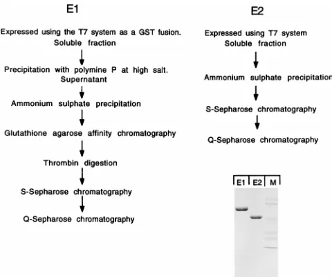
soluble form and purified both proteins to apparent homoge-neity (Fig. 1). Both the E1 and E2 proteins were expressed by using the expression vector pET 11C (32). The E1 protein was expressed as an N-terminal GST fusion protein. After partial purification, the GST portion was cleaved off with thrombin protease, resulting in a native E1 protein with a five-amino-acid extension at the N terminus. As reported previously, E1 in crude form is tightly associated with DNA, forming high-mo-lecular-weight DNA-protein complexes (28). To obtain E1 protein in monomeric form with high specific activity for DNA binding, we performed a precipitation of nucleic acids with polyimin P at high salt concentration to remove associated DNA (6). E1, partially purified only by glutathione-agarose chromatography, had 100- to 1,000-fold-lower DNA binding activity than highly purified E1, most likely due to the presence
of large quantities of contaminating E. coli DNA. The E2
protein was expressed without fusions by using pET 11C. To generate soluble highly active E2 protein, it was necessary to
grow the cultures below 208C. The protein was purified by two
on November 9, 2019 by guest
http://jvi.asm.org/
conventional column chromatography steps to apparent homo-geneity as described in Materials and Methods.
DNase footprint analysis delineates sequences required for binding of E1.To delineate the sequences involved in binding of E1 and E2, DNase I footprint analysis was performed with several different templates in the presence of purified E1 and E2 proteins as shown in Fig. 2. In this experiment, four differ-ent templates were used: (i) the wt minimal ori (7914-27) that
contains the A1T-rich region, the binding site for E1, and the
wt low-affinity E2 binding site 12; (ii) a deletion mutant that
lacks the A1T-rich region (2A1T); (iii) a substitution mutant
where the E2 binding site was replaced with a high-affinity E2 binding site with different sequence (E2 BS9); and (iv) a de-letion mutant that lacks the E2 binding site altogether (7914-15). DNase I footprint analysis was carried out by using probes with the top strand labeled. Binding by E1 alone to these different templates showed only very small differences in the levels of E1 protein required to produce a footprint (compare
the first four lanes in each set). Deletion of the A1T-rich
sequence resulted in a slightly (twofold) reduced ability to bind E1. Deletion or substitution of the E2 binding site immediately adjacent to the palindromic sequence had no effect on E1 binding. The protections observed with E1 alone in all cases
was centered over the uniqueHpaI restriction site at the center
of the palindrome and extended in both directions beyond the BPV sequence and into the polylinker sequence of the vector. The extended protections on both sides of the palindrome observed for E1 alone appeared to have little or no sequence
specificity, since deletion of either the A1T-rich region or the
E2 binding site resulted in only very slight reductions of E1 binding.
In the presence of E2, several effects on binding of E1 could be observed. Protections could be obtained by E1 and E2 at concentrations where neither of the proteins alone footprinted (data not shown). Furthermore, the footprint generated in the presence of E2 was substantially smaller than the footprint generated by E1 alone (compare, for example, the first nine lanes in Fig. 2). These effects of E2 were observed only when an E2 binding site was present on the template. For the
tem-plate 7914/15, which lacks an E2 binding site, the level of E1 required for protection was the same (4 ng) in the absence and presence of E2 and the extent of the footprint was also the same, demonstrating that E2 had no apparent effect on E1 binding in the absence of an E2 binding site. Finally, the smaller E1-E2 footprint appeared even at concentrations of E1 where an E1 footprint could be observed in the absence of E2 (compare the footprint on the 7914/27 probe at 4 ng of E1 in the absence and presence of E2), indicating that the formation of the E1-E2-ori complex was dominant over the formation of the E1-ori complex.
Gel mobility shift assays demonstrate that E1 binds in a different form in the presence of E2. To study the physical properties of these DNA-protein complexes, we wanted to use a gel mobility shift assay. It has previously been demonstrated that binding of E1 and E2 to the ori can be detected in a gel mobility shift assay under special conditions (19, 26). This method uses the cross-linking agent glutaraldehyde to stabilize
[image:3.612.59.299.68.268.2]FIG. 1. Expression and purification of the E1 and E2 proteins. The E1 and E2 proteins were expressed inE. coliand purified to apparent homogeneity as shown schematically. After the final step of purification, the material was ana-lyzed by sodium dodecyl sulfate-polyacrylamide gel electrophoresis and stained with Coomassie brilliant blue. Lane M, size markers.
FIG. 2. DNase I footprint analysis of ori deletion mutants. Probes were generated from four different ori constructs: the wt minimal ori (7914-27),
2A1T, which lacks the A1T-rich region,1E2 BS9, where the wt E2 binding site has been replaced with a high-affinity E2 binding site, and 7914-15, where the E2 binding site has been deleted. For each template, DNase I footprint analysis was carried out either in the presence of E1 alone or in the presence of both E1 and E2, using the indicated quantities (in nanograms) of E1 and E2. The templates used for generation of the different probes are shown schematically at the bottom. In all cases, protection of the top strand is shown. E1 BS, E1 binding site.
on November 9, 2019 by guest
http://jvi.asm.org/
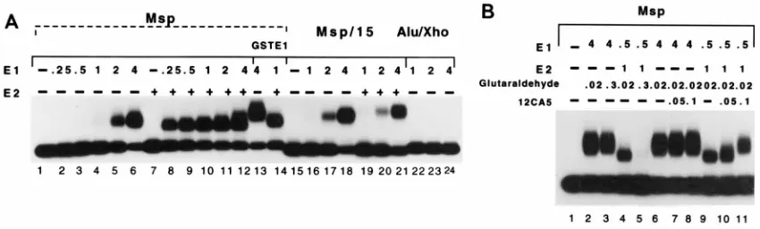
the complexes in combination with separation of complexes on agarose gels. We used this method with some modifications (see below). As shown in Fig. 3A, an E1 complex can readily be detected on the ori probe Msp under these conditions (lanes 1 to 6). When E2 was added to the binding reactions, two major changes could be observed (lanes 8 to 12). First, consistent with the results seen in the DNase I footprint assays, a complex could be observed at considerably lower concentrations of E1. Second, the mobility of the complex that appeared in the presence of E2 was increased significantly. E2 alone under these conditions did not give rise to a detectable complex (lane 7). To demonstrate that these complexes contained E1, we substituted E1 for a GST-E1 fusion protein (lanes 13 and 14). Under these conditions, a reduction of the mobility of both the E1 complex and the E1-E2 complex was observed, indicating that E1 was present in both complexes. To determine if for-mation of the faster-migrating complex required an E2 binding site, we used the probe Msp/15, where the E2 binding site had been deleted. With this probe, the faster-migrating complex was not formed (lanes 19 to 21) although formation of the slower-migrating complex was observed at concentrations of E1 similar to those used for the Msp probe (compare lanes 4 to 6 to lanes 16 to 18). To demonstrate that the E1 binding site was required for the formation of both complexes, we used the
probe Alu/Xho, which contains an Xholinker inserted at the
center of the palindrome. This probe failed to give rise to either complex (lanes 22 to 24). These results are consistent with the observations made by Lusky and coworkers (19) and demonstrated that the results obtained in the mobility shift assay corresponded well to the results obtained in DNase foot-print analysis.
In the course of these experiments, we had observed that the concentration of glutaraldehyde used for cross-linking ap-peared to affect the two complexes differently (Fig. 3B). At the level of glutaraldehyde used in other studies, i.e., 0.3%, the slower-migrating E1 complex was readily detected (lane 3). However, the faster-migrating complex could not be detected under these conditions (lane 5). Reduction of the level of glutaraldehyde used in the cross-linking reaction to 0.02% showed the presence of also the faster-migrating complex (lane 4). This inhibition is likely to be caused by sensitivity of the E2 protein to glutaraldehyde, since DNA binding of E2 alone is severely inhibited by cross-linking with glutaraldehyde (data not shown). To demonstrate that E2 protein was present in the
faster-migrating complex, a gel shift was generated in the pres-ence of E2 protein tagged with the hemagglutinin (HA) epitope (37). Addition of the monoclonal antibody 12CA5 (9), which is directed against the HA epitope, gave rise to a super-shift of the faster-migrating complex (lanes 9 to 11) but did not affect the E1 complex (lanes 6 to 8). Thus, two distinct com-plexes can form on the ori sequence. One of these, the faster-migrating complex, is composed of both E1 and E2 protein and forms through cooperative binding. The other complex mi-grates more slowly, contains E1 alone, and forms only at higher concentrations of E1. Taken together, these results are consis-tent with the footprint analysis and indicate not only that the cooperative binding leads to binding of E1 at lower concentra-tions but also that two kinds of E1-containing ori complexes can form.
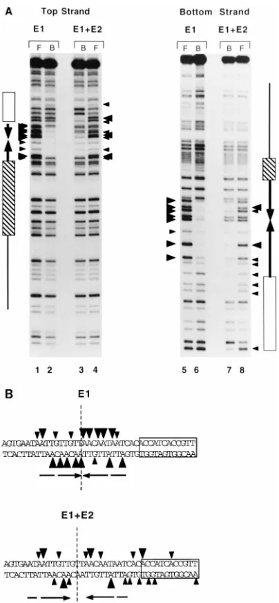
E1 in the two complexes recognizes overlapping but not identical sites.To determine the DNA sequence requirement for the formation of the two complexes, we performed inter-ference analysis. After modification of the probe with DEPC, gel shift analysis was performed; shifted bands were extracted from the gel, cleaved with piperidine, and analyzed on se-quencing gels. The results are shown in Fig. 4. DEPC modifies
A and G residues and generates an A1G ladder. The
inter-ference patterns for the two complexes showed significant dif-ferences. For the E1-ori complex, interference was observed only in the region of the palindrome, while the E1-E2-ori complex, as expected, also showed interference over the E2 recognition sequence. The interference over the E2 binding site was virtually identical to that produced by E2 alone, and we therefore concluded that these interferences corresponded to binding by E2 (data not shown). In addition, the numbers and positions of bases whose modification generated interfer-ence were different also in the region of the palindrome. For example, the center of symmetry of interference for the trim-eric E1-ori complex was at the center of the palindrome. The center of symmetry for the E1-E2-ori complex was a palin-drome interrupted by three nucleotides centered slightly up-stream of the center of symmetry for the E1-ori complex. Thus, the patterns of interference for the two complexes were re-lated: the interference obtained from the E1-E2-ori complex was a subset of the interference for the E1-ori complex and contained no additional points of interference that could not be accounted for by the binding of E2. These results indicated that E1 required fewer contact points in the presence of E2
FIG. 3. (A) Gel mobility shift analysis of complexes formed in the presence of E1 and in the presence of E1 and E2. Gel mobility shift assays were performed with three different probes. Msp contains the wt ori sequence, Msp/15 has a deletion that removes E2 binding site 12, and Alu/Xho contains the wt ori sequence with an Xholinker inserted into the uniqueHpaI restriction site at the center of the E1 binding site. The probes were tested for the ability to form complexes in the presence of E1 alone or in the presence of E1 and E2. The quantity of E1 used in each binding reaction is indicated in nanograms. E2 was added at 1 ng per binding reaction. (B) Glutaraldehyde is inhibitory for the formation of the faster-migrating complex which contains E2. Gel mobility shift assays were performed to determine the effects of the cross-linker glutaraldehyde on the two different E1-containing complexes. The complexes were formed and treated with either 0.3 or 0.02% glutaraldehyde as indicated. To determine if E2 was present in the faster-migrating complex, E2 tagged with the HA epitope was used for the formation of the faster-migrating complex, and a monoclonal antibody (12CA5) directed against the HA tag was added.
on November 9, 2019 by guest
http://jvi.asm.org/
[image:4.612.96.521.70.198.2]than in the absence of E2 and that E1 therefore appeared to be capable of binding in two different ways to the same DNA sequence. These results also demonstrated that the extended protections that could be observed in the DNase footprint assays are not due to sequence specific contacts of E1 with the DNA.
Point mutations in the ori.The sum of these results indi-cated that all specific sequences involved in E1 binding were located within the palindromic sequence. We therefore gener-ated single-point mutations at all positions within this
se-quence. We chose to maintain the G1C content of the ori to
avoid effects caused by changes in melting temperature of the ori and consequently generated transversions, converting A to
T, T to A, G to C, and C to G. These point mutants were tested in the context of the intact minimal ori in three different assays: (i) for E1-ori complex formation in gel shift assays, (ii) for E1-E2-ori complex formation in gel shift assays, and (iii) for DNA replication in vivo in a transient replication assay. Be-cause we wanted to make very precise comparisons of the effects of a given mutation on these different activities, we designed assays that would very accurately measure E1 binding and DNA replication of the mutant oris.
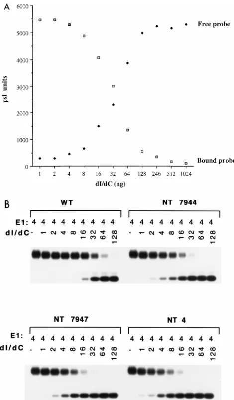
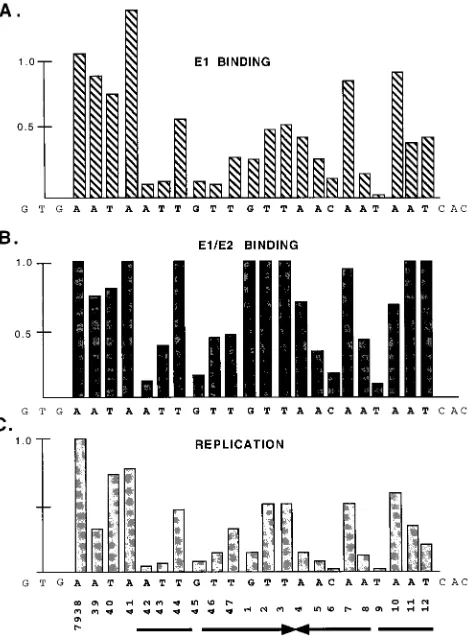
Formation of the E1-ori complex on mutant oris.Binding of E1 to the ori shows an extremely nonlinear response to titra-tion of E1. To measure binding of E1 to the different point mutations in ori, we therefore used a competition assay where we could keep the quantity of E1 protein constant. In these assays, a fixed amount of probe was mixed with increasing quantities of the nonspecific competitor poly(dI-dC), and a fixed amount of E1 was added. After cross-linking with glutar-aldehyde, the samples were analyzed in a gel shift assay; free probe and the shifted complexes were quantitated for each competitor concentration and plotted as a function of poly(dI-dC) concentration. An example of such a competition assay is shown in Fig. 5A for the wt ori. The concentration of poly(dI-dC) where the two curves intersect can be determined accu-rately and represents the 50% competition point, which can be used for comparison of the different mutants. The ability to withstand competitor relative to the wt ori gives a measure of the binding affinity of the various mutant oris. An example of this assay for three mutants and the wt ori is shown in Fig. 5B. The results from testing all of the mutants in this assay are shown in the bar graph in Fig. 8A. Surprisingly, the majority of these mutations had a substantial effect on E1 binding. All but six of the mutants were significantly (greater than twofold) reduced in their ability to bind E1. One of these exceptions, mutant 7941, had an approximately 40% increased ability to bind E1. This particular mutation extends the palindromic sequence by three nucleotides, indicating that perhaps the pal-indromic nature of the site is important for sequence recogni-tion by E1. The rest of the mutarecogni-tions fell roughly into two categories: mutations that had a two- to fourfold effect on binding (nine positions), and seven positions (7942, 7943, 7945, 7946, 6, 8, and 9) that had large effects (greater than fivefold reduction) on E1 binding. These severe mutants are symmet-rically arranged and correspond well to positions that showed strong interference in the DEPC interference analysis (Fig. 4).
[image:5.612.78.280.67.503.2]Formation of the E1-E2-ori complex on mutant oris.The formation of the E1-E2-ori complex showed a linear response to titration of increasing quantities of E1 in the presence of an excess of E2. We therefore could measure the ability to bind E1 in the E1-E2-ori complex by simple E1 titrations. The results from binding assays for seven mutants are shown in graphical form in Fig. 6, and the results for all the mutants are summarized in Fig. 8B. The formation of the E1-E2-ori com-plex showed a dependence on the palindromic sequence sim-ilar to that for formation of the E1 complex, with some notable differences. A total of nine mutations had substantial effects (greater than twofold) on E1-E2 binding. The positions impor-tant for E1-E2-ori complex formation consist of a subset of the positions that are important for formation of the E1-ori com-plex. The five mutations that had severe effects on E1-E2-ori complex formation (greater than 50% reduction in binding 7942, 7943, 7945, 5, 6, and 9) were all positions that also had large effects on E1 binding, in agreement with the results from the interference analysis. However, the majority of the muta-tions that had intermediate effects on E1 binding had very small or undetectable effects on formation of the E1-E2-ori complex.
FIG. 4. DEPC interference analysis of the E1-ori and E1-E2-ori complexes. (A) The wt ori probe was denatured and modified with DEPC. After reanneal-ing, the probe was used for gel mobility shift assays to generate the E1-ori and E1-E2-ori complexes. The shifted complexes and the free probe were extracted from the gel, cleaved with piperidine, and analyzed on a sequencing gel. Arrow-heads indicate the positions of interference. Lanes: F, free probe; B, bound probe. (B) Summary of the interference results obtained with the two complexes.
on November 9, 2019 by guest
http://jvi.asm.org/
Replication of mutant oris.The mutants that were tested for binding were also tested for the ability to replicate in the transient replication assay. We performed these transient rep-lication assays in the presence of a wt ori construct as an internal standard in order to accurately quantitate the replica-tion activity of the different mutants. This wt ori plasmid is
identical to the test plasmids except that anEcoO109
restric-tion site which is present in the pUC19 backbone has been destroyed. After harvesting of low-molecular-weight DNA, the
material was digested withDpnI,XbaI, andEcoO109. The wt
plasmid gives rise to a 2.9-kb band since this plasmid was
linearized by XbaI and lacks a site for EcoO109, while the
mutants which are cleaved also by EcoO109 give rise to a
smaller band of 2.5 kb. To quantitate the replication activity of a given mutant, a sample of the mix that was used for trans-fection was loaded on the gels as a marker after digestion with
XbaI andEcoO109. The ratios between the wt and mutant oris
were then compared before and after replication. An example of these assays for some of the mutants is shown in Fig. 7, and a summary and the quantitations are shown in Fig. 8C. All mutants showed reduced ability to replicate compared to the wt ori, and some of the more severe mutants failed to show detectable replication. Taken together, these results showed a very strong correlation between the ability of a particular mu-tant to bind E1 either in the E1-ori complex or in the E1-E2-ori complex and to replicate in the transient replication assay. A majority of the mutations that had an effect on DNA replica-tion can be accounted for by the effect on binding of E1 and E2. However, some exceptions exist; mutant 7939, which has a very small effect on both ori complex formation and E1-E2-ori complex formation (90 and 75%, respectively, of the wt level) has a substantial effect on DNA replication (40% of the wt level). Similarly, the mutation at nt 7, which has close to wt activity for binding of E1 and E2 (90 and 95%, respectively of wt activity), only shows 50% replication activity, indicating that some nucleotide positions, in addition to the positions required for the formation of the E1-ori and E1-E2-ori complexes, are important for DNA replication activity.
DISCUSSION
The results in this study demonstrate that the cooperative binding of E1 and E2 proteins to the BPV ori is not simply a quantitative effect. The complex formed on the ori by E1 alone (E1-ori) and the complex formed by the combination of E1 and E2 (E1-E2-ori) are clearly qualitatively different by a number of criteria: (i) the E1-E2-ori complex in a mobility shift assay has greater mobility than the E1-ori complex, indicating lower molecular weight; (ii) interference analysis demonstrates that binding of the two complexes to the BPV ori involve
[image:6.612.59.294.68.470.2]overlap-FIG. 5. (A) Competition assay for binding of E1 to the wt ori. Gel mobility shift assays were performed with a constant level of E1 protein (4 ng) in the presence of increasing concentration of the nonspecific competitor poly(dI-dC). Both bound and free probe were quantitated and plotted as a function of concentration of poly(dI-dC). The point where the two curves intersect repre-sents the 50% competition point, which was used for comparison of different mutant mutant oris. psl, photostimulated luminescence. (B) Determination of binding affinities of ori mutants by poly(dI-dC) competition. Binding of E1 to the different ori mutants was determined by performing gel mobility shift assays using a constant quantity of E1 (4 ng) in the presence of increasing quantities of the nonspecific competitor poly(dI-dC). Free probe and the shifted complex were quantitated; the 50% competition point was determined for each mutant and compared to that for the wt ori. A summary of the results is shown in Fig. 8A.
FIG. 6. Effects of point mutations in the ori on E1-E2 binding. Formation of the E1-E2-ori complex was measured in gel mobility shift assays by incubating the different mutant probes with a constant amount of E2 (1 ng) and increasing quantities of E1 protein. The quantity of shifted probe for each mutant as well as for the wt was quantitated and plotted as a function of E1 concentration. The results for seven of the mutants are shown. The fraction of shifted probe com-pared to the wt was determined at three concentrations of E1 and averaged; a summary of the results is shown in Fig. 8B.
on November 9, 2019 by guest
http://jvi.asm.org/
[image:6.612.336.532.70.249.2]ping but nonidentical sequences; and (iii) the mutational anal-ysis demonstrates that the E1-ori and E1-E2-ori complexes are affected differentially by certain mutations within the palin-dromic sequence, indicating that E1 in the E1-E2-ori complex binds to a subset of the sequences required for E1-ori complex formation. These results together indicate that E1 in the E1-E2-ori complex constitutes a subset of E1 in the E1-ori com-plex. A likely possibility is therefore that E1 by itself is capable of binding in a multimeric form and that together with E2, a smaller number of E1 molecules can stably bind to the DNA. These results are in good agreement with our recent molecular mass determination of the E1-ori and E1-E2-ori complexes, which indicated that E1 in the E1-ori complex binds as a trimer but that E1 together with E2 binds as a monomer (27).
The results from the interference and mutational studies
demonstrate that E1 in both the E1-E2-ori and the E1-ori complexes bind to the same sequence element. Consequently, a very interesting question is how the same short sequence can be used for binding of E1 in its different forms. Although we do not have a definitive answer to this question, analysis of the interference and mutational results can yield some interesting clues. The mutational analysis in itself provides little insight into how E1 recognizes and binds to the ori; the results are very complex. However, when the results from the mutational anal-ysis are considered in conjunction with our data concerning the stoichiometry of binding of E1 in the two different complexes, the mutational analysis becomes easier to interpret.
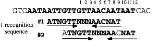
We previously observed that when high-resolution hydroxyl radical footprints were compared, protections resulting from binding of E1 in the E1-E2-ori complex constituted a subset of the protections observed with the E1-ori complex. E1 in the E1-E2-ori complex protected a discrete set of bases exclusively on one face of the DNA helix. The protections observed with the E1-ori complex exhibited two additional features: a precise duplication of the protection observed with the E1-E2-ori com-plex, but shifted by three nucleotides such that two-thirds of the circumference of the DNA helix was strongly protected, and a weaker protection completing the circumference of the helix. These results indicated that in the E1-E2-ori complex, E1 was bound on one face of the helix and that in the E1-ori complex, additional E1 was bound in virtually identical fashion to another face of the helix. A logical conclusion from this observation is that the mutations in the E1 binding site that affect the E1-E2-ori complex represent a minimal sequence element required for binding of E1 and that the additional mutations that affect the formation of the E1-ori complex represent positions that are required for binding of additional E1 molecules. Thus, the element ATNGTTNNNAACNAT, as defined by the mutagenesis (Fig. 8B), would represent the minimal sequence required for binding of E1 in the presence of E2. This sequence includes two copies of the pentanucle-otide sequence ApyAAPy that has been proposed to be in-volved in binding of E1 by analogy to the T-antigen binding sites in the simian virus 40 ori (21).
One interpretation of the precise duplication of protection observed in the hydroxyl radical footprints is that a second E1 recognition sequence is present but shifted three nucleotides relative to the E1 recognition sequence used for E1 binding in the E1-E2-ori complex. When the sequence of the ori is ex-amined, an element closely related to the sequence defined by mutagenesis as required for binding of E1 in the E1-E2-ori complex can be found overlapping the first sequence but shifted by three nucleotides (Fig. 9). This second sequence element is in the position predicted by the hydroxyl radical footprints. Thus, a model for binding of E1 based on these results would indicate that the E1 binding site in the BPV ori consists of two separate, overlapping recognition sequences for E1.
[image:7.612.63.299.372.690.2]FIG. 7. Replication of point mutants in the transient replication assay. Ori plasmids with the indicated point mutations were cotransfected together with the wt ori into cell line 4.15, which expresses the E1 and E2 proteins. At the indicated time points, low-molecular-weight DNA was harvested and digested with a mixture ofDpnI, XbaI, andEcoO109 and analyzed by Southern blotting. The wt ori plasmid lacks a restriction site forEcoO109 and gives rise to a band of 2.9 kb, while the mutant oris generate a band of 2.5 kb. Each lane M corresponds to a sample of each input mixture of wt and mutant plasmid used in the transfections.
FIG. 8. Effects of ori point mutations on E1 binding, E1-E2 binding, and DNA replication. Shown is a summary of the effects of the point mutations on formation of the E1-ori complex (A), formation of the E1-E2-ori complex (B), and DNA replication (C). The wt level is in all cases set at 1.0.
on November 9, 2019 by guest
http://jvi.asm.org/
With this model in mind, a comparison of the effects of mutations on binding of E1 in the E1-E2-ori complex and E1-ori complexes becomes more informative. The most strik-ing differential effect of mutations in the E1 bindstrik-ing site is that mutation of three nucleotides in the center of the E1 binding site (nt 1 to 3), which have little or no effect on E1-E2-ori complex formation (Fig. 8B), show substantial effects on E1-ori complex formation (Fig. 8A). According to the model, al-though these positions do not affect recognition sequence 1, because of the three-nucleotide shift, these nucleotides are part of recognition sequence 2. These three positions would therefore be important only for binding by E1 on the side face of the helix and thus would affect E1-ori complex formation only. A further interesting difference is the effect of mutations at nt 11 and 12. Mutations at these positions have no effect on formation of the E1-E2-ori complex but show a significant effect on E1-ori complex formation. This result is consistent with the notion that these positions are not included in recog-nition sequence 1 but are part of recogrecog-nition sequence 2. Thus, mutation of these nucleotides would affect binding of the sec-ond E1 molecule only and thus would influence only E1-ori complex formation. This model for E1 binding also accommo-dates and explains some of the observations from the interfer-ence analysis: as shown in Fig. 4, the centers of symmetry are different for binding of the E1-E2-ori complex and the E1-ori complex. The interrupted palindrome that we observed for the E1-E2-ori complex is clearly consistent with binding of E1 to recognition sequence 1. The change of symmetry observed for the E1-ori complex can be easily explained as resulting from the combined symmetries of recognition sequences 1 and 2.
Thus, from the sum of these results, we propose the follow-ing model for how E1 binds to the origin of replication. In the presence of E2, E1 can bind to the recognition sequence AT-NGTTNNNAACNAT. Binding of E1 to this site requires an interaction with the E2 protein bound to the adjacent E2 binding site. At higher concentrations of E1 and in the absence of E2, a second binding site is recognized by E1. This second binding site overlaps the first binding site but is shifted by three nucleotides, placing it on a different face of the DNA helix. Based on molecular weight determinations, we have previously proposed that E1 in the E1-ori complex binds as a trimer (27). In this study, we see no direct indication that binding of a third E1 molecule requires a specific DNA sequence, and maybe protein-protein interactions with two E1 molecules that are bound to the DNA are sufficient for formation of an E1 trimer. Finally, this model for E1 binding can also accommodate a number of different papillomavirus oris; similar overlapping sequence arrangements appear to be a general feature of many papillomavirus oris.
The symmetrical arrangement of important bases observed both in the mutational analysis and in the interference analysis indicates that the palindromic nature of the E1 binding site is not fortuitous and that the twofold rotational symmetry that we observe in the recognition sequence is important for binding of E1. This conclusion is supported by the conservation of partial or complete palindromic E1 binding sites in a large number of papillomavirus oris and also by other studies. Holt and Wilson
concluded that the palindromic sequence was important for binding of E1, based on mutational analysis of the 18-bp pal-indromic sequence (14). Mendoza and coworkers (22) gener-ated several insertion mutants in the center of the E1 palin-drome, demonstrating that replication and to some extent also binding of E1 were dependent on both the phasing and the spacing of the two halves of the palindrome relative to each other. Surprisingly, the ori could retain some replication activ-ity even when 10 or 20 bp were inserted into center of the palindrome, indicating that the two halves of the palindrome may represent units that function with some degree of inde-pendence. At first glance, this result would appear to be at odds with the model for E1 binding that we are proposing, where E1 binds across the center of the palindrome. However, since the levels of replication that were observed were quite low, replication may have resulted from a low residual level of E1 binding to these altered sites. Mendoza et al. (21) observed binding of E1 to these altered sites but did not address in what form E1 bound to the mutant templates.
Our previous experiments have demonstrated that point mu-tations in the E1 binding site that affected both formation of the E1-E2-ori complex and the E1-ori complex, and also rep-lication, could be rescued for E1-E2-ori complex formation as well as for replication by the introduction of a high-affinity E2 binding site (26). These mutants were not rescued for E1-ori complex formation. We concluded from those experiments that the formation of the E1-E2-ori complex was essential for replication, while the formation of the E1-ori complex was not: the mutants that were defective for E1-ori complex formation replicated at close to wt levels in the presence of a high-affinity E2 binding site. This conclusion would appear to be contra-dicted by the mutational analysis presented here; the effects of the point mutations on E1-ori complex formation and DNA replication correlate very well. Also, several of the point mu-tations in the E1 binding site that can form the E1-E2-ori complex at wt or close to wt levels show reduced levels of replication (comp. Fig. 8B and C). The simplest explanation is that the abilities to form both the E1-ori complex and the E1-E2-ori complex are important for replication and that the formation of the E1-ori complex in vivo takes place with the E1-E2-ori complex as a required precursor. Since a high-affin-ity E2 binding site can rescue binding of E1 in the E1-E2-ori complex, formation of the E1-ori complex, via the E1-E2-ori complex as a precursor, would obviously also be affected by the high-affinity E2 binding site. In support of this idea, we have recently demonstrated that the E1-E2-ori complex is a pre-ferred substrate for the formation of the E1-ori complex (25). Thus, E1 in the E1-E2-ori complex appears to function as “seed” for the formation of the E1-ori complex.
[image:8.612.105.257.73.114.2]Comparison of the results obtained in this study to a previ-ous mutational analysis demonstrate a general agreement in the instances where direct comparisons can be made but also some notable differences. Overall, Holt and Wilson (14) ob-served generally smaller effects of mutations on both E1 bind-ing and DNA replication and also a less obvious correlation between E1 binding and DNA replication. None of the mu-tants tested in that study was reduced more than 5-fold for replication or more than 10-fold for E1 binding. We believe that the level of E1 and E2 expressed in the transfected cells may affect the severity of a given mutant and that high levels of E1 and E2 may mask the effects of mutations in the ori and thus explain some of the differences concerning the replication results. It seems likely that the transient expression of E1 and E2 that was used by Holt and Wilson results in higher levels of E1 and E2 than the stable E1- and E2-expressing cell line that was used in this study. The overall difference observed for E1
FIG. 9. Proposed overlapping recognition sequences for E1 in the BPV or-igin of replication. See text for details.
on November 9, 2019 by guest
http://jvi.asm.org/
binding is harder to explain. For example, some of the mutants that have the most severe effects on E1 binding in our hands (nt 7943 and 9 are reduced more than 10-fold for E1 binding) have modest (2- to 3-fold) effects in the other study, while other mutants (e.g., nt 2 and 3) have similar effects (2- to 3-fold reduction) on E1 binding in both studies. However, significant differences exist in the assays that were used to measure E1 binding. Holt and Wilson used an immunoprecipitation assay and crude E1 extract to measure E1 binding, and it is not clear in what form E1 binds to DNA under these conditions.
An earlier study used similar approaches to study the E1-containing complexes that can form on the BPV ori (19). Superficially, the results from these studies appear similar; i.e., two different complexes were observed, E1 alone gave rise to a complex that showed lower electrophoretic mobility than the complex formed in the presence of E2, and also cooperative DNA binding could be observed between the two proteins. However, some of the characteristics of the complexes ob-served in these studies are clearly different. Generation of the two complexes by Lusky and coworkers (19) required much higher concentrations of the E1 and E2 proteins. In addition, the authors could observe effects on complex formation by addition of ATP and magnesium, while under our conditions of complex formation no significant effects can be observed with the exception of effects on complex half-lives (data not shown). Finally, estimates of the stoichiometry of binding differ significantly; Lusky et al. estimated that the E1 complex con-tained 10 to 15 molecules of E1, while our recent results indicate that the E1-ori complex contains 3 molecules of E1 (27). At the moment we cannot entirely resolve these differ-ences; however, it seems likely that the complexes observed in these studies in fact are related but that differences in the conditions used for the generation of the complexes are re-sponsible for the different properties. Possible reasons for these differences include the use of different expression sys-tems and purification procedures for the E1 and E2 proteins as well as different levels of glutaraldehyde in the cross-linking reactions. Further analysis of the complexes formed under these different conditions will be required to resolve the rela-tionship between these different complexes.
ACKNOWLEDGMENTS
We thank M. Berg for critical reading of the manuscript. This work was supported by grant CA 13106 from NIH.
REFERENCES
1.Androphy, E. J., D. R. Lowy, and J. T. Schiller.1987. Bovine papillomavirus E2 trans-activating gene product binds to specific sites in papillomavirus DNA. Nature (London)325:70–73.
2.Aurora, R., and W. Herr.1992. Segments of the POU domain influence one another’s DNA-binding specificity. Mol. Cell. Biol.12:455–467.
3.Benson, J. D., and P. M. Howley.1995. Amino-terminal domains of the bovine papillomavirus type 1 E1 and E2 proteins participate in complex formation. J. Virol.69:4364–4372.
4.Blitz, I., and L. Laiminis.1991. The 68-kilodalton E1 protein of bovine papillomavirus is a DNA binding phosphoprotein which associates with the E2 transcriptional activator in vitro. J. Virol.65:649–656.
5.Bonne-Andrea, C., S. Santucci, P. Clertant, and F. Tillier.1995. Bovine papillomavirus E1 protein binds specifically DNA polymerasea but not replication protein A. J. Virol.69:2341–2350.
6.Burgess, R. R.1991. Use of polyethyleneimine in purification of DNA binding proteins. Methods Enzymol.208:3–10.
7.Cheng, I., J. I. Workman, R. E. Kingston, and T. J. Kelly.1992. Regulation of DNA replication in vitro by the transcriptional activation domain of GAL4-VP16. Proc. Natl. Acad. Sci. USA89:589–593.
8.Cheng, L., and T. Kelly.1989. Transcriptional activator nuclear factor I stimulates the replication of SV40 minichromosomes in vivo and in vitro. Cell59:541–551.
9.Field, J., J.-I. Nikawa, D. Broeke, B. MacDonald, L. Rodgers, I. A. Wilson, R. A. Lerner, and M. Wigler.1988. Purification of a RAS responsive adenylyl
cyclase complex fromSaccharomyces cerevisiaeby use of an epitope addition method. Mol. Cell. Biol.8:2159–2165.
10. Gillette, T. G., M. Lusky, and J. A. Borowiec.1994. Induction of structural changes in the bovine papillomavirus type 1 origin of replication by the viral E1 and E2 proteins. Proc. Natl. Acad. Sci. USA91:8846–8850.
11. Hawley-Nelson, P., E. J. Androphy, D. R. Lowy, and J. T. Schiller.1988. The specific DNA recognition sequence of the bovine papillomavirus E2 protein is an E2-dependent enhancer. EMBO J.7:525–531.
12. He, Z., B. T. Brinton, J. Greenblatt, J. A. Hassel, and C. L. Ingles.1993. The transactivator proteins VP16 and Gal 4 bind replication factor A. Cell73:
1223–1232.
13. Holt, S. E., G. Schuller, and V. G. Wilson.1993. DNA binding specificity of the bovine papillomavirus E1 protein is determined by the sequences con-tained within an 18-base-pair inverted repeat element at the origin of rep-lication. J. Virol.68:1094–1102.
14. Holt, S. E., and V. G. Wilson.1995. Mutational analysis of the 18-base-pair inverted repeat element at the bovine papillomavirus origin of replication: identification of critical sequences for E1 binding and in vivo replication. J. Virol.69:6525–6532.
15. Li, R., J. Knight, G. Bream, A. Stenlund, and M. Botchan.1989. Specific recognition nucleotides and their DNA context determine the affinity of E2 protein for 17 binding sites in the BPV genome. Genes Dev.3:510–526. 16. Li, R., and M. Botchan.1993. The acidic transcriptional activation domains
of VP16 and p53 bind the cellular replication protein A and stimulate in vitro BPV-1 DNA replication. Cell73:1207–1221.
17. Li, R., and M. Botchan.1994. Acidic transcription factors alleviate nucleo-some-mediated repression of DNA replication of bovine papillomavirus type 1. Proc. Natl. Acad. Sci. USA91:7051–7055.
18. Lusky, M., and E. Fontane.1991. Formation of the complex of bovine papillomavirus E1 and E2 proteins is modulated by E2 phosphorylation and depends on sequences within the carboxyl terminus of E1. Proc. Natl. Acad. Sci. USA88:6363–6367.
19. Lusky, M., J. Hurwitz, and Y.-S. Seo.1994. The bovine papillomavirus E2 protein modulates the assembly but is not stably maintained in a replication-competent multimeric E1-replication origin complex. Proc. Natl. Acad. Sci. USA91:8895–8899.
20. McBride, A. A., H. Romanczuk, and P. M. Howley.1991. The papillomavirus E2 regulatory proteins. J. Biol. Chem.266:18411–18414.
21. Melendy, T., J. Sedman, and A. Stenlund.1995. Cellular factors required for papillomavirus DNA replication. J. Virol.69:7857–7867.
22. Mendoza, R., L. Gandhi, and M. Botchan.1995. E1 recognition sequences in the bovine papillomavirus type 1 origin of DNA replication: interaction between half sites of the inverted repeats. J. Virol.69:3789–3798. 23. Mohr, I. J., R. Clark, S. Sun, E. J. Androphy, P. MacPherson, and M.
Botchan.1990. Targeting the E1 replication protein to the papillomavirus origin of replication by complex formation with the E2 transactivator. Sci-ence250:1694–1699.
24. Muller, F., Y.-S. Seo, and J. Hurwitz.1994. Replication of bovine papillo-mavirus type 1 origin-containing DNA in crude extracts and with purified factors. J. Biol. Chem.269:17086–17094.
25. Sanders, C., and A. Stenlund.Unpublished observation.
26. Sedman, J., and A. Stenlund.1995. Co-operative interaction between the initiator E1 and the transcriptional activator E2 is required for replicator specific DNA replication of bovine papillomavirus in vivo and in vitro. EMBO J.14:6218–6228.
27. Sedman, J., and A. Stenlund.1996. The initiator protein E1 binds to the bovine papillomavirus origin of replication as a trimeric ring-like structure. EMBO J.15:5085–5092.
28. Seo, Y.-S., F. Muller, M. Lusky, and J. Hurwitz.1993. Bovine papillomavirus (BPV)-encoded E1 protein contains multiple activities required for BPV DNA replication. Proc. Natl. Acad. Sci. USA90:702–706.
29. Seo, Y.-S., F. Muller, M. Lusky, E. Gibbs, H.-Y. Kim, B. Phillips, and J. Hurwitz.1993. Bovine papilloma virus (BPV)-encoded E2 protein enhances binding of E1 protein to the BPV replication origin. Proc. Natl. Acad. Sci. USA90:2865–2869.
30. Spalholz, B. A., A. A. McBride, T. Sarafi, and J. Quintero.1993. Binding of bovine papillomavirus E1 to the origin is not sufficient for DNA replication. Virology193:201–212.
31. Spalholz, B. A., Y. C. Yang, and P. M. Howley.1985. Transactivation of a bovine papillomavirus transcriptional regulatory element by the E2 gene product. Cell42:183–191.
32. Studier, F., and B. Moffatt.1986. Use of bacteriophage T7 RNA polymerase to direct selective high-level expression of cloned genes. J. Mol. Biol.189:113–130. 33. Sturm, R., T. Baumruker, B. R. Franza, and W. Herr.1987. A 100 kD HeLa cell octamer binding protein (OBP 100) interacts differently with two separate oc-tamer-related sequences within the SV40 enhancer. Genes Dev.1:1147–1160. 34. Syrjanen, K., L. Gissman, and L. G. Koss (ed.).1987. Papillomaviruses and
human disease. Springer-Verlag, New York, N.Y.
35. Thorner, L., D. Lim, and M. Botchan.1993. DNA binding domain of bovine papillomavirus type 1 E1 helicase: structural and functional aspects. J. Virol.
67:6000–6014.
36. Ustav, M., and A. Stenlund.1991. Transient replication of BPV-1 requires
on November 9, 2019 by guest
http://jvi.asm.org/
two viral polypeptides encoded by the E1 and E2 open reading frames. EMBO J.10:449–457.
37. Ustav, M., E. Ustav, P. Szymanski, and A. Stenlund.1991. Identification of the origin of replication of bovine papillomavirus and characterization of the viral origin recognition factor E1. EMBO J.10:4321–4329.
38. Ustav, E., M. Ustav, P. Szymanski, and A. Stenlund.1993. The bovine papillomavirus origin of replication requires a binding site for the E2 tran-scriptional activator. Proc. Natl. Acad. Sci. USA90:898–902.
39. Wilson, V. G., and J. Ludes-Meyers.1991. A bovine papillomavirus
E1-related protein binds specifically to bovine papillomavirus DNA. J. Virol.
65:5314–5322.
40. Yang, L., I. Mohr, E. Fouts, D. A. Lim, M. Nohaile, and M. Botchan.1993. The E1 protein of the papillomavirus BPV-1 is an ATP dependent DNA helicase. Proc. Natl. Acad. Sci. USA90:5086–5090.
41. Yang, L., R. Li, I. Mohr, R. Clark, and M. R. Botchan.1991. Activation of BPV-1 replication in vitro by the transcription factor E2. Nature (London)
353:628–633.
42. zur Hausen, H.1991. Viruses in human cancers. Science254:1167–1173.