
A PROSPECTIVE STUDY
Dissertation Submitted to
THE TAMIL NADU DR.M.G.R. MEDICAL UNIVERSITY
in partial fulfillment of the
regulations
for the award of the degree of
M.D. (GENERAL MEDICINE)
BRANCH - I
STANLEY MEDICAL COLLEGE
THE TAMIL NADU DR.M.G.R. MEDICAL UNIVERSITY
CHENNAI, INDIA
This is to certify that this dissertation entitled TO STUDY THE
IMMUNE AND ALLERGENIC PROFILES OF ATOPIC
ASTHMA PATIENTS is the bonafide original work of
Dr.N.JOTHIMURUGAN in partial fulfillment of the requirement for
MD (Branch -I) GENERAL MEDICINE examination of the Tamil Nadu
Dr.MGR Medical University to be held in March 2008.
Dr. S. RAMASAMY M.D. Addl. Prof.of Medicine
Govt.Stanley Medical College Hospital, Chennai-600001
Prof. S.NATARAJAN M.D. Head Of The Department, Department Of Medicine, Govt. Stanley Medical College &
Hospital, Chennai-600001
Dr. MYTHILI BASKARAN DEAN
I, Dr.N.JOTHI MURUGAN, solemnly declare that this dissertation
"TO STUDY THE IMMUNE AND ALLERGENIC PROFILES OF
ATOPIC ASTHMA PATIENTS"is a bonafide record of work done by me in
the Department of Medicine, Government Stanley Medical College and
Hospital, Chennai under the guidance of Prof.Dr.S.RAMASAMY, M.D.,
Addl. Prof. of Medicine, Government Stanley Medical College and Hospital,
Chennai – 600 001.
This dissertation is submitted to the Tamilnadu Dr.M.G.R.Medical
University, Chennai in partial fulfillment of the University regulations for the
award of MD Degree (General Medicine) Branch-I, Examination to be held in
March 2008.
Place: Chennai
Stanley Medical College for permitting me to carryout this study.
With sincere gratitude, I wish to acknowledge the expert guidance
and precise suggestions of my Head of the Department and Chief
Prof.Dr.S.NATARAJAN. M.D., Prof.DEENADAYALAN M.D.
(Former Chief), and Prof.S.RAMASAMY,M.D, without whose guidance
this study would not have been possible, and my co-guides Asst.
Professors Dr.ASHOKKUMAR, Dr.GEETHA, Dr.SURENDRAN,
Dr.SAMUEL DINESH without whom this work would not be in the
present shape, I am deeply indebted to and highly grateful to them.
I am deeply indebted to and highly grateful to Dr.BALARAM
GHOSH, Ph.D, scientist F, Deputy Director, Institute Of Genomics and
Integrative Biology, Delhi, Dr.U.MABALIRAJAN, pursuing PhD in
AIIMS, Mr.AMRENDAKUMAR M..Sc. pursuing PhD in, Institute Of
Genomics and Integrative Biology, Mr.SATISH DEVARAPU M.Sc.
pursuing PhD in AIIMS.
I wish to thank all my co-post graduates Dr.DEIVASIGAMANI,
Dr.SATHYABABU, Dr.LAVANYA, Dr.MARIANJUDE VIJAY,
Dr.SAMANTHA, Dr.HARIHARAN and Dr.BETSY without their
SL.
NO. TITLE PAGE NO.
1. INTRODUCTION 1
2. REVIEW OF LITERATURE 3
3. AIM AND OBJECTIVES 22
4. PATIENTS AND STUDY METHODS 23
5. OBSERVATION AND RESULTS 35
6. DISCUSSION 46
7. SUMMARY 59
8. CONCLUSIONS 60
9. LIMITATIONS AND SUGGESTIONS 61
BIBLIOGRAPHY
ANNEXURES
MASTER CHART
INTRODUCTION
Nearly 300 million people in the world suffer from asthma(1)
.Asthma is a common chronic inflammatory disease of the airways, with
considerable heterogeneity both in its phenotype and in the underlying
pathophysiology(2). Both intrinsic or non-atopic and extrinsic or atopic
cases of asthma are known. Extrinsic asthma is mainly a childhood
disorder though the age of onset can vary; whereas intrinsic asthma is
observed where the age of onset is above 45 yrs and is mainly due to the
age induced changes in the lung function(3). The majority of asthmatics
(around 80%) are atopic, with manifestations of allergic diathesis
including clinical allergy to aeroallergens and foods, or subclinical
allergy apparent by skin test reactivity to allergen or elevated serum IgE.
Approximately 7-10% of the Indian population, particularly children, is
affected by atopic asthma (4, 5) .It has been estimated that 34% of the man
days in India are lost due to airway related disorders, of which asthma is a
major cause (6) .Asthma and atopy are considered complex traits, with
evidence of both heritable and environmental factors contributing to their
pathogenesis. Atopy is known to be a strong predisposing factor in the
development of asthma(7). Atopic asthma is a hereditary disease and is
pathogenesis is complex, management of the disease is also difficult. But
prevention of the disease is possible if the allergic status of patients is
known. However, due to the risk of anaphylaxis the allergy test is not
being performed routinely in asthma clinics managing a lot of new as
well as old patients. Due to this, poor asthma patients are not aware of
these allergies and do not know the status of allergen positivity they have.
In contrast, skin prick test is a test which carries less risk rate of
anaphylaxis due to minimum possibility of allergen entry into systemic
circulation (8) .
With this background, this study emphasizes the importance of the
skin prick test in asthmatics and tries to correlate with the immune
response in these patients. In addition, it tries to understand the
prevalence of the allergen positivity in these patients and it also explores
REVIEW OF LITERATURE
DEFINITION AND DESCRIPTION OF ASTHMA:
Asthma is a disorder defined by its clinical, physiological, and
pathological characteristics. The predominant feature of the clinical
history is episodic shortness of breath, particularly at night, often
accompanied by cough. Due to complexity of its nature it is very difficult
to define asthma. The following definition has been given by Global
Initiative for Asthma (9)
“Asthma is a chronic inflammatory disorder of the airways in
which many cells and cellular elements play a role. The chronic
inflammation is associated with airway hyperresponsiveness that leads to
recurrent episodes of wheezing, breathlessness, chest tightness, and
coughing, particularly at night or in the early morning. These episodes
are usually associated with widespread, but variable, airflow obstruction
within the lung that is often reversible either spontaneously or with
treatment”.
It is a chronic inflammatory disorder of the airways in which many
cells and cellular elements play a role, in particular, mast cells,
cells . In susceptible individuals, this inflammation causes recurrent
episodes of wheezing, breathlessness, chest tightness, and coughing,
particularly at night or in the early morning(10). These episodes are usually
associated with widespread but variable airflow obstruction that is often
reversible either spontaneously or with treatment. The inflammation also
causes an associated increase in the existing bronchial hyper
responsiveness to a variety of stimuli.
ASTHMA DIAGNOSIS:
Asthma diagnosis is done on the basis of spirometry in individuals
identified by certain key indicators outlined in the Clinical Practice
Guidelines of Expert Panel Report-2, National Asthma Education and
Prevention Program (2002) as shown in Box 1. Typically in spirometry,
the maximal volume of air forcibly exhaled from the point of maximal
inhalation (forced vital capacity, FVC) and the volume of air exhaled
during the first second of the FVC (forced expiratory volume in 1 second,
FEV1) is measured.
• Airflow obstruction is indicated by reduced FEV1 and
FEV1/FVC values relative to reference or predicted values.
A reduced ratio of FEV1/FVC (i.e., <65 percent) indicates
reduced FVC with a normal FEV1/FVC ratio suggests a
restrictive pattern.
• Significant reversibility is indicated by an increase of ≥12
percent or 200 mL in FEV1 after inhaling a short-acting
BOX 1. KEY INDICATORS FOR CONSIDERING A DIAGNOSIS OF ASTHMA
Consider asthma and performing spirometry if any of these
indicators are present.* These indicators are not diagnostic by
themselves, but the presence of multiple key indicators increases the
probability of a diagnosis of asthma. Spirometry is needed to establish a
diagnosis of asthma.
• Wheezing—high-pitched whistling sounds when breathing out—
especially in children. (Lack of wheezing and a normal chest
examination do not exclude asthma.)
• History of any of the following:
— Cough, worse particularly at night
— Recurrent wheeze
— Recurrent difficulty in breathing
— Recurrent chest tightness
• Reversible airflow limitation and diurnal variation as measured by
using a peak flow meter, for example:
• Symptoms occur or worsen in the presence of:
— Exercise
— Viral infection
— Animals with fur or feathers
— House-dust mites
(in mattresses, pillows, upholstered furniture, carpets)
— Moulds
— Smoke (tobacco, wood)
— Pollen
— Changes in weather
— Strong emotional expression (laughing or crying hard)
— Airborne chemicals or dusts
— Menses
• Symptoms occur or worsen at night, awakening the patient.
*Eczema, hay fever, or a family history of asthma or atopic
ROLE OF GENES AND ENVIRONMENT:
Heritability estimates of asthma vary between 36 and 79%.
Allergen sensitization and subsequent exposure, dependent on both
genetic and environmental factors, are widely understood to influence the
development of atopic asthma. An association between total serum IgE
level and asthma prevalence has been recognized in asthma patients of all
ages (11, 12) .The linkage of total IgE levels to chromosome 5q highlights
the importance of genetic factors in the development of sensitization(13,
14). In addition, the level of environmental exposure to allergen is likely to
be important because sensitization to different allergens in different
patient populations has been associated with asthma(15, 16) and airway
hyperresponsiveness(17). The most prominent of these include Alternaria
in children living in a desert environment(15) ,dust mite in adolescents
living in central Virginia (16), and cat allergen in older men.
ALLERGY:
Without doubt, the casual correlation has been found between
allergy and atopic asthma. These two phenotypes are components of
atopy. Allergies affect around 30% of the adult population and 40% of
children. The prevalence, severity and complexity of allergy in the
for primary prevention or at least for delaying the onset of asthma by
avoiding allergens. The great majority of general practitioners have
received little allergy teaching as students and no extra postgraduate
training.
Allergy is a ‘hypersensitivity’ reaction, or exaggerated sensitivity,
to substances which are normally tolerated. Such substances are known as
allergens. Examples of common allergens include peanuts, milk, cats,
horses, medicines and grass pollens. These allergens trigger the
production of a harmful antibody, immunoglobulin E (IgE). In an allergic
reaction, the interaction between the IgE and the allergen causes the
release of inflammatory chemicals such as histamines and leukotrienes.
These cause symptoms such as sneezing, itches, rashes and falls in blood
pressure; they may also cause airway narrowing, which leads to shortness
of breath and wheezing, and swelling which, if in the mouth, throat or
airway, causes severe difficulty in breathing (19) .Sometimes symptoms
are caused by other mechanisms, where IgE is not involved. These are
often described as ‘intolerances’ to, for example, foods or medicines.
Allergy practice deals with both IgE mediated and non-IgE-mediated
reactions (20). Allergic symptoms vary greatly. An individual may have a
asthma, eczema and hay fever); swellings on the skin; or sickness The
most extreme reaction of all is anaphylaxis.
INDOOR AND OUTDOOR ALLERGENS:
Though, both genetic and environmental factors are involved in the
complex pathogenesis of asthma, genetic components are very complex
due to the heterogeneity of every population. For example, one gene
which is involved in asthma pathogenesis in a particular population, the
same gene may not be significant in another population. These ethnicity
variations are due to migration status of the population and many other
factors(21). Similarly environmental factors are also varies population to
population. But it is predominantly determined by the current
environment. There are some important environmental factors such as air
pollution and some allergens. Among these, air pollution is having a
major role in extrinsic asthma. In allergens, there are indoor and outdoor
allergens. It has been shown by birth-cohort studies that sensitization to
house dust mite allergens, cat dander, dog dander and aspergillus mold
are independent risk factors. However, sensitization depends on the
allergen, the dose, the time of exposure, the age, and probably genetics as
For some allergens, such as those derived from house dust mites
and cockroaches, the prevalence of sensitization appears to be directly
correlated with exposure (23). So identification of the allergic status will be
definitely helpful in preventing or at least delaying the onset of
symptoms.
Common indoor allergens are following:
1. Dust mites and their faeces
2. Dander from pets and other animals having fur
3. Fungi
4. Cockroaches
PREVENTION OF ALLERGY:
Asthma has a significant effect not only on individuals but also on
their families, and society also bears the brunt. Though there is no cure
for asthma, appropriate management most often results in the
achievement of control.
For this, there are few requirements:
1) Patient should know that to which allergens they are allergic.
recognized by the patients themselves but some invisible
allergens may need help.
2) Awareness of avoiding the allergen to which they are
allergic.
3) Encourage patients atopic asthma to do a family screening
by which hidden or subclinical allergy can be traced out.
To evaluate this, this study has been performed to understand the
level of awareness patients have and Skin Prick Tests (SPT) has been
performed to their probands and blood relatives of them. Further,
Immunoglobulin E measurement was performed not only for the proband
but for relatives of the proband also.
Though pharmacologic interventions to treat established asthma are
highly effective in controlling symptoms and improving quality of life,
measures to prevent the development of asthma, and to avoid and reduce
exposure to risk factors should be implemented wherever possible. This
area is a focus of intensive research, but until such measures are
developed prevention efforts must primarily focus on prevention of
asthma symptoms and attacks. Due to combined genetic and
asthma until a method is available to screen at the level of prenatal level
and/or perinatal stages even before allergen sensitization. Though gene
therapy has potential advantages, at the moment it is in an experimental
stage due to the involvement of multiple known and unknown genes.
Also, heterogeneity is very common among various populations which
could restrict the generalized treatment modality even if it comes into
widespread use.
Once allergen sensitization has occurred, it is difficult to prevent
it.However, there are still a few possibilities to prevent or postpone the
occurrence of asthma. Whether H1-antagonists (antihistamines) or
allergen-specific immunotherapy can prevent the development of asthma
in children who have other atopic diseases, still remains an area of
investigation (24). But this approach is not validated experimentally. So,
definitely this study will definitely give some clues which can act as tools
for future possible fruitful prevention even in early childhood.
So, if we find environmental triggers, it will be helpful to control
the symptoms or delay the onset of asthma. Triggers are referred to a
variety of factors which cause asthma exacerbations. They are many
factors including allergens, viral infections, pollutants, and drugs.
known to cause symptoms) improves the control of asthma and reduces
medication needs (25). In the case of other factors (e.g., allergens, viral
infections and pollutants), avoidance is best solution.
INDOOR ALLERGENS:
Among the wide variety of allergen sources in human dwellings are
domestic mites, furred animals, cockroaches, and fungi.
ARTHROPODS:
DOMESTIC MITES:
Dust mites are not visible to naked eyes. They are eight legged
arachnids and 0.3 mm long. Biologically they are related to ticks, spiders
and scabies mites. They stay in the dust accumulating areas like beddings,
carpets, fabrics and furnitures. Skin scales of human are the major source
of food for house dust mites. Beddings are the major source of their
infestation. There will be 10 to 1000 mites per gram of house dust. They
require optimum conditions of temperature and humidity to survive and
breed. They are mostly found in domestic houses rather than public
COCKROACH:
An allergy to cockroaches is similar to an allergy to dust mites. The
cockroach allergens stem from their shed outer coverings (cuticles), their
saliva, their eggs, and their feces. Cockroach allergens can be found in
house dust and bedding. Avoidance measures for cockroaches include
eliminating suitable environments (restricting havens by caulking and
sealing cracks in the plasterwork and flooring, controlling dampness, and
reducing the availability of food), restricting access (sealing entry sources
such as around paperwork and doors), chemical control, and traps (27).
Various studies suggest that exposure to cockroach allergens is one
of the most important risk factors for asthma in inner-city households. It
was reported that asthma morbidity was highest in children with both a
positive skin test response and a high exposure to the cockroach allergen
in the bedroom by National Cooperative Inner-City Asthma Study (27).
Further, it was also found that the most common sensitization was to
cockroach allergen, with 47% of the subjects sensitized. Cockroach
sensitization was also associated with a significant reduction in FEV1 in
this population in elder asthma patients with asthma in New York City(28).
These evidences indicate that reducing exposure to cockroach allergens
MOTH:
In our environment, insects such as butterfly, moth, caddis fly, and
chironomid were predominant and most abundant in the spring and
autumn (29). Antibody responses against silkworm wing and caddis fly
wing were higher than those against silkworm body and caddis fly body
respectively, indicating that the wing components, such as the
microscopic scales and hair, are antigenic for asthmatic patients.
MOSQUITO:
Mosquito bites produce cutaneous reactions such as pruritic wheals
and delayed papules. It also causes arthus-type local and systemic
symptoms but anaphylactic reactions are very rare. Experimental
evidence suggest that the various bite reactions result from sensitization
to the mosquito saliva injected into the skin during feeding. Cutaneous
sensitization to mosquito bites can be divided into five different stages
ranging from the stages of immediate whealing and delayed bite papules,
to the stage of non-reactivity. No desensitization treatment is generally
available for mosquito allergy but it has recently been shown that
cetrizine, a potent non-sedating antihistamine, is effective against the
POLLENS:
There are many type of pollens depending on the type of plant:
grasses, weeds and trees. Pollen allergy is very is common in people with
asthma. It may worsen asthma symptoms during the pollen seasons
(usually in spring and early summer or during the dry season in tropical
regions).It can cause outbreaks of asthma attacks after thunderstorms. It is
usually caused by imported grasses, weeds and trees, which are wind
pollinated. It is not usually caused by highly flowered plants as they
produce less pollen (which is transported by bees) than wind pollinated
plants. The importance of pollens causing pollinosis is well established
and documented in India (31).
NEEM POLLEN:
Though neem tree has lot of medicinal properties its pollens act as
allergens and allergy to the same has been reported. Neem trees are
prevalent in south India and their pollination period is from April to June(31).
EUCALYPTUS POLLEN:
Eucalyptus is also a medicinal plant, the products of which have
been shown to have various anti-inflammatory properties. In addition
eucalyptus allergy has also been reported either in the form of pollen or
also prevalent in south India, it has two pollination periods per year; one
is between February and March and the other is between July to October(31).
PARTHENIUM POLLEN:
Congress grass (Parthenium hysterophorus) is a common weed
growing wild in the urban and semi urban environs of India (32).
Parthenium allergy is caused by direct and indirect contact with
Parthenium hysterophorus which was introduced into India accidentally
from the United States 20 years ago. Its distribution required optimum
environmental conditions such as Indian climate. It causes severe contact
dermatitis in a lot of people, which becomes chronic after longer
exposure. During the dry season in India the mature plants crumble to a
fine dust, which is scattered by the wind and becomes disseminated
throughout the countryside. Permanent contact with this dust is the source
of thes so called "airborne contact dermatitis" (33).
RICINUS COMMUNIS:
Ricinus communis (castor bean) is a plant species, Euphorbiaceae
family, common in warm regions of the world. Though the allergenicity
of its seed is well known, references are scarce regarding the role played
by its pollen as a pneumo-allergen. Castor bean pollen is an allergen
MOULD ALLERGENS
It is prevalent in houses that are damp and have visible mould and
can increase the risk of wheezing in some people. It can be avoided by
removing visible mould by cleaning with bleach or other mould reduction
cleaners, ensuring adequate natural ventilation, sealing leaks in
bathrooms and roofs, removing indoor pot plants (which promote mould
growth) and not working with garden compost or mulch. Although
humidity promotes mould growth, dehumidifiers have been shown not to
be helpful in asthma control. Reduction in fungal exposure can
reasonably be expected to improve health. Removal of moisture from the
indoors and proper maintenance of air filters can help in prevention and
elimination of fungi from the home environment. Areas of present
contamination can be cleaned with a dilute bleach solution, which kills
viable colonies and removes their mycelia. If fungal contamination is not
addressed early, substantial damage can occur, requiring professional
remediation(35).
ASPERGILLUS FUMIGATUS:
Asthma has a significant impact on individuals, their families, and
society. Although there is no cure for asthma, appropriate management
most often results in the achievement of control. Fungal exposure has
been associated with exacerbations from asthma and the number of fungal
spores can best be reduced by removing or cleaning mold laden objects.
In tropical and subtropical climates, fungi may grow on the walls of the
house due to water seepage and humidity. To avoid this, the walls could
be tiled or cleaned as necessary. Air conditioners and dehumidifiers may
be used to reduce humidity to levels less than 50% and to filter large
fungal spores. However, air conditioning and sealing of windows have
also been associated with an increase in fungal and house dust mite
allergens. Allergic bronchopulmonary aspergillosis (ABPA) is a complex
disease, which is triggered by hypersensitivity for allergens of Aspergillus
fumigatus (36). .The clinical manifestations of the disease include
remission and exacerbation, and also include the appearance of central
bronchiectasis and progress to terminal stages with the presence of
pulmonary fibrosis (37)
HOUSE DUST:
House dust is not a pure allergen but it is a mixture of many
proteins predominantly biological contaminants such as house dust mite,
spores of fungi and danders of pet. Complete avoidance of pet allergens is
environments outside the home, including schools, public transportation,
and cat-free buildings. Although removal of such animals from the home
is encouraged, even after permanent removal of the animal it can be many
months before allergen levels decrease and the clinical effectiveness of
AIM AND OBJECTIVES
AIM:
• To study the immune and allergenic profiles of atopic
asthma.
OBJECTIVES:
1. To study the allergen positivity predisposition in our
environment.
PATIENTS AND METHODS
RECRUITMENT OF THE STUDY POPULATION:
This study was a hospital based study, wherein, subjects were
enrolled from the Allergy Clinic, Stanley Medical College, Chennai.
Allergy clinic is the centre where most of the patients who complained of
allergy related symptoms were referred from various other departments.
PERIOD OF STUDY:
January 2006 to January 2007 (thirteen months).
DESIGN OF STUDY:
Prospective study.
SELECTION OF SAMPLE:
Cases – ninety (90)
Controls-thirty six(35)
THE STEPS INVOLVED IN CARRYING OUT THE STUDY WERE:
1. Phenotyping and recruitment of patients.
2. Skin prick test for allergy
4. Serum isolation and IgE estimation
5. Statistical analysis
1. PHENOTYPING AND RECRUITMENT OF PATIENTS:
The probands were patients with asthma who visited the Allergy
Clinic and fulfilled the following inclusion criteria:
1.Asthma diagnosed as per the definition of the American thoracic
society ,1991 ie)
Significant reversibility is indicated by an increase of ≥12 percent
or 200 mL in FEV1 after inhaling a short-acting bronchodilator
(American Thoracic Society 1991).
2.A family history of asthma. (With either parent affected
preferably first generation).
3.Age group were between 11-65 years.
4.Patients were recruited only after getting consent.
These probands were recruited based on the evaluation of clinical,
family and environmental details using a standardized questionnaire (38).
The questionnaire was designed to collect all the relevant information
family members. Patients were clinically assessed and examined for a
reported history of breathlessness, wheezing and allergic symptoms
including conjunctivitis, rhinitis and dermatitis.
Exclusion criteria included:
1. Known chronic disease such as tuberculosis patients and
symptoms suggestive of tuberculosis.
2. Age group of below 11 and above 65.
3. Patients who were not giving consent
4. Patients having negative family history.
5. Patients with history of smoking.
6. Known tropical pulmonary eosinophilic patients/other
parasitic infections and symptoms suggestive of tropical
pulmonary eosinophilia/other parasitic infections in the past
three years.
In this study, patients were recruited from allergy clinic of Stanley
medical college. Ethical approval was obtained from the ethical
committee of Stanley Medical College(annexure A) . Written informed
participating in the study after explaining the objectives of the study (See
Annexure B).
METHODOLOGY:
As a first step, screening phase was performed with short
Questionnaire (see Annexure C) through which patients who were
diagnosed as asthma were divided into two categories. First group being
the patients who had a family history of asthma. Positive family history
includes the history of wheezing and asthma related symptoms to the
parents, grandparents or siblings. Patients who were having negative
family history were excluded from further studies.
A detailed questionnaire was utilized to include the clinical history,
economy, sanitation, the geographical region of origin and other
important informations(38). Only individuals with a family history of
asthma/atopy were included in the study.
Spouses of probands were considered as healthy volunteers
(referred to as normal controls) who answered negatively to a screening
questionnaire for respiratory symptoms and on the basis of the criteria of
having no symptoms or history of allergic diseases. Further, it was
confirmed by allergy test for the spouses with their consent. Pulmonary
consent was obtained. Individuals having a history of smoking and
parasitic/helminthic infestations in the past three years were excluded
from the study.
To determine awareness status of hereditary nature of asthma and
to explore the hidden or sub clinical allergy status of the relatives,
proband’s relatives were recruited for both SPT and immune studies.
Severity statuses of the probands were determined following the National
Asthma Education and Prevention Program (Expert Panel Report-2,
2002) guidelines.
Based on the questionnaire and reported histories of alternative
clinical symptoms and therapeutic interventions, patients with
tuberculosis, parasitic infestations and chronic pulmonary diseases were
excluded. The diagnosis of asthma was supported by pulmonary function
test (PFT) in those co-operative with the testing and with supportive
evidence in the form of high Absolute Eosinophils Count (AEC), total
IgE etc.
At the time of enrollment, all the patients were subjected to a
detailed clinical examination. In those who were co-operative with the
testing, pulmonary function test (PFT) was carried out using a computer
Written informed consent was obtained from all parents for
withdrawing blood samples from family members.
ASTHMA DIAGNOSIS AND CLINICAL PHENOTYPING
Probands who were diagnosed as asthma patient by clinical
examination undervent clinical tests such as presence of BHR (defined as
FEV1/FVC below 80% at the time of attack and improvement by
bronchodilator, skin prick test and total serum IgE associated with a
positive response to at least one of the following questions depending
upon the age of the patient (Have you ever had attacks of breathlessness
at rest with wheezing (mainly in case of children)? Have you ever had an
asthma attack at night? Have you ever got admitted to the hospital for
asthma? Are you taking any asthma therapy?). Bronchial
hyperresponsiveness (BHR) was measured in patients who had not taken
medication for at least 12 hrs before the investigation.
2. SERUM ISOLATION AND IGE ESTIMATION:
BLOOD SAMPLE COLLECTION:
Eight-ten ml blood was obtained from individuals using
vaccutainers (BD Biosciences, San Jose, CA, USA). For the sera
(BD Biosciences). Serum was separated by incubating whole blood
obtained at room temperature (24-27oC) for 2-3 hrs.
Total serum IgE was estimated as a quantitative measure of the
atopicity of the patients and their parents. Whole blood obtained in a
glass Vaccutainer (BD Biosciences) was incubated at room temperature
(24 - 27°C) for 4 – 6 hours to separate the serum. Total serum IgE was
estimated using a sandwich ELISA based assay procedure, as per
manufacturer’s instructions (Bethyl Laboratories, Montgomery, Texas,
USA) and as used in a previous Indian study on asthma and atopy (45).
Optical Density (OD) was measured at 490 nm and plotted against the
standard concentration of h-IgE (SOFT-MaxPro, Molecular Devices,
ELISA Reader, Minnesota, and USA). Data analysis was carried using
the software Softmax Pro, Ver.2.0 (Molecular Devices, Minnesota, USA).
Total serum IgE levels were expressed as International Units (IU). One
IU is equivalent to 6.5ng/ ml of IgE. IgE values were represented as log10
-transformed data for analysis purposes. Total serum IgE was estimated
using a sandwich ELISA based assay procedure, as per manufacturer’s
instructions (Bethyl Laboratories, Montgomery, Texas, USA) (44). Briefly,
ELISA plates were coated with the capture antibody (1:100) at saturating
(50mM Tris, 0.14M NaCl, 0.05% Tween-20), post coat solution (50mM
Tris, 0.014M NaCl, 1% BSA) was added to the wells. After half an hour,
the wells were washed three times with TBS and 100µl of the serum to be
tested was added. Following this, the plates were incubated at 37oC for 1
hr. After the incubation period, the plates were washed five times using
TBS. 1:1000 dilution of the detection antibody (IgG-HRP conjugate) was
added to the plates which were incubated at 37oC for 1 hr. After 7 final
washes with TBS, the color development was carried out using OPD
substrate (40 mg/ml in Sodium Citrate Buffer, Sigma Aldrich, St Louis,
USA). OD was measured at 490 nm and plotted against the standard
concentration of h-IgE (SOFT-MaxPro, Molecular Devices, ELISA
Reader, Minnesota, USA). Data analysis was carried out using the
Softmax Pro, Ver2.0, software (Molecular Devices, Minnesota, USA).
Total serum IgE levels were expressed as International Units/ml (IU/ml)
of the serum (39). One international unit is equivalent to 6.5 ng/ml of IgE.
The IgE values were log10 transformed for any further analyses.
3. SKIN PRICK TEST:
Skin prick test is easy to perform and results are obtained
practitioner who has training in the technique and in the interpretation of
results.
The test is based on the introduction of purified allergens into the
dermis resulting in an IgE-mediated response, which is characterized by
an immediate wheal and flare reaction. When the allergen is introduced
into the skin on a previously sensitized individual, IgE molecules on the
surface of a mast cell are bridged and degranulation of the mast cell
occurs. Pre-formed granules containing histamine are released followed
by progressive infiltration of the dermis by eosinophils and neutrophils
which have been attracted to the site by chemotactic factors.
SPT can be performed on the volar or inner aspect of the forearms
avoiding the flexures and the wrist areas. The procedure should be
explained to the patients. The skin should be cleaned with spirit or 70%
ethanol. A grid is marked with a pen at 2 cm intervals and a drop of the
relevant allergen placed on the arm at the end of each line. The pattern
should follow corresponding list of allergens used for easy identification.
A lancet with 1 mm point should be used to prick the skin through
the drop. With the so-called "prick through drop" method it is
needle should be wiped with dry gauze between each prick, in order to
prevent carry-over of allergens.
The allergen should be blotted off the test site. Reactions should
occur within 10-15 minutes after which the results can be assessed. A
positive and negative control must be included in each series of tests. The
negative control solution is the diluent used to preserve the allergen
extract. The positive control solution is a 1 mg/ml histamine
hydrochloride solution.
Several types of allergens such as indoor, outdoor, food, mould
allergens were selected and used for the skin prick test with both negative
and positive controls. They were House dust mite, Azadirachta indica,
Eucalyptus, Parthenium hysterophorus, Moong dal, Alternaria tenuis
Ricinus communis, cocus nucifera, Albizzia lebbeck, Cockroach,
Aspergillus fumigates, Mosquito, Moth, and Grain dust rice. Allergenic
extracts in 50% glycerinated buffer were used for the test (40). Histaminic
acid phosphate (1mg/ml) and 50% glycerinated buffer was used as
positive and negative controls, respectively. Allergens, positive and
negative controls were purchased from ALL CURE PHARMACEUTICALS,
4. GRADING OF ALLERGY (41-44):
As described previously (41-44), SPT was done and was graded. A
reaction of 3 mm greater than the negative control is regarded as positive.
Grading may be expressed as a percentage of the positive histamine
control or may be measured as follows:
+ No wheal, 3 mm flare
++ 2-3 mm wheal with flare
+++ 3-5 mm wheal with flare
++++ >5 mm wheal, may have pseudopodia
Clinical data on the diagnosis of asthma and atopic diseases or
other respiratory disorders, their duration, skin problems, types and doses
of medications, and history of tobacco smoking were obtained by filling
up a detailed questionnaire. Details of economic status, environmental
factors, and geographical region of origin were also noted. Individuals
having history of active smoking in the past three years and/or suffering
from parasitic or helminthic infestation were excluded from the study.
The affection status was also noted for all the family members recruited
STATISTICA ANALYSIS: Data are expressed as mean ± SEM.
This was done with JMP Statistical software. Significant differences
between two groups were estimated using unpaired Student t-test.
Statistical significance was set at p < or = 0.05.
ETHICAL COMMITTEE APPROVAL:
The study was submitted for the approval of the ethical committee
meeting held at Govt. Stanley Hospital and approval was obtained
OBSERVATIONS AND RESULTS
IgE VALUES IN PATIENTS AND CONTROLS
As mentioned in methods IgE was measured in sera and it was
compared between patients and controls.
TABLE:1
Groups IgE in IU/ml SIGNIFICANCE
Patients (n= 90) 236.8 ± 28.3
Controls (n=35) 153.7 ± 27.6
P=0.0642
NOT SIGNIFICANT
p VALUE=0.0642.Patients show higher IgE than controls though it
is not statistically significant.
[image:40.612.109.518.254.361.2]GENDER DIFFERENCE IN IGE LEVELS:
TABLE 2:
S.NO. SEX IgE in IU/ml SIGNIFICANCE
1. FEMALES(n=55) 206 + 32.716
2. MALES(n=35) 287 + 51.824
P=0.1713 NOT SIGNIFICANT
Here it is seen that males have higher IgE values than females
ECONOMIC STATUS AND IgE LEVELS;
TABLE 3:
ECONOMY TOTAL NO. IgE in IU/mL SIGNIFICANCE
<1500 n=64 285 + 39.532
1500-3000 n=26 160 + 31.028
>3000 - -
P=0.0699 NOT SIGNIFICANT
It shows that poor socioeconomic status people have higher IgE
values as the p value is 0.0699.
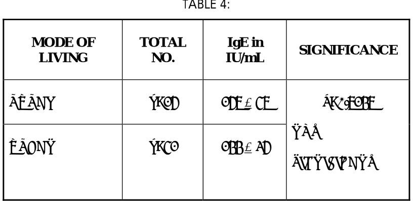
MODE OF LIVING AND IgE LEVELS:
TABLE 4:
MODE OF LIVING
TOTAL NO.
IgE in
IU/mL SIGNIFICANCE
RURAL n=26 267 + 57
URBAN n=52 244 + 36
P=0.7247
NOT
SIGNIFICANT
p value shows 0.7247.
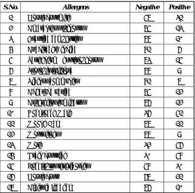
FREQUENCY OF ALLERGEN POSITIVITY:
TABLE 5:
S.No. Allergens Negative Positive
1 House dust mite 57 31
2 Amaranthus spinosus 75 13
3 Brassica campestris 78 10
4 Azadirachta indica 81 6
5 Parthenium hysterophorus 73 15
6 Prosopis juliflora 78 9
7 Ricinus communis 81 7
8 Adhatoda vasica 75 12
9 Aspergilus fumigatus 76 12
10 Cockroach Male 36 51
11 Moong Dal 77 11
12 Mosquitoes 77 9
13 Moth 32 56
14 Grain dust rice 30 58
15 Eucalyptus tereticornis 58 30
16 House dust 67 21
17 Albizzia lebbeck 76 10
This data clearly shows that cockroach, moth and grain dust rice
have shown more positivity than other allergens.
Prosopis juliflora, ricinus communis, adhatoda vasica, aspergillus
GENDER DIFFERENCE IN ALLERGEN POSITIVITY:
The difference was calculated after normalization by gender ratio.
(Difference between allergy positivity was divided by gender ratio). As
[image:43.612.106.507.234.662.2]shown in Table 6, females have more tendency for allergen positivity.
Table 6 – Female/Male ratio for SPT positivity:
Allergen Female/Male
House dust mite 1.7
Amaranthus spinosus 1.3
Brassica campestris 1.3
Azadirachta indica 3.5
Parthenium hysterophorus 2.3
Prosopis juliflora 0.9
Ricinus communis 0.4
Adhatoda vasica 1.7
Aspergilus fumigatus 1.7
Cockroach Male 1.0
Moong Dal 1.0
Mosquitoes 0.7 Moth 0.9
Grain dust rice 0.9
Eucalyptus tereticornis 0.9
House dust 0.9
Females and males showed equal rate of allergy positivity for most
of the allergens. However, females have shown the predominant
positivity for House dust mite, Amaranthus spinosus, Brassica
campestris, Azadirachta indica, Parthenium hysterophorus, Adhatoda
vasica, and Aspergilus fumigatus.
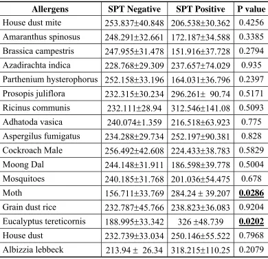
[image:44.612.112.502.295.671.2]ALLERGY POSITIVITY AND IGE LEVELS:
Table 7. Comparison of Total Ig E levels with allergen positivity:
Allergens SPT Negative SPT Positive P value
This table shows p value of 0.0286 for moth and p value of 0.0202
for eucalyptus.both are statistically significant.
SKIN PRICK TEST GRADING AND IGE LEVELS:
HOUSE DUST MITE:
TABLE 8:
S.NO. LEVELS IgE IN IU/mL SIGNIFICANCE
1. 1+ 435 + 166
2. 2+ 122 + 30.13
3. 3+ 328 + 111.43
4. 4+ 406 + 76.91
P=0.2693 NOT SIGNIFICANT
p value =0.2693 .Though 4+ shows higher IgE ,it is not statistically
significant.
AMARANTHUS SPINOSUS:
TABLE 9:
S.NO. LEVELS IgE IN IU/mL SIGNIFICANCE
1. 1+ 145 + 53.87
2. 2+ 362 + 158.08
P=0.2214
NOT SIGNIFICANT
Higher IgE for 2+ skin prick test grading though it is not
BRASSICA CAMPESTRIS:
TABLE 10:
S.NO. LEVELS IgE IN IU/mL SIGNIFICANCE
1. 1+ 131 + 39.74
2. 2+ 524 + 297.81
P=0.0543 SIGNIFICANT
p value =0.0543 .significant difference is there for this allergen.
AZADIRACHTA INDICA:
TABLE 11:
S.NO. LEVELS
IgE
IN
IU/mL
SIGNIFICANCE
1. 1+ 272 + 79.77
2. 2+ 584 + 522.82
3. 3+ 865
P=0.0432 SIGNIFICANT
p value =0.0432 shows significant correlation between skin prick
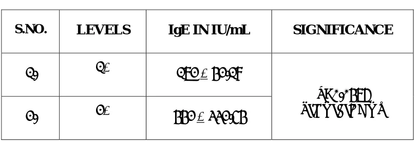
PARTHENIUM HYSTEROPHORUS:
TABLE 12:
S.NO. LEVELS IgE IN IU/mL SIGNIFICANCE
1. 1+ 172 + 42.18
2. 2+ 442 + 332.54
P=0.0476 SIGNIFICANT
p value=0.0476 .It is statistically significant..
PROSOPIS JULIFLORA:
TABLE 13:
S.NO. LEVELS IgE IN IU/mL SIGNIFICANCE
1. 1+ 300 + 89.29
2. 2+ 588 + 518.4
P=0.3288 SIGNIFICANT
Here the p value =0.3288.higher levels of skin prick test Shows
[image:47.612.106.510.376.476.2]TABLE 14:
COCKROACH:
S.NO. LEVELS IgE IN IU/mL SIGNIFICANCE
1. 1+ 336 + 111.25
2. 2+ 288 + 77.5
3. 3+ 288 + 107.43
P=0.9082
NOT SIGNIFICANT
p value =0.9082.it’s not significant statistically.
MOONG DAL:
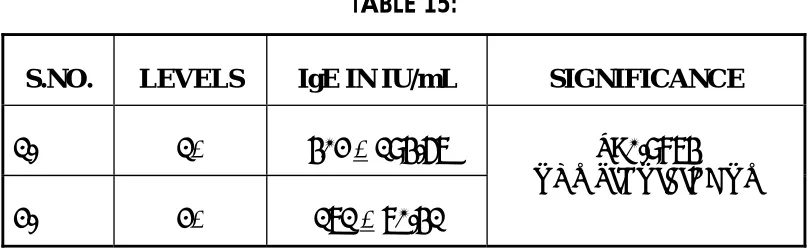
TABLE 15:
S.NO. LEVELS IgE IN IU/mL SIGNIFICANCE
1. 1+ 402 + 194.67
2. 2+ 171 + 70.41
P=0.9874 NOT SIGNIFICANT
Skin prick test grading for moong dal and IgE values are not
[image:48.612.104.510.399.523.2]MOSQUITO:
TABLE 16:
S.NO. LEVELS IgE IN IU/mL SIGNIFICANCE
1. 1+ 406 + 195.43
2. 2+ 240 + 95.65
P=0.7106 NOT SIGNIFICANT
p value =0.7106 .No correlation was found statistically.
MOTH:
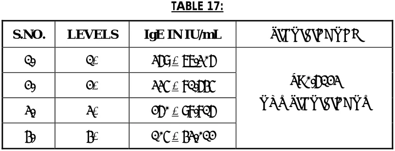
TABLE 17:
S.NO. LEVELS IgE IN IU/mL SIGNIFICANCE
1. 1+ 369 + 77.306
2. 2+ 335 + 81.645
3. 3+ 260 + 98.816
4. 4+ 105 + 43.012
P=0.6123 NOT SIGNIFICANT
p value=0.6123
[image:49.612.103.509.336.492.2]GRAIN DUST RICE:
TABLE 18:
S.NO. LEVELS IgE IN IU/mL SIGNIFICANCE
1. 1+ 332 + 111.94
2. 2+ 256 + 55.04
3. 3+ 276 + 134.39
P=0.8293 NOT SIGNIFICANT
p value=0.8293.Not significant statistically.
[image:50.612.107.509.345.481.2]HOUSE DUST:
TABLE 19:
S.NO. LEVELS IgE IN IU/mL SIGNIFICANCE
1. 1+ 398 + 111.94
2. 2+ 173 + 105.62
P=0.318
NOT SIGNIFICANT
p value =0.318.No significance was found statistically.
ALBIZZIA LEBBECK:
TABLE 20:
S.NO. LEVELS IgE IN IU/mL SIGNIFICANCE
1. 1+ 614 + 259.39
2. 2+ 258 + 134.36
P=0.2314
NOT SIGNIFICANT
[image:50.612.102.509.560.679.2]DISCUSSION
IGE IN PATIENTS AND CONTROLS:
The range of age for Patients and controls were 11 to 65 years. So
as shown in Table I, the IgE levels of patients were higher than normal
control individuals though it is not statistically significant.
GENDER DIFFERENCE IN IGE LEVELS:
Our study shows that males have higher IgE values as compared to
females The p value is 0.1713, which is not significant statistically.
This is really interesting and controversial because many atopic
symptoms are having maternal inheritance. For example, recently a study
has shown that mitochondrial haplotype is associated with IgE levels(45).
Consequently gender variation in IgE levels could be due to the following
mechanisms: 1) Genetic 2) Hormonal and 3) Environmental(46). At the
time of birth, genetic factors have a main role in determining a higher IgE
levels in male rather than female infants (47). In adults, a higher exposure
to environmental factors in males as compared to females could have a
role in causing higher IgE levels in males (48). This is true not only for
the indoor environment like dust mite and cats, with males having higher
specific IgE antibodies for these antigens as well. (49).
ECONOMIC STATUS AND IGE LEVELS:
Our study also shows that people frpm the lower socioeconomic
strata have higher IgE levels ,though not significant statistically.
There was an inverse correlation between IgE levels and economic
status. . However this was not found to be statistically significant. The
reasons for this are not clear, but it could be due to effective disease
management in the higher socioeconomic groups in contrast with an
irregular treatment and a lack of awareness in lower socioeconomic
groups. To illustrate this point it was observed during the course of this
study that poorer patients attended the allergy clinic for treatment only
after a protracted period after the onset of an acute attack, did not avail of
any emergency treatment.
MODE OF HOUSING AND IGE VALUES:
Our study shows no correlation between IgE and the mode of
This is really surprising because there is much evidence to suggest
that pollution and contamination can act as triggering factors for the
development of an asthma attack (50). This could be explained by the
Hygiene hypothesis. The hygiene hypothesis considers that there is an
inverse association between family size and manifestations of atopy in
early life to childhood. (51, 52) .Along with these changing patterns of
microbial exposure, the decline in naturally occurring infectious diseases
and some vaccinations (eg, against tuberculosis), form the background for
this hypothesis
Exposure to microbes, either in the form of active infection or as a
sub clinical infection may initiate protective responses(53). The derivatives
of microbes, which are primarily recognized by the innate immune
system, may drive protective responses, especially at the cytokine level.
This exposure may play a critical role in the shaping of the immune
response when encountered at important stages during the maturation of
immune responses. This could result in the development of immune
tolerance to potential allergens. A major basis for the hypothesis is that
improved hygienic conditions in Western or developed countries results
in less infection-driven or microbial pressure during early but critical time
found in our population. However this needs well regulated study design.
Th1 responses are predominantly mediated by interferon (IFN)-gamma
and interleukin (IL)-12 productions, whereas Th2 responses are primarily
associated with IL-4, IL-5, IL-13 (and IL-10) production (54). In
association with reductions or altered exposures to infectious agents or
their components, it is proposed that Th2 immunity, predominating from
birth, dominates through critical childhood periods, resulting in the higher
incidence of atopy and asthma. Various studies have advanced the theory
that fecal contamination of the environment (and possibly infections such
as hepatitis A), and unhygienic food handling may similarly protect
against development of atopy. However, results from this study are not
sufficient enough to support the hypothesis.
FREQUENCY OF ALLERGEN POSITIVITY:
Our study shows a higher rate of allergen positivity with
cockroach, moth and grain dust rice as compared to other allergens.
In this study, no signs of anaphylaxis was found while performing
SPT. As shown in the Figure, cockroach, moth, and grain dust rice
showed high incidence of allergy positivity. This indicates that outdoor
allergens such as pollens have only a smaller role in causing asthmatic
have a predominant role in determining the prevalence of allergic
symptoms. The cockroach allergy was first recognized in 1960s and
identified as an underlying cause of asthma morbidity (55).
Moreover these results suggest that moth allergy is also a factor of
particular importance in causing allergy-induced bronchial asthma in our
patients. As moths are attracted readily by artificial lights and often fly
into houses, these insects are especially suspect as important factors in
extrinsic asthma (56).
Very importantly, grain dust rice allergy also is of particular
importance in our population, as rice is the staple cereal of this region
with rice being cleaned indoors in many households. Furthermore it is
seen that the prevalence ofthisallergy is almost equal in males as well as
in females as shown in the Figure. This indicates that the allergen may
spread throughout the house during the process of rice cleaning which is
done before cooking rice.
In addition to these major allergens, house dust mite, eucalyptus
tereticornis and house dust are the other important antigens showing
allergen positivity. Among these, house dust mite allergy is unique in that
it is a universal health problem. House dust mite allergy has been
involved predominantly in the development of dust allergy Later it
was observed that mites are the major source of allergens for an asthma
and allergic rhinitis. The two most important species are
Dermatophagoides pteronyssinus and Dermatophagoides farinae. Since
mites live and thrive in many sites throughout the house, they are difficult
to reduce and impossible to eradicate. No single measure is likely to
reduce exposure to mite allergens, and single chemical and physical
methods aimed at reducing mite allergens are not effective in reducing
asthma symptoms in adults. An integrated approach including barrier
methods, dust removal, and reduction of microhabitats favorable to mites
has been suggested, although its efficacy at reducing symptoms has only
been confirmed in deprived populations with a specific environmental
exposure.
Biological agents have an important role in causing indoor air
pollution. The indoor air environment has changed with the introduction
of soft furniture’s, carpets etc. There is an increased indoor relative
humidity and decreased ventilation due to these modernizations. However
due to these changes airborne allergens have increased significantly.
Indoor air is the mixture of physical, chemical and biological
The major biological contaminants are dust mites and their feces,
dander from animals and molds. In this study it is found that predominant
allergens are indoor allergens rather than pollens which are outdoor
allergens. So in contrast to our belief, it is very much essential to control
indoor allergens rather than focus on outdoor allergens in our population.
The relationship between human health and indoor air environment is an
area of growing interest. It needs further detailed investigations.
GENDER DIFFERENCE IN ALLERGEN POSITIVITY:
Females and males showed equal rate of allergy positivity for most
of the allergens. However, females have shown the predominant
positivity for House dust mite, Amaranthus spinosus, Brassica
campestris, Azadirachta indica, Parthenium hysterophorus, Adhatoda
vasica, and Aspergilus fumigatus.
This predominance was very common in various pollens and house
dust mite. House dust mite allergy predominance can be explained by
increased staying time in indoor environment. In contrast, it was not true
for other indoor allergens. But the predominance of pollen allergens in
ALLERGY POSITIVITY AND IGE LEVELS:
To determine the participation in allergy in the production of total
IgE levels, the comparison was done between allergy positivity and total
IgE levels. Only Moth and eucalyptus allergy have shown the significant
correlation between the allergy positivity and total IgE levels. The
reasons for this are not clear at the moment.
SPT GRADING AND IGE LEVELS:
HOUSE DUST MITE:
Higher IgE levels were found with lower grading. However the
levels were not correlated with serial grading of SPT positivity. This
indicates that quantity of allergens entering into the body is not
equivalent to the production of IgE levels. The reason for controversial
relation between allergy grading and IgE levels is not clear. Similar
controversial reports have been found in the dose response relationship
between symptoms and dust mite allergen exposure (59). Marks et al have
found that the concentration of mite allergens is not higher in the houses
AMARANTHUS SPINOSUS:
There was a good correlation between IgE levels and SPT
positivity, that is lower IgE levels were correlated with lower allergy
grading and vice versa .
BRASSICA CAMPESTRIS:
Almost similar correlation which was found with Amaranthus
spinosus was observed with this allergen also.
AZADIRACHTA INDICA:
Surprisingly, the similar trend which was observed in brassica
campestris pollen was found with this allergen.
PARTHENIUM HYSTEROPHORUS AND PROSOPIS JULIFLORA:
It is very interesting that there is a good correlation between allergy
grading and IgE levels in case of pollens. This dose relationship is not
found in literature, undoubtedly it might give various interesting findings
if we explore in detail.
COCKROACH:
Interestingly there was no correlation was found between allergy
grading and IgE levels. A similar relation was observed in house mite
MOONG DAL:
There was an opposite correlation found between in allergy grading
and IgE levels in case of this food allergy. The reasons are not known. It
could be due to tolerance mechanisms. Because this kind of mucosal
tolerance has been found in case of mice model of asthma when chicken
egg ovalbumin has been given orally(61)
MOSQUITO AND MOTH:
Surprisingly, a similar observation which was found with mosquito
allergy was observed in case of moth allergy. It is very interesting that
controversial relationship or inverse correlation has been observed in case
of in case of insect allergies such as house dust mite, mosquito and moth.
This could be due to the following reasons:
1) Tolerance
IMMUNOTOLERANCE:
As shown in the above Figure, high dose allergens are in fact act as a
therapeutic modality (62). When the allergens enter into our body mostly
through mucosal surfaces, innate mucosal immune system, represented by
epithelial and other mucosal cells and their products, is able to recognize
them by pattern recognition receptors such as Toll-like receptors, CD14
and others. These local and systemic immune responses are later replaced
by inhibition, by induction of mucosal (oral) tolerance. Characteristic
players of mucosal immunity are: well developed innate defense
mechanisms, characteristic populations of unique types of lymphocytes,
the mucosal organized tissues ('common mucosal system') and
preferential induction of inhibition of the responses to non dangerous
antigens (mucosal tolerance). Various chronic diseases, including allergy,
may occur as a result of genetically or environmentally induced changes
in mechanisms regulating mucosal immunity and tolerance. These
mechanisms lead to cause the impairment of mucosal barrier function,
disturbed exclusion and increased penetration of microbial, food or
airborne antigens into the circulation and further lead to exaggerated and
generalized immune responses to allergens (63, 64).
Recently, a mammalian homologue of an enzyme associated with
parasitic infection in lower organisms, including amphibians and
insects(65) and (66), has been shown to be expressed in Th2-mediated
inflammation such as asthma (67). Acidic mammalian chitinase (AMCase)
is a 50-kDa protein that contains a 30-kDa N-terminal catalytic domain
that can hydrolyse chitin, a polymer of N-acetylglucosamine (68). Chitin is
not expressed in mammalian systems but is abundant in the structural
coatings of fungi (69), the exoskeleton of many arthropods (70) and parasitic
nematodes (71). AMCase is a member of the glycosyl hydrolase 18 family
GRAIN DUST RICE , HOUSE DUST AND ALBIZZIA LEBBECK:
All these allergens do not show any significance with skin prick
test grading and ige values.The opposite correlation has been found with
house dust allergy. This could be due to the fact that house dust is the
mixture of many biological mixtures including dust mite allergens.
FAMILY ANALYSIS:
Many patients are unaware of the reasons for frequent sneezing.
But if the allergy had been diagnosed earlier, it could be controlled by
avoiding those allergens. For this reason, the relatives of the patients were
recruited for the study. The non-blood related individuals such as spouses
of the probands were taken as normal individuals for the comparison of
IgE levels. Blood related individuals were recruited for the diagnosis of
allergy irrespective of whether they had allergy related symptoms or not.
Our preliminary analysis has shown that 20% of the individuals who had
mild allergic symptoms showed positive allergy for at least one allergens.
10 % of the individuals who did not have any allergy related symptoms
showed positive allergy to at least one allergen. As atopy is a hereditary
disease, it seems that allergy checkup for at least a common allergen for
SUMMARY
The present study is an attempt to understand the immune and
allergenic profiles of atopic asthma patients.
The sample in this study consisted of 90 patients, who visited the
allergy clinic in Stanley Medical College hospital for a period of twelve
months.
This study concentrates mainly on the importance of skin prick test
in bronchial asthma patients. This test reveals the allergy status of the
individual,and this knowledge of the offending allergens can be used to
avoid them.
Our study reveals lesser IgE in female patients compared to male
patients and IgE levels are very high in low socio-economic groups
compared to high socioeconomic groups.
The incidence of allergenicity is more common in females when compared to males.Moth and eucalyptous allergens show significant association between allergy positivity and IgE levels.
Allergens like brassica campestris, Azadirachta indica,parthenium
hysterophorus shows higher IgE values with higher skin prick test
CONCLUSION
1. The allergic status of the individual can be assessed with the help of skin prick test. This information can be used for avoidance of allergens.
2. Female patients have lesser IgE compared to male patients
3. There is an inverse correlation between IgE levels and socio economic status.
4. Cockroach, Moth, and Grain dust rice are shown the high allergy positivity while azadirachta indica, prosopis juliflora, ricinus communis and mosquitoes are shown the lower allergy positivity.
5. Females have more tendency for allergen positivity. This predominance was very common with respect to various pollens and house dust mite.
6. There was a significant association between allergy positivity and IgE levels in case of moth allergy and eucalyptus allergy.
LIMITATIONS AND SUGGESTIONS
1. Skin prick test is a safe procedure, which can be performed
in any allergy clinic with minimum precautions due to the
lesser chance of anaphylaxis.
2. SPT test could help in revealing hidden allergies.
3. Ideally the allergen grading has to be compared with allergen
specific IgE. But for this, we need a purified powdered
allergen.
4. The results are better if we have ruled out the other
conditions which increase the serum IgE levels, like other
parasitic /helminthic infections.
5. The study will be better if we might have compared
pulmonary function test with IgE levels and skin prick test to
1)
Bosquet J, Bosquet PJ, Godard P, Duares J-P.july. 2005 Thepublic health implications of asthma. Bulletin of the World
Health Organisation.83:7/548.
2)
Peters SP.2003, Aug . Heterogeneity in the pathology andtreatment of asthma. Am J Med.18;115 Suppl 3A:49S-54S.
3)
Walker C.1993. The immunology of extrinsic and intrinsicasthma. Agents Actions Suppl.;43:97-106.
4)
Chhabra SK, Gupta CK, Chhabra P, and Rajpal S. 1998.Prevalence of bronchial asthma in schoolchildren in Delhi. J.
Asthma.35:291-296.
5)
Jindal SK, Gupta D, Aggarwal AN, Jindal RC, Singh V .2000.Study of the prevalence of asthma in adults in North India using
a standardized field questionnaire. J. Asthma. 37:345-351
6)
Smith KR (2000) Inaugural article: national burden of diseasein India from indoor air pollution. Proc. Natl. Acad. Sci. U. S.
A. 97:13286-13293
7)
Renwick DS,Connolly MJ.1997 Persistence of atopic effects onairway calibre and bronchial responsiveness in older adults. Age
4:145-8
9)
Bethesda .2007.National Heart LaBI. Global initiative forasthma. Global strategy for asthma management and prevention. Bethesda: National Institutes of Health.
10)
Ghosh B, Sharma S, Nagarkatti R.2003. "Genetics of asthma:current research paving the way for development of
personalized drugs" Ind J. Med Res. 117:185-97.
11)
Burrows B, Lebowitz MD, Barbee RA, Cline MG. 1991Findings before diagnoses of asthma among the elderly in a
longitudinal study of a general population sample. J. Allergy
Clin. Immunol. 88:870-877
12)
Sears MR, Burrows B, Flannery EM, Herbison GP, Hewitt CJ,Holdaway MD. 1991 Relation between airway responsiveness
and serum IgE in children with asthma and in apparently
normal children. N. Engl. J. Med. 325:1067-1071
13)
Marsh DG, Neely JD, Breazeale DR, Ghosh B, Freidhoff LR,Ehrlich-Kautzky E, Schou C, Krishnaswamy G, Beaty TH.
1994 Linkage analysis of IL4 and other chromosome 5q31.1
markers and total serum immunoglobulin E concentrations.



![Enantioselective N heterocyclic carbene catalyzed formal [3+2] cycloaddition using α aroyloxyaldehydes and oxaziridines](data:image/gif;base64,R0lGODlhAQABAIAAAP///wAAACH5BAEAAAAALAAAAAABAAEAAAICRAEAOw==)