A STUDY ON
SEETHA KAZHICHAL
Dissertation submitted to
THE TAMILNADU DR. M.G.R MEDICAL UNIVERSITY
Chennai-32
For the partial fulfillment of the requirements to the Degree of
DOCTOR OF MEDICINE (SIDDHA)
(Branch IV - Kuzhanthai Maruthuvam)
Department of Kuzhanthai Maruthuvam
GOVERNMENT SIDDHA MEDICAL COLLEGE
PALAYAMKOTTAI – 627 002.
MARCH 2008
CONTENTS
Page. No
INTRODUCTION
1
AIM
AND
OBJECTIVES
3
REVIEW OF SIDDHA LITERATURES
5
REVIEW
OF
MODERN
LITERATURES
25
MATERIALS
AND
METHODS
44
RESULTS
AND
OBSERVATIONS
46
DISCUSSION
67
SUMMARY
73
CONCLUSION
75
ANNEXURES
¾
PREPARATION AND PROPERTIES OF TRIAL MEDICINE
76
¾
BIOCHEMICAL ANALYSIS OF TRIAL MEDICINE
81
¾
ANTI MICROBIAL STUDY OF TRIAL MEDICINE
84
¾
PHARMACOLOGICAL ANALYSIS OF TRIAL MEDICINE
85
¾
LABORATORY DIAGNOSIS OF SHIGELLA AND ENTAMOEBA
HISTOLYTICA
97
¾
PROFORMA OF CASE SHEET
102
CERTIFICATE
Certified that I have gone through the dissertation submitted by
---a student of Fin---al M.D.(s), Br---anch IV, Kuzh---andh---ai M---aruthuv---am of this college ---and
the dissertation does not represent or reproduce the dissertation submitted and
approved earlier.
Place: Palayamkottai
Date: Professor and
Head of the Department, (PG)
Br IV, Kuzhanthai Maruthuvam,
Govt. Siddha Medical College,
ACKNOWLEDGEMENT
First of all, I thank God for his blessings to complete my dissertation
work successfully.
My foremost thanks to The Vice Chancellor, The Tamil Nadu Dr.M.G.R.Medical University-Chennai , for giving permission to undertake this dissertation.
I contribute my thanks to Dr.M.Thinakaran M.D(s)., The Principal,Govt. Siddha Medical College, Palayamkottai for granting permission
and facilities to complete this dissertation.
I owe a deep sense of gratitude to Dr.R.Patrayan M.D (s)., The Head of the department , Dr.N.Chandra Mohan M.D (s)., Lecturer, Department of Kuzhanthai Maruthuvam , Govt.Siddha Medical College, Palaymkottai for their
encouragements , valuable suggestions and necessary guidance during this
study.
I take immense pleasure in thanking Dr.P.Sivagami M.D(s)., Lecturer, Department of Magalir and sool Maruthuvam, G.S.M.C,Palayamkottai for
their valuable opinions regarding this study.
I am grateful to Dr.Kadhir Subramaniam M.D.,D.C.H., Professor and Head of the Department, Dr.M. Mathivanan M.D., D.C.H., Asst.Professor, Dept. of Pediatrics, Tirunelveli Medical College, Palayamkottai for their
advice during the dissertation period.
The genuine interest shown by Mr.Kalaivanan M.Sc., M.Phil., Lecturer and staffs of the Dept. of Pharmacology in carrying out the
My sincere thanks to Prof. Mrs. N.Naga Prema M.Sc.,M.Phil., H.O.D and the Technical experts of Dept. of Bio-Chemistry for their help in Bio
chemical analysis of the trial medicine.
My sincere thanks to Dr.Napolean B.Sc., M.D., Microbiologist, Malar diagnostic centre , palayamkottai for helping me to carryout the culture studies
and anti microbial assay of the trial medicine.
INTRODUCTION
Medicine is an art of fundamental importance to the healthy
survival of humanity . Siddha, one of the ancient system of medicine has got a
holistic history of origin . Being a science of life, it helps the world by not only
giving solutions to health problems but also by paving the way to attain the
ultimate aim of the life.
The word "Siddha" comes from 'Siddhi' which means perfection or healthy bliss. It generally refers to the Astamaa siddhi i.e, the eight
supernatural powers. Those who attained these powers are known as the
siddhars. The basic principle of siddha system is 96 Thathuvas of which
panchapootha theory and Mukkutra theory are very important.
The Universe is composed of five elements viz.,
Earth,Water,Fire,Air,Ether (Mann, Neer, Neruppu, Kaatru and Aakayam). The
human anatomy, physiology, pathology of disease, materials for the
treatment and the food for sustenance all fall with in the five elemental
categories.
The pathology in siddha system depends upon the Mukkutra theory
viz., vatha ,pitha and kaba.The normal order of vatha , pitha, kaba is in
proportion of 1 : 1/2 : 1/4 respectively.
This is stated in the following verses.
“ Upr<gqb!uikl<!lik<kqjv!obie<xigqz<!
!!!!!!kpr<gqb!hqk<kf<!ke<eq!zjvuisq!
!!!!!!npr<Gr<!ghf<kiemr<gqOb!giOzicz<!
!!!!!!hqxr<gqb!sQui<g<Gh<!hqsogie<X!lqz<jzOb
”
Imbalance results in disease.
This can be inferred from the following Thirukkural,
“lqgqEl<!GjxbqEl<!Ofib<!osb<Bl<!FiOzii<!
!utq!Lkzi!w{<{qb!&e<X/
”
-
kqVut<Tui</
The clinical methods through which the correct diagnosis made out
are Envagai thervugal. They are Naadi, sparissam, Naa, Niram, Mozhi, Vizhi,
Malam, and Moothiram.
Kuzhandhai Maruthuvam is a branch of medical science of siddhars,
which deals with the diseases and treatment of child. In Kzhandhai
Maruthuvam, the diseases of children are broadly classified into Agakarana
Noigal and Purakarana Noigal.
SEETHA KAZHICHAL,one of the three kazhichal noigal occuring in infants and children due to varied Aetiology is one among the health
hazards, that a society faces frequently. The Aetiological factors (In take of
improperly cooked food stuffs, Drinking impure water,living in over crowded
areas), clinical features of the disease (Bloody mucoid stools,abdominal
pain,fever,painful defaecation) explained in the siddha literature are more or
less related to Amoebic and Bacillary dysentry described in modern system of
Medicine.
AIM AND OBJECTIVES
Seetha kazhichal, of which the signs and symptoms are related to
dysentery in modern aspect is a major health hazard in the developing
countries like India. It forms one of the major causes of sickness among
infants and children, which causes a heavy economic burden on health
services.
India is a country, having large population in the world, where people
of different socio economic status are found. Poor children who live in densely
areas with poor sanitary facilities, lack of personal and environmental hygiene
are the common victims of this disease. If proper attention has not been
given, it may lead to many complications like dehydration, Rectal
prolapse,Septicaemia etc.,
Objectives:
To explore most efficacious drug for seetha kazhichal.
To have a clinical trial on seetha kazhichal affected children with Atthi Pinju chooranam.
To evaluate the disease seetha kazhichal clinically by careful
examination on aetiology, clinical features, differential diagnosis,
investigations, diagnosis, treatment, diet, prognosis, complications etc.
To collect the literary evidences regarding the disease seetha
kazhichal as per siddha system.
To make comparative study of this disease with morden
To evaluate Biochemical and pharmocological analysis of the drug.
To evaluate efficacy of trial medicine on anti microbial activity by invitro
studies.
Control of disease by creating awareness of proper hygiene.
Being a herbal preparation, Trial medicine is safe and drugs are easily
REVIEW OF SIDDHA LITERATURE
Eyal (Definition):
ubqX!gMk<K!ncg<gc!sqxqK!sqxqkiObEl<!nz<zK!ubqx<Xg<!gMh<H!
nkqglqe<xq!ntU!gmf<OkEl<?!sQkg<gm<Ml<!GVkqBl<!%cObEl<!gpqBl</
!Seetha kazhichal means the dysentery due to specific inflammation
and ulceration of the mucus lining of large intestine resulting in evacuation of
stools mixed with mucus and blood (T.V. sambasivam pillai. 1978).
Verupeyarkal (synonyms):
¾ Amakazhichal,
¾ Seethapethy
¾ Kadduppu kazhichal,
¾ Seetharathapethy
¾ Vayettru kaduppu,
¾ Vayettrulaivu
¾ Seetha ratha Kazhichal
¾ Seetha Kaduppu
¾ Ratha Kaduppu
¾ Ama pethy
¾ Amaratha pethy
¾ Giragani
¾ Girani
¾ Seetha athisaram
¾ Seetha ratha girani
¾ Kuzhanthai seetha pethy
¾ Vayettru kottal
gpqs<sz<!Ofib<!ujggt
<!
(Classification):
!
“Seetha Kazhichal” is a disease which occurs both in children and adults. It has been described as one of the Kazhichal noi in various Siddhaliteratures.
In Kuzhandhai Maruthuvam, it is classified under Kazhichal vaguppu,
where as it has been described separately in siddha maruthuvam.
Various classifications of kazhichal noi, which have been described in
several siddha texts, are given below,
1. In Kuzhandhai Maruthuvam three types of Kazhichal noikal have been
described
i. Mantha Kazhichal
ii. Kana Kazhichal
iii. Ama Kazhichal (Seetha Pethy)
At the same time,
i. Veppu Kazhichal
ii. Raththa Kazhichal
iii. Athisara Kazhichal
iv. Kaduppu Kazhichal
v. Porumal Kazhichal
vi. Pachilai Kazhichal
vii. Vidaa Kazhichal
have also been mentioned in the treatment of Kazhichal noikal in
2. In T.V.Sambasivam pillai dictionary, the following Kazhichal noikal have
been mentioned.
i. Seetha Kazhichal (passage of mucus)
ii. Raththa Kazhichal (passage of Blood)
iii. Sala Kazhichal (watery diarrhoea)
iv. Soba Kazhichal (Diarrhoea with great weakness and exhaustion)
v. Vaeludai Kazhichal (white diarrhoea)
vi. Vayettu Kazhichal (gastrogenic diarrhoea)
vii. Sangara Kazhichal (diarrhoea with various symptoms)
3. In Jeeva Rakshamirtham, the following Kazhichal noikal are given
i. Raththa Kazhichal
ii. Sala Kazhichal
4. Two types of “Kazhichal” have been described in pararaja sekaram
balaroga nidhanam
i. Vayettru Kaduppu
ii. Vayettrulaivu
5. In Athma rakshamirutham also called Vaidya Sara Sangirakam fifteen
types of Kazhichal noikal have been classified.
“osiz<ZgqOxe<!gpqs<sz<ujg!Oki]f<ke<je!
!!!!!!!Spqlif<k!gpqs<sozes<!osh<hziGl<!
!!!!!ouz<ZgqOxe<!hix<gpqs<sz<!uvm<gpqs<sz<!
!!!!!!!uQxie!uif<kqbqe<xe<!gpqs<sziGl<!
!!!!!Hz<ZgqOxe<!g{g<gpqs<sz<!lif<kg<gpqs<sz<!
!!!!!!!Hgpie!Nlk<kqe<!gpqs<sziGl<!
!ogiz<Zgqe<x!szg<gpqs<sz<!ouKh<Hg<gpqs<sz<!
!!!!!!!%xie!vk<kk<kqe<!gpqs<sziOl”
!!!!!!!nh<hOe!ohiVlzqe<!gpqs<sziGl<!
!OhiOlkie<!sQvk<kg<!gMh<HuiGl<!
!!!ohiz<zik!gpqs<soze<X!filolb<Kl<!
!kiOlkie<!hs<sqjzg<!gpqs<sziGl<!
!!!!!!!!sii<uie!uqmg<gpqs<sz<!six<xziGl<!
!fiOlkie<!osie<OeiOl!gpqs<sz<!lii<g<gl<!
!!!!!!fuqe<xqm<mii<!hizVg<G!fuqe<xqm<miOv”!
i. Suzhimantha Kazhichal
ii. Paal Kazhichal
iii. Varat Kazhichal
iv. Vaanthi Kazhichal
v. Kana Kazhichal
vi. Maantha Kazhichal
vii. Ama Kazhichal
viii. Sala Kazhichal
ix. Vethuppu Kazhichal
x. Raththa Kazhichal
xi. Athisara Kazhichal
xii. Porumal Kazhichal
xiii. Raththa Kaduppu
xiv. Pachilai Kazhichal
xv. Vida Kazhichal
6. In Noi nidhanankal, ten types of Kazhichal noikal are given
i. Moola Kazhichal
ii. Vadha girani
iii. Pitta girani
iv. Seetha girani
v. Vatha pitta girani
vi. Pitta Sethuma girani
vii. Vatha Seetha girani
viii. Thontha girani
ix. Vayettru Kaduppu
7. According to Agathiyar vaidya Kaavium 1500, Kazhichal is classified in to
six types.
“gpqs<soze<x!gqvi{qbqOz!uqklixh<hi!
!
!!
!g{<m!hqk<kl<!nez<!uikl<!uiBuiGl<!
!
!!!!npqs<soze<x!JbfQI!&e<Xr<%c!!
!
!
!nh<hOe!Ohkqg<Gl<!hzf<kie<!OhiGl<!
!
!!!!okpqs<soze<x!uiBkie<!OlgOhkq!
!
!
!kqxlie!&zk<kqe<!OkimOhkq!
!
!!!!hpqs<soze<x!sr<gie!Ohkqobie<X!
!
!
!hivh<hi!uiBouie<X!NXlis<Os”
i. Vatha Kazhical
ii. Pitta Kazhical
iii. Kaba Kazhical
iv. Moola Kazhical
v. Sangana Kazhical
vi. Mega Kazhical
8.Same classification has been given in Thirumoolar Vaidhyam”
Karukkidai 600.
“gpqs<sz<!gqvi{q!gi[l<!uqkl<!OgT!
!
!
!!npqs<sqb!hqk<k!l{z<!uik!jlblil<!
!!!!!osPs<sqb!uiB!OsIf<kqju!&e<xiOz!
!
!!!!!!hpqs<ose!Ohkqg<Gl<!hiI!ohzl<!OhiGOl”!
“ohzlie!Olgk<kqx<!hqxf<kokiV!Ohkq!
!
!
!!Gzlie!&zk<kqx<!ogicbokiV!Ohkq!
!!
!
!Sglie!uiBuix<!sr<gqk<okiV!Ohkq!
!
!
!!UzliekiXl<!uGk<k!LjxbiOl”
From the above, many authors describe the types of Kazhical noikal.
Noi varum Vazhi (Etiology):
The causes for seethakazhichal mentioned in various siddha texts are
follows,
i.Intake of food stuffs which are not easily digestable.
ii.Intake of excessive pungent and sour tasted food stuffs.
iii.Taking large amount of sweets, mutton and improperly cooked foodstuffs.
iv.Drinking impure water like sunaineer and karchunna neer.
v.Wandering in hotsun and exposure to cold air.
vi.Living in over crowded areas.
vii.Suffering from seetha suram.
viii.Improper treatment for “Athisara Noi”
The above mentioned causes are stated in the following verses.
“lioee<x!ubqx<xqz<!lf<klqVg<Gl<!OhiK!
!!lih<h{<m!lKvr<gt<!lr<jg!Ogi]<c!
!
!!
Doee<x!lilqsr<gt<!Ougih<h{<ml<!
!
!
!!d{<mkix<!gqvi{q!uf<Kx<huqg<Gr<!g{<mib<”!!
!!!!!!!
!
.!B,gqsqf<kil{q!
“kieig!d{<miGl<!uqkk<jkg<!Ogtib<!
!
!
!!kv{qkeqx<!GtqIs<sqBme<!uqmsk<Kk<kiEl<!
Okeig!lqGkQeq!Hsqk<kiZl<!
!
!
!!kqv{<m!seg<%m<mk<kqz<!OhiukiZl<!
lieie!sQkSvr<!gi[l<OhiKl<!
!
!
!!lgk<kie!-f<OfiB{<mi!ole<X!
Ogi{ie!Fiz<keqOz!ohiqObii<!osie<eii<!
!
!
!!ogix<xuOe!bkqEjmb!G{k<jkg<!OgOt”!
“Guru naadi Nool” explains the causative organism and the
pathogenesis of the disease.
“OgTlqeqg<!gqVlqbiz<!uf<k!gqvi{qjbk<kie<!
!
!
!!gqVjhBme<!&zk<kqz<!OuU!ogi{<M!
fiTlK!gqVlqbkqe<!Gmjzs<!Sx<xq!
!
!
!!vk<k!L{<miR<!SOvi{qkk<kiz<!lzLr<gm<c!
lQTuK!uib<U!ose<X!uqvuqk<kiEl<!
!
!
!!uqvuqbr<Og!gzf<kqVg<gqz<!gqVlqobz<zil<!
OgTlK!hzuqklib<g<!gpqBl<!hizi<!
!
!
!!Gc!ogMk<k!gqVlq!osb<k!gqvi{qkiOe”!
Due to excessive heat the pathogenic micro organisms (Kirumigal)
multiplies in large numbers in the intestine. They make the stools dry,
decomposed and producing foul smelling gases (vayu). Then it produces
Kazhichal.
Murkuri Gunangal (premonitory symptoms):
Head ache, nausea, pain in the abdomen, burning sensation in the
anus, tenesmus due to increased peristalitic movement are the symptoms
produced in the initial stage of the disease.
Pothukuri Gunangal (General Signs and Symptoms):
Following the premonitory symptoms, there is passing of loose stools
containing small amounts of mucus and blood, pain in the abdomen and
burning sensation in the anal region are aggravated.
Besides passing of mucus and blood, frequent scanty stools are
present. During that time intense abdominal pain is observed. Due to severe
pain, the patient will be always in sitting posture. The patient may pass loose
stools many times in a day. If it is not controlled by proper treatment the
tongue becomes dry, and symptoms of muppini will occur and may be fatal.
The above mentioned features are stated in ‘siddha maruthuvam’.
The following symptoms and signs occur in vayettru kaduppu.
-Mh<Hg<!gMk<K!ubqXjtk<kqm<ctgqs<!sQklix<xQf<K!
LMg<gqg<!KbvLme<!&zf<Okie<xq!lzLl<!gpqf<kqVg<Gl<!
nMk<Okive<e!lVuVg<Gl<!lxOu!br<g!olzqf<K!uVl<!
okiMg<Gl<<ubqx<Xg<gMh<ohe<X!osiz<Zr<!G{r<gtqjubiOl”!
!
!
!
!
!
.!hvvisOsgvl<!)hizOvig!fqkiel<*!
Patient have gripping pain in the lower abdomen, with irritation in and
around the anal region, rectal tenesmus with loose stools, poor appetite and
weakness of the body due to excessive blood loss in stools.
The same features have been described in Agathiyar 2000
“-Mh<Hg<!gMk<K!ubqXjtf<K!-tGs<!sQklix<xQf<K!
LMgg<Gk<kq!Lg<gq!KbvLlib<!d{<{i!lzOl!gpqf<kmr<Gl<!
!!
nMk<Okive<ef<!kjef<Okmi!kxOu!olzqf<K!uVf<okiMg<Gl<!
!!
ubqx<Xg<!gMh<ohe<X!osie<Oeif<!osb<Bl<!Kbi<g{<Om”!
.ngk<kqbi<!3111
!The following Kurigunangal have been described for “Vayettru ulaivu”
“uVf<kqMl<!ouKh<Hg<giBl<!ubqXjtf<kqMf<kQe<osz<zi!
!!!!Kvk<kqM!Lxr<g!ouim<miKt!lzr<gpqf<K!OsiVl<!
!!!!ohiVk<okzir<!gpZl<!H{<Ohix<!ohiVg<ogi{i!fMg<gr<%xz<!
!!!!ohiVk<kqMr<!gpqs<sz<sQkl<!ouXubqx<XjtuqkiOl”!
-hvvisOsgvl<!)hizOvig!fqkiel*<!
Patient is having fever with abdominal pain, loss of appetite, loose
motion with mucus, general weakness and shivering.
In chronic stage, there is regurgitation of milk and anaemia, fever,
“d{<mhi!ozkqovMg<Gl<!dmz<hz!Lpg<gr<!gim<Ml<!
!!!!g{<MOl!vk<k!sizs<!Svlqgqf<kqVg<Gl<!Oleq!
!!!!g{<MOsi<!olipqBf<!kip<f<K!gioziM!jgBfQk<K!
!!!!uq{<cc!ziole<X!uqtl<hqei<!Lequi<!kiOe”!
.hizuigml<!
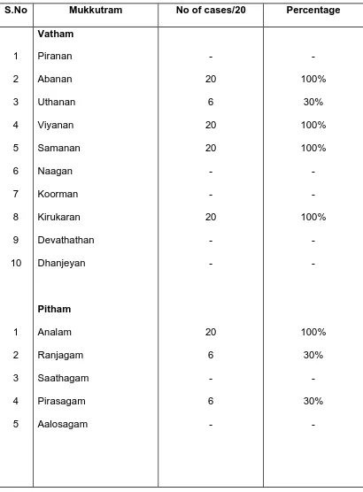
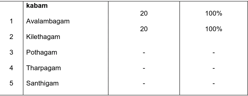
MUKKUTRA VERUPADUGAL (PATHOLOGY)
According to siddha system of medicine, diseases are produced due to
derangements in Thridoshas (i.e) Vatham, pitham and kabam.
The siddha concepts of pathology of Seetha kazhichal have been
described in ‘Thirumoolar Vaidhyam’ karukkidai 600.
“gpqs<sz<!gqvi{q!gi[l<!uqkl<OgT!
!
!!npqs<sqb!hqk<kl<!njzuikl<!Jblil<!
!
!osPs<sqb!uiB!Osi<f<kju!&e<xiz<!
!
!!!hpqs<ose!Ohkqg<Gl<!hii<!ohzl<!OhiGOl”!
!
!
!
!
!
.!kqV&zi<!juk<kqbl<!gVg<gqjm!711
According to siddha system of medicine, diseases are produced due to
derangement in in Thridoshas (i.e.,) Vatham, Pitham and Kabam.
In “Seetha Kazhichal” due to various causes stated above, the pitha
kuttram is vitiated from its normal condition. This in turn stimulates Abanan, a
type vatha. Also, chenneer (blood) and kaba kuttram are affected.
Vitated pitham along with kabam causes ulceration in the intestine and
produces passage of loose stools with blood and mucus
Pain in the abdomen and tenesmus are produced mainly due to vitiated
vayu. Finally all the trithathus are deranged from their normal positions and
Piniyari Muraimai (Diagnosis):
In siddha medicine, diagnosis of a disease is made up on the following
principles.
1. Poriyaal arithal (Inspection)
2. Vinaathal (Interrogation)
3. Pulanaal arithal (Palpation)
Pori are the five organs of perception namely nose, tongue, eyes, ears
and skin.
Pulan are the five objects of senses namely smell, taste, sight,
sensation and sound.
Poriyaal arithal and pulanaal arithal goes hand in hand with concept of
examing the patient’s pori and pulan with that of the physician’s pori and
pulan.
By Vinaathal, the physician knows about the patient’s name, age,
native place, socio economic status, family history, dietetic habits etc. If it is
infant or child or unable to talk (deaf and dumb and in other diseased
conditions) the particulars are obtained from his/her relatives or parents i.e,
informer.
Poriyaalarithal, pulanaalarithal and vinaathal are effected through eight
special methods of investigation (Envagai Thervugal)
Envagai Thervugal:
Envagai Thervugal is considered to be physician’s instruments.
“fic!hiqsl<!fi!fqxl<!olipq!uqpq!
!
!!!lzl<!&k<kqvlqju!lVk<KuviBkl<”!
!
.!Okjvbi<!
Naadi (Pulse)
Sparisam (Palpation)
Niram (Colour of Skin)
Mozhi (Speech)
Vizhi (Eyes)
Malam (Stools)
Moothiram (Urine)
Naadi (Pulse):
Naadi is an important observation for diagnosis and prognosis. Naadi is
responsible for the existence of life and can be felt one inch below the wrist on
the radial side by means of palpation with the tips of index, middle and ring
finger corresponding to vatham, pitham and kabam.
Normally the three humors vatham, pitham and kabam exist in the ratio
1: ½: ¼.
Derangement in these ratio leads to various disease entities and is
best diagnosed by feeling the naadi.
Naddi nadai in Seethakazhichal:
“kjph<hie!hqk<kk<kqZ]<{r<!ogi{<miz<!
!
!!]bl<!nk<kqSvl<!ouKh<Hs<!sk<kq!Ge<ll<!!
!
!gjph<hie!ohiXk<KjtU!nkqsivr<gt<!
!
!!gMh<HmOe!ubqx<Xuzq!&zuiB!
!
!-jth<higq!B,{<lXk<kz<!fig<gsh<H!
!
!!-vuqz<!geUmOe!sr<giv!Okiml<!
ujph<hie!hbqk<kqbOfi!obiqU!kigl<!
!
!!uf<k[gqz<!hzhq{qg<Gl<!ujgbkiOl”!
!
!
!
!
!!!!!!!!!!!!.!skgfic!
“okif<kqk<k!sqOzx<hek<kqz<!uib<U!%ck<!
!
!Kmi<f<k!Ge<ll<!ofR<sjmh<H!Suisgisl<!
!!!uf<kqk<k!Gvz<!keqOz!UXk<kzQjt!
!
!uPuPh<H!fQVxz<!lzk<kqz<!sQkl<!
!!!ouf<kqvkl<!ogiPk<kz<!Gk<Kk<!kqlqi<uqbikq!
!
!uQs<SOmOe!uzqobm<Mf<!kqvm<js!hi{<M!
!!!nf<kqk<k!GXGXh<H!lbg<gl<!uqg<gz<!
!
!Nehz!hq{qBl<!uf<kmXf<!kiOe”!
!
!
!
!
!!!!!!!!!!.!skgfic!
Thonthamana kabam with vayu produces motion mixed with mucus.
Naadi Nadai for Grani is also responsible for “Seetha Kazhichal”
“sqxh<hie!hqk<kk<kqz<!uik!fic!
!!!!!!!!!OsiqZXf<kiK!fm<mLkv!hQjm!
!
!djxh<higs<!osiqbijlg<Ge<!lR<$jz!!
!!
!!!!Bx<x!Svr<gqvi{q!ubqx<xqjvs<sz<!lf<kl<!
!!!! !njxh<hie!Yr<giv!HxfQi<<g<Ogiju!
!
!!! Nbis!lqvg<g!oliM!lbg<g!&i<s<js!!
!
!Ljxg<gib<U!uq]!uQg<gl<!&zuib<U!
!!!!!!
Lvmie!Ofib<!hzU!LMGl<!h{<Oh”!
.skgfic!
In pitha vatham Naadi, Grani is produced.
When there is aggravated vatha naadi the disease Grani is produced.
“uikolEl<!ficbK!Okie<xqz<!ouh<H!
sQklf<koliM!ubqX!ohiVlz<!kqvm<sq!uib<U!
!!!!sQkLXr<gqvi{q!lOgikvl<!fQvijl!
!!!!!kqvt<!uib<U!$jz!uzqgMh<Hk<!kQjv!
!!!!fQkLXr<gq!VlqGe<ll<!n{<muikl<!
!!!!!fqjzBl<!fQi<g<gqiqs<svr<gt<!kf<K!Olgl<!
!!!!Ohkgli!Lkvhq{q!&zOvigl<!
!!!!!Ohs!ouGhq{qgTOl!ohiVtkiOl”!
Sparisam (Palpation):
By sparisam, the temperature of skin (heat or cold), smoothness,
roughness, Hardness, sweat, dryness, swelling, tenderness, ulcers, and
pigmentation can be examined.
In “Seetha Kazhichal” dryness of the body, raised body temperature,
tenderness in the abdomen, sometimes liver enlargement is present.
Naa (Tongue):
In the examination of tongue, colour, coating, wetness, or dryness,
deviation movements, fissures, variation in taste, condition of teeth and gums
are carefully noted.
In “Seetha Kazhichal” coated tongue shows loss of appetite and
indigestion.
Niram (Colour):
Colours indicating vatham, pitham, kabam and thridhosas, cyanosis,
pallor, yellowish, discoloration of the body are noted.
In “Seetha Kazhichal” pallor of the body is present.
Mozhi (Speech):
In the examination of mozhi, the pitch of voice (high or low), laughing,
slurring, speech in hallucination, crying, breathlessness or wheezing and
incompleteness while talking may be noted.
In seetha kazhichal mozhi may be affected.
Vizhi (Eye):
Both sensory and motor disturbances are noted. Colour, inflammation,
ulceration, lacrimation, sharpness of vision, response of the pupil to light may
In the case of seetha kazhichal, sunken eyes and pallor of eyes
sometimes noted.
Malam (Faeces):
In the examination of malam, Niram (Colour) Nurai (froth), Erugal
(Solid) Elagal (Semi solid or liquid), quantity (increased or decreased), smell
can be noted. Other examinations like presence of blood, mucus, undigested
matter in the stools and odour can also be noted.
In “seetha kazhichal” the malam may be liquid or semisolid, Bulky or
scanty in quantity, bright red or dark brown in colour, sometimes gives
offensive odour containing mucus and blood.
Moothiram (Urine):
In the examination of Urine, colour, odour, quantity of Urine, the
presence of froth, deposits, blood, and pus, abnormal constituents such as
sugar, protein etc. and frequency of urination can be noted.
In “Seetha Kazhichal”, the quantity is slightly diminished and yellow in
colour.
Neerkuri:
“uf<k!fQIg<!giqobjm!l{l<!Fjv!wR<soze!
!jxf<kqb!Ztju!bjxGK!LjxOb”!!
–sqk<k!lVk<Kuir<gs<SVg<gl<!
!
According to this verse, the general features of urine are niram, edai,
manam, nurai and enjal.
1 Niram indicates the colour of the urine voided
2 Edai indicates the specific gravity of the urine.
4 Nurai indicates the frothy nature of urine voided.
5 Enjal indicates the quantity of urine.
Collection of urine for Neikuri:
“nVf<Kli!xqvkLl<!nuqOvi!klkib<!!
!!!!!!n0gz<!nzIkz<!ngizU,{<!kuqIf<kpx<!
!Gx<xtuVf<kq!dxr<gq!jugjx!
!!Ncg<gzsk<!kiuqOb!giK!ohb<!
!okiVL%i<k<kg<!gjzg<Gm<hM!fQiqe<!
!!!!!!!fqxg<Gxq!ofb<g<Gxq!fqVlqk<kz<!gmOe”!
.Okjvbi<!
Prior to the day of examination, the patient is asked to take a regular
and balanced diet without any derangement in amount and quality. The
patient is allowed to have a good sleep. In the next early morning, the urine
first voided is collected in a glass container for analysis.
The analysis should be carried out in one and half hours.
A drop of gingelly oil is dropped into a wide vessel containing the urine
and is kept in the bright light in a calm place without shaking. The
derangement of three thathus is studied by nature of oil on the surface of
urine.
“ nvoue!fQ{<cce<!n0Ok!uikl<!
!NpqOhix<!hvuqe<!n0Ok!hqk<kl<!
!Lk<okik<K!fqx<gqe<!olipquke<!ghOl”!
!
!
!
!
!
!
!
.!Ofib<!fimz<!Lkz<!higl<!
Oil spreading like snake indicates Vatham.
Oil spreading like a ring indicates pitham.
Complications:
“d{<miGl<!Ohkqkie<!dg<gqvlib<g<!g{<miz<!
!!dk<klOe!GmZg<Gt<!Kuivr<!g{<M!
!fe<xie!Gmz<!su<Uk<!kihqkOl!g{<M!
!!fqzlie!=vzqz<kie<!sQg<gm<c!ogit<Tl<!
!h{<mie!-v{Lzi<f<K!Gmx<SVr<gq!eig<giz<!
!!htqs<ose<X!lzhf<kl<!d{<mi!lh<hi!
!sq{<mie!sqOzm<Mls<!su<U!nPgqh<!Ohieiz<!
!!sqxh<HmOe!Svh<Hg<!g{<M!-xh<hie<!kiOe”!
!!
.!ngk<kqbi<!G{uigml<!
From the above verses, it is clear that severe bedhi leads to perforation
and inflammation of the colon, liver abscess, constipation and obstruction.
Sometimes it may end fatally.
“hi{<M!hqvOlgl<!he<uik!$jzGe<ll!
!Ou{<mi!
]bR<se<eq!ou{<Osijh.!fQ{<m
!
!nkqfQOv!gilijz!biehq{q!kl<L!
!ekqsivli!gikxq”
.g{<[silqbl<!
If the above diseases are associated with Grani it may lead to a fatal
outcome.
“sf<kq!uqmOsijhsii<!Ge<ll<!fQiqpqU!
!Ke<Er<!gqvi{q!Svl<!Ohkq!!he<Ehqv!
!Olgl<!sblqux<Xt<!&s<S!uqg<gz<!Olz<uQg<gl<!
!NgqZbqi<!Ohilxq”
!!
!
!
!
!
.!g{<[silqbl<
If the Grani is associated with dropsy, hiccup, dyspnoea, it would be
Prognosis:
“Seetha Kazhichal” is a curable one with proper medicine at proper
time. If it is not treated with proper medicine, it leads to severe discomfort,
ulceration of colon causing passage of excessive amount of blood and mucus.
Pulse appears weak,eyes become sunken and there is dryness of tongue.
Pallor of the body due to excessive loss of blood which leads to muppini.
Finally end in fatal condition. (Shanmugavelu 1988, kuppusamy mudaliar
1987)
Ofib<g<!g{q<h<H!uquikl<!
(Differential diagnosis):
Seetha kazhichal should be differentiated from other Kazhichal noikal.
They are
1) Maantha kazhichal:
“uif<kq!hqvif<kq!&i<sjsbkib<!uib<f<K!GvZR<!sQ{qk<Kg<!
!!gib<f<K!Oleq!ouKouKh<hib<g<!jggiz<!Gtqi<f<K!uzqB{<mil<!
!!Osi<f<K!gpqB!lzf<kiEl<!sQi<ogm<cVg<Gl<!hzuqklib<h<!
!!Ohif<k!lif<kg<!gpqs<szqK!ohiz<ziokeOu!Hge<xeOv”!
!!
!
!
!!!!!!!!!!!!!!!!!!.hizuigml<!
In maantha kazhichal following symptoms and signs were seen.
Vomiting, loss of consciousness, hoarseness of voice, dryness of skin,
fever, coldness of limbs, convulsions and different types of loose stools.
2)Kanakazhichal:
“sQkr<gpqb!lzr<gpqBl<!kqVl<hqg<!ogm<m!hiz<!OhiOz!
!Ohikg<!gpqBr<!gxqk<!k{<{Qi<!OhiZr<!jgBr<!giz<!Gtqi<f<K!
!gijkbjmg<Gl<!ouKh<H{<mil<!jgbqx<hqt<jt!kr<giK!
!Ogikibqf<kg<!G{r<g{<miz<!GzUlqke<Ohi<!gpqg{Ol”!
.!hizuigml<!
In kana kazhichal the following signs and symptoms may be present.
Stools may be mucus or bulky or curdy milk or curry water. Coldness of the
hands and legs, deafness, fever, restlessness.
Seetha kazhichal should also be differentiated from vatha kazhichal,
pitha kazhichal, kaba kazhichal, mukkutra kazhichal and oozhi noi.
Maruthuvam (Treatment):
&e<xqozie<X!bi<f<kjk!Le<evxqf<K!
Lf<kqbkje!obipqk<kqM!lVf<kqM!
k{qBl<!Ofibqe<!kf<kqvlqKOu!
Oh{qg<!g{qk<kqce<!hqxuib<!hqe<!G{l<”!
!!
!
!
.!Ofib<!fimz<!Lkz<!higl<!
!
In Siddha system of medicine, the principle of treatment is bringing
back the vitiated thathus to their normal position. This is clear from the above
verses.
Line of treatment:
1. In the disease seetha kazhichal, the vitiated Azhal Kuttram and
Keelnokku Kaal should be brought to their normal positions.
2. Specific medicine for arresting the passage of loose stools with blood
and mucus.
A large numbers of medicines are stated in different literatures. Among
them an economical and efficacious medicine is “Atthi Pinju chooranam”. It is administered with buttermilk three times a day.
Dose: - 250mg – 1gram (The dose varies with age and adjusted
according to the condition of the patient and severity of the disease).
The method of preparation and other details regarding the medicine
hk<kqbi!hk<kqbl<
(Diet regimen):
In infants breast feeding should be appreciated. It prevents
dehydration also.
“
-Vf<Oki]l<!Ohig<Gl<!lqgx<!gqiqs<svf<!kQi<g<Gl<!
!!nVf<K!lVf<kqeK!hi{l<!.!ohiVf<Kl<!
!!nR<sek<kqx<giG!lxz<!uxm<sq!fQr<gquqMl<!
!!hR<sqec!liki<!Ljzh<hiz<”!
.hkii<k<k!G{sqf<kil{q
Cow’s butter milk, buffalo’s butter milk and goat’s milk are useful in
“seetha kazhichal” These are stated in the following stanzas.
“uQg<g!lOgikv!Lt<!uQXGe<ll<!hi{<M!hqk<kf<!
!!!kig<G!lVf<kqm<m!kkqsivoliM!–!%g<GvOz!
!!!lixk<!kqiqOki]!lf<k!l{x<xigl<Ohil<!
!!!uQxiuqe<!OliVg<G!olb<”!
!!
!
!
!
!
.hkii<k<k!G{sqf<kil{q!!
!
!“kigr<!gqvi{q!gzg<gpqs<sz<!gilijz!
!!Ngr<!Gjm!HPU!lx<Xh<Ohi!–!Oliglqz<zik<!
!!Okuilqi<k!LliR<!sQi<!lieqmi<!klg<G!
!!&uilVf<okVjl!Olii<”!
.hkii<k<k!G{sqf<kil{q!
!
“out<tim<Mh<!hiZg<G!Oluqb!fx<xQhelif<!
!kt<tiM!uik!hqk<kR<!sif<klil<!.!dt<tqjvh<Hs<!
!sQklkqsivR<!sqOz]<l!lXl<!H{<{iXl<!
!uik!gqOzsLl<!Ohilib<f<K”!
hkii<k<k!G{!sqf<kil{q!
Nelpori gangi or nelpori water is useful for “seetha kazhichal”. It
“ofx<ohiiqjbk<!kqe<xiz<!ofMf<kigl<!uif<kq!lf<kl<!
lx<hqk<kl<!uik!lk!&i<s<js!–!hx<hzuil<!
OhkqbVsqbqju!OhVzjg!uqm<omipqBl<!
sikqlm!lbqOz!six<X”!
.G{himl<!&zqjg!uGh<H!
!!
!
In pararajasekaram, the following stanza mentioned the dietregimen of vayettrulaivu.
“uvG!OsiXm!ez<oz{<o{b<!juk<k!fQi<s<!OsiX!OliVl<!
!!kvlqG!lqvs!uijp!kir<gqb!geqB!fe<xil<!
!!HvlqG!LSm<jmg<gQjv!ohiVf<kqb!gxqBfe<xil<!
!!DvlqG!OliVr<%c!B{<cc!ZjtU!OhiOl”!
hvvis!Osgvl<!hizOvigl<!!
The following diet should be avoided. These are karamani keerai, kattu
parangi leaves, leaves of perum payaru, Agathi leaves, katharikai and fishes.
“givil{qg<!gQjv!gim<Mh<!hxr<gqbqjz!
! !Ohvil<!ohVl<!hbx<xqe<!Ohiqjzgt<!.!sQvii<!
! !ngk<kqbVr<!gk<kqiqg<gib<!NbqjpOb!lQe<gt<!
! !hjgk<kkqg!OhkqkVl<!hii<”!
.hkii<k<k!G{sqf<kil{q!
!
!
!
!
!
Prophylaxis:
1 Personal hygiene plays a major role in the prevention of the disease
“seetha Kazhichal”. Avoiding uncooked or half cooked foods, fruits and
vegetables without washing helps in the prevention of disease.
2 Personal hygiene should be maintained.
3 Hand washing before eating, nail cutting, use of foot wears etc.
4 Toilet should be used for defaecation.
REVIEW OF MODERN LITERATURES
Dysentery is an acute inflammation of the large intestine characterized
by diarrhoea with blood and mucus in the stools.
Dysentery results from “Entero invasive” micro organisms that
penetrate through the mucosa and cause inflammation of intestinal wall.
Bacteria, fungi, protozoa and virus play a major role.
Bacteria : Shigella(S.Sonnei,S.Flexneri, S.boydii, S.dysentriae) E-coli (Enterotoxigenic, Enteropathogenic)
Salmonella
Staphylococcus
Campylobacter
Protozoa : Entamoeba histolytica, Giardia lamblia etc.
Virus : Rota virus, Norwalk and allied viruses.
Dysentery is mainly 2 types;
1) Bacillary dysentery
BACILLARY DYSENTERY BY SHIGELLA (Shigellosis)
Bacillary dysentery is an acute infection of the bowel caused by the
organisms belonging to the genus shigella. This disease is more common in
infants than in adults.
Shigella is so named after ‘shiga’, who in 1896 isolated the first number
of this genus from epidemic dysentery in Japan.
Shigella is non motile gram negative bacilli belonging to the family
Enterobacteriaceae and consists of four main pathogenic groups.
1) S.dysenteriae(Group A)
2) S.Flexneri(Group B)
3) S. Boydii(Group C)
4) S.Sonnei(Group D)
The genus is characterized by its ability to invade the intestinal
epithelial cells and produce highly potent toxins that irreversibly inhibit
eukaryotic cell protein synthesis by a specific enzyme action.
Epidemiology:
Bacillary dysentery is endemic all over the world. It occurs in epidemic
form wherever there is a crowded population with poor sanitation and has
been a constant accompaniment of wars and natural catastrophes. Epidemics
in civilian communities are associated with poverty.
Infection with shigella occurs most often during warm months in
temperate climates and during rainy season in tropical climates. Both sexes
are equally affected and in endemic among preschool children in tropical
Infection is rare in first six months. Breast milk, which in endemic areas
contains antibodies to both virulence plasmid coded antigens and
lipopolysaccharides may partially explain the age related incidence.
S.dysentriae occurred in South India in the years 1974-78 and in the
eastern parts of India and Bangladesh in mid 1980’s.
S.dysentriae serotype I tends to occur in massive epidemics. It shows
special predilection for child population.
Mode of Transmission:
The only source of infection are human beings.The mode of
transmission may be as follows;
1) Direct through contaminated fingers-hand to mouth infection(Faeco
oral route)
2) Through contaminated water and food or drinks.
3) Through fomites such as door handles;water tapes,lavatory seats
4) Through flies which may transmit the infection as mechanical
vectors.
5) Through contaminated water when used to irrigate or wash
vegetables.
6)
The spread is boosted by the low level of personal hygiene and environmental sanitation level.Pathogenesis:
Infection occurs by ingestion. The minimum infective dose is low, as
few as 10-100 bacilli being capable of initiating the disease, probably because
they survive gastric acidity better than other enterobacteria. Their pathogenic
The bacilli infect the epithelial cells of the villi in the large intestine and
multiply inside them, spreading laterally to involve adjacent cells and
penetrating into the lamina propria.
Inflammatory reaction develops with capillary thrombosis, leading to
necrosis of patches of epithelium, which slough off, leaving behind transverse
superficial ulcers. Bacteremia may occur in severe infections, particularly in
malnourished children.
Morphology:
In severe bacillary dysentery, the colonic mucosa becomes hyperemic
and edematous, enlargement of lymphoid follicles creates small projecting
nodules. Within the course of 24 hours, fibro suppurative exudate first
patchily, then diffusely covers the mucosa and produces a dirty grey yellow
pseudo-membrane.
The inflammatory reaction within the intestinal mucosa builds up, the
mucosa becomes soft and friable and irregular superficial ulcerations appear.
If the infection is severe, large tracts may be denuded leaving only islands of
preserved mucosa.
Histologically, there is predominantly mononuclear leukocytic infiltrate
within the lamina propria, but the surfaces of the ulcers are covered with an
acute, suppurative, neutrophilic reaction accompanied by congestion, marked
edema, fibrin deposition and thrombosis of small vessels.
Incubation period:
Clinical features:
After ingestion of shigella there is an incubation period of several days
before symptoms ensue. Characteristically severe abdominal pain, high fever,
emesis, anorexia, generalized toxicity, urgency and painful defaecation occur.
The diarrhoea may be watery and large volume initially evolving into
frequent small volume bloody mucoid stools. Physical examination may show
abdominal distension and tenderness, hyperactive bowel sounds and a tender
rectum on digital examination. Chronic diarrhoea is uncommon except in
malnourished infants. Only about 10% patients have diarrhoea persisting for
more than 10 days.
Neurological findings are among the most common extra intestinal
manifestation of bacillary dysentery occurring in as many as 40% of
hospitalized infected children.
They are,
¾ Convulsions
¾ Lethargy
¾ Head ache
¾ Confusion
¾ Nuchal rigidity
¾ Hallucination
The cause of neurological findings is not known.Hypocalcemia and
hyponatraemia may be associated with seizures in a small number of
patients. Most important complication is dehydration with its attendant risk of
Complications:
Significant complications are dehydration, convulsions, Haemolytic
uremic syndrome, sepsis, Disseminated intravascular coagulation, rectal
prolapse, toxic megacolon, pseudo membranous colitis, cholestatic hepatitis,
conjuctivits, iritis, corneal ulcer, arthritis, Reiter’s syndrome, cystitis,
myocarditis and vaginitis.
Diagnosis:
Essentials of diagnosis:
Abdominal colic with bloody diarrhoea
Fever and malaise
Faecal leucocytes
Peripheral blood leucocytosis
Isolating the bacillus from faeces
Stool culture is considered to be the golden standard
Rectal swab
Examination of stools:
Macroscopic examination:
The macroscopic appearance of the stool will assist in the diagnosis.
The colour of the faeces is often pink, with no foul smell, blood and mucus
intimately mixed.
Microscopic examination:
Microscopically there are plenty of cellular exudates, bacteria, swollen
polymorphonuclears with distinctive ring like nuclei, red cells and
macrophages. Bacteriological cultures should be obtained as a routine in
Fresh faeces should be inoculated without delay or transported in a
suitable medium such as sach’s buffered glycerol saline, Ph 7-7.4 for
culturing, selective media like s.s.agar, Xyloselyseine-deoxycholate (XLD)
agar or Hekton enteric (HE) agar is used.
Indentification is confirmed by slide agglutination with polyvalent and
monovalent sera.
Fluorescent antibody technique has been employed for the direct
identification of shigellae in faeces but it is complicated by antigenic cross
actions and non specific fluorescence.
Prognosis:
This is usually good except in young and debilitated infants and those
with septicemia.
Prevention:
As bacillary dysentery is exclusively human infection transmitted by
faeco-oral route, control consists essentially in improving environmental
sanitation. Health education with an emphasis on washing hands with soap
after each defaecation is of paramount importance.
Decontamination of water supplies, use of sanitary latrines, protection
of food preparation and its storage can all reduce the primary and secondary
transmission of shigella.
Breast feeding decreases the risk of symptomatic shigellosis and
lessens its severity in infants who acquire infection despite breast feeding.
Meticulous attention to standards of personal hygiene and supervision
of hygiene in young children are necessary for the prevention and control of
AMOEBIASIS –AMOEBIC DYSENTERY BY
ENTAMOEBA HISTOLYTICA
Infection with protozoa, Entamoeba histolytica is the major parasitic
infection in causing mortality and morbidity. The incidence is 20% less as
compared to the adult. Protozoan infection of the intestine cause a wide
variety of clinical symptoms ranging from asymptomatic carrier state to severe
disease associated with pathological lesion in the gastrointestinal tract.
Distribution:
Human infection with Entamoeba histolytica is prevalent world wide.
Endemic foci are particularly common in tropics and areas with low
socio-ecnomic and sanitary standards.
WHO report about 10% of the world population is affected by
E.histolytica.
Etiology:
Entamoeba histolytica is the only pathogenic organism of amoebic
dysentery. The organism can exist in nature as a cyst or a trophozoite. Cysts
are oval or round, asymmetrical with four nuclei. They are easily destroyed by
most disinfectants and by heating to 550C but may survive chlorination of water and in water at low temperature.
Five other species of non pathogenic amoeba may infect the human
gastrointestinal tract. They are Entamoeba hartmanni, Entamoeba gingivalis,
Epidemiology:
The prevalence of amoebic infection world wide varies from 5 to 81%
with highest frequency in tropics. Humans are the major reservoir.This
infection is associated with 500 million of cases symptomatic diseases and an
annual mortaility of 40,000 to 1, 00,000 deaths per year.
Amoebic dysentery due to invasion of Intestinal mucosa occurs
in 1-17% of infected subjects. Dissemination of the parasite to internal organs
is less common in children than adults. The pattern of infection varies in
different parts of world.. Infection acquired in India, Mexico, Durban and South
Africa is apparently more virulent than that from other location.
Although 50-90% of population in tropics and subtropical countries
harbor infection, few only suffer.
Mode of Transmission:
Transmission is by faeco-oral route. Food and drinks contaminated
with Entamoeba histolytica cysts are the most common means of infection.
Untreated water, human faeces used as fertilizers are the important
source of infection.
Food handlers carrying amoebic cyst play a role in spreading the
infection. Since cyst survive for over 45 minutes under the finger nails, it is
easy to imagine extensive spread of infection.
Raw vegetable irrigated by contaminated water convey infection.
Epidemic outbreaks can occur in institutions such as mental hospitals
Vectors:
Flies, cockroaches and rodents are capable of carrying the cysts and
contaminating foods and drinks.
Incubation period: About 3-4 weeks.
Habitat:
Trophozoites of E.histolytica live in the mucous and submucous layers
of the large intestine of man.
Pathogensis:
Pathogenic lesions caused by E.histolytica are included into two heads,
1. Primary or intestinal lesion
2. Secondary or Metastatic or Extra intestinal lesions.
When the amoeba attaches to the colonic epithelium, lyse colonic epithelial
cells and invade the bowel wall. Amoeba proteins that may be involved in
tissue invasion include,
1. A lecithin on the surface of parasite, that binds to the carbohydrate on the
surface of colonic epithelial cells.
2. A channel forming protein that contains an amphipathic helix that induces
pores in the plasma membrane of colonic epithelial cells and lyses them.
3. Cysteine proteinases which are able to break down proteins of the extra
cellular matrix.
Intestinal lesion:
Cysts of E.histolytica are the infective form of the organism that resists
of the lower small intestine and the other form, trophozoite is liberated. The
trophozoite penetrate the mucous membrane in regions of maximal fecal
stasis i.e. caecum, ascending colon, and rectosigmoid colon.
The amoeba fanout laterally to create a flask shaped ulcer with a
narrow neck and base. As the lesion progresses, the overlying surface
mucosa are deprived of its blood supply and sloughs formed. The earliest
amoebic lesion show neutrophilic infiltrate in the mucosa, which later develop
into ulcers which contain few host inflammatory cells and areas of extensive
liquefactive necrosis. The mucosa between the ulcers is often normal or mildy
inflammed. As uncommon lesion is the amoeboma, a napkin like constrictive
lesion which represents a focus of profuse granulation tissue response to the
parasite and it is sometime mistaken for a colonic tumour.
Extra intestinal lesion:
About 40% of patients with amoebic dysentery, parasites penetrate
portal vessels and embolize to the liver to produce solitary or less often
multiple discrete abscesses.
Amoebic liver abscesses have a scanty inflammatory reaction at their
margins and shaggy fibrin lining. Because of haemorrhage into the cavities,
the abscesses are sometimes filled with a chocolate coloured, odourless,
pasty material. As it enlarges they produce pain by pressing the liver capsule
and can be visualized, by ultrasound.
Metastatic lesion in other organs includes pulmonary amoebiasis,
Clinical Features:
The disease may occur as an acute or chronic illness and symptoms
may vary from mild gastric upsets to acute fulminant types of dysentery. The
most common clinical manifestions are due to local invasion of the intestinal
epithelium and dissemination to the liver.
Intestinal amoebiasis:
1. Asymptomatic infection
2. Acute or subacute or recurring dysentery
3. Chronic amoebic dysentery
4. Acute surgical amoebiasis
Extra intestinal amoebiasis:
1. Amoebic liver abscess
2. Amoebic hepatitis
3. Vague recurrent abdominal pain
4. Asymptomatic cyst passers
Intestinal amoebiasis:
Intestinal amoebiasis may occur within 2 week of infection or be
delayed for months. The onset is usually gradual with colicky abdominal pain
and frequent bowel movement (5-8 movements /day). Diarrhoea is frequently
associated with tenesmus. Stools are blood stained and contain fair amount
mucus with few leucocytes. Fever documented in only one third of cases.
Tenderness along the colon, usually more marked over the caecum and
1 .Asymptomatic infection:
Most of the infected individuals are asymptomatic and cysts are found
in their faeces.
2. Acute or subacute or recurring dysentery:
The acute type of illness is sudden in onset with vomiting and
diarrhoea and passage of blood and mucus. Blood when present is usually
separate, being seldom mixed with mucus or faecal matter. The sub acute
cases mimic picture of ulcerative colitis.
3. Chronic amoebic dysentery:
Chronic amoebic dysentery is common in patients with anaemia (due
to blood loss from intestinal haemorrhage), prostrations, emaciation,
dehydration and edema due to protein malnutrition. These children have
recurrent episodes of dysentery and become irritable, wasted and their growth
is interfered. A significant proportion of kwashiorkor cases with loose dysentric
stools have shown amoebae.
4.The acute surgical amoebiasis:
These cases with partial or complete intestinal obstruction perforation
or peritonitis and intussusception are encountered infrequently. Rectal ulcer
and fistula formation or prolapse of rectum are important features.
Extra intestinal amoebiasis: 1) Amoebic abscess of liver:
It constitutes the most important complication, though less frequent in
children. The onset is ofen insidious but the presence of fever, rigor, night
sweats, weight loss and upward enlargement of liver indicates the
Fluroscopy may reveal an elevated and immobile right hemidiaphragm.
Aspiration of the abscess may yield a thick chocolate coloured material in
which E.histolytica are rarely found because amoebae primarily localize in the
wall of the abscess cavity.
2) Amoebic hepatitis:
Liver involvement develops in about 5% of these with amoebic
dysentery. Amoebic hepatitis is perhaps met with more frequently among
children. There is pain in the right lower chest and liver is enlarged and
tender. There may be associated amoebic ulceration of the colon and often
the trophozoites may be recovered in the stools or from these lesions. The
association of hepatomegaly along with the detection of E.histolytica in stool
and the response to therapy is considered sufficient for the diagnosis of
amoebic hepatitis.
3) Vague recurrent abdominal pain:
Cases of vague recurrent abdominal pain in childhood without
diarrhoea have sometimes been found due to amoebiasis. This is on the basis
of finding the amoebae in the stools and the exclusion of the other more
common causes of abdominal pain in childhood and finally by the response to
specific therapy.
4) Asymptomatic cyst passers:
Asymptomatic cases may have acquired the infection without any overt
symptoms of the disease. They constitute a potential danger to the community
Complications Amoeboma
Toxic megacolon
Extra Intestinal Extension to liver, lung, spleen and brain
Local perforation
Peritonitis
Diagnosis:
Essentials of diagnosis:
Diarrhoea with blood and mucus
Evidence of colitis
Pain and tenderness
Detecting the organism in stool samples for trophozoites and cysts.
Sigmoidoscopy
Endoscopy and biopsy when stool samples are negative.
Indirect haemagglutination test
Examination of stools:
The diagnosis of amoebic colitis is established by examination of wet
mounts of the stool specimen. The pre-requisites for obtaining a greater
number of positive results are
1) Stools must have been freshly passed and the bloody or mucoid portion
should be picked out for microscopic examination.
2) More number of specimens (Atleast six) should be examined (single stool
examination reveals only 1/6 to 1/3 of the total infection)
Formed stools are microscopically examined initially in saline and
iodine mounts for amoebic cysts. If there is any delay in examination of stool,
a portion of the specimen may be refrigerated for few hours at 40celsius or placed in polyvinylalcohol and 10% formalin.
Serologicaltest:
Serologic tests may also be helpful if the stool examinations are
inconclusive. Four tests are available. They are indirect haemaggultination
assay (IHA), Agar gel diffusion (AGD). ELISA and counter immuno
electrophresis (CIEP). Where as IHA tests are persistently positive for upto 10
years after an attack of amoebic colitis, the other tests typically negative
within 6 to 12 months of an episode of colitis. Patients with amoeboma are
usually seropositive.
Sigmoidoscopy:
Sigmoidoscopy is performed in cases where clinical evidence is strong
but stools are negative. The edge of colonic, ulcers are scrapped and
examined for the presence of trophozoites.
Barium enema:
It may be required to distinguish other forms of chronic colitis from
amoebic dysentery.
Differential diagnosis:
Amoebiasis should be considered in the differential diagnosis of every
case of diarrhoea. The commonest condition to be differentiated is bacillary
dysentery.
Other conditions like ulcerative colitis, tuberculous enteritis, crohn’s
Prognosis:
With the early detection and good treatment of both the diseases, the
prognosis is generally good. The prognosis is less favorable in the case of
ruptured liver abscesses and amoebic abscess of brain (this is rare in adults
and children)
Prevention:
Eradication of vectors such as houseflies. Hygenic practices such
as keeping food covered, filtration and boiling water etc.
Avoiding consumption of raw vegetables can reduce the incidence
of amoebiasis.
Those cooking for large number of people must periodically
undergo stool examinations for detecting asymptomatic cyst
passers who are the reservoirs of infection.
Proper sanitary disposal of human excreta
Maintaining good personal hygiene like hand washing with soap
after defaecation.
Differences between amoebic and bacillary dysentery
S.N Amoebic Dysentery Bacillary dysentery 1 2 3 4 5 6 7 8 Epidemiology Incu. period Onset Age Course Symptoms and signs Dehydration, prostration Complicatio-ns and outcome Chronically endemic (Occasionally epidemic) Variable
Often insidious, poor health prior to
attack
Rare in children (But becoming
frequent)
Chronic and prone to remissions and
exacerbation
Tenesmus not so marked, thickening
of colon, ascending and transverse
colon
Not marked
Liver abscess or hepatitis surgical
amoebiasis including perforation.
Fatal outcome due to exhaustion,
liver abscess or intestinal
haemorrhage.
Acute epidemic disease
(occasionally endemic)
A week or less
Oftenacute, even explosive or
hyperacute, good health prior
to attack.
Common in children
Acute (Few days)
Severe tenesmus due to
rectum being involved
frequently. No thickening of
colon.
Well marked
Due to exhaustion, dehydration
Difference between amoebic and bacillary stools
S.No Amoebic Stools Bacillary Stools1
2
3
4
5
6
7
8
9
10
Naked eye
An appreciable amount of faecal
matters
Blood appears dark brown
“Altered”
Peculiar characteristic foul smell
Acid to litmus
Microscopy
An appreciable amount of faecal
matter
RBC tend to be clumped
Pus cells and macrophages
virtually absent E.H.Veg present
Common intestinal bacteria seen in
wet preparation
Flagellates commonly seen
Charcot leydon crystals often
present
Very little fecal matter - chiefly
exudates
Blood bright red
No foul smelling
Alkaline to litmus
Chiefly exudates
RBC discrete
The presence of pus cells and
macrophages are characteristic
feature
No bacteria seen in wet preparation
Flagellates usually absent.
Charcot leydon crystals not a
MATERIALS AND METHODS
The clinical study on seetha kazhichal was carried out in the out-patient
and in-patient department (postgraduate) of kuzhanthai maruthuvam at
government siddha medical college palayamkottai.
Selection of cases
Twenty cases of both sexes 12 male,8 female in the age group
between 3 years to twelve years were selected from the out patient
department and admitted in the post-graduate kuzhanthai maruthuvam ward.
The diagnosis was confirmed by clinical and laboratory criteria.
Study of siddha clinical diagnosis
The following siddha methods of diagnosis were employed:
poriyalarithal, pulanaalarithal, mukkutra nilai, ezhu udal thathukkal, envagai
thervugal, neerkuri, neikuri etc.,
Evaluation of clinical parameters:
During admission the patients had passage of loose stools
frequently. The loose stools were often mixed with blood and mucus and
associated with lower abdominal pain and tenesmus.
Patients having signs of severe dehydration and in need of
Clinical investigations:
Stools examination:
Stools were examined macroscopically for
Niram(colour), Nurai(froth), Erugal(solid), Elgal(semisolid or liquid) and
microscopically for ova, cyst, trophozoites of entamoeba histolytica,
occultblood, culture for shigellasp etc.
Routine blood and urine examinations were done for all cases.
Case proforma:
All clinical signs and symptoms of seetha kazhichal, history of
present and past illness, personal history, nutritional history, family history,
immunizational history, laboratory investigations and management methods
were systemically recorded in a proforma for analysis.
Administration of trial medicine:
The trial medicine used in the study is “Atthi Pinju chooranam”. Preparation and properties, biochemical analysis, pharmacological studies
RESULTS AND OBSERVATIONS
Results were obsevered with regard to the following features:
1. Age distribution
2. Sex distribution
3. Religion distribution
4. Socio economic status
5. Food habits
6. Kaalam
7. Paruvakaalam
8. Thinai
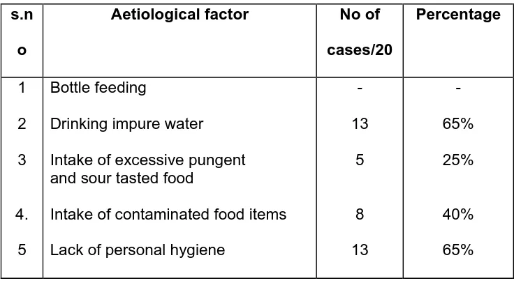
9. Aetiological factors
10. Duration of illness
11. Clinical presentation
12. Signs and symptoms
13. Reference to mukkutram
14. Ezhu udal kattugal
15. Envagai thervugal
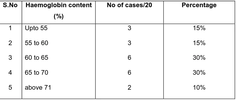
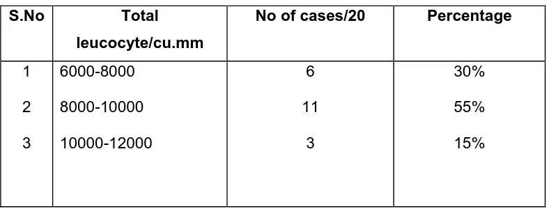
16. Haemotological profile.
17. Microscopic examination of stool and culture.
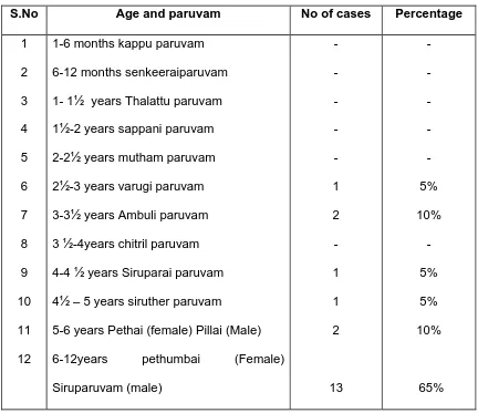
Table: 1 Age Distribution
S.No Age and paruvam No of cases Percentage 1 2 3 4 5 6 7 8 9 10 11 12
1-6 months kappu paruvam
6-12 months senkeeraiparuvam
1- 1½ years Thalattu paruvam
1½-2 years sappani paruvam
2-2½ years mutham paruvam
2½-3 years varugi paruvam
3-3½ years Ambuli paruvam
3 ½-4years chitril paruvam
4-4 ½ years Siruparai paruvam
4½ – 5 years siruther paruvam
5-6 years Pethai (female) Pillai (Male)
6-12years pethumbai (Female)
Siruparuvam (male) - - - - - 1 2 - 1 1 2 13 - - - - - 5% 10% - 5% 5% 10% 65%
Among the 20 cases 65% of cases in the age group of 6-12 years, 15% in the
age group of 3-4 years and 20% in the age group of 4-6 years.
Table: 2 Distribution of sex
S.No Sex Percentage No of cases/20 1 2 Male Female 60% 40% 12 8
[image:52.612.106.545.563.645.2]Table: 3 Religion Distribution
S.No Religion No of cases/20 Percentage 1 2 3 Hindu Christian Muslim 18 2 - 90% 10% -
Out of 20 cases 90% belonged to Hindu and 10% cases belonged to
[image:53.612.106.521.310.416.2]Christian.
Table: 4 Socio economic status
S.No Socio Economic Status No of cases/20 Percentage 1 2 3 Poor Middle Rich 16 4 - 80% 20% -
Out of 20 cases 80% cases belonged to poor socio economic status and
20% of cases belonged to middle class.
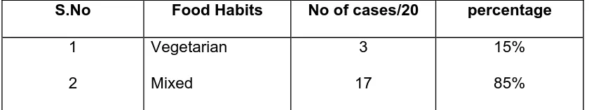
Table: 5 Distribution according to food habits
S.No Food Habits No of cases/20 percentage 1 2 Vegetarian Mixed 3 17 15% 85%
According to food habits 85% of cases had mixed diet and 15% had
[image:53.612.104.524.520.599.2]Table: 6 Distribution according to kaalam
S.No Kaalam No of cases /20 Percentage 1 2 3 Vatha Kaalam Pitha kaalam Kaba kaalam 20 - - 100% - -
100% cases were from vatha kaalam because the clinical study was
carried out in children under the age of 12.
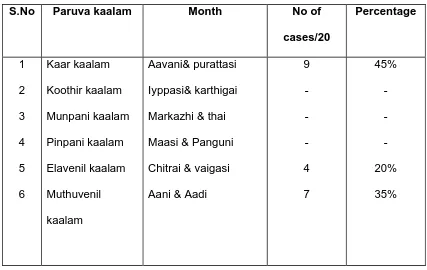
Table: 7 Distribution according to paruva kaalam
S.No Paruva kaalam Month No of cases/20 Percentage 1 2 3 4 5 6 Kaar kaalam Koothir kaalam Munpani kaalam Pinpani kaalam Elavenil kaalam Muthuvenil kaalam Aavani& purattasi Iyppasi& karthigai
Markazhi & thai
Maasi & Panguni
Chitrai & vaigasi
Aani & Aadi
9 - - - 4 7 45% - - - 20% 35%
45% of cases were recorded in Kaar kalam, 20% of cases in Elavenil
[image:54.612.103.530.319.589.2]