Vol.67, No. 10 JOURNALOFVIROLOGY, OCt. 1993,p.5976-5988
0022-538X/93/105976-13$02.00/0
Copyright © 1993,AmericanSocietyforMicrobiology
Characterization of Chimeric
Full-Length Molecular Clones of
Aleutian Mink Disease Parvovirus (ADV):
Identification of
a
Determinant Governing Replication of ADV in Cell Culture
MARSHALL E. BLOOM,* BRADLEY D. BERRY, WU WEI, SYLVIA PERRYMAN,ANDJAMES B. WOLFINBARGER
Laboratory ofPersistent ViralDiseases, NationalInstituteofAllergy andInfectious Diseases, Rocky Mountain Laboratories, Hamilton, Montana59840
Received 22 March 1993/Accepted 1 July 1993
TheADV-G strain of Aleutian minkdisease parvovirus (ADV) is nonpathogenic for mink but replicates
permissivelyincell culture, whereas the ADV-Utah1strain is
highly
pathogenic forminkbutreplicates poorlyin cell culture. In ordertorelatethesephenotypic differences toprimarygenomicfeatures, weconstructeda series of chimeric plasmids between a full-length replication-competent molecular clone of ADV-G and
subgenomicclones ofADV-Utah1representingmapunits(MU) 15to88.After transfection oftheplasmidsinto cell culture and serial passage of cell lysates, we determined that substitution of several segments of the
ADV-Utah1genome(MU 15to54 and 65to73) withinaninfectious ADV-G plasmiddidnotimpairtheability
oftheseconstructs toyield infectiousvirus invitro. LikeADV-G, the virusesderived fromthese
replication-competent clones caused neither detectable viremia 10days after inoculation noranyevidence of Aleutian
disease inadult mink. On theotherhand, other chimeric plasmidswereincapableofyielding infectiousvirus andwerethereforereplicationdefective invitro. The MU 54to65 EcoRI-EcoRVfragmentofADV-Utah 1was
the minimal segment capableofrendering ADV-G replication defective. Substitution ofthe ADV-G EcoRI-EcoRVfragmentintoareplication-defective clonerestoredreplicationcompetence,indicating that this 0.53-kb portion ofthegenome, whollylocatedwithin sharedcoding sequencesforthecapsidproteinsVP1 andVP2, containedadeterminantthatgovernsreplicationincell culture. When cultures of cellswerestudied5daysafter transfection with replication-defective clones, rescue of dimeric replicative form DNA and single-stranded
progeny DNA could not bedemonstrated. This defectcould not be complemented bycotransfection witha
replication-competent construction.
Strains of the nondefective Aleutian mink disease parvo-virus (ADV)differdramatically in avariety of ways (2, 19, 40). Pathogenicstrains of ADV, suchasADV-Utah1,cause severe disease in mink that is influenced bythe age of the animals at the time of infection (2, 13). In response to
ADV-Utah1infection,newbornminkuniformly succumbto afulminantinterstitial pneumonitis accompaniedby permis-sive viral replication in alveolar type 2 cells (2, 3, 6), but adult mink infected with the same virus develop a fatal chronic disease characterized byapersistent butrestricted infection (2, 5, 13, 19, 60). This adult or classical form of disease is associated with avariety ofimmunological aber-rations, includingimmunecomplexnephritis, hypergamma-globulinemia, and plasmacytosis (2, 13, 34, 60). On the other hand, the cell culture-adapted ADV-G strain shows little evidence ofreplicationinvivo and doesnot causedisease in adult minkeven afterinjectionoflargeamountsof virus (2, 12, 20). In addition, injection of newborn mink kits with ADV-Gproduces only limitedreplication, very mild pulmo-nary lesions, and no clinical disease (2). Whereas ADV-G replicates permissively in susceptible cell cultures (18), attempts to propagate the ADV-Utah 1 strain serially have beengenerally unsuccessful (20, 41). These findings indicate that differences in the primary sequence of ADV must controlboth the abilityof a particularADV strain to
repli-catein vivo and itspathogenicity.
The complete sequence and transcription map of the
*Correspondingauthor.
nonpathogenic ADV-G strain are known (12, 14), and a full-length molecular clone has been developed (12). When this construct,denotedpXV-B-4, is transfected into suscep-tible CRFKcells, infectious ADVcanbe rescued, and this rescued virus faithfully recapitulates the parent ADV-G in thatitproductively infects CRFK cells but isnonpathogenic for adult mink (12). The sequence of ADV-G has been compared with thepartialsequenceof several otherviruses, notablythepathogenicADV-Utah 1 (14, 37).These molec-ular clones, corresponding to approximately 76% of the genome, were derived directly from infected mink organs, avoiding any in vitro selection. The comparisons suggest that ADV-G is greater than97% relatedattheDNAlevelto
theseviruses, and fewer than 75 nucleotide differenceswere
found between theseADVstrains.
In orderto hypothesizehow aparticularsequence varia-tion might influence replication and pathogenicity, it is necessary to understand genomic organization. Nondefec-tive parvoviruses such as ADV are small, nonenveloped icosahedral viruses withsingle-stranded DNAgenomes ap-proximately 5,000nucleotides(nt)inlength(30).Thegenetic organizationof theseviruses isrelatively simple,and that of ADV resembles that of the others (4, 12, 14). There are
major left and right open reading frames and several smaller open reading frames in the mid-genome. Two overlapping capsid proteins (VP1 and VP2) are specified by the right open reading frame, and expression of these proteins is controlledbyapromoterlocatedatmap units(MU)36to38 (P36)(4, 14,28).Alarge nonstructuralprotein(NS1),coded forby the left open reading frame (14, 29), is driven by a
5976
on November 9, 2019 by guest
http://jvi.asm.org/
CHIMERIC CLONES OF ADV 5977
constitutively active promoter at MU 4 (P4) (4, 14) and serves several functions. NS1 is absolutely required for genomic replication and also trans activates the P36 pro-moter (32, 42, 47, 67, 82). In addition, NS1 is required for the excision and replication of parvoviral genomes contained in molecular clones (68). Smaller nonstructural proteins, such as NS2, arise by splicing of the amino-terminal portion of NS1 with smaller open reading frames in the middle of the genome (4, 14, 29). The role of NS2 in replication is less clear, but it appears to regulatereplication and virus produc-tion in a cell type-specific fashion (24, 48, 55).
In recent years, detailed studies on other parvoviruses havealso found that apparently minor sequence differences can be correlated with pronounced biological differences. For example, although canine parvovirus (CPV) and feline panleukopenia virus (FPV) are 98% related by DNA se-quence and highly cross-reactive antigenically (25, 56-58, 76), they exhibit distinct patterns of host cell tropisms in vitro (76) and, in addition, differ in the ability to replicate and causedisease indogsand cats, respectively(76). Allotropic strains of minute virus of mice (MVM) provide another example of closely related parvoviruses having different properties.Theprototypic MVM(p)andimmunosuppressive MVM(i) areserologically indistinguishable andexhibit 97% DNAsequenceidentity,yet eachstrain is restricted for viral growth in the reciprocal host cell at some stage in viral replication subsequent to initial entry (9, 70, 73). These examples clearly demonstrate that parvoviruses as closely related as ADV-G and ADV-Utah 1 can exhibit markedly differentbiological characteristics.
Experiments to assign apparently minor differences in genome structure to observedvariability in viral replication and host cell tropism have been performed with the MVM and CPV/FPV models. By the construction of viruses chi-meric for selected segments of the genome, it is apparent that differences in host range, celltropism, and pathogenic-ity can be accounted for by very limited aminoacidchanges (9-11, 25, 35, 56, 76). Surprisingly, perhaps, for MVM and CPV, the segment of the viral genome responsible for this phenomenon seems toresidue in theportionof the genome coding for the virion capsid proteins (11, 25, 56). Similar studies with porcine parvovirus havesuggested that deter-minants in the nonstructural coding regions may also play a role in some instances (79).
In the experiments reported in this article, we replaced segmentsofan infectious ADV-G molecular clone with the corresponding portions of clones from pathogenic strains. These chimeric constructions were assayed after transfec-tion into cell cultures for the capacity to yield infectious virus. Virus derived from suchreplication-competent clones
wasfoundtobenonpathogenicfor adult mink.Replacement ofa531-ntsegment in thecapsid proteingene of ADV-Gwas
sufficient to render full-length molecular clones of ADV incapableofgeneratinginfectious virussubsequentto
trans-fection. When cultures transfected with these replication-defective plasmids were studied, it was apparent that the levels ofDNAreplicativeformDNA, progeny strand DNA, and, insome cases, capsid proteinwere greatly reduced.
MATERIALSANDMETHODS
Cells, sera, and viruses.ADV-G and viruses derived from infectious molecular clones ofADV were propagated and assayed in Crandell feline kidneycells (CRFK)asreported before (12, 20, 31, 62).Thepreparation and assay of
ADV-Utah 1 have been described before
(19, 26).
The anti-ADV murine monoclonal antibodies 30, 68, P-2-1, and 163 have been described and characterized pre-viously (64). The anti-ADV murine monoclonal antibody Y-2-9 was prepared by standard methods (64). All react against both ADV capsid proteins VP1 and VP2. Murine monoclonal antibody 355, reactive against rabies virus gly-coprotein, waskindly provided byDonLodmell.
Molecularcloning techniques. Standard molecularbiology techniqueswereused forcloning,DNAsequencing, restric-tionenzymedigestion, ligation, linkerinsertion,agarosegel electrophoresis, and plasmidbatchpreparation (12, 14, 51, 69).
Full-length molecular clones were successfully
trans-formed into andamplifiedinEschenichia coliJC8111(recBC sbcBrecf) (12, 23).Transformationefficienciesandyieldsof full-lengthcloneswerelow,typicallyin the range of 25to200 ,ugofplasmidperliter ofculture, evenwhen chloramphen-icolamplificationwasused.Otherplasmidswerepropagated
asdescribedpreviouslyinE. coliJM109(12,
14).
To facilitate manipulations, the MU 100 EcoRI site of pXV-B-4wasdeleted andreplacedwitha
unique BglII
site. This construction, pXXI-Q-3-15, behavedexactlythe same aspXV-B-4inall respects.For the preparation of chimeric virus
constructions,
the described andsequenced molecular clonesrepresenting
the 3,455-nt BamHI-HindIII (MU 15 to 88) segment of ADV-Utah 1(pUT1-IX-G) (14,
15)
orthe ADV-Utah 1-likeclone derived from the Danish ADV-K strain(pKl) (12, 37)
wereused. These two clones are identical except for a
single-nucleotide deletion in pUT1-IX-G ofan A at nt 806, inter-rupting the left open reading frame. Apreviously reported C-A difference between thesetwoisolates at nt3100was a
sequencing error (37). Although one of the chimeric
con-structions herein described(pXX-B-1) usedasegmentfrom pKl,forsimplicityweshallrefertoall the constructionsas
havingbeen made betweenADV-G and ADV-Utah 1. PlasmidpXX-B-1wasconstructedbyreplacingthe MU 15
to 88 BamHI-HindIII segment ofpXV-B-4 with the
corre-spondingsegmentfrom ADV-Utah 1.PlasmidspXVII-T-1-2 and pXVII-T-2-2 contained the XhoI-EcoRV and the EcoRV-BstEII
fragments, respectively.
ClonespXXI-Q-2-2,
pXXIII-S-2, and pXXII-K1 were prepared by
exchanging
the MU 30 to69
(AflII),
MU 15to 54(BamHI-EcoRI),
and MU54to65(EcoRI-EcoRV) fragments,respectively,
of thepathogenic
strain for those of ADV-G inplasmid
pXXI-Q-3-15. The structure of these constructions was verified by restriction enzyme analysis and by DNA sequencing. The maintenance of the unstable right-hand
(MU 100)
hairpin
wasconfirmedbyrestriction enzymeanalysis.Plasmids destined for transfectionwerestored in
aliquots
asethanolprecipitates. Priortouse,the DNAwaspelleted
andresuspendedinsterile deionized water, and the
concen-trationwasverifiedbyagarose
gel
electrophoresis.
Because thecircularplasmids migrated
withamobility
similartothat ofduplexmonomeric ADVreplicative
formDNA,
insomeexperiments
DNAs weredigested
with the enzymeBglI
(whichdoesnot cutADV) priorto usein transfection. This had no effect on the abilityof these molecules to transfect cultures(data
notshown).
Transfections. DNA transfections
(with
5 ,ug ofplasmid
DNA, unless noted)were done as describedpreviously
in 35-mm 6-wellplatesforanalysisofnucleic acidsorproteins
(12,38).The transfected cultureswereharvested after 5
days
ofincubationat
31.8°C.
When DNAs were to be tested for
infectivity,
transfec-tions were performed in individual 35-mm dishes. At theVOL. 67,1993
on November 9, 2019 by guest
http://jvi.asm.org/
5978 BLOOM ET AL.
time of harvest, cell pellets were collected in 1 ml of medium, freeze-thawed, sonicated, and serially passaged in 25-cm2 flasks before assay.
When DNAs were to be transfected in vivo (33, 71, 80) into adult sapphire mink, both an intranasal and an intra-peritoneal route were used on sedated animals. For intrana-saltreatment, 5 ,ul of precipitatecontaining 10,ugofplasmid DNA, 6.5 ,ug of DNA prepared from an ADV-Utah 1-in-fected mink kit lung, or 2.7 ,g of normal lung DNA was instilled into each nostril (i.e., 10,ul per mink). For intra-peritoneal treatment, 10 ,ul of the same precipitates was diluted to 100
pl
with sterile phosphate-buffered saline and injected.Toverify the number of fluorescence-forming units (FFU) permicrogram of DNA for each experiment, transfections of SO-,I samples were performed in four-well Labtek chambers or the wells of 24-well culture dishes containing coverslips. At thetime of harvest, these specimens were acetone fixed and stained for ADV antigens with a direct fluorescein isothiocyanate conjugate prepared from infected mink sera (20). The number of cells displaying ADV antigens per chamber was counted and used to estimate the FFU per microgram for the various DNA samples.
SDS-PAGE and immunoblot analysis. Sodium dodecyl sulfate-polyacrylamide gel electrophoresis (SDS-PAGE) and immunoblotting for ADV proteins were performed as de-tailed in previouspublications (1, 15, 21, 53), but the signal was developed with a chemiluminescent substrate (ECL; Amersham, Chicago, Ill.). This method amplified the sensi-tivity by at least a factor of 10 over standard chromogenic substrates.
Nucleic acid isolation and Southern blot
analysis.
For preparation of whole-cell DNA (72), cell pellets or tissue samples were lysed in 0.01 M Tris (pH 8.0) containing 1% Sarkosyl and 100 ,ug of RNase (boiled to inactivate extrane-ous nucleases) per ml and incubated for 3 h at 37°C. Following addition of proteinase K to 200 ,ug/ml, cellular DNAin the samples was sheared through a 27-gauge needle, and theincubation was allowed to proceed for another 3 h at 37°C. DNA was ethanol precipitated in the presence of oysterglycogen (74) afterphenol-chloroform extraction. The DNAwascollected bycentrifugation and resuspended in 30RI
of 0.01 M Tris (pH 8.0)-0.001 M EDTA (TE buffer). For preparation of DNase-resistant, single-stranded (SS) virion DNA (17, 20), the cell pellets were freeze-thawed three times and sonicated before a 3-h incubation at 37°C in 0.01 MMgCl2-20,ug ofRNase per ml-200 ,ug ofDNase per ml. Followinginactivation of the DNase with 0.02 M EDTA and the addition of 1 ,ug of salmon sperm DNA as the carrier, the samples were incubated at 37°C with 1% Sarkosyl and 500 ,ug of proteinase K per ml for 3 h. DNA was recovered afterphenol-chloroform extraction and ethanol precipitation in the presence of oyster glycogen by dissolution in 30,ulof TE.Whole-cell DNA was isolated from a sapphire mink kit lung 12 days after injection with ADV-Utah 1 or from a normal mink lung. The ADV genome content was deter-mined bySouthern blothybridization with a known standard DNA preparation and found to be approximately 5 x 10⁸ genomes per ,ug (18, 22). This DNA contained both replica-tive form and SS DNA (data not shown). The normal lung
DNAdid not contain detectable ADV DNA sequences. Samples containing equivalent amounts of DNA were electrophoresed in 0.7% agarose gels, alkaline blotted onto Hybond-N membranes, and hybridized as before with 32p_ labeledpositive-sense ADV probes (22).
Hybridization with ADVtype-specificoligonucleotides end labeled with
[_y-32P]ATP
was performed as reported in previ-ous publications (4, 16, 51). The type-specific oligonucleo-tides were based on the sequences reported (14) for the positive-sense strand of the ADV-G and ADV-Utah 1 "hy-pervariable regions." The ADV-Goligonucleotide sequence, spanning nt 3093 to 3131, was5'-GAAAAAGTTGCAACA
GAAACACTAACCTGGGATGCAGTA-3'. The correspond-ing ADV-Utah 1 oligonucleotide sequence was5'-CAAAAG ATGGGGCAGGAGAGCAATTAGAATGGACTGGTA CA-3'.
In vivo infectivity and
pathogenicity.
For determination of the in vivo infectivity and pathogenicity ofchimericviruses, adult female sapphire mink, housed and maintained under standard conditions, were injected intraperitoneally with105
FFU of the indicated virus or with the indicated cell lysate (18, 26). Serum samples were collected 10, 30, and 60 days after infection and assayed for anti-ADV antibodies by counterimmunoelectrophoresis (18). At the time of nec-ropsy, animals were inspected for the typical lesions of Aleutian disease (18, 34).Viremia at 10 days after infection was used as a surrogate for in vivo viral replication in some experiments (18, 22, 34, 63). Virus was concentrated from 200
,ul
of serum by ultracentrifugation for 30 min at 80,000 rpm in a Beckman Airfuge. The pellet was suspended by sonication in 10 p1 of sterile TE buffer, denatured for 30min by the addition of 190 ,ul of 1.5 M NaCl-0.5 M NaOH, and applied as100-,ul
duplicates to Hybond-N membranes under vacuum with a hybridization manifold with a 96-well dot-blot format. As standards, serial twofold dilutions from 20 to 0.15 pg of a nearly full-length segment of recombinant ADV DNA were prepared in TE containing salmon sperm DNA, denatured, and applied to manifold wells in parallel with the unknown samples. The wells were allowed to fill and empty twice with 1.5 M NaCl-0.5 M NaOH. Filters were removed from the manifold, rinsed in 2x SSPE (lx SSPE is 0.18 M NaCl, 10 mM NaPO4, and 1 mM EDTA [pH7.7]),baked at80°C
under vacuum, and reacted with a32P-labeledpositive-sense ADV RNA probe as described before (22). The lower level of detection was 0.15 pg of ADV DNA, which corresponds to 7 x10⁵
genomes per ml (18, 22).RESULTS
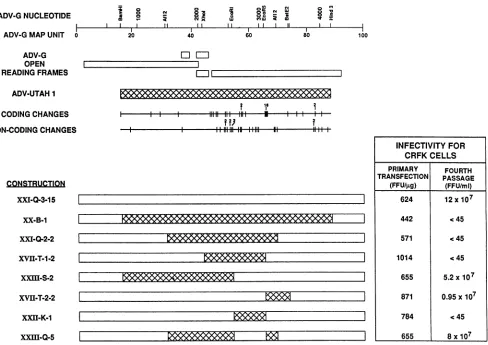
Construction of full-length chimeric clones. The develop-ment of an infectious molecular clone of the nonpathogenic ADV-G strain of ADV, designated pXV-B-4, has already been described (12). In order to prepare chimeric full-length clones, this clone was modified to prepare pXXI-Q-3-15, which had the same characteristics as the parent clone. Segments from clones of the pathogenic ADV-Utah 1 strain (14) were exchanged for the corresponding segment in an infectious ADV-G clone. A series of full-length chimeric constructions were prepared and are depicted schematically in Fig. 1.
Infectivity ofchimeric full-length clones for CRFK cells.
The infectivity of the constructions was assessed by trans-fecting CRFK cells with purified plasmid DNA, serially passaging lysates of the infected cells a total of four times, and titering the final material in CRFK cells to see whether infectious ADV had been rescued (Fig. 1). Of the eight clones tested and compared with our initial infectious clone (pXV-B-4), four were able to initiate a productive infection. Lysates from cells transfected with clones pXXI-Q-3-15, pXXIII-S-2, and pXVII-T-2-2 all contained chimeric virus
J. VIROL.
on November 9, 2019 by guest
http://jvi.asm.org/
CHIMERIC CLONES OF ADV 5979
ADV-GNUCLEOTIDE ADV-GMAPUNIT 0
a 0 0 0
20
C' CDi I ^, ;i
II II II II
40 60
O
80
ADV-G OPEN READINGFRAMES
ADV-UTAH1
CODING CHANGES NON-CODING CHANGES
jigl
i ll ll lllll H 111
|
||~
11111|'1111 iiXXI-Q-3-15
XX-B-1
XXI-Q-2-2
XVH-T-1-2
XXIII-S-2
XVII-T-2-2
XXII-K-1
XXIII-Q-5
FIG. 1. Schematic of chimeric ADV plasmids. The chimeric ADV plasmids used in thisstudyarealigned underamapof the ADVgenome identifying theopenreading frames and the restrictionenzymerecognition sites used in preparing the chimeric plasmids. Theextentof the ADV-Utah1sequenceused,aswellascodingandnoncodingsequencedifferences,arealso shown. The infectivity for CRFK cells of each
of theclonesassayed either5days after primary transfection (expressedasFFUpermicrogram of plasmid)orafter four serialpassagesof
celllysates(expressedasFFUpermilliliter of celllysate) is also indicated in tabular form adjacenttothe relevantconstruction. The initial replication-competentADV-G clonepXV-B-4had thesamestructureaspXXI-Q-3-15 and isnotshown.
infectious for CRFK cells and were therefore defined as
replicationcompetent.Furthermore, therewas nosignificant difference in the virus titer attainedbyanyof these viruses
compared with that by the virus derived from
pXV-B-4-transfected cells. Thus, pXV-B-4 was omitted from subse-quentstudies.
No virus was rescued from lysates of cells transfected with theremainingfive clones, whichwere therefore repli-cation defective. On examination of the structure ofthese fiveclones, the minimalcommonfeatureappearedtobe the
presence of the ADV-G MU 54 to 65,EcoRI-EcoRV
seg-ment. In other words, any construct that contained an EcoRI-EcoRV fragmentfrom ADV-Utah 1 was replication defective. Interestingly, theEcoRV-BstEII segment,which contains thehypervariable region,hadnosuch effect. Thus,
somedeterminant located within theMU 54 to65 segment
governedtheabilityofafull-lengthclone of ADVtoproduce
infectious virussubsequent totransfection of CRFK cells. In order to confirm that the MU 54 to 65 segment was
controlling the abilityof theseclones toinitiate productive replication, the ADV-GMU 54to65 piecewasputinto the
replication-defective constructionpXXI-Q-2-2 (Fig. 1). The
resulting construction, pXXIII-Q-5, wasreplication
compe-tent.
All of the clonesproducedADVantigens inCRFKcells after primary transfection (Fig. 1). There was no obvious correlation between the level of FFU per microgram after
primarytransfection and the abilityof the constructions to generate infectious virus.
Comparison ofreplication-competent viruses derived from chimeric full-length molecular clones in CRFK cells. The
findings described in the previous section indicated that virus infectious for CRFK cells could be derived from several of the chimeric full-lengthclones. In order to com-pare severalparameters of viralreplicationin CRFKcells,
cultures were infected atequivalent multiplicities of infec-tion withvirus stocks derived from these
replication-compe-tentclones and subjectedtoanalysis.
Western immunoblotanalysis (Fig. 2A)showed that both
capsid proteinsVP1 and VP2aswellasthemajor nonstruc-tural protein NS1 (21, 54)were synthesized byall of these
replication-competent chimeric viruses. The molecular
masses of NS1 were the same for all theviruses, but the
capsid proteinsof XVII-T-2appearedtobe 1 to 2 kDalarger
than those of the other preparations. The only unique
sequenceof XVII-T-2 comparedwith the other
replication-competentviruses resides within theAfllI-BstEII segment. A similar difference between the capsid proteins of
ADV-100
INFECTIVITY FOR CRFKCELLS
PRIMARY FOURTH TRANSFECTION PASSAGE
(FFU/4g) (FFU/mI)
624 12x107
442 C45
571 C45
1014 . 45
655 5.2x10⁷
871 0.95x10⁷
784 .45
655 8x10⁷
m
MM
W...L
MM
K66Wi
m
VOL.67, 1993
on November 9, 2019 by guest
http://jvi.asm.org/
[image:4.612.69.557.73.418.2]5980 BLOOM ET AL.
A.IMMUNOBLOT B.WHOLE CELL DNA C.SINGLE STRANDED VIRION DNA
LO
o m
aT
,L C)° > x x > x 0
E x x x x x E
LO
C?
r a
x x x
LO 04
6
C.
6-1
R 5 R
x x x
VP1\
VP2
-
NS1--DD DNA
l ;T T T~~ SS DNA
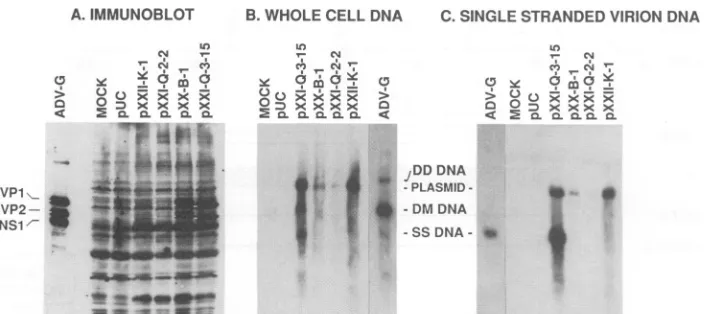
FIG. 2. Analysis of CRFK cells infected with replication-competent chimeric ADV viruses. Cultures of CRFK cellswereinfected withthe indicated replication-competent chimeric virus (fourth passage)atanapproximate multiplicity of infection of 1 FFU/cell. After 3 days of incubation at31.8°C, aliquots of the culturewereanalyzed. (A) Cell pelletswerelysed in Laemmlisample buffer, resolvedonSDS-7.5% polyacrylamide gels, transferred tonitrocellulose membranes, and immunoblotted with high-titered mink anti-ADVserum. SpecificADV signalsweredeveloped byachemiluminescent method with peroxidase-conjugated protein Aasthereportermolecule. The locations ofthe capsid proteins VP1 and VP2 and the major nonstructural proteinNS1areindicated. (B and C) Equivalentamountsof whole-cell (B) andSS virion (C) DNAwereisolated as detailed in Materials and Methods and electrophoresed in 0.7% agarosegels. Following denaturing, the samplesweretransferredtoHybond-N membranes by alkaline transfer and hybridizedtopositive-sense ADV RNA probes labeled with32P.
The locations of duplex monomeric (DM) and duplex dimeric (DD) replicative form DNAaswellasSS virion DNAareindicated.
Utah 1 and ADV-G has been noted previously (8, 64), and this findingsuggeststhat some sequencewithin that region mayaccountfor the difference.
Southern blotanalysis (Fig. 2B)showed that cells infected
byeachof the viruses contained both duplex monomeric and duplex dimeric replicative formDNA(17, 18, 22). In addi-tion,SSvirionDNA, definedasDNase-resistantDNA,was
alsopresent inthese cells (Fig. 2C) (20).
Pathogenicity of chimeric viruses for adult mink. The results detailed in the previous sections indicated thatwe could exchange certain genomic segments between patho-genic and nonpathogenic types of ADV withoutnoticeably affecting replication in CRFK cells. We wished to assess
whether the chimeric viruses were nonpathogenic, like
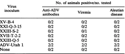
ADV-G, or pathogenic for adult mink, like ADV-Utah 1. Virus stockswerepreparedfrom thereplication-competent chimeras XXI-Q-3-15, XXIII-S-2, XVII-T-2-2, and
XXIII-Q-5and inoculated into adultsapphire mink. None of these mink exhibited signs of Aleutian disease, nor did they
develop detectable anti-ADVantibody (Table 1).
We also looked for direct evidence ofinvivo viral
repli-cationby assessingviremia 10daysafterinfection (18, 22).
None of the animals injected with either the ADV-G
con-structXXI-Q-3-15or anyof the chimeric viruses had detect-ableviremia,whereas minkinjectedwithpathogenic ADV-Utah 1 did (Table 1).
The failure of these animals to mount an ADV
anti-body response or to develop viremia at 10 days strongly
suggested that in vivo viral replication is extremely limited
or does not occur. This result was identical to that noted
previouslyforpXV-B-4 (12).Becauseoneof these chimeric
viruses, XVII-T-2,waschimeric forthe hypervariable region
(Fig. 1),thisresult alsosuggested that themerepresenceof the ADV-Utah 1 hypervariable region in an ADV-G virus wasnotsufficienttorender the virus pathogenic.
Comparison of replication-defective full-length molecular clones inCRFKcells. Anumberof the full-length molecular
clones describedabove anddepicted in Fig. 1were
replica-tion defective, as they were unable to generate infectious virus in CRFK cells. To try to elucidate the block to
replication, we prepared cultures of cells transfected with several of theseplasmidsandcompared selected parameters ofreplicationwith those in cells transfectedwith the repli-cation-competent XXI-Q-3-15.
Oncloseexamination,the pattern ofimmunofluorescence of thereplication-defectiveclonesdifferedslightlyfrom that of the replication-competent clone (data not shown). Cells transfected with replication-competent pXXI-Q-3-15 exhib-itedapatterntypical of ADV-Gand the
[image:5.612.123.499.77.244.2]replication-compe-tent clones; that is, both nuclear and cytoplasmic antigen signals,withsomeof thecytoplasmicsignalbeingofafinely granular nature (54). On the other hand, the replication-defective clones induced primarily a nuclear signal. Only pXX-B-1 DNA yielded a readily observable cytoplasmic signal. Although similar numbers of cells contained ADV
TABLE 1. Invivoanalysis of viruses derived from replication-competentADV chimeric molecularclonesa
No.of animalspositive/no. tested Virus
inoculum Anti-ADV . Aleutian
antibodies Viremia disease
XV-B-4 0/2 0/2 0/2
XXI-Q-3-15 0/2 0/2 0/2
XXIII-S-2 0/2 0/1 0/2
XVII-T-2-2 0/2 0/2 0/2
XXIII-Q-5 0/2 0/2 0/2
ADV-Utah 1 2/2 2/2 2/2
None 0/2 0/2 0/2
a Adultsapphire minkwereinoculatedintraperitoneally with105 FFUof the indicated viruspreparation orwith 10' 50% mink infectious doses of ADV-Utah 1. Thedevelopmentofanti-ADV antibodieswithin 60days after infectionwasassessedby counterimmunoelectrophoresis. The presence of viremiawasdetermined 10daysafter infectionbydot-blothybridizationas described in Materials and Methods. The presence of typical lesions of Aleutian diseasewassoughtatnecropsy 60 to90days after infection.
C)
2 x< x
Ln N
6
1
-J. VIROL.
on November 9, 2019 by guest
http://jvi.asm.org/
[image:5.612.320.558.565.668.2]CHIMERIC CLONES OF ADV 5981 A. IMMUNOBLOT B. WHOLE CELL DNA C. SINGLE STRANDEDVIRION DNA
c0J CMJ
> 2SO m?)XXXX9 O DXXXXCm =
Oxxxxxfl x
TICLC LC L LC LC
VPI\ VP2-NS1'
b .
* U*
w .
LO
0 0Z<>Ex x
iDD
DNA-PLASMID
--DM DNA -SSDNA- *
---_
-- w L
,'
FIG. 3. AnalysisofCRFKcells transfected withreplication-competent and replication-defectivechimericADVplasmids. Cultures of CRFKcells in 35-mm culture wellsweretransfected with 5 A±goftheindicated replication-competentandreplication-defective plasmids (previously digested withBglItolinearizethesupercoiledplasmids), glycerolboosted after 4h,and incubatedat31.8°Cfor5 days. Replicate samplesfrom each transfectionwereprocessedasdescribed in Materialsand Methods and the legendtoFig. 2 for(A) immunoblot, (B) whole-cell DNA, and(C)SSvirionDNA.The location of the 7.6-kb linearized residual input plasmid (B and C) and the locations ofVP1and VP2(A)areindicated.
antigen after transfection with pXXI-Q-2-2 and pXXII-K-1, thesignalwasweaker and localizedprimarilytothe nucleus. Western blot analysis (Fig. 3A) of the transfected cells revealed that the levels of NS1 directed by the replication-defective clones were similar to that of the replication-competent clone, but that the relative levels of capsid proteins VP1 and VP2were reduced(21, 54), although VP1 and VP2 could be detected inlysatesofpXX-B-1-transfected cells and the molecular masses were greater than for the corresponding proteins from the ADV-G clone(8, 64). The capsid proteins from pXXI-Q-2-2 and pXXII-K-1 were
present at avery reduced level comparedwith those from pXXI-Q-3-15 and pXX-B-1. Because the capsid proteins werenoted in cells transfected withpXX-B-1, these results suggested that the replication defect could not be linked solelyto impaired capsid protein synthesis.
Southern blot analysis of DNAisolated from cells
trans-fected5days earlier withreplication-competentor-defective plasmids also revealed differences (Fig. 3B and C). Cells transfected with the replication-competent pXXI-Q-3-15 contained DNA species that comigrated with replicative formduplexmonomeric DNA andSS virion DNA(Fig. 3B) (17, 18, 22); thus,therewasevidence that the ADV genome had beenrescued andamplified from theplasmid. Further-more,although9.6-kbduplexdimericreplicativeform DNA could not be identified, a band corresponding to residual linearizedplasmid (7.6kb)wasevident. In contrast,onlythe residual plasmid was present in cells transfected with the replication-defectiveconstructs. Neitherduplexmonomeric
norSS DNA could beclearly identified in whole-cell DNA samplespreparedfrom the severalreplication-defective chi-mericclones, anda smearofsignalfrom DNArepresenting residualinput plasmidmadeitimpossibletojudgewhethera
smallamountofduplexmonomeric DNAwaspresent. AnalysisofDNase-resistant SS virion DNA(Fig. 3C) (20) indicated that virion DNAwasreadilyapparentin pXXI-Q-3-15-transfected cells but not in parallel samples from the replication-defective clones. Interestingly, even though thesesamples had been digestedwith DNase, some of the input plasmidwasstill present. Thesefindings suggestedthat therescue andamplificationofADVreplicative form DNA
aswellasthegenerationofencapsidatedvirion DNAwere
severely impaired in cells transfected with the replication-defectiveclones.
Taken together, these results suggested that therewas a severeblockto rescueandamplificationof ADV sequences from these replication-defective clones and that levels of capsid proteins were also reduced. The facts that ADV antigenwasinduced and thatNS1 was synthesized implied that these plasmids were capable of entering transfected cells andarrivingat acellularsite wherethey couldserve as
initial transcriptional templates.
Inoculationofadult sapphireminkwith viral and plasmid DNA.The results described in theprevious sections revealed thatfull-length chimeric molecular clones containing the MU 54to65 segment ofADV-Utah 1werereplication defective in CRFK cells. Thus, we were unable to propagate virus from these clones in vitro andtestthepathogenicity of these constructions for mink. Work with avarietyof other virus systemsreveals that either viral DNA orfull-length molec-ularplasmid clones canbeintroduced directly into animals
by
several methods and infections can beinitiatedthereby
(33, 71, 80). We also showed previously that purified repli-cative forms ofADV-Gwereinfectious in cell culture (12). In order to determine whether pathogenic virus might be rescued from replication-defective ADV clones, we pre-pared DNA precipitates and inoculated them directly into adult sapphire mink. As a control, we also injected a
precipitate fromwhole-cell DNA from an ADV-Utah 1-in-fected mink kit lung. ThisDNA sample contained 5 x 10⁸ ADV genomes per ,ugof whole-cell DNA and consisted of both virion and replicativeform DNA(18, 22). Noneof the animals injectedwith any of the DNA preparations devel-opedanysignsof Aleutiandiseaseoranyanti-ADVantibody duringthe90-dayperiodof observation(datanotshown).As aresult,weconcluded that theseDNAs were notinfectious for adultsapphire mink under theconditions used. The fact that the DNAfrom theinfected minklungwas also appar-entlynoninfectious makesit impossible to draw any infer-encesabout theplasmids tested.
Reactivity of chimeric constructions with anti-ADV mono-clonal antibodies. Previous work with murine monoclonal antibodiesagainstADVcapsid proteinsindicates that there
are distinct antigenic differences between strains ofADV,
VOL. 67,1993
on November 9, 2019 by guest
http://jvi.asm.org/
[image:6.612.135.491.79.236.2]5982 BLOOM ET AL.
ADV-GNUCLEOTIDE ADV-GMAPUNIT 0
M o
m
V-Il 20
11 I 11
40 60
mmi
ADV-G OPEN READINGFRAMES
ADV-UTAH I CODINGCHANGES NON-CODING CHANGES
CONSTRUCTION
XXI-Q-3-15
XXHI-S-2
XVII-T-2-2
XXIII-Q-5
MOCK
pXXI-Q-3-15
pXX-B-1
pUC
, liI
1 ii1i111 i I I HI
l~~~~~~~
11 f l l Ei~~~~~
Hilll HW Hi H iiallCHIMERIC VIRUS
RK~
r-
I
CHIMERIC PLASMID
l-7
X
-0
N m X
+-C
+-I_I_I-_
:
_
I_
I_
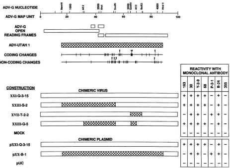
-FIG. 4. Reactivities of chimeric ADV viruses andplasmidswith anti-capsid proteinmonoclonal antibodies. Cultures of CRFK cells in
eight-chamberLabtek slideswereeither infected with theindicatedchimericADV virusfor3daysortransfected with theindicated ADV chimericplasmidfor5days.The cultureswerefixed withacetone, reacted with the various monoclonalantibodysupernatants, anddeveloped withfluorescein-conjugated proteinA. Forconvenience,thestructuresof the several viruses andplasmidsarerepresentedand relatedtothe ADV-G and ADV-Utah 1genomes.
although the location of the specific epitopes defined by
these reagents is unclear (64). Because several of the chi-meric viruses and plasmids described in previous sections and depicted in Fig. 1 were chimeric for segments of the
capsid codingregion,wetestedapanelof thesemonoclonal antibodiesbyimmunofluorescenceto seewhetherwecould map their reactivitiestospecific regions (Fig. 4). Several of the monoclonal antibodies(30,Y-2-9,andB-24)reacted with all the chimeric viruses;however,three(68, 163,andP-2-1)
exhibited specific patterns of staining. Antibody 68 was
positive only againstvirusescontaininganEcoRV-AflIl(MU 65 to 69) segment derived from ADV-G; this piece of the genomecontains the hypervariable region of therightopen
reading frame. Antibody 163 reacted only with viruses
containing theADV-GAflII-BstEII (MU69to73)segment, and antibodyP-2-1 reactedspecificallywithviruses having
the ADV-Utah 1AflII-BstEII piece. The control antibody 355, reactive against the rabies virus glycoprotein, was
uniformly negative.
The same panel of monoclonal antibodies were tested
against CRFK cells transfected 5 days earlier with pUC, pXXI-Q-3-15,andpXX-B-1 (Fig.4).Thestainingpatternfor cells transfected with the ADV-GplasmidpXXI-Q-3-15was identical to that of the derivativevirus; all themonoclonal antibodies except MAb P-2-1 and the control MAb 355
reacted. Furthermore, the ADV-G-reactive antibodies 163 and 68 did not stain positive cells in pXX-B-1-transfected cells,whereas P-2-1 did.
Attemptedrescueofreplication-defective plasmid by repli-cation-competent plasmid. In afinal set ofexperiments,we wishedtodeterminewhether virus froma replication-defec-tive construction(pXX-B-1)couldbe rescuedby cotransfec-tion withareplication-competentone(pXXI-Q-3-15).CRFK cells weretransfected with a mixture ofthe twoplasmids,
and lysates of the transfected cells were serially passaged four times.CRFK cellsweretheninfected withavirus stock
preparedfromthis material andanalyzedfor thepresenceof
ADV-Utah1 DNAsequencesby performing Southernblots with oligonucleotides specific for either ADV-G or ADV-Utah 1 (Fig. 5). As a control for the presence of the ADV-Utah 1 sequences, a parallel sample was prepared
from cells infected with thereplication-competent XVII-T-2-2virus,which contains the ADV-Utah1 target sequence
(Fig. 1).NoADV-Utah 1signalwasdetected in DNA from cells infectedwithvirus derived from the cotransfection but was readily apparent in DNA from XVII-T-2-2-infected cells. ADV-Gsequenceswerepresentincells infected with eitherXXI-Q-3-15 virusorvirus derived from the cotrans-fection.
We also looked for the presence ofADV-Utah 1 capsid
80 100
REACTIVITYWITH
MONOCLONAL ANTIBODY
r--l
J.VIROL.
on November 9, 2019 by guest
http://jvi.asm.org/
[image:7.612.80.543.74.413.2]CHIMERIC CLONES OF ADV 5983
ADV-UTAH1 PROBE ADV-G PROBE
WHOLECELL VIRION WHOLECELL VIRION
DNA DNA DNA DNA
o0
m
a
-m
-2
x
3
2
x3x
xE
x
ax
2x
3
x
-DM
DNA--SSDNA-'
FIG. 5. Hybridization of DNA from ADV-infected cells with ADV-G- and ADV-Utah 1-specific oligonucleotides. Individual 35-mmdishesofCRFK cellsweretransfected with 5 pg(total)of the
replication-competent plasmid pXXI-Q-3-15 DNA, a mixture of
pXXI-Q-3-15 and replication-defective pXX-B-1 DNA, or
replica-tion-competent pXVII-T-2 DNA as described in previous figure
legends. Cell pelletswere collected, lysed, and serially passageda
total offourtimes.Following titrationof thefinalpassage material,
cultureswereeithermockinfected orinfectedatamultiplicity of 1 for 3 days, at which time whole-cell and SS virion DNA was
prepared. Equivalent amounts of DNA were resolved on 0.7%
neutral agarose gels and alkaline blotted onto Hybond-N
mem-branes.Duplicate membraneswere hybridizedwith oligonucleotide
probesspecific for either the ADV-Gorthe ADV-Utah1 hypervari-ableregionendlabeledwith [-y-32P]ATP.
antigens in the same preparations with the type-specific monoclonal antibodies detailedin aprevious section. There
was no evidence of ADV-Utah 1 antigen in cells infected
with virus derived from the cotransfected cells (data not
shown).
Theseexperimentssuggested that virus froma
replication-defectiveconstruction couldnotbe rescued by the presence ofareplication-competent virus.
DISCUSSION
In this study, wecharacterized full-length molecular chi-mericclonesofADV. Specifically,wereplacedsegmentsof
afull-lengthinfectiousmolecularclone of thenonpathogenic ADV-G (12) with the corresponding pieces from a
patho-genic strain ofADV (14) and examinedthe abilityof these constructions to initiate a productive infection in cell cul-tures.
Several of the chimeric constructions containing ADV-Utah 1sequenceswerereplicationcompetentinCRFK cells
(Fig. 1). Included in this groupwere clones that contained fromADV-Utah1 allbutthe172amino-terminal residuesof NS1 (XXIII-S-2), the VP1-specific region of the capsid
proteins(XXIII-S-2 and XXIII-Q-5),and the uniqueportions of NS2 and the putative NS3 (XXIII-S-2 and XXIII-Q-5). None of these replication-competent constructions were
pathogenic for adult mink, nor did they cause detectable viremiaat10days afterinfection (Table 1).Thus, it appeared thatsubstantialportionsoftheADV-Utah1genomedidnot
conferpathogenic potentialortheabilitytoreplicatetohigh
titer in vivo in adult mink. Analysis of infection by these viruses in highlysusceptible minkkits (3, 26) is in progress. A number of other clones could not initiate in vitro infection and were replication defective in CRFK cells. This replication defect made it impossible to inject mink with virus stocks prepared from these constructions. Attempts to bypass in vitro replication in CRFK cells and deliverplasmid DNA directly in vivo were unsuccessful (data not shown). Furthermore, this block to replication in vitro could not be overcome by cotransfection with a replication-competent clone, suggesting that thedefectcould not becomplemented in trans in vitro (Fig. 5).
The experiments indicated that a determinant
governing
the ability of ADVto replicate inCRFK
cells mapped to a531-bp EcoRI-EcoRV fragment spanning nt 2553 to 3084 (Fig. 1) (12, 14, 15). No construction containing this MU 54 to 65 piece from ADV-Utah 1 was replication competent in CRFKcells. Comparison of ADV-G and ADV-Utah 1(Fig. 6)reveals only 14 nucleotide differences in this segment, and thus some change resulting from one or more of these differences likely controls replication in CRFK cells.
Inspection of the ADV sequence(12, 14) andtranscription map (4) indicates that the mid-genomic AATAAA polyade-nylation motif (nt 2564) and poly(A) addition site (nt 2589) are present at the 5' end of this fragment (Fig. 6). This polyadenylation site is utilized for productionof R2 and RX, which code for NS2 and a putative NS3, respectively (4). NS2 has been shown toregulate MVMpermissivity in a cell type-specific fashion (24, 48, 55). Furthermore, the mid-genomic polyadenylation site has been implicated inlimiting replication of B19 in nonpermissive cells (50). However, neither the polyadenylation motif (AATAAA) nor the poly(A) addition site differ between ADV-G and ADV-Utah 1, although there are several noncoding nucleotide changes apparent 3' to the poly(A) site. Poorlydefined sequences 3' to the actual poly(A) addition site have also been implicated in regulating polyadenylation in some systems (46, 52, 81), and detailed comparison of transcription and gene expres-sion of replication-competent and replication-defective mo-lecular clones may allow us to determine whether such noncoding changes are involved in the phenomena that we have reported here.
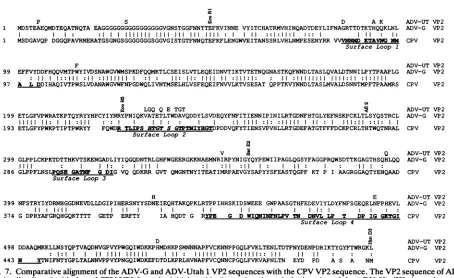
The MU 54 to 65 portion of the genome is wholly con-tained within the shared coding sequence for the capsid proteins VP1 and VP2 (4, 12). Furthermore, there are four amino acid differences between ADV-G and ADV-Utah 1 in this region (Fig. 6), and it is possible that one or more of these coding changes are involved in regulating permissivity. A role for the capsid proteins in governing the ability of ADV to replicate in CRFK cells might be reasonable when recent elegant studies on CPV/FPV (25, 56, 61, 76) and MVM(i)/MVM(p) (9, 10, 24) are considered. In both cases, tropism and permissivity for particular cell types are regu-lated by short sequences in the capsid genes, and in both cases, two or three amino acids acting coordinately are required for the phenotypic change. Because the crystallo-graphic structure of CPV has been determined (77), it has been possible to map these sequences to short loops of amino acids on the surface of the virus particle near the three-fold axis of symmetry. Although no crystallographic data are available for ADV, when the ADV VP2 sequence is optimally aligned with the CPV VP2 (65) (Fig. 7), the four coding changes observed in the ADV EcoRI-EcoRV seg-ment appear clustered near surface loop 1 of the CPV sequence, extremely close to the critical CPV residuesN-93 and A-103. Thus, it may be reasonable to speculate that for
VOL.67, 1993
on November 9, 2019 by guest
http://jvi.asm.org/
[image:8.612.72.287.77.262.2]5984 BLOOM ET AL.
(Eco Rl) G C ADV-UT DNA
2553 GAATTCAAAGTAATAAACAATGAAGTGTATATTACTTGTCACGCTACTAGAATGGTACACATTAACCAAGCTGAC ADV-G DNA 50 E F K V I N N E V Y I T C H A T R M V H I N Q A D ADV-G VP2
T A GC AA
2628 ACAGACGAATACTTGATATTTAATGCTGGTAGAACTACTGATACCAAAACACATCAGCAAAAACTAAACTTAGAA
75 T D E Y L I F N A G R T T D T K T H Q Q K L N L E
D A K
T G
2703 TTTTTTGTATATGATGATTTTCACCAACAAGTAATGACACCTTGGTATATAGTAGATAGCAACGCTTGGGGTGTA
100 F F V Y D D F H Q Q V M T P W Y I V D S N A W G V
F
ADV-UT DNA ADV-G DNA ADV-G VP2 ADV-UT VP2 ADV-UT DNA ADV-G DNA ADV-G VP2 ADV-UT VP2
G G ADV-UT DNA
2778 TGGATGAGTCCTAAAGACTTTCAACAAATGAAAACACTGTGTAGTGAAATTAGTTTGGTTACTTTGGAACAAGAA ADV-G DNA
125 W M S P K D F Q Q M K T L C S E I S L V T L E Q E ADV-G VP2
G C ADV-UT DNA
2853 ATAGACAATGTAACCATAAAAACTGTAACAGAAACCAACCAAGGTAACGCATCTACCAAGCAATTCAACAATGAC ADV-G DNA
150 I D N V T I K T V T E T N Q G N A S T K Q F N N D ADV-G VP2
2928 TTAACTGCGTCGTTACAGGTTGCTTTAGATACTAACAACATACTGCCATATACTCCAGCTGCGCCGTTGGGGGAA ADV-G DNA
175 L T A S L Q V A L D T N N I L P Y T P A A P L G E ADV-G VP2
3003 ACACTGGGCTTTGTTCCTTGGAGAGCAACCAAACCAACCCAATATAGGTATTATCATCCATGTTACATTTACAAC 200 T L G F V P W R A T K P T Q Y R Y Y H P C Y I Y N
3078 225
(Eco R5) (A) A GGG G G AT GAA AC GTAC G
rTAGTrGT R Y P N I Q K V A T E T L T W D A V Q D D Y L S V
L G Q Q E T G T
G C
3153 GATGAACAGTACTTTAACTTTATTACTATAGAGAACAACATACCTATTAACATTCTCAGAACGGGAGATAACTTT
250 D E Q Y F N F I T I E N N I P I N I L R T G D N F
ADV-G DNA ADV-G VP2 ADV-UT DNA ADV-G DNA ADV-G VP2 ADV-UT VP2 ADV-UT DNA
ADV-G DNA
ADV-G VP2
(Afl 2)
3228 CATACAGGCTTGTATGAGTTTAACAGTAAACCATGTAAACTAACCTTAAGCTATCAAAGTACACGTTGCTTGGGG ADV-G DNA
275 H T G L Y E F N S K P C K L T L S Y Q S T R C L G ADV-G VP2
3303 CTACCTCCTCTCTGCAAACCAAAGACAGATACAACACACAAAGTAACCTCAAAAGAAAACGGAGCTGACCTAATT ADV-G DNA 300 L P P L C K P K T D T T H K V T S K E N G A D L I ADV-G VP2
3378 TACATACAAGGACAAGATAATACCAGACTAGGTCACTTTTGGGGTGAGGAAAGAGGTAAGAAAAACGCAGAGATG ADV-G DNA
325 Y I Q G Q D N T R L G H F W G E E R G K K N A E M ADV-G VP2
G (BSt E2) ADV-UT DNA
3453 AACAGAATTAGACCTTACAACATAGGTTACC ADV-G DNA
350 N R I R P Y N I G Y ADV-G VP2
[image:9.612.119.497.73.456.2]V ADV-UT VP2
FIG. 6. Comparisonof ADV-G and ADV-Utah 1 DNA and amino acid sequences between the EcoRI and BstEIIrecognitionsites. The DNAsequence andsingle-letter aminoacid translationof ADV-G and ADV-Utah 1(ADV-UT)werecompared for the spanbetweenthe EcoRIandBstEIIrecognition sites.Simplesequencedifferencesarenoted with theappropriateletter;thedeletion(A)andaddition(*)of A residues in theADV-Utah1sequencecorrespondingtoADV-Gnt3094 and3113,respectively,areindicated. TheAATAAApolyadenylation signal (nt2564to2569),the site of actualpoly(A)addition(TAat nt2585to2586),andthecleavagesites forEcoRI,EcoRV,AflIl,andBstEII
areemphasizedinboldface.
ADV-G/ADV-Utah 1,thesecoding changes acting singlyor
coordinately may regulate permissivity in cell culture.
Ex-periments are under wayto mutatethese four residues and observe the effectonreplication.
Eight of the nineamino acid differences found in the MU 65 to 73, EcoRV-BstEII
fragment
(hypervariable region)
appeared clustered in a position likely to correspond with CPVsurfaceloop2(Fig.6and7) (77). Although exchangeof thisregion byitselfdidnotrenderinfectious clones replica-tiondefective, thepronouncedvariability noted in thisarea
might still have some undetermined biological importance, perhapsan asyetundefinedepitope.
Our results suggested that the block to rescue of the replication-defective plasmids was characterized by a virtual absence of encapsidated progeny strands and a failure of amplification ofreplicative form DNA (Fig. 3). This result seemed slightly different from the MVM(i)/MVM(p) situa-tion, inwhich the blocktoreplication canbeovercome by transfection of the allotropic genome (36). In the MVM system,the accumulation of progeny SSDNAisdependent
oncapsid protein production (43, 66,75), and the genomes
are thought to becaptured within preformed capsids (39), whichcanself-assembleintheabsence of genomesynthesis (28). However, the replication defect reported here cannot
be relatedsimplytocapsid protein synthesis,because both VP1 and VP2 were produced in cells transfected with pXX-B-1 (Fig. 3A), although virus was not propagated. Perhaps there aresubtle differences in cellular localization and transport which we have not yet identified. Alterna-tively, capsid formation or stabilityin CRFK cells may be impairedif the propercis-actingsequenceis absent. Never-theless, the mechanism by whichacis-actingsequence in the capsid coding sequences mightcausethisremains obscure. Beforewe canconcludethatonly thecapsidgenes control ADV permissivityfor CRFK cells, it will be necessaryto
analyze constructions that contain the portions of ADV-Utah 1 not used in these experiments. The preliminary sequenceobtained from anearly full-length clone of ADV-Utah 1 (59) suggests a very limited number of sequence differences in the portionof the genome spanningthe
left-J. VIROL.
on November 9, 2019 by guest
http://jvi.asm.org/
CHIMERIC CLONES OF ADV 5985
P S D A K
1 MDSTEAEQMDTEQATNQTA EAGGGGGGGGGGGGGGGGVGNSTGGFNNTTEFKVINNE VYITCHATRMVHINQADTDEYLIFNAGRTTDTKTHQQKLNL
: :IIIIIIIII III III IIIIIII : II ::III:I ::: I I : 1:
1 MSDGAVQP DGGQPAVRNERATGSGNGSGGGGGGGSGGVGISTGTFNNQTEFKFLENGWVEITANSSRLVHLNMPESENYRR VVVNNDM KTAVWG NK
Surface Loop 1
ADV-UT VP2 ADV-G VP2 CPV VP2
F ADV-UT VP2
99 EFFVYDDFHQQVMTPWYIVDSNAWGVWMSPKDFQQMKTLCSEISLVTLEQEIDNVTIKTVTETNQGNASTKQFNNDLTASLQVALDTNNILPYTPAAPLG ADV-G VP2
: 11 1::1|1 :11 :1111111 :1: II: 11: 1111 11 :111:1: : 11 :11111111 1111:11 :1:1111
97 A LDDIHAQIVTPWSLVDANAWGVWFNPGDWQLIVNTMSELHLVSFEQEIFNVVLKTVSESAT QPPTKVYNNDLTASLMVALDSNNTMPFTPAAMRS CPV VP2
r LGQ QE TGT
199 ETLGFVPWRATKPTQYRYYHPCYIYNRYPNIQKVATETLTWDAVQDDYLSVDEQYFNFITIENNIPINILRTGDNFHTGLYEFNSKPCKLTLSYQSTRCL
11111 II: I 11 :111 :: II: : :1 1111 :1: :11111 11: 111:11 ::I:
193 ETLGFYPWKPTIPTPWRYY FQWDR TLIPS MTGTS GTPTNIYHGTDPDDVQFYTIENSVPVHLLRTGDEFATGTFFFDCKPCRLTHTWQTNRAL
Surface Loop 2
V 0Q
299 GLPPLCKPKTDTTHKVTSKENGADLIYIQGQDNTRLGHFWGEERGKKNAEMNRIRPYNIGYQYPEWIIPAGLQGSYFAGGPRQWSDTTKGAGTHSQHLQQ
lIIII : -I 1: : II :1 :11 :11 1: : 1I I
286 GLPPFLNSLPQSE GATNF G DIG VQ QDKRR GVT QMGNTNYITEATIMRPAEVGYSAPYYSFEASTQGPF KT P I AAGRGGAQTYENQAAD Surface Loop 3
ADV-UT VP2
ADV-G VP2 CPV VP2
ADV-UT VP2
ADV-G VP2
CPV VP2
399 NFSTRYIYDRNHGGDNEVDLLDGIPIHERSNYI
11: 1:11 11
374 G DPRYAFGRQHGQKTTTT GETP ERFTY
YSDNE
E
EIEQHTAKQPKLRTPPIHHSKIDSWEEE GWPAASGTHFEDEVIYLDYFNFSGEQELNFPHEVL
I I 1: 1 : : : :
IAHQDT G RYPE G D WIQNINNLPV TN DNVL LP T DP IG GKTGI Surface Loop 4
D ADV-UT VP2
498 DDAAQMKKLLNSYQPTVAQDNVGPVYPWGQIWDKKPHMDHKPSMNNNAPFVCKNNPPGQLFVKLTENLTDTFNYDENPDRIKTYGYFTWRGKL ADV-G VP2
: 1:1 1 11 1111 111111 I 11: 111111 11 IIIIIII:: III 11 11
443 N YTNIFNTYGPLTALNNVPPVYPNGQIWDKEFDTDLKPRLHVNAPFVCQNNCPGQLFVKVAPNLTN EYD PD A S A NM CPV VP2
FIG. 7. Comparative alignment of the ADV-G and ADV-Utah 1 VP2sequenceswith the CPV VP2sequence.The VP2sequenceof ADV-G wasoptimally aligned with that of CPV VP2 from the initial methionine codon through the location of the ADV HindlIl site and compared with the correspondingsegmentof the ADV-Utah 1 (ADV-UT) VP2. Variations between the ADV-G and ADV-Utah 1sequences arespecified by noting the variant residues. The boundaries of the defined CPV surface loops 1 through 4arenoted and emphasized in boldface, and the apical residuesareitalicized. The ADVEcoRI, EcoRV, AflII, BstEII, and HindIII sitesarenoted.
handpalindrome inboard to the BamHI site but somewhat
more heterogeneity between theHindIII (MU 88) site and theright-handend of thegenome.Studiesarealsoplannedto
investigate a potential role for these regions of the ADV-Utah 1genome.Arole for the NS1proteininthe hostrange ofporcine parvovirushas beenproposed (79), soit remains apossibilitythat someof these regions arealsoinvolved in
influencing the permissivity and pathogenicity of ADV.
Furthermore, thenatureof the blocktoreplication reported here resembles that seenforNS2mutantviruses in certain
celltypes(4, 7, 27, 49).It hasrecentlybeenreportedforH-1 parvovirus that NS2 regulates viral gene expression via a sequence in the 3' untranslatedregionof thecapsid protein
mRNA (49). Infact, the 3' untranslated region of the ADV
capsid protein mRNA (R3) is located to the right of the
HindIII siteat MU88, and thus the ADV-Utah 1sequence for thisregionisnotrepresentedinthe chimeras described in thisreport.Perhaps,in additiontothedeterminantwehave
mappedinthe EcoRI-EcoRVsegment, there will alsoprove tobe anotheronecontrolledbyanNS2-responsivesequence in the3' untranslatedportion of thegenome.
It is temptingtospeculatethat thesamegenomicfeatures that constrain these constructions from permissively repli-catingincell culturemaybe relatedtopathogenicity.Studies that have defined reciprocal determinants for MVM(i)/ MVM(p) (10, 35, 36, 73)andCPV/FPV (25, 56, 61, 76) have been facilitatedbythe availabilityof cell lines nondiscrimi-natory to these various features. However, although the
pathogenic strains replicate permissively in vivo in the alveolartype2cells of newborn mink kits(6), areliable cell culturesystemfully permissivefor thereplicationof patho-genicstrains of ADV hasnotbeen identified (20, 41, 44, 45,
62). The lack of such a cell line has obviously hampered
someofourinvestigations.
Finally, these constructions enabled usto mapthe
reac-tivity for several anti-ADV monoclonal antibodies (64).
Monoclonal antibody 68 reacted specifically with the
EcoRV-AflII segment. Because the only coding changes in thisfragmentareinthehypervariable region (Fig. 6) (14)and because this monoclonal antibody reacts in the particle-dependent counterimmunoelectrophoresis (64), the relevant
epitope likelyresidesontheparticlesurface. Thesefindings may in fact bolster our suggestions that the hypervariable
regionmaybelocated in the ADVequivalentof surfaceloop
2 of CPV. Monoclonal antibodies P-2-1 and 163 reacted
reciprocallywith theAflII-BstEII fragments ofADV-G and
ADV-Utah 1. Theonly coding difference in thissegmentis
replacementof an ADV-G isoleucine withan ADV-Utah 1 valine (Fig. 6). Examination of the ADV-CPV amino acid
alignment (Fig. 7) placesthis residuenearCPV surfaceloop
3, anotherpotentiallocation foran epitope-controlling resi-due.
Furthermore, capsid proteins from viruses (e.g.,
XVII-T-2)orplasmids (e.g., XX-B-1) containingtheADV-Utah 1
AflII-BstEII fragment were 1 to 2 kDa larger in molecular massthan those from other constructions(Fig. 2A and3A).
Thissuggestedthat the ItoVtransition in thisfragmentnot
onlycontrolled theepitopefor antibodies P-2-1 and 163 but also effected a change in apparent molecular mass not
accounted forby achange ofan I (131.2 Da)to a V(117.1 Da). Perhaps the I to V transition causes aconformational
change that yields aberrant migration in the SDS-PAGE system (78).
Insummary,the construction of chimericADV clones has ADV-UT VP2 ADV-G VP2 CPV VP2 VOL.67,1993
on November 9, 2019 by guest
http://jvi.asm.org/
[image:10.612.85.539.74.352.2]5986 BLOOM ET AL.
enabledus to map a determinant within thecapsid protein-coding region that governs replication in cell culture. Al-though it is asyet unclear how thisdeterminantfunctions in vitroandwhether the samegenomic region governs in vivo replication and pathogenicity, these studies have provided information that will facilitate further examination of the structure-function relationships of the ADVgenome.
ACKNOWLEDGMENTS
During the course of these studies, Bradley D. Berry was sup-ported by generous grants from the MinkFarmers' Research Foun-dation.WuWei was aSpecialVolunteeron leavefrom the Chinese Academy of Agricultural Sciences, Jilin, People's Republic of China.
We thank Gary Hettrick and Robert Evans for assistance in draftingthefigures and thescientificstaff ofLPVD forcrucialand illuminating scientific discussions. Animal care was provided by William Anderson and other members of the RMLAnimal Care Unit underthesupervisionof Don Dale.
REFERENCES
1. Aasted, B., R. E. Race, and M. E. Bloom. 1984.Aleutian disease virus, aparvovirus, is proteolytically degraded during in vivo infection in mink. J. Virol. 51:7-13.
2. Alexandersen, S. 1990.Pathogenesis of disease caused by Aleu-tian mink disease parvovirus.ActaPathol. Microbiol. Immunol. Scand. 98(Suppl. 14):1-32.
3. Alexandersen, S., and M. E. Bloom. 1987. Studies on the sequential development of acute interstitial pneumonia caused by Aleutian diseasevirus in mink kits. J. Virol. 61:81-86. 4. Alexandersen, S., M. E. Bloom, and S. Perryman. 1988. Detailed
transcriptionmapofAleutian mink disease parvovirus. J. Virol. 62:3684-3694.
5. Alexandersen, S., M. E. Bloom, and J. Wolfinbarger. 1988. Evidence ofrestrictedviral replication in adult mink infected with Aleutian disease of mink parvovirus. J. Virol. 62:1495-1507.
6. Alexandersen, S., M. E. Bloom, J.Wolfinbarger, and R. E. Race. 1987. In situmolecularhybridization fordetection of Aleutian mink disease parvovirus DNA by usingstrand-specific probes: identification of target cells for viral replication in cell cultures andin mink kitswithvirus-induced interstitial pneumonia. J. Virol.61:2407-2419.
7. Alexandersen, S., J. Hau, B. Aasted, and0. M. Poulsen. 1985. Thin-layer counter current line absorption immunoelectro-phoreticanalysis ofantigens and antibodies to Aleutian disease virus-a mink parvovirus. Electrophoresis6:535-538. 8. Alexandersen, S., A. Uttenthal-Jensen, and B. Aasted. 1986.
Demonstration ofnon-degraded Aleutian disease virus (ADV) proteins in lung tissue fromexperimentally infected mink kits. Arch. Virol.87:127-133.
9. Ball-Goodrich, L. J., R. D. Moir, and P. Tattersall. 1991. Parvoviraltargetcellspecificity: acquisition of fibrotropism by a mutant of the lymphotropic strain of minute virus of mice involves multiple amino acid substitutions within the capsid. Virology184:175-186.
10. Ball-Goodrich, L. J., and P. Tattersall. 1992. Two amino acid substitutions within the capsid are coordinately required for acquisitionoffibrotropism by thelymphotropicstrain ofminute virus of mice. J. Virol.66:3415-3423.
11. Barbis, D. P.,S.-F. Chang, and C. R.Parrish. 1992. Mutations adjacenttothedimple of the canineparvovirus capsid structure affect sialic acid binding. Virology191:301-308.
12. Bloom, M. E., S.Alexandersen, C. F. Garon, S.Mori,W. Wei, S. Perryman, and J. B. Wolfinbarger. 1990. Nucleotide se-quenceof the 5'-terminal palindrome of Aleutian mink disease parvovirusandconstruction of an infectious molecular clone. J. Virol. 64:3551-36556.
13. Bloom, M. E., S.Alexandersen, S. Mori, and J. B. Wolfinbarger. 1989. Analysis of parvovirus infections using strand-specific hybridization probes.Virus Res. 14:1-26.
14. Bloom, M.E., S. Alexandersen, S. Perryman, D. Lechner, and J. B. Wolfinbarger. 1988. Nucleotide sequence and genomic organization of Aleutian mink disease parvovirus (ADV): se-quence comparisons between a nonpathogenic and a pathogenic strain ofADV. J. Virol. 62:2903-2915.
15. Bloom, M. E., O.-R. Kaaden, E. Huggans, A. Cohn, and J. B. Wolfinbarger. 1988. Molecular comparisons of in vivo- and in vitro-derivedstrains ofAleutian disease of mink parvovirus. J. Virol. 62:132-138.
16. Bloom, M. E., D.Lechner, D.L.Wiedbrauk, and J. B. Wolfin-barger. 1987. Analysis of molecularly cloned DNA reveals minordifferences among three virusstrains of Aleutian disease parvovirus. Arch.Virol. 92:175-181.
17. Bloom, M. E., L.W.Mayer, and C. F. Garon. 1983. Character-ization of theAleutian disease virusgenomeand its intracellular forms. J. Virol.45:977-984.
18. Bloom, M. E., R. E.Race, B. Aasted, and J. B. Wolfinbarger. 1985.Analysis of Aleutian diseasevirusinfection in vitro and in vivo:demonstration ofAleutian disease virus DNA in tissues of infected mink. J. Virol. 55:696-703.
19. Bloom, M. E., R. E. Race,W. J.Hadlow, and B. Chesebro. 1975. Aleutian disease of mink: theantibodyresponse ofsapphireand pastel mink to Aleutian disease virus. J. Immunol. 115:1034-1037.
20. Bloom, M. E., R. E. Race, and J. B. Wolfinbarger. 1980. Characterization of Aleutian disease virus as a parvovirus. J. Virol.35:836-843.
21. Bloom, M. E., R. E. Race, and J. B. Wolfinbarger. 1982. Identification of a nonvirion protein ofAleutian disease virus: minkwith Aleutian disease have antibody to bothvirion and nonvirion proteins. J. Virol.43:608-616.
22. Bloom, M. E., R. E. Race, and J. B. Wolfinbarger. 1987. Analysis of Aleutian disease of minkparvovirus infection using strand specifichybridization probes. Intervirology 27:102-111. 23. Boissy, R., and C. Astell. 1985. AnEscherichia colirecBCsbcB
recFhostpermits thedeletion-resistant propagationof plasmid clonescontaining the5'-terminal palindrome of minute virus of mice. Gene35:179-185.
24. Brownstein, D. G., A. L. Smith, E. A. Johnson, D. J. Pintel, L. K. Naeger, and P. Tattersall. 1992. The pathogenesis of infection with minute virus of mice depends onexpression of the small nonstructural protein NS2 and on the genotype of the allotropicdeterminants VP1 and VP2. J. Virol. 66:3118-3124. 25. Chang, S.-F., J. Y. Sgro, and C. R. Parrish. 1992. Multiple
amino acids in the capsid structure of canine parvovirus coor-dinatelydetermine the canine host range andspecific antigenic and hemagglutinationproperties. J. Virol.66:6858-6867. 26. Chesebro, B., M. Bloom, W. Hadlow, and R. Race. 1975.
Purification andultrastructure of Aleutian disease virus of mink. Nature (London)254:456-457.
27. Chesebro, B., M. Bloom, K. Wehrly, and J. Nishio. 1979. Persistence of infectious Friend virus in spleens of mice after spontaneous recovery from virus-induced erythroleukemia. J. Virol. 32:832-837.
28. Clemens, D. L., J. B.Wolfinbarger, S. Mori, B. D.Berry,S. F. Hayes, and M. E. Bloom. 1992. Expression of Aleutian mink disease parvovirus capsid proteins by a recombinant vaccinia virus: self-assembly of capsid proteins into particles. J. Virol. 66:3077-3085.
29. Cotmore, S. F., L. J.Sturzenbecker,and P.Tattersall. 1983. The autonomous parvovirusMVMencodes two nonstructural pro-teins inaddition to its capsidpolypeptides. Virology 129:333-343.
30. Cotmore, S. F., and P. Tattersall. 1987. The autonomously replicatingparvoviruses. Adv. Virus Res. 33:91-174.
31. Crandell, R. A., C. G. Fabricant, and W. A. Nelson-Rees. 1973. Development, characterization, and viral susceptibility of a feline (Felix catus) renal cell line (CRFK). In Vitro9:176-185. 32. Doerig, C., B. Hirt, P. Beard, and J.-P.Antonietti. 1989. Minute
virus of mice non-structural protein NS-1 is necessary and sufficient fortrans-activationof theviral p39promoter. J. Gen. Virol.69:2563-2573.
33. Dubensky, T. W., B. A. Campbell, and L. P.Villarreal. 1984. J. VIROL.
on November 9, 2019 by guest
http://jvi.asm.org/
CHIMERIC CLONES OF ADV 5987 Direct transfection of viral andplasmidDNAinto the liveror
spleenof mice. Proc. Natl. Acad. Sci. USA 81:7529-7533. 34. Eklund,C.M.,W.J.Hadlow,R.C.Kennedy,C. C.Boyle,and
T. A.Jackson. 1968. Aleutiandiseaseofmink:propertiesof the
etiologicagent andthe hostresponses.J.Infect.Dis.118:510-516. 35. Gardiner, E. M., and P. Tattersall. 1988. Mapping of the
fibrotropic and lymphotropic host range determinants of the
parvovirusminute virus ofmice.J.Virol. 62:2605-2613. 36. Gardiner,E.M., and P.Tattersall. 1988. Evidence that
devel-opmentally regulatedcontrol ofgeneexpression byaparvoviral allotropicdeterminant is particle mediated. J. Virol. 62:1713-1722.
37. Gottschalk, E., S. Alexandersen, A. Cohn, L. Poulsen, M. E.
Bloom, and B. Aasted. 1991. Nucleotidesequence analysisof Aleutian mink disease parvovirus shows that multiple virus typesarepresentininfectedmink. J.Virol. 65:4378-4386. 38. Graham,F.L.,and A.J.Van der Eb. 1973. Anewtechniquefor
theassayofinfectivityof human adenovirus 5 DNA.Virology
52:456-467.
39. Gunther, M., and B. Revet. 1978. Study of Kilham rat virus
nucleoprotein complexes extracted from infected rat cells, p. 469-477.In D. C. Ward and P.Tattersall(ed.), Replicationof mammalian parvoviruses. Cold Spring Harbor Laboratory,
ColdSpring Harbor,N.Y.
40. Hadlow,W.J.,R. E.Race,and R.C.Kennedy.1983.
Compar-ativepathogenicityof four strains of Aleutian diseasevirus for
pastelandsapphiremink. Infect.Immun.41:1016-1023. 41. Hahn,E.C.,L.Ramos,andA.J.Kenyon.1977. Expressionof
Aleutian mink disease antigen in cell culture. Infect. Immun. 15:204-211.
42. Hanson, N. L., and S. L. Rhode, m. 1991. Parvovirus NS1 stimulates P4expression by interaction with the terminal re-peatsandthrough DNAamplification. J.Virol. 65:4325-4333. 43. Hermonat,P.L.,M. A.Labow,R.Wright,K.I.Berns,and N.
Muzyczka. 1984. Genetics of adeno-associatedvirus: isolation andpreliminarycharacterizationofmutantsofadeno-associated virustype2mutants.J. Virol. 51:611-619.
44. Kanno, H., J.B.Wolfinbarger,and M.E.Bloom. 1992. Identi-fication of Aleutian mink disease parvovirus transcripts in
macrophagesofinfectedadultmink. J. Virol.66:5305-5312. 45. Kanno, H.,J.B.Wolfinbarger,and M. E. Bloom.1993.Aleutian
mink disease parvovirus infection ofmink peritoneal
macro-phages and human macrophage cell lines. J. Virol. 67:2075-2082.
46. Kennedy, I. M., J. K. Haddow, and J. B. Clements. 1990.
Analysisof human papillomavirustype 16latemRNA 3'
pro-cessing signals
invitroandinvivo.J. Virol. 64:1825-1829. 47. Legendre, D.,andJ. Rommelaere.1992.Terminalregionsof theNS-1 protein of the parvovirus minute virus of mice are in-volved in cytotoxicityand promotertransinhibition. J. Virol. 66:5705-5713.
48. Li, X.,and S. L.Rhode,III. 1991.NonstructuralproteinNS2of
parvovirus H-1is required for efficientviral protein synthesis
and virusproductioninratcellsin vivo and invitro. Virology
184:117-130.
49. Li, X., and S. L. Rhode, III. 1993. The parvovirusH-1 NS2
proteinaffects viral geneexpressionthroughsequencesin the 3' untranslatedregion. Virology194:10-19.
50. Liu,J.M.,S. W.Green,T.Shimada,and N.S.Young.1992. A blocktofull-length transcriptmaturation in cellsnonpermissive
forB19parvovirus. J. Virol.66:4686-4692.
51. Maniatis, T., E. F.Fritsch, andJ. Sambrook. 1982. Molecular
cloning: alaboratorymanual. ColdSpringHarborLaboratory,
ColdSpring Harbor,N.Y.
52. Manley, J. L. 1988. Polyadenylation of mRNA precursors. Biochim.Biophys.Acta 950:1-12.
53. Mayer,L.W.,B.Aasted,C. F.Garon,and M. E. Bloom.1983. Molecularcloningofthe Aleutian disease virus genome: expres-sion of Aleutian disease virusantigensbya recombinant
plas-mid. J. Virol. 48:573-579.
54. Mori, S., J. B.Wolfinbarger, N. Dowling,W.Wei, and M. E. Bloom. 1991. Simultaneous identification of viralproteinsand nucleic acids in cells infected with Aleutian mink disease
parvovirus. Microb.Pathogen.9:243-253.
55. Naeger, L. K., J. Cater, and D. J. Pintel. 1990. The small nonstructuralprotein (NS2) of theparvovirusminute virus of mice is required for efficient DNA replication and infectious virus production in a cell-type-specific manner. J. Virol. 64: 6166-6175.
56. Parrish, C. R., C. F. Aquadro, and L. E. Carmichael. 1988. Canine hostrangeandaspecific epitopemapalong withvariant sequencesin the capsid proteingeneof canineparvovirusand relatedfeline, mink, andraccoon parvoviruses. Virology166: 293-307.
57. Parrish, C. R., L. E. Carmichael, and D. F. Antczak. 1982. Antigenic relationships between canine parvovirustype2,feline panleukopenia virus and mink enteritisvirususingconventional antisera and monoclonal antibodies. Arch. Virol.72:267-278. 58. Parrsh, C. R., C.W.Leathers,R. Pearson, and J. R. Gorham.
1987.Comparisons of feline panleukopenia virus, canine parvovi-rus, raccoonparvovirus, and mink enteritis virusand their patho-genicity for mink and ferrets. Am. J. Vet. Res. 48:1429-1435. 59. Perryman, S., J. B. Wolfinbarger, andM. E. Bloom.
Unpub-lished data.
60. Porter, D. D. 1986. Aleutian disease: a persistent parvovirus infection of mink with a maximal but ineffective hostimmune response.Prog.Med.Virol. 33:42-60.
61. Porter,D.D.,andA. E. Larsen.1974. Aleutian disease of mink. Prog. Med.Virol. 18:32-47.
62. Porter,D.D.,A. E.Larsen,N. A.Cook,H.G. Porter,andS. L. Suffin. 1977. Isolation of Aleutian diseasevirus of minkin cell culture. Intervirology 8:129-144.
63. Porter, D. D., A. E. Larsen, and H. G. Porter. 1969. The
pathogenesis of Aleutian disease of mink. I. In vivo viral replication and the host antibodyresponse toviral antigen. J. Exp. Med. 130:575-589.
64. Race, R. E., B. Chesebro, M. E. Bloom, B. Aasted, and J.
Wolfinbarger. 1986. Monoclonal antibodies against Aleutian disease virusdistinguishvirusstrains and differentiate sites of virus replication from sites ofviral antigen sequestration. J. Virol.57:285-293.
65. Reed, A. P., E. V. Jones, and T.J. Miller. 1988. Nucleotide sequence and genome organization of canine parvovirus. J. Virol.62:266-276.
66. Rhode,S.L., III.1976. Replicationprocessof the parvovirus H-1.V. Isolation andcharacterizationoftemperature-sensitive H-1 mutants defective in progeny DNA synthesis. J. Virol. 17:659-667.
67. Rhode, S. L., m. 1985. trans-Activation of parvovirus P38 promoterbythe 76Knoncapsid protein.J.Virol.55:886-889. 68. Rhode,S.L.,m. 1989.Bothexcisionandreplication of cloned
autonomousparvovirusDNArequirethe NS1(rep) protein.J. Virol. 63:4249-4256.
69. Rich, I. N., C.Vogt, andS. Pentz. 1988. Erythropoietingene
expressionin vitroandinvivodetectedby in situ hybridization. Blood Cells14:505-520.
70. Ron, D.,andJ.Tal. 1985. Coevolution ofcellsandvirusesas a mechanism for thepersistenceoflymphotropicminute virus of micein L-cells.J. Virol. 55:424-430.
71. Seeger, C., D. Ganem, and H. E. Varmus. 1984. The cloned genome ofground squirrel hepatitisvirus is infectious in the animal. Proc.Natl.Acad. Sci.USA81:5849-5852.
72. Spalholz,B.A.,and P. Tattersall. 1983. Interaction ofminute virus of mice with differentiatedcells: strain-dependenttarget cell specificity is mediated by intracellular factors. J. Virol. 46:937-943.
73. Tattersall, P.,andJ.Bratton.1983. Reciprocal productiveand restrictive virus-cell interactions of immunosuppressive and prototypestrainsofminute virus of mice. J.Virol.46:944-955. 74. Tracy,S. 1981.Improved rapid methodologyfor the isolationof nucleic acids from agarosegels.Prep.Biochem. 11:251-268. 75. Tratschin, J. D., I. L. Miller, andB.J.Carter. 1984. Genetic
analysis ofadeno-associated virus: properties of deletion mu-tantsconstructed in vitro and evidence foranadeno-associated virusreplicationfunction.J. Virol.51:611-619.
76. Truyen, U., and C. R. Parrish. 1992. Canine and feline host VOL.67, 1993
on November 9, 2019 by guest
http://jvi.asm.org/
5988 BLOOM ET AL.
ranges of canine parvovirus and feline panleukopenia virus: distinct host cell tropisms of eachvirus in vitro and in vivo. J. Virol. 66:5399-5408.
77. Tsao, J., M.S. Chapman, M. Agbandie, W. Keller, K. Smith, H. Wu,M. Luo, T. J. Smith, M. G. Rossmann, R. W.Compans,and C. R. Parrish.1991. The three-dimensionalstructureof canine parvovirus and its functional implications. Science 251:1456-1464.
78. Tullis, G. E., L. R. Burger, and D. J. Pintel. 1992. The trypsin-sensitiveRVER domain in thecapsid proteinsof minute virus of mice is required for efficient cell binding and viral infection but notfor proteolytic processing in vivo. Virology 191:846-857.
79. Vasudevacharya, J., and R. W. Compans. 1992. The NS and capsidgenes determine the host rangeofporcine parvovirus. Virology 187:515-524.
80. Willems, L., D. Portetelle, P. Kerkhofs, G. Chen, A. Burny, M. Mammerickx, and R. Kettman. 1992. In vivo transfection of bovineleukemia provirus intosheep. Virology 189:775-777. 81. Wilson-Gunn,S.I.,J. E. Kilpatrick, and M. J. Imperiale.1992.
Regulated adenovirusmRNA3'-end formation inacoupledin vitrotranscription-processingsystem.J.Virol. 66:5418-5424. 82. Yang, Q., A. Kadam, and J. P. Trempe. 1992. Mutational
analysis of the adeno-associated virusrep gene. J. Virol. 66: 6058-6069.
J. VIROL.