INCIDENCE AND PREDISPOSING FACTORS
OF BIRTH TRAUMA IN A TERTIARY CARE
HOSPITAL: A PROSPECTIVE STUDY
Dissertation submitted to
THE TAMILNADU DR.M.G.R MEDICAL UNIVERSITY
In partial fulfillment of the regulations
For the award of the degree of
M.D.BRANCH- VII
PAEDIATRICS
GOVT. STANLEY MEDICAL COLLEGE
THE TAMILNADU DR. M.G.R. MEDICAL UNIVERSITY, CHENNAI,
INDIA
.
CERTIFICATE
This is to certify that the dissertation entitled “INCIDENCE AND PREDISPOSING
FACTORS OF BIRTH TRAUMA IN A TERTIARY CARE HOSPITAL:A
PROSPECTIVE STUDY” is a bonafide original work of Dr. R. SENTHIL PRABHU, in
partial fulfillment of the requirements for M.D.BRANCH VII (PAEDIATRICS) examination
of the Tamilnadu Dr. M.G.R. Medical University to be held in march 2009.
.
DEAN
Govt. Stanley Medical College Chennai-600 001
DIRECTOR
Institute of social pediatrics Govt. Stanley Medical College
DECLARATION
I, DR.R.SENTHIL PRABHU, solemnly declare that dissertation titled, “INCIDENCE
AND PREDISPOSING FACTORS OF BIRTH TRAUMA IN A TERTIARY CARE
HOSPITAL: A PROSPECTIVE STUDY” is a bonafide work done by me at Institute of
Social Pediatrics, Govt. Stanley Medical College, Chennai-1 during the period of October 2007
to September 2008 under the supervision of my Prof. DR. M.L.VASANTHAKUMARI, M.D,
D.C.H, Director, Institute of Social Pediatrics, Govt. Stanley Medical College, Chennai. The
dissertation is submitted to Tamilnadu Dr. M.G.R. Medical University, towards partial
fulfillment of requirement for the award of M.D. Degree(Branch-VII) in pediatrics.
Place: Chennai
ACKNOWLEDGEMENT
I owe my thanks to the Dean Dr. MOHANASUNDARAM, M.D, Phd, Govt. Stanley
Medical College, Chennai, for granting permission to conduct this study at Govt. RSRM
Hospital, Royapuram, Chennai.
I thank my respected Prof. Dr. M.L.VASANTHA KUMARI, M.D, D.C.H, Professor,
Institute of Social Pediatrics, Govt. Stanley Medical College for having been very much
supportive and encouraging for conduct of this study.
I thank Prof. Dr. SUJATHA SRIDHARAN, M.D, D.C.H., Additional Professor,
Institute of Social Paediatrics for guiding me to conduct this study.
I also thank Prof. Dr. KARUNAKARAN, M.D, D.C.H, Additional Professor, Institute
of Social Pediatrics, Prof. Dr. SUNDARI, M.D, D.C.H, Professor, Dept. of Neonatology,
Govt. RSRM Hospital for their invaluable suggestions & support.
I would like to offer my gratitude to the Registrar, Dr.C.N.KAMALARATHINAM,
M.D, D.C.H, for his kindness and gratitude.
I offer my special thanks to my Asst. Prof. Dr. M.A.ARAVIND, M.D (Paed), for his
invaluable help and suggestions throughout my study.
I also thank my Assistant Professors Dr. J.GANESH, M.D., D.C.H., Dr. ANBU, M.D,
EKAMBARANATH, M.D(Paed), Dr. RADHIKA, M.D (Paed)., Dr. KUMAR, D.C.H., for
their critical reviews and suggestions.
I also thank Prof. Dr. STEPHEN ABRAHAM SURESHKUMAR, M.D., D.C.H.,
D.M., Professor & HOD, Dept. of Pediatric Neurology and Prof. Dr. JOHN SOLOMON,
M.D., D.C.H., Professor, Dept. of Pediatric Haematology & Oncology, Institute of Social
Pediatrics for their valuable suggestions.
I also thank Mr. A.VENKATESAN, Lecturer in statistics, Madras Medical College,
Chennai for his invaluable help in analyzing the values.
I am greatly indebted to all my co- postgraduates who have been the greatest source of
encouragement, support, enthusiasms, criticism and friendly concern and timely help.
Last but not the least I owe my sincere thanks and gratitude to all the children and their
CONTENTS
SL.NO TITLE PAGE NO
1. INTRODUCTION 1
2. REVIEW OF LITERATURE 3
3. AIM OF THE STUDY 41
4. MATERIALS & METHODS 42
5. OBSERVATION & RESULTS 44
6. DISCUSSION 61
7. CONCLUSION 72
8. LIMITATIONS 74
9. RECOMMENDATIONS 75
10. BIBLIOGRAPHY
ANNEXURES
I. PROFORMA
II. MASTER CHART
III. KEY TO MASTER CHART
INTRODUCTION
The concepts of health are important to Obstetrics. Labor is an intensive care situation.
The woman and her unborn infant are at potential risk from unpredictable acute emergencies.
In the context of raising level of expectation, knowledge and medico legal problems, it is the
right of every prospective parent to be blessed with normal newborn. However there are
clinical situations inherent to that particular pregnancy when birth injuries are expected. These
inherent factors could be maternal, fetal, type of assisted deliveries and lastly the experience of
the health worker conducting the delivery.
The 19th century was witness to many detailed autopsy clinical studies relating birth
trauma to fetal presentation and mode of delivery. Despite a declining incidence due to
improvements in obstetrical care and prenatal diagnosis, birth injuries remain a significant
cause of morbidity and mortality.
The significance of birth injuries may be assessed by review of mortality data. In 1981,
birth injuries ranked 6th among major causes of neonatal death, resulting in 23.8 deaths per
100,000 live births71. During ensuing decade because of refinements in obstetric techniques and
the increased use of caesarean deliveries over difficult deliveries dramatic decline occurred in
birth injuries as a cause of neonatal death. Statistics for 1993 revealed a reduction to 3.7 deaths
per 100,000 live births72. The most recent figures available for 2005, the mortality rate in USA
were 0.6 per 100,000 live births9.
The overall incidence of birth trauma reported from USA ranges from 6-8 injuries per
nearly impossible as the majority of deliveries are still conducted by unskilled, self acclaimed
birth attendants and even in tertiary medical institutions, autopsies are seldom performed. The
Indian literature is deplete with information on birth trauma. Guha et al 1970, observed the
incidence of BT as 6.8 per 1000 live births28.
Injuries to the infant that result from mechanical forces (i.e., compression, traction)
during the birth process are categorized as mechanical birth trauma47. Factors responsible for
mechanical injury may coexist with hypoxic-ischemic insult; one may predispose the infant to
the other. Nearly one half are potentially avoidable with recognition and anticipation of
obstetric risk factors. Infant outcome is the product of multiple factors.
Many injuries such as soft tissue trauma are minor and self limiting but others such as
liver lacerations, SGH or large subdural haemorrhage can be life threatening and require
prompt recognition and intervention. Mechanical BT can result in both physical and neuro
REVIEW OF LITERATURE
BIRTH TRAUMA DEFINITION:
An impairment of the infant’s body function or structure due to adverse influences that
occurred at birth. This definition is coined by National Vital Statistics, U.S.A37.
Birth injuries may be avoidable, or they may be unavoidable and occur despite skilled
and competent obstetric care, as in an especially hard or prolonged labor or with an abnormal
presentation. Fetal injuries related to amniocentesis and intrauterine transfusions and neonatal
injuries after resuscitation procedures are not considered birth injuries23. However, injuries
related to the use of intrapartum monitoring of the fetal heart rate and collection of fetal scalp
blood for acid-base assessment are included.23
INCIDENCE: Morbidity: 6 - 8 per 1000 live births (USA)57.
Mortality: 0.6 per 100,000 live births37.
Despite a reduction in related mortality rates, birth injuries still represent an important
source of neonatal morbidity68 and neonatal intensive care unit admissions. Of particular
concern are severe intracranial injuries after combined methods of vaginal delivery
(vacuum-assisted and forceps delivery) and failed attempts at operative vaginal delivery61.
The clinician should consider the broad spectrum of birth injuries in the differential
diagnosis of neonatal clinical disorders. Although many injuries are mild and self-limited,
others are serious and potentially lethal. The birth process is a blend of compression,
complicates this event, such intrapartum forces may lead to tissue damage, edema,
hemorrhages, or fractures in the neonate. The use of obstetric instrumentation may further
amplify the effects of such forces or may induce injury alone. Under certain conditions,
cesarean delivery can be an acceptable alternative but does not guarantee an injury-free birth.
PREDISPOSING FACTORS : 54
11. Prima gravida
12. Cephalo pelvic disproportion, small maternal stature, maternal pelvic anomalies
13. Prolonged or rapid labor
14. Deep transverse arrest of descent of presenting part of the fetus
15. Oligohydramnios
16. Abnormal presentation (breech)
17. Use of midcavity forceps or vacuum extraction
18. Versions and extractions
19. Very low birth weight infant or extreme prematurity
20. Fetal macrosomia
22. Fetal anomalies
Birth injuries can be divided into two categories based on their etiology47.
1) Anoxic birth trauma, insults from hypoxia and ischemia,
2) Mechanical birth trauma, insults from mechanical forces during the process of
labor and delivery.
TYPES OF BIRTH TRAUMA:
1) Head & Neck injuries
2) Cranial Nerve, Spinal cord and Peripheral nerve injuries
3) Bone injuries
4) Intra abdominal injuries
1) HEAD & NECK INJURIES:
INJURIES RELATED TO INTRAPARTUM FETAL MONITORING:
Continuous monitoring of the fetal heart rate and the intermittent sampling of fetal scalp
blood for determination of acid-base status often are used to monitor the fetus during labor.
Injuries Related to Direct Fetal Heart Rate Monitoring23:
Direct monitoring of the fetal heart rate during labor depends on application of an
electrode to the fetal scalp or other presenting part. Superficial abrasions, lacerations, and
hematomas can occur rarely at the site of application of the electrode. These complications
require no specific therapy beyond local treatment. Rarely, abscesses of the scalp may follow
application of scalp electrodes. These abscesses usually have been sterile and have required
only local treatment. Systemic signs or symptoms require evaluation for possible septicemia.
Injuries Related to Fetal Scalp Blood Sampling23:
Fetal biochemical monitoring requires puncture of the presenting part, usually the scalp,
with a 2-mm blade. Major complications that may occur rarely are excessive bleeding and
accidental breakage of the blades. The bleeding can be stopped by pressure, but on occasion
this may require sutures. Rarely blood replacement may be required. The second major
complication has been breakage of the blade within the fetal scalp. Removal soon after delivery
has been recommended to prevent secondary infection.
EXTRA CRANIAL INJURIES:
CAPUT SUCCEDANEUM :
Caput succedaneum, a frequently observed lesion, is characterized by a vaguely
demarcated area of edema over that portion of the scalp that was the presenting part during a
vertex delivery. Serum or blood or both accumulate above the periosteum in the presenting part
during labor. This extravasation results from the higher pressure of the uterus or vaginal wall
on those areas of the fetal head that border the caput. The soft swelling is usually a few
millimeters thick and may be associated with overlying petechiae, purpura, or ecchymoses.
After an especially difficult labor, an extensive caput may obscure various sutures and
fontanelles. Usually no specific treatment is indicated. A caput succedaneum usually resolves
within several days.
CEPHALHEMATOMA:
Cephalhaematoma is an infrequently seen subperiosteal collection of blood overlying a
cranial bone. The incidence is 0.4% to 2.5% of live births23; the frequency is higher in male
infants and in infants born to primiparous mothers. Hughes8 et al found 0.5% as incidence of
cephalhaematoma. Thacker 67KE et al found 2.5% as incidence of cephalhaematoma in study
conducted over period of 10 years. A cephalhaematoma is caused during labor or delivery by a
rupture of blood vessels that traverse from skull to periosteum. Repeated buffeting of the fetal
skull against the maternal pelvis during a prolonged or difficult labor and mechanical trauma
caused by use of forceps in delivery has been implicated. The bleeding is sharply limited by
lines. The bleeding usually occurs over one or both parietal bones. Less often it involves the
occipital bones and, very rarely, the frontal bones. The overlying scalp is not discolored.
Because subperiosteal bleeding is slow, the swelling may not be apparent for several hours or
days after birth. The swelling is often larger on the second or third day, when sharply
demarcated boundaries are palpable. The cephalhaematoma may feel fluctuant and often is
bordered by a slightly elevated ridge of organizing tissue that gives the false sensation of a
central bony depression. In 1974, Zelson et al73 noted an underlying skull fracture in 5.4% of
Cephalhaematomas. These fractures are almost always linear and nondepressed. X ray skull
indicated if skull fracture suspected.
Therapy is not indicated for the uncomplicated cephalhaematoma. Significant
hyperbilirubinemia also may result, necessitating phototherapy or other treatment of jaundice.
The most common associated complications are skull fracture and intracranial hemorrhage.
Routine incision or aspiration of a cephalhaematoma is contraindicated because of the risk of
introducing infection. If a local infection is present, surgical drainage and specific antibiotic
therapy should be instituted. Most cephalhaematomas are resorbed within 2 weeks to 3 months,
depending on their size; most of these are resorbed by 6 weeks. In a few patients, calcium is
deposited, causing a bony swelling that may persist for several months and, rarely, up to 1½
years.
SUBGALEAL HAEMORRHAGE :
Subgaleal hemorrhage (SGH) is a collection of blood in the soft tissue space between
10,000 deliveries23, with an even higher incidence after instrumental deliveries. Ng and et al53
have reported an incidence of 64 per 10,000 deliveries when vacuum extraction is performed
compared with an overall incidence of 0.8 per 1000 deliveries.
The most common predisposing factor is difficult instrumental delivery, particularly
midforceps delivery and vacuum extraction.27,59 The risk of SGH may be reduced by use of
softer silicone vacuum cups instead of the original rigid metallic ones.13 Other factors include
coagulopathies,16,61 prematurity, macrosomia, fetal dystocia, and precipitous labor.37 The loose
connective tissue of the subgaleal space can accommodate as much as 260 ml of blood.59 SGH
may result from an associated skull fracture or rupture of an interosseous synchondrosis
(primarily between the parietal bones), in turn causing injury to major intracranial veins or
sinuses. Another possible mechanism results from distortion of or traction on emissary veins
bridging the subdural and subgaleal spaces.27
Early manifestations may be limited to pallor, hypotonia, and diffuse swelling of the
scalp. The development of a fluctuating mass straddling cranial sutures, fontanelles, or both is
highly suggestive of the diagnosis.27 Because blood accumulates beneath the aponeurotic layer,
ecchymotic discoloration of the scalp is a later finding.45 This often is associated with pitting
edema and progressive posterior spread toward the neck and lateral spread around the ears,
frequently displacing the ears anteriorly. Periorbital swelling and ecchymoses also are
commonly observed.59 Eventually, hypovolemic shock and signs of cerebral irritation develop.
Massive lesions can cause extracranial cerebral compression, which may lead to rapid
SGH should be considered in infants who show signs of hypoperfusion and falling
hematocrit after attempted or successful vacuum delivery, even in the absence of a detectable
fluctuant mass. Standard radiographs of the skull may identify possible associated fractures. CT
scanning may demonstrate abundant epicranial blood, parieto-occipital bone dehiscence, bone
fragmentation, and posterior cerebral interhemispheric densities compatible with subarachnoid
hemorrhage.27 Prompt restoration of blood volume with fresh frozen plasma or blood is
essential. If the bleeding continues, gentle compression wraps may be applied to the head;
however, the value of this therapy is only anecdotal.[8] In the presence of continued
deterioration, surgery may be considered as a last resort. A bicoronal incision allows for
exposure of the subgaleal space. Bipolar cauterization of any bleeding points can then be
accomplished, and a drain can be left in the subgaleal space. Nearly 25% of infants with SGH
die.35,59 More experience with aggressive and timely surgical intervention may help to improve
outcomes.
CRANIAL INJURIES:
SKULL FRACTURES:
Fracture of the neonatal skull is uncommon because the bones of the skull are less
mineralized at birth and thus more compressible. In addition, the separation of the bones by
membranous sutures usually permits enough alteration in the contour of the head to allow its
passage through the birth canal without injury. Padmini et al55 reported an incidence of 0.18 per
1000 live births. Skull fractures usually follow a forceps delivery or a prolonged, difficult labor
promontory, or ischial spine. They have also been described after a vacuum extraction
delivery.32 Most of the fractures are linear. Depressed fractures almost always result from
forceps application. However, they may occur spontaneously after cesarean section21,25 or
vaginal delivery without forceps. Factors that have been implicated include pressure on the
fetal skull by a maternal bony prominence (e.g., sacral promontory) or uterine fibroid, a fetal
hand or foot, or the body part of a twin. Occipital bone fractures usually occur in breech
deliveries as a consequence of traction on the hyperextended spine of the infant when the head
is fixed in the maternal pelvis. Linear fractures over the convexity of the skull frequently are
accompanied by soft tissue changes and cephalhaematoma.
Uncomplicated linear fractures over the convexity of the skull usually do not require
treatment. Fractures at the base of the skull often necessitate blood replacement for severe
hemorrhage and shock in addition to other supportive measures. If cerebrospinal fluid
rhinorrhea or otorrhea is present, antimicrobial coverage is indicated to prevent secondary
infection of the meninges. Small (less than 2 cm) “Ping-Pong” fractures may be observed
without surgical treatment. Loeser et al43 reported on three infants with depressed skull fractures
in whom spontaneous elevation of the fractures occurred within 1 day to 3½ months of age.
Several nonsurgical methods have been described for elevation of depressed skull fractures in
certain infants.
Simple linear fractures usually heal within several months without sequlae. Basal
fractures carry a poor prognosis. When separation of the basal and squamous portions of the
occipital bone occurs, the outcome is almost always fatal; surviving infants have an extremely
when treatment is early and adequate. When therapy is delayed, especially with a large
depression, death may occur from pressure on vital areas of the brain.
INTRA CRANIAL INJURIES:
INTRACRANIAL HEMORRHAGE:
The incidence of symptomatic ICH in term infants is approximately 5.1 to 5.9 per
10,000 live births.62,68 Traumatic ICH include Epidural(EDH), Subdural(SDH),
Subarachnoid(SAH), and less commonly intraventricular, intracerebral, or intracerebellar. Risk
factors include forceps delivery, vacuum extraction, precipitous delivery, prolonged second
stage of labor, and macrosomia.60,62,68 The incidence of birth associated ICH has decreased in
recent years, secondary to discontinuation of midforceps deliveries and softer more pliable
vacuum extractor devices.62 The most common presenting symptoms are apnoea and seizures.60
Although symptoms may not be evident immediately after birth, 87% of infants with ICH
become symptomatic within 48 hours of birth.
EPIDURAL HAEMORRHAGE:
The incidence is rare, occurring in only 2.2% of autopsies with an ICH.66 EDH primarily
arises from injury to middle meningeal artery, and is frequently associated with a skull fracture
or a cephalhaematoma.52 The rarity of EDH in newborns is due to the absence of middle
meningeal artery groove in the neonatal cranial bones, making the artery less susceptible for
injury. Although some investigators have postulated that the low incidence of EDH may also be
finding in autopsy specimens that the dura was easily separated from the periosteum.66 Clinical
manifestations include diffuse neurological symptoms with increased ICP and bulging
fontanalle, or more localized symptoms such as lateralizing seizures and eye deviation.
Diagnosis by cranial CT, showing high density lentiform lesion in the temporo parietal region.
Most infants require surgical drainage; however Negishi et al described 4 infants in whom
nonsurgical treatment was successful.52
SUBDURAL HAEMORRHAGE:
SDH is the most frequent ICH related to BT. Polina et al60 reported that SDH accounted
for 73% of intracranial birth injuries in term newborns. The incidence of SDH ranges from 2.9
per 10,000 live births in spontaneous deliveries to 8 to 10 per 10,000 live births in vacuum and
forceps deliveries.68 SDH arises from traumatic tearing of veins and venous sinuses at 4
possible locations. The most common locations for SDH are tentorial and interhemispheric.60
Occipital osteodiastesis is associated with breech delivery. Respiratory symptoms such as
apnoea and dusky episodes are the initial clinical findings in 40% to 60% 0f infant.60 Seizures,
focal neurological deficits, lethargy and other neurological symptoms are the initial clinical
manifestations. SDH over cerebral convexity tend to produce focal neurologic dysfunction.
Posterior fossa SDH are more likely to produce signs of increased ICP, including apnoea,
unequal pupils, eye deviation and coma. The onset of symptoms is usually within 24 hours of
birth, but some infants may not become symptomatic until 4 to 5 days after delivery.33, 58
Cranial CT is the procedure of choice. Many neonates can be treated conservatively.58
compression.58 In one series, 53% of patients with posterior fossa SDH required surgical
evaluation. Occasionally VP shunt placement is required, due to progressive hydrocephalus.
The long term prognosis depends on the size of the lesion and the presence of intraparenchymal
lesions. In a series of 45 infants without an associated intracerebral haemorrhage, Hayashi et al
31found that 70% were neurologically normal at follow up. In a series of 15 patients with
posterior fossa SDH with follow up at mean of 4.5 years, Perrin et al58 found that 20% were
mildly developmentally delayed, 13% were moderately delayed, and 20% were severely
delayed.
SUBARACHNOID HAEMORRHAGE:
The incidence of symptomatic SAH ranges from 1.3 per 10000 live births in
spontaneous vaginal deliveries to 2 or 3 per 10,000 live births in vacuum and forceps
deliveries.68 Increased incidence occurs with prematurity and asphyxia1. In neonates, SAH is
caused by rupture of the bridging veins of the subarachnoid space or small leptomeningeal
vessels. Haemorrhage from ruptured intra cranial aneurysm or bleeding AV malformation is
very rare in newborns. Although SAH may be asymptomatic, the most common presentation is
seizures, often occurring on the 2nd day life24. Neurologic examination is usually normal during
inter ictal periods; however irritability or depressed level of consciousness may be present.
Underlying contusion may cause focal neurologic signs. Diagnosis is confirmed by cranial CT.
Unless bleeding is massive, SAH in term neonates resolves without intervention30. Usually
there are no long term sequelae if underlying cortical injury and hypoxic injury are not
present1,24. If the SAH is large, posthaemorrhagic hydrocephalus may develop; therefore,
Face
FRACTURES AND DISLOCATIONS OF FACIAL BONES :
Facial bone fractures may occur during passage through the birth canal, during forceps
application and delivery, and during obstetric manipulation (most often the Mauriceau
maneuver for delivery of the fetal head in a breech presentation). Manipulation may result in
mandibular fractures and mandibular joint damage but is rarely severe enough to cause
separation of the symphysis of the mandible. Fracture of the nose may result in early
respiratory distress and feeding difficulties.
The most frequent nasal injury is dislocation of the cartilaginous part of the septum
from the vomerine groove and columella. The reported incidence is 0.6% to 0.9% of
deliveries8. This may result from intrauterine factors such as a uterine tumor or persistent
pressure on the nose by fetal small parts or during delivery from pressure on the nose by the
symphysis pubis, sacral promontory, or perineum. The presence of nasal septal dislocation may
be differentiated from the more common normal variant of a misshapen nose by a simple
compression test, in which the tip of the nose is compressed18. Fractures of the maxilla, lacrimal
bones, and nose warrant immediate attention because they unite quickly, with fixation in 7 to
10 days. Nasal trauma frequently requires extensive surgery. While waiting, the pediatrician
should provide an oral airway to relieve respiratory distress.65
Eyes
abnormal presentation, in dystocia from cephalopelvic disproportion, or as a result of
inappropriate forceps placement in normal deliveries. Most of the injuries are self-limited and
mild and require no specific treatment. But significant ocular injury occurs in approximately
0.19% of deliveries47. Subconjuctival haemorrhage may be found after a difficult delivery but
often is noted after easy, completely uncomplicated deliveries. This finding is considered to
result from increased venous pressure in the infant’s head and neck, produced by obstruction to
venous return consequent to compression of the fetal thorax or abdomen by uterine contractions
during labor.40 If the infant is otherwise well, management consists of reassuring the parents.
The blood is usually absorbed within 1 to 2 weeks. Trauma at birth may result in retinal
hemorrhage, hyphema, or vitreous hemorrhage, with retinal hemorrhage the most common. The
cause is most likely compression of the fetal head, resulting in venous congestion. The fetal
head is compressed two to four times more forcefully than other fetal parts during the second
stage of labor. Retinal hemorrhage is more common in primiparous deliveries and after forceps
or vacuum extraction; it is rare after cesarean section. It may occur in normal deliveries. It
usually disappears within 1 to 3 days (occasionally 5 days) with no residual effects. Rarely,
hemorrhages may take as long as 21 days to resolve.
Ears
The proximity of ears to the site of application of forceps makes them susceptible to
injury at birth. Most of the injuries are mild and self-limited, but serious injuries may occur
because of slipping or misplacement of forceps. Abrasions must be cleansed gently to minimize
the risk of secondary infection. Hematomas of the external ear, if not treated promptly, liquefy
auricle may be repaired by the pediatrician if they are superficial and involve only skin. If the
laceration involves cartilage, surgical consultation should be obtained because of the tendency
toward postoperative perichondritis, which is refractory to treatment and leads to subsequent
deformities.
Injury to the Sternocleidomastoid Muscle:
Injury to the sternocleidomastoid muscle is designated muscular torticollis, congenital
torticollis, or sternocleidomastoid fibroma. Incidence36 0.4%. The birth trauma theory suggests
that the muscle or fascial sheath is ruptured during a breech or difficult delivery involving
hyperextension of the muscle. A haematoma develops and is subsequently invaded by fibrin
and fibroblasts with progressive formation of scar tissue and shortening of the muscle. A mass
in the midportion of the sternocleidomastoid muscle may be evident at birth, although usually it
is first noted 10 to 14 days after birth. It is 1 to 2 cm in diameter, hard, immobile, fusiform, and
well circumscribed; there is no inflammation or overlying discoloration. The mass enlarges
during the following 2 to 4 weeks and then gradually regresses and disappears by age 5 to 8
months. A transient torticollis produced by contracture of the involved muscle appears soon
after birth. The head tilts toward the involved side, and the chin is somewhat elevated and
rotated toward the opposite shoulder. The head cannot be moved passively into normal
position.
Treatment should be instituted as early as possible. The involved muscle should be
stretched to an overcorrected position by gentle, even, and persistent motion with the infant
fully corrected, surgery should be considered to prevent permanent skull and cervical spine
deformities. Akazawa et al4 reported favorable results after partial resection. Most infants
treated conservatively show complete recovery within 2 to 3 months. If surgery is necessary
and if it is performed early, the facial asymmetry will disappear almost entirely. Infants treated
before their first birthday has a better outcome than those treated later, regardless of the type of
treatment. Nonsurgical treatment after 1 year is rarely successful.
2) NERVE CRANIAL NERVE, PERIPHERAL AND SPINAL CORD INJURIES:
BRACHIAL PALSY:
Incidence of brachial palsy decreased from 1.56 per 1000 live births in 1938 to 0.38 per
1000 in 1962, reported by Adler and Patterson in New York. Subsequent reports have
suggested an increase in incidence, possibly because of increase in mean birth weight. Bennett
and Harrold reported an incidence of 0.61 per 1000 in the United Kingdom in 1976, Greenwald
et al reported an incidence of 2.0 per 1000 in USA in 1984, Sjoberg et al reported an incidence
of 1.9 per 1000 in Sweden in 1988, and in 1990 al-Rajeh et al reported an incidence of 1.19 per
1000 in Saudi Arabia, Evans jones et al, 1998-99 reported an incidence of 0.4 per 1000 live
births in UK.
3 main forms of brachial palsy 23 occur, depending on the site of injury: (1)
Duchenne-Erb, or upper arm, paralysis, which results from injury of the C5 & C6 and is by far the most
common(90%); (2) Klumpke, or lower arm, paralysis, which results from injury of the eighth
cervical and first thoracic roots and is extremely rare(<1%); and (3) paralysis of the entire arm,
Most cases of brachial palsy follow a prolonged and difficult labor culminating in a
traumatic delivery. The affected infant is frequently large, relaxed, and asphyxiated and thereby
vulnerable to excessive separation of bony segments, overstretching, and injury to soft tissues.
Injury of the fifth and sixth cervical roots may follow a breech presentation with the arms
extended over the head; excessive traction on the shoulder in the delivery of the head may
result in stretching of the plexus. The same injury may follow lateral traction of the head and
neck away from one of the shoulders during an attempt to deliver the shoulders in a vertex
presentation. More vigorous traction of the same nature results in paralysis of the entire arm.
The mechanism for isolated lower arm paralysis is uncertain; it is thought to result from
stretching of lower plexus nerves under and against the coracoid process of the scapula during
forceful elevation and abduction of the arm. Excessive traction on the trunk during a breech
delivery may result in avulsion of the lower roots from the cervical cord. In most patients the
nerve sheath is torn and the nerve fibers are compressed by the resultant hemorrhage and
edema. Less often the nerves are completely ruptured and the ends severed, or the roots are
avulsed from the spinal cord with injury to the spinal gray matter. One study, while confirming
the well-known association of shoulder dystocia and brachial plexus injury in macrosomic
infants, also identified an increased incidence of other malpresentations in low- and
normal-weight infants with brachial plexus injury.26
The infant with upper arm paralysis holds the affected arm in a characteristic position,
reflecting involvement of the shoulder abductors and external rotators, forearm flexors and
supinators, and wrist extensors. The arm is adducted and internally rotated, with extension at
abducted, it falls limply to the side of the body. Moro, biceps, and radial reflexes are absent on
the affected side. There may be some sensory deficit on the radial aspect of the arm, but this is
difficult to evaluate in the neonate. The grasp reflex is intact. Any signs of respiratory distress
may indicate an accompanying ipsilateral phrenic nerve root injury.
Lower arm paralysis involves the intrinsic muscles of the hand and the long flexors of
the wrist and fingers. The hand is paralyzed, and voluntary movements of the wrist cannot be
made. The grasp reflex is absent; the deep tendon reflexes are intact. Sensory impairment may
be demonstrated along the ulnar side of the forearm and hand. Frequently, dependent edema
and cyanosis of the hand and trophic changes in the fingernails develop. After some time there
may be flattening and atrophy of the intrinsic hand muscles. Usually an ipsilateral Horner
syndrome (ptosis, miosis and enophthalmos) also is present because of injury involving the
cervical sympathetic fibers of the first thoracic root. Often this is associated with delayed
pigmentation of the iris, sometimes of more than 1 year’s duration.
When the entire arm is paralyzed, it is usually completely motionless, flaccid, and
powerless, hanging limply to the side. All reflexes are absent. The sensory deficit may extend
almost to the shoulder.
A careful radiographic study of the shoulder, including an examination of the lower
cervical spine, clavicle, and upper humerus, should be made to exclude tearing of the joint
capsule, fracture of the clavicle, and fracture, dislocation, or upper epiphyseal detachment of
the humerus.
on prevention of contractures while awaiting recovery of the brachial plexus. During the last
decade, this approach has been replaced by a more comprehensive program that combines
initial conservative management with closer follow-up and earlier decision regarding surgical
intervention. This is best represented by the care plan developed by Shenaq et al64. If
improvement in deltoid, biceps, and triceps function has not occurred by the third month of life,
functional outcome without surgery is unlikely. Consequently, a decision for surgery should be
made by the end of the third month, followed by primary brachial plexus exploration during the
fourth month.
Initial surgical intervention beyond 12 months of age at the level of the cervical root
alone has resulted in disappointing outcomes. However, when infants referred at this age have
been offered a combined cervical root and infraclavicular exploration with neurolysis, graph
reconstruction, and nerve transfer of appropriate elements in both anatomic compartments,
improved outcomes have been noted. Blaauw and Slooffhave reported their experience with
transfer of pectoral nerves to the musculocutaneous nerve in 25 patients, 22 of whom had upper
root avulsions. Seventeen patients, including one who went to surgery at 3 months of age, had
excellent outcomes, five had fair outcomes, and two were considered treatment failures. This
aggressive approach has resulted in up to 90% of patients demonstrating useful function of
muscle groups above the elbow. Function below the elbow has been characterized by 50% to
70% recovery because of the increased distance required for nerve regeneration.
Although most (93% to 95%) infants achieve return of function with conservative
management, the remainder with persistent deficits may go on to development of long-term
approximately 90% of these children. Later treatment reduces this number to 50% to 70%. To
avoid missing the window of opportunity for timelier and more successful treatment, infants
with brachial plexus palsy should be referred to centers that have an established comprehensive
management protocol.
FACIAL NERVE PALSY:
The incidence of facial nerve palsy ranges from 1.8 to 7.5 per 1000 live births.
Traumatic facial nerve palsy most often follows compression of the peripheral portion of the
nerve, either near the stylomastoid foramen, through which it emerges, or where the nerve
traverses the ramus of the mandible. The nerve may be compressed by forceps, especially when
the fetal head has been grasped obliquely. The condition also occurs after spontaneous
deliveries in which prolonged pressure was applied by the maternal sacral promontory. This
condition may occur rarely with simultaneous ipsilateral brachial plexus palsy, most likely
secondary to compressive forces during delivery.19 Contributing factors include prolonged
second stage of labor and midforceps delivery. Traumatic facial nerve palsy may follow a
contra lateral injury to the CNS, such as a temporal bone fracture, or hemorrhage, tissue
destruction, or both to structures within the posterior fossa. This CNS injury is less frequent
than peripheral nerve injury.
Central paralysis is a spastic paralysis limited to the lower half or two thirds of the
contra lateral side of the face. Usually other manifestations of intracranial injury appear most
often sixth cranial nerve palsy.
When the infant is at rest, the only sign may be a persistently open eye on the affected side,
caused by paralysis of the orbicular muscle of the eye. With crying, the findings are the same as
in a central facial nerve injury, with the addition of a smooth forehead on the involved side.
Because the tongue is not involved, feeding is not affected.
A small branch of the nerve may be injured, with involvement of only one group of
facial muscles. Paralysis is then limited to the forehead, eyelid, or mouth. Peripheral paralysis
caused by nerve injury distal to the geniculate ganglion may be accompanied by a
hematotympanum on the same side.
Central and peripheral facial nerve palsies must be distinguished from nuclear agenesis
(Möbius syndrome). The latter frequently results in bilateral facial nerve palsy; the face is
expressionless and immobile, suggesting muscle fibrosis. Other cranial nerve palsies and
deformities of the ear, palate, tongue, mandible, and other bones may be associated with
Möbius syndrome. Congenital absence or hypoplasia of the depressor muscle of the angle of
the mouth also may simulate congenital facial palsy and has been associated with an increased
incidence of other congenital anomalies.
No specific therapy is indicated for most facial palsies. If the paralysis is peripheral and
complete, initial treatment should be directed at protecting the cornea with an eye pad and
instilling 1% methylcellulose drops every 4 hours. The functional state of the nerve should be
followed closely. Falco and colleagues22 proposed the comprehensive approach; neuro surgical
repair should be considered only after lack of resolution during 1 year of observation.
require several weeks or months. Electro diagnostic testing is beneficial in predicting recovery;
repeatedly normal nerve excitability indicates a good prognosis, but decreased or absent
excitability early in the course suggests a poor outlook. The subsequent appearance of muscle
fibrillation potentials indicates nerve degeneration. The prognosis in surgically treated infants
worsens with increasing age at treatment.
RECURRENT LARYNGEAL NERVE PALSY:
Recurrent laryngeal nerve injury causes vocal cord paralysis. 5% to 26% of congenital
vocal cord paralysis is due to BT16, 47. Unilateral vocal cord paralysis may be a consequence of
excessive traction on the head during a breech delivery or lateral traction with forceps in a
cephalic presentation. The recurrent laryngeal branch of the vagus nerve in the neck is injured.
The left side is involved more often because of this nerve’s lower origin and longer course in
the neck. Bilateral paralysis may be caused by peripheral trauma involving both recurrent
laryngeal nerves, but more frequently it is caused by a CNS insult such as hypoxia or
hemorrhage involving the brainstem.
An infant with a unilateral paralysis may be completely free of symptoms when resting
quietly, but crying is usually accompanied by hoarseness and mild inspiratory stridor. When
associated with difficulty in feeding and clearing secretions, concurrent involvement of the
12th (hypoglossal) cranial nerve should be suspected. Bilateral paralysis results in more severe
respiratory symptoms. At birth the infant may have difficulty in establishing and maintaining
spontaneous respiration; later, dyspnea, retractions, stridor, cyanosis, or aphonia may develop.
improvement. Gentle handling and frequent small feedings aid in keeping the infant quiet and
minimizing the risk of aspiration. Bilateral paralysis necessitates immediate tracheal intubation
to establish an airway. Tracheostomy is required subsequently in most patients. Laryngoscopic
examinations then should be performed at intervals to look for evidence of return of vocal cord
function; early extubation may be attempted if complete return occurs within a short time.
Unilateral paralysis usually resolves rapidly without treatment, and complete resolution occurs
within 4 to 6 weeks. The prognosis for bilateral paralysis is more variable.
PHRENIC NERVE PARALYSIS:
Phrenic nerve paralysis results in diaphragmatic paralysis and rarely occurs as an
isolated injury in the neonate. Most injuries are unilateral and 75% are associated with an
ipsilateral upper brachial plexus palsy8,20. The most common cause is a difficult breech
delivery. Lateral hyperextension of the neck results in overstretching or avulsion of the third,
fourth, and fifth cervical roots, which supply the phrenic nerve.
The first sign may be recurrent episodes of cyanosis, usually accompanied by irregular
and labored respirations. In a severe injury, tachypnea, weak cry, and apneic spells may occur.
Radiographs show the apparent elevation of the diaphragm. Early diagnosis can be confirmed
by real-time ultrasonographic examination of the diaphragm, which reveals abnormal motion of
the affected hemidiaphragm. Fluoroscopy should be reserved for the equivocal case.
Infants require only nonspecific medical treatment. The infant should be positioned on
the involved side, and oxygen should be administered for cyanosis or hypoxemia. Intravenous
progressive oral or gavage feedings may be started. Antibiotics are indicated if pneumonia
occurs. Infants with more severe respiratory distress, particularly those with bilateral phrenic
nerve palsy, may require assisted ventilation shortly after delivery. de Vries Reilingh et al20
have reviewed their experience with 23 infants who incurred phrenic nerve injury as neonates.
Infants who had not recovered diaphragmatic function after 30 days of conservative treatment
did not demonstrate spontaneous recovery thereafter. Infants in this category should be
considered candidates for plication of the diaphragm early in the second month of life. Many
INJURIES TO THE SPINE AND SPINAL CORD:
Birth injuries to the vertebral spine and spinal cord are rarely diagnosed. It is not certain
whether the low incidence is real, reflecting improved obstetric techniques, or represents a
tendency for postmortem examination to overlook spine and spinal cord lesions. Incidence
reported 0.14 per 10,000 live births47. These injuries almost always result from breech
deliveries,15 especially difficult ones in which version and extraction were used. Other
predisposing factors include brow and face presentations, dystocia (especially shoulder),
prematurity, primiparity, and precipitous delivery. Although cesarean delivery has been
recommended as optimal for infants in breech presentation with a hyperextended head,
Maekawa et al44 documented spinal cord injury after cesarean section. Difficulty in delivery of
the shoulders in cephalic presentations may result in a similar mechanism of injury. The lower
cervical and upper thoracic regions are most often involved, but occasionally the entire length
of the spinal canal contains a heavy accumulation of blood.
Affected infants may follow one of four clinical patterns. Those in the first group are
either stillborn or in poor condition from birth, with respiratory depression, shock, and
hypothermia. They deteriorate rapidly; death occurs within several hours, often before
neurologic signs are obvious. The second group consists of infants who at birth may appear
normal or show signs similar to those of the first group; these infants die after several days. The
brachial plexus is involved in approximately 20% of all cases. The third group, with lesions at
the seventh cervical to first thoracic vertebra or lower, comprises infants who survive for long
periods, some for years. Paraplegia noted at birth may be transient. Infants in the fourth group
procedure that provides a direct image of the spinal cord and clearly is the most reliable
modality available to evaluate presumptive cervical spinal cord injury in the infant.41,48
Treatment is supportive and usually unsatisfactory. The infant affected at birth requires
basic resuscitative and supportive measures. Infants who survive present a therapeutic
challenge that can be met only by the combined and interested efforts of the pediatrician,
neurologist, neurosurgeon, urologist, psychiatrist, orthopedist, nurse, physical therapist, and
occupational therapist. The prognosis varies with the severity of the injury. Most severe injuries
result in death shortly after birth. Infants with cord compression from vertebral fractures or
dislocations or both may recover with reasonable return of function if prompt neurosurgical
removal of the compression is performed. Infants with mild injuries or partial transections may
recover with minimal sequelae. MRI evidence of hemorrhage in the cervical spinal cord
portends a poor neurologic outcome.48 If MRI reveals extensive edema in multiple spinal cord
segments without concurrent hemorrhage, complete recovery is possible.48 Many die in infancy
3) INJURIES TO INTRA-ABDOMINAL ORGANS:
Intra-abdominal injuries are rare and involve rupture or sub capsular hemorrhage into
the liver, spleen, or adrenal gland. Liver injury is the most common of these injuries. In an
analysis of 783 neonatal deaths, French et al found that 15% had large hepatic sub capsular
hematomas or a hemoperitoneum from a ruptured liver47. When screening ultrasounds are
performed, the incidence of adrenal hemorrhage is 1.9 per 1000 live births47. The incidence of
intra-abdominal injury is increased in complicated deliveries, prematurity, and presence of
hepatosplenomegaly, coagulation disorders, and asphyxia. Three potential mechanisms lead to
intra-abdominal injury: (1) direct trauma, (2) compression of the chest against the surface of the
spleen or liver, and (3) chest compression leading to tearing of the ligamentaous insertions of
the liver or spleen.
Clinical presentation depends on the degree of blood loss. With hepatic or splenic
rupture, patients develop sudden pallor, hemorrhagic shock, abdominal distention, and
abdominal discoloration. Presentation of a liver rupture with scrotal swelling and discoloration
has been described. Sub capsular hematomas may present more insidiously, with anemia, poor
feeding, tachypnea, and tachycardia. Signs and symptoms may be delayed as a sub capsular
hematoma gradually enlarges and then subsequently ruptures, leading to an acute deterioration.
Adrenal hemorrhage may present as a flank mass.
Diagnosis is best made by abdominal ultrasound. Computed tomography is also useful,
but requires transport of an often critically ill infant. Abdominal radiographs may how
hemoperitoneum is present. Treatment begins with volume replacement and correction of any
coagulopathy. If the infant is hemodynamically stable and a sub capsular hematoma is present,
conservative management is indicated. With rupture or hemodynamic instability, a laparotomy
is required to control the bleeding. Patients with adrenal hemorrhage may require hormone
replacement therapy.
4) BONE INJURIES:
Fracture of the Clavicle:
The clavicle is the most frequently fractured bone during labor and delivery. Most
clavicular fractures are of the greenstick type, but occasionally the fracture is complete. The
incidence is 0.3% to 2.9% of newborns48.The major causes of clavicular fractures are difficult
delivery of the shoulders in vertex presentations and extended arms in breech deliveries.
Vigorous, forceful manipulation of the arm and shoulder usually has occurred. However,
fracture of the clavicle may also occur in infants after apparently normal labor and delivery.39
Most often a greenstick fracture is not associated with any signs or symptoms but is first
detected after the appearance of an obvious callus at 7 to 10 days of life. Thus the majority of
neonatal clavicular fractures are diagnosed at discharge or at the first follow-up visit.38
Complete fractures and some greenstick fractures may be apparent shortly after birth;
movement of the arm on the affected side is decreased or absent. Deformity and, occasionally,
discoloration may be visible over the fracture site with obliteration of the adjacent
supraclavicular depression as a result of sternocleidomastoid muscle spasm. Passive movement
irregularity along the clavicle. Moro reflex on the involved side is characteristically absent.
Radiographs confirm the diagnosis of fracture.
Therapy is directed toward minimizing the infant’s pain. The affected arm and shoulder
should be immobilized with the arm abducted more than 60 degrees and the elbow flexed more
than 90 degrees. A callus forms, and pain usually subsides by 7 to 10 days, when
immobilization may be discontinued. Prognosis is excellent, with growth resulting in
restoration of normal bone contour after several months.
Fracture of the Humerus:
After the clavicle, the humerus is the bone most often fractured during the birth process.
Incidence 0.05 per 1000 live births48. The most common mechanisms responsible are difficult
delivery of extended arms in breech presentations and of the shoulders in vertex presentations.
Besides traction with simultaneous rotation of the arm, direct pressure on the humerus also is a
factor. This may account for the occurrence of fracture of the humerus in spontaneous vertex
deliveries.
The fractures are usually in the diaphysis. They are often greenstick fractures, although
complete fracture with overriding of the fragments occasionally occurs. A greenstick fracture
may be overlooked until a callus is noted. A complete fracture with marked displacement of
fragments presents an obvious deformity that calls attention to the injury. Often the initial
manifestation of the fracture is immobility of the affected arm. Palpation reveals tenderness,
crepitation, and hyper mobility of the fragments. The ipsilateral Moro response is absent.
The affected arm should be immobilized in adduction for 2 to 4 weeks. This may be
accomplished by maintaining the arm in a hand-on-hip position with a triangular splint and a
Velpeau bandage, by strapping the arm to the chest, or by application of a cast. The prognosis
is excellent. Healing is associated with marked formation of callus. Moderate overriding and
angulation disappear with time because of the excellent remodeling power of infants. Complete
union of the fracture fragments usually occurs by 3 weeks.
Fracture of the Femur:
Fracture of the femur is by far the most common fracture of the lower extremity in the
newborn. Incidence 0.13 per 1000 live births50. Fracture of the femur usually follows a breech
delivery when the leg is pulled down after the breech is already partially fixed in the pelvic
inlet or when the infant is improperly held by one thigh during delivery of the shoulders and
arms. Femoral fracture even may occur during cesarean delivery.5 Usually an obvious
deformity of the thigh is seen; as a rule the bone breaks transversely in the upper half or third,
where it is relatively thin. Less often the injury may not be appreciated until several days after
delivery, when swelling of the thigh is noted; this swelling may be caused by hemorrhage into
adjacent muscle. The infant refuses to move the affected leg or cries in pain during passive
movement or with palpation over the fracture site. Radiographs almost always show overriding
of the fracture fragments.
Optimal treatment is traction-suspension of both lower extremities, even if the fracture
is unilateral. The legs are immobilized in a spica cast; with Bryant traction the infant is
the mattress. The legs are extended and the thighs flexed on the abdomen. The weight of the
infant’s body is enough to overcome the pull of the thigh muscles and thereby reduce the
deformity. The infant is maintained in this position for 3 to 4 weeks until adequate callus has
formed and new bone growth has started. During the treatment period, special attention should
be given to careful feeding of the infant and to protection of bandages and casts from soiling
with urine and feces. The prognosis is excellent; complete union and restoration without
shortening are expected.
Dislocations:
Dislocations caused by birth trauma are rare. Often an apparent dislocation is actually a
fracture displaced through an epiphyseal plate. Because the epiphyseal plate is radiolucent, a
fracture occurring adjacent to an unmineralized epiphysis gives a radiographic picture
simulating a dislocation of the neighboring joint. This type of injury has been termed
pseudodislocation.29 Because the humeral and proximal femoral epiphyses are usually not
visible on radiographs at birth, a pseudodislocation can occur at the shoulder, elbow, or hip. A
true dislocation resulting from birth trauma is that involving the radial head. This has been
associated with traumatic breech delivery.
Epiphyseal Separations:
As with dislocations, epiphyseal separations are rare. They occur mostly in primiparity,
dystocic deliveries, and breech presentations, especially those requiring manual extraction or
version and extraction. Any delivery associated with vigorous pulling may predispose the
Usually on the second day the soft tissue over the affected epiphysis develops a firm swelling
with reddening, crepitus, and tenderness. Active motion is limited, and passive motion is
painful. Early radiographs will show only soft tissue swelling, with occasional superolateral
displacement of the proximal femoral metaphysis. After 1 to 2 weeks, extensive callus appears,
confirming the nature of the injury; during the third week, subperiosteal calcification appears.
If possible, treatment should be conservative. Closed reduction and immobilization are
indicated within the first few days before rapidly forming fibrous callus prevents mobilization
of the epiphysis. The hip is immobilized in the frog-leg position as in congenital dislocation.
Poorly immobilized fragments of the proximal or distal femur may require temporary fixation
with a Kirschner wire45. Union usually occurs within 10 to 15 days. Untreated or poorly treated
epiphyseal injuries may result in subsequent growth distortion and permanent deformities such
as coxa vara. Mild injuries carry a good prognosis.
5) INJURIES TO SOFT TISSUES:
Erythema and Abrasions:
Erythema and abrasions frequently occur when dystocia has occurred during labor as a
result of cephalopelvic disproportion or when forceps have been used during delivery. The
affected areas should be kept clean to minimize the risk of secondary infection. These lesions
usually resolve spontaneously within several days with no specific therapy.
Petechiae are observed more frequently after breech deliveries. Petechiae are probably
caused by a sudden increase in intrathoracic and venous pressures during passage of the chest
through the birth canal. No specific treatment is necessary. Traumatic petechiae usually fade
within 2 or 3 days.
Ecchymoses:
Ecchymoses may occur after traumatic or breech deliveries. The incidence is increased
in premature infants, especially after a rapid labor and poorly controlled delivery. No local
therapy is necessary. The rise in serum bilirubin that follows severe bruising may be decreased
by the use of phototherapy. The ecchymoses usually resolve spontaneously within 1 week.
Subcutaneous Fat Necrosis:
Subcutaneous fat necrosis is characterized by well-circumscribed, indurated lesions of
the skin and underlying tissue. The cause of subcutaneous fat necrosis is uncertain, although
obstetric trauma is considered a possibility. Many affected infants are large and have been
delivered by forceps or after a prolonged, difficult labor involving vigorous fetal manipulation.
They occur on the cheeks, neck, back, shoulders, arms, buttocks, thighs, and feet, with relative
sparing of the chest and abdomen. The lesions vary in size from 1 to 10 cm; rarely, they may be
more extensive. They are irregularly shaped, hard, plaquelike, and nonpitting. The overlying
skin may be colorless, red, or purple. The affected areas may be slightly elevated above the
adjacent skin; small lesions may be easily moveable in all directions. There is no local
These lesions require only observation. Surgical excision is not indicated. The lesions
slowly soften after 6 to 8 weeks and completely regress within several months. Affected
infants should be followed closely during the first 6 weeks for potential development of
hypercalcemia. It is important to treat this complication without delay to prevent central
nervous system (CNS) and renal sequelae.17
Lacerations:
Accidental lacerations may be inflicted with a scalpel during cesarean section. They
usually occur on the scalp, buttocks, and thighs, but they may occur on any part of the body. If
the wound is superficial, the edges may be held in apposition with butterfly adhesive strips.
Deeper, more freely bleeding wounds should be sutured with the finest material available,
preferably 7–0 nylon. Rarely, the amount of blood loss and depth of wound require suturing in
the delivery room. After repair, the wound should be left uncovered unless it is in an area of
potential soiling, such as the perineal area; in such locations the wound should be sprayed with
protective plastic. Healing is usually rapid, and the sutures may be removed after 5 days.
AIM OF THE STUDY
1. To study the incidence, morbidity & mortality of birth trauma.
MATERIALS & METHODS
Study design: Prospective, case control study.
Study place: Govt. RSRM Hospital, Stanley Medical College, Chennai.
Study period: October 2007-September 2008.
Inclusion criteria:
All babies with Mechanical Birth Trauma delivered in RSRM hospital during the study
period.
Exclusion criteria:
1. Still births
2. Anoxic Birth Trauma
3. Caput Succedaneum
Methodology:
• All babies born during the study period were examined for mechanical BT & included as cases in this study.
• Detailed antenatal history & intrapartum history will be obtained for the cases.
• This is followed by complete physical examination & relevant investigations done for obtaining diagnosis.
• Cases were followed till discharge to record the morbidity & mortality.
• The first normal baby born after 12 A.M daily during the study period was taken as controls.
• Detailed antenatal & intrapartum history were obtained for controls.
• The babies diagnosed as BT were treated based on standard protocols.
• If needed specialist opinion were obtained for diagnosis & treatment.
Statistical methods used:
• Incidence, morbidity pattern & mode of treatment were given in frequencies and their percentage.
• Maternal weight, parity, weight, height, oxytocin use, duration of labor, shoulder dystocia, mode of delivery, neonatal variables like sex, maturity, size of the, birth weight and resuscitation requirements were analyzed using Pearson chi-square test and Yates corrected chi-square test(X2).
• Obstetrical complications, late referral, presentation and asphyxia were analyzed using Normal test (Z test).
• Risk factors for accident were identified using multivariate logistic regression analysis.
OBSERVATION & RESULTS
Total deliveries during the study period were 12826, from which 12968 babies were
born. 233 cases of still birth were recorded. So the study group constitutes 12735 babies. Out of
12735 babies examined, 283 babies having 288 birth injuries were found as per inclusion
criteria. 5 neonates had more than one injury. The incidence of birth trauma was 22.22 per 1000
live births.
Head and neck injuries were commonest with 253(88%) injuries, followed by skin and
soft tissue injuries 17(6%), nerve injuries 14(5%), bone injuries 4(1%). No intra abdominal
[image:45.612.61.476.401.641.2]injury has been recorded.
Table I. Incidence of Birth Trauma based on types
Type of birth trauma Cases* n=283
Incidence % n=12735
Incidence per 1000 live
births
1.Head & Neck injuries 248(88) 1.95 19.5
2.Skin & Soft tissue injuries 17(6) 0.13 1.3
3.Nerve injuries 14(5) 0.11 1.1
4.Bone injuries 4(1) 0.03 0.3
5.Intra abdominal injuries 0 0 0
INCIDENCE OF BIRTH TRAUMA
88% 5% 1%6%
1.HEAD & NECK 2.NEURAL 3.ABDOMINAL 4.BONE 5.SOFT TISSUE
Considering the individual injuries Subconjuctival haemorrhage recorded in 107(37%)
babies with an incidence of 0.84% of live births. Cephalhaematoma found in 72 (25%) babies,
an incidence of 0.57%. In Head & Neck injuries, abrasions, ecchymoses, laceration found in
31(10.8%), 23(7.9%), 8(2.8%) babies an incidence of 0.24%, 0.18% & 0.06% respectively.
Soft tissue contusion and laceration recorded in 13(4.51%), and 4(1.39%) respectively, an
incidence of 0.13% when combined together. SGH found in 6(2.08%) neonates an incidence of
0.47 per 1000 live births. Auricle injury noted in 5(1.74%), an incidence of 0.4 per 1000 live
births. Sternomastoid tumor found in 1(0.35%) baby, an incidence of 0.08 per 1000 live births.
Brachial palsy found in 10(3.47%), an incidence of 0.8 per 1000 live births. Out of this total
brachial palsy found in 3(30%) cases. Facial nerve palsy found in 4(1.39%) neonates an
incidence of 0.31 per 1000 live births. In bony injuries Fracture clavicle, femur, humerus
recorded in 1(0.35%), 2(0.7%), 1(0.35%) babies respectively an incidence of 0.08, 0.16, 0.08
Table II. Morbidity pattern in birth trauma
S. No Injury n * Incidence
%
Incidence per 1000 live births
1. Sub conjuctival haemorrhage 107(37) 0.84% 8.4
2. Cephalhaematoma 72(25) 0.57% 5.7
3.. H & N abrasion 31(10.8) 0.24% 2.4
4. H & N ecchymoses 23(7.9) 0.18 1.8
5. Soft tissue contusion 13(4.51) 0.102% 1.0
6. Brachial palsy 10(3.47) 0.079% 0.8
7. H & N laceration 8(2.8) 0.06% 0.6
8. Sub galeal haemorrhage 6(2.08) 0.047% 0.47
9. Auricle injury 5(1.74) 0.039% 0.4
10. Facial N palsy 4(1.39) 0.031% 0.31
11. Soft tissue laceration 4(1.39) 0.031% 0.31
12. Fracture femur 2(0.7) 0.016% 0.16
13. Fracture clavicle 1(0.35) 0.008% 0.08
14. Fracture humerus 1(0.35) 0.008% 0.08
15. Sternomastoid tumor 1(0.35) 0.008% 0.08 * Figures in parenthesis indicate percentage.
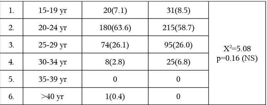
Table III. Distribution of maternal age in birth trauma
S. No
Age group Cases* n=283 Controls*
n=366