0022-538X/94/$04.00+0
Copy,right ©D 1994,AmericanSocietyforMicrobiology
A
Ca2+/Calmodulin-Dependent
Protein
Kinase, CaM
Kinase-Gr,
Expressed
after
Transformation
of
Primary
Human B
Lymphocytes
by
Epstein-Barr
Virus (EBV) Is
Induced
by the EBV
Oncogene
LMP1
GEORGE MOSIALOS,' SILVA H. HANISSIAN,2 SATYAJAWAHAR,2 LISAVARA,' ELLIOYT KIEFF,'
ANDTALALA. CHATILA2*
Departments ofMedicineandMicrobiology andMolecularGenetics, HarvardMedicalSchool,'
andDivision ofImmunology, The Children'sHospital, and Department ofPediatrics,
Harvard MedicalSchool,2Boston, Massachusetts 02115 Received8 October 1993/Accepted8 December 1993
CaM kinase-Gr is a multifunctional Ca2+/calmodulin-dependent protein kinase which is enriched in neurons and T lymphocytes. The kinase is absent from primary human B lymphocytes but is expressed in
Epstein-Barr virus (EBV)-transformed B-lymphoblastoid cell lines, suggesting that expression of the kinase
can beupregulated by an EBVgene product(s). We investigated the basis of CaM kinase-Gr expression in EBV-transformed cells and the mechanisms that regulate its activity therein by usinganEBV-negative Burkitt
lymphoma cell line, BJAB, and BJAB cellsconvertedtoexpression ofindividual EBV proteins bysingle-gene transfer.CaMkinase-Gr expressionwasupregulatedin BJAB cells by EBVlatent-infection membrane protein
1 (LMP1) but not by LMP2A or by nuclear proteins EBNA1, EBNA2, EBNA3A, and EBNA3C. In
LMP1-convertedBJAB cells, the kinasewasfunctional andwasdramatically activateduponcross-linking of surface immunoglobulin M. Overlapping cDNA clones that encode human CaM kinase-Grweresequenced, revealing 81%aminoacid identitybetween therat and human proteins. Transfection of BJAB cells withanexpression
constructforthe humanenzymeresulted inafunctional kinase whichwasshownbyepitope taggingtolocalize primarily to cytoplasmic and perinuclear structures. Induction of CaM kinase-Gr expression by LMP1 provides thefirst example ofa Ca2+/calmodulin-dependent protein kinase upregulated byaviral protein. In
view of the key role played by LMP1 in B-lymphocyte immortalization by EBV, these findings implicate CaM kinase-Gras a potential mediator of B-lymphocyte growth transformation.
Ca2+ isakey secondmessengerthatmediatesabroadarray ofcellular responses inducedby diverse extracellular stimuli, such as growth factors, hormones, neurotransmitters, etc.
Many of the effects of elevated free intracellular Ca2+ are
transmitted by the Ca2+ receptor calmodulin which, upon
bindingCa2+, activates adiversefamily of effector molecules.
An important component of the Ca2+ signalling cascade in mamalian cells is afamily ofserine-threonine-specific, Ca-'/ calmodulin-dependent protein kinases, whose members have beenimplicated inabroadarrayof cellular processes, includ-ing neuronal development and plasticity, cellular contractility and motility, carbohydrate metabolism, cell cycle progression, and gene expression (reviewed in reference 11). Ca2+/calm-odulin-dependent protein kinases are classified into two
groups. Members of the first group, which include myosin
light-chain kinase, phosphorylase kinase, and the Ca2+/calm-odulin-dependent protein kinasestypeIII,arededicatedtothe phosphorylation of onespecific substrate and hence serve to
regulate single important cellularprocesses. Members of the second group, which include the Ca2+/calmodulin-dependent protein kinase types Ia, Tb, and II and
Ca2+/calmodulin-dependent protein kinase type Gr (CaM kinase-Gr), are multifunctional protein kinases that have broad substrate specificities and serve to regulate a wide array of cellular processes (35).
CaMkinase-Gr is unique among the multifunctional Ca2/
calmodulin-dependent protein kinasesin that itsexpression is
*Correspondingauthor.Mailingaddress:Division of Immunology-Rheumatology, Department of Pediatrics, Washington University
School of Medicine, One Children's Place, St. Louis, MO 63110.
largely restricted to neurons and T lymphocytes. The kinase waspurified and cloned fromratbraintissue, where it exists in two monomeric isoforms, a 65,000-Mr ox polypeptide and a 67,000-Mr polypeptide.These two isoforms share adomain
structure common to other Ca2+/calmodulin-dependent
pro-tein kinases that includes a catalytic domain, a calmodulin-binding domain,andanassociative domain whichmay serveto
mediate interactions with other proteins (14, 23, 28, 30). The
twoisoforms of CaM kinase-Grare encodedbyasinglegene, which also codes for calspermin, a testis-specific calmodulin-binding protein which comprises the calmodulin-binding and associative domains but lacks thecatalytic domain. Calspermin is generated by alternate transcription initiation and
alterna-tivesplicing of internal exons (14, 23, 28,30).
CaM kinase-Gr is widely distributed in therat brain andis particularly enriched in cerebellar granule cells, from which theenzymederives itsname(6,29). In peripheral tissues, CaM kinase-Gr is highly expressed in developing Tlymphocytes in the thymus, where its levels are comparable to those in the cerebellum(5). The kinase is also expressedatlowlevels inthe spleen andtestesbutis undetectable in several other tissues(5, 23).
In human lymphoid tissues, CaM kinase-Gr is exclusively found in T lymphocytes; it is most abundant in immature thymocytes, and its levels drop byseveralfold in thecourseof T-cell maturation (9).The humanenzymehastwomonomeric isoformswith sizes of57 and 60 kDa. The catalyticactivity of the humankinasewasdramatically upregulated upon
engage-mentof T-cellantigen receptormolecules by ligands, indicat-ing that CaM kinase-Grisacomponent oftheCa2+signalling
pathway in Tlymphocytes.
1697
on November 9, 2019 by guest
http://jvi.asm.org/
1698 MOSIALOS ET AL.
While the kinase was not detected in primary human B
lymphocytes, it is expressed in Epstein-Barr virus (EBV)-transformedB-lymphoblastoidcell lines (9), raisingthe
possi-bility that this kinase is important for EBV-mediated
B-lymphocyte growthtransformation. Intransforming primaryB
lymphocytes,EBVexpressessixnucleoproteins(EBNAs),two
integral membrane proteins (LMPs), and two small RNA
species (EBERs) (16, 19). EBNA LP, EBNA2, EBNA3A,
EBNA3C, and LMP1 are essential forprimary B-lymphocyte growth transformation, while EBNA3B, LMP2, and the EBERs are not (16, 19). In this study, we examined the
capacityof these EBV genestoinduce CaMkinase-Gr expres-sion in a kinase-negative, non-EBV-infected Burkitt's lym-phoma (BL) cell line, BJAB. Induction of CaM kinase-Gr
expression was shown to be mediated by EBV LMP1. The LMP1-induced kinase was found to be activated by the
in-crease in the intracellular
Ca2+
concentration which followscross-linking of surface immunoglobulins. A cDNA which encodes both isoforms of the kinasewasderived from EBV-infected BLcells,sequenced,andexpressedin BJAB cells. The recombinant kinases localized to thecyoplasmic and
perinu-clearareasandwerealso activatedupon
signalling
via surfaceimmunoglobulinmolecules.
MATERIALSAND METHODS
Cells. BJAB is an
EBV-negative
BLcell line(24).
BJAB clonesstably expressing
LMP1, LMP2A, both LMP1 andLMP2A, EBNA1, EBNA2, EBNA3A,orEBNA3Cwere gen-erated as
previously
described(21,
25,35).
Jurkat human leukemic T-cell clone JE6.1wasobtained from the AmericanTypeCulture Collection
(Bethesda,
Md.).Human EBV-trans-formedB-lymphoblastoid
cell lineswere prepared and main-tained aspreviously
described(37).
Immunoblotting. For Westernblot
(immunoblot)
analysis,cellswere
lysed
onice for 15 min in abuffer(cell lysis
buffer)
containing
25 mMHEPES(N-2-hydroxyethylpiperazine-N'-2-ethanesulfonic
acid;
pH7.5),
0.5%NonidetP-40,50mMNaCl,
25 mM NaH2PO4, 25 mM NaF, 12.5 mM Na4P207, 2 mM
EDTA, 2 mM ethylene
glycol-bis(P-aminoethyl
ether-N,N,
N',N'-tetraacetic acid
(EGTA),
1 mM Na3VO4, 100 ,ug ofphenylmethylsulfonyl fluoride per ml, and 1
jig
each of the protease inhibitorsleupeptin,
pepstatin, chymostatin,
andan-tipain
perml. Thelysateswerethen cleared of nuclei and other insolublematerialby
centrifugation
at16,000
xgfor 30 minat4°C. Supernatant
samples
containing
200 ,ug ofprotein
wereboiled in sodium
dodecyl
sulfate(SDS)-containing
samplebuffer,
subjected todenaturing
polyacrylamide gelelectro-phoresis, and then electroblotted onto
polyvinylidene
difluo-ride(PVDF)
membranes(Millipore
Corp.,Bedford, Mass.).
The filters were then incubated for 1 h at 23°C in blotting
buffer
(10
mMTris-HCl[pH 7.5],
150mMNaCl,2mMEDTA,0.05%Tween
20)
containing5% nonfat milkandthen rinsed withblottingbuffer and incubated for 3 h at230C
with either CaMkinase-Gr-specific
polyclonalrabbit antiserumat a1:200 dilution in blottingbuffer (9) orwith anti-LMP1 monoclonalantibody
S12at a1:2dilution inblottingbuffer(21).Thefilterswere washed, and the LMP1 immunoblotswere additionally incubated for 1 h at 23°C with affinity-purified rabbit
anti-mouse immunoglobulin G (IgG) (Cappel) at 1
jig/ml
inblottingbuffer. Thefiltersweresubjectedtoafinal incubation with
I251-labeled
protein G (New England Nuclear) at 1 to 2,uCi
per membrane for 1 h at 230C and then washed free of125I-labeled
protein G and dried. Radiolabeled bands werevisualizedby autoradiography. For some CaM kinase-Gr
im-munoblots, alkaline phosphatase-conjugated goat anti-rabbit
IgG (Cappel)wasused as a secondary antibody. These blots
were developed with nitroblue tetrazolium and 5-bromo-4-chloro-3-indolylphosphate.
Immunoprecipitation and gel electrophoresis. Cells were
washed and thensuspended at 2.5 x 10⁷ cells per ml in RPMI 1640 mediumsupplemented with 10% fetal calf serum, incu-batedat37°C for 10min, and treated as indicated in thefigure legends. The treatment was terminated by pelleting the cells for5sinamicrocentrifuge andlysing them for 15 minonice in lysis buffer. Nuclei and insoluble material were removedby
centrifugation, and the supernatantswerepreclearedovernight
at4°C with 25 ,u of packed protein G-Sepharose(Boehringer MannheimBiochemicals) precoated with normal rabbitserum.
CaM kinase-Gr was then precipitated by incubating the pre-cleared lysates for 4 h at 4°C with 25 ,u of proteinG-Sepharose precoated with anti-CaM kinase-Gr serum. The beads were washed four times with lysis buffer, and the
immunoprecipi-tatesweresolubilized andsubjected to denaturing acrylamide gel electrophoresis as previously described (9).
Assay of CaM kinase-Gr
activity
in immunoprecipitates. CaM kinase-Gr immunoprecipitates were prepared as de-scribed above, except that the kinase-containing beads were washed twice in abuffercontaining 25 mM HEPES (pH 7.5), 0.5 mMEDTA, 0.5 M NaCl, 100 ,ug of phenylmethylsulfonyl fluoride per ml, and 1 ,ug each of the protease inhibitors leupeptin, pepstatin, chymostatin, and antipain per ml and then washed two additional times in this buffer but without NaCl. The beadswerefinallyresuspended in 100[LI
of the last buffer and used for enzyme assays. Assays of purified CaM kinase-Gractivitywerecarriedoutfor 10 min at23°C inafinal volume of 100 ,ul containing 50 ,u of the bead-wash buffer suspension and 50RI
of a reaction mixturecontaining 20 mM HEPES (pH 7.5), 10 mM MgCl2, 50 ,uM ATP, 1 jiCi of [_y-32P]ATP, a 20 jiM concentration of the substrate peptidesyntide-2 (Bachem), and either 5 mM CaCl2 and 600 nM calmodulin (Pharmacia) or 5 mM EGTA. The reaction was terminatedby spotting 25 1.1 of the mixture onto P81 phospho-cellulose filters (Whatman). The filters were washed three times in0.5%phosphoric acid,allowedtoairdry, and counted invials containing scintillation fluid.
cDNA
library
screening. A lambda gtlO cDNA library prepared from EBV-positive BL cell line BL41/B95-8 (1) was screened for isolation ofa human lymphoidcDNA for CaMkinase-Gr. Approximately 150,000 phage plaques were
screened. Theprimary screeningwasperformedwithaprobe made from a 454-base-pair (bp) fragment ofa human CaM kinase-Gr cDNA correspondingto bp +727 to +1171 ofrat
CaM kinase-Gr (23). The probe was generated by PCR
amplification (9) andwas labeled with a random-hexanucle-otideDNA-labelingkit(BoehringerMannheimBiochemicals).
The phage plaqueswerelifted with colony-plaque screen filters
(NEN), andthefilterswerehybridized at42°C for 18to24 h
in abuffercontaining 50% formamide,6 x SSPE (1x SSPE is 0.18MNaCl, 10 mMNaPO4, and 1 mMEDTA[pH7.7]), 1% SDS, 1x Denhardt'ssolution,and 100
jig
of sonicatedsalmon sperm DNA per ml. The filters were washed twice underhigh-stringency conditions for 30 min each timeat 65°Cina
buffercontaining0.2x SSPE, 1% SDS, and0.05%sodium PP1 and then subjected to autoradiography. Positive clones were
plaque purified. Certain positive clones were also screened with aprobespanning bp +1 to +752 ofratCaMkinase-Gr
cDNA (23). Lambda DNA was prepared from the positive clones. Inserts were excised with EcoRI digestion and sub-clonedintoplasmid pSG5(Stratagene) forsequencing.
Over-lappingclones weresequenced withprimerssynthesizedonthe basis of a newly generated sequence (Sequenase kit; United
J. VIROL.
on November 9, 2019 by guest
http://jvi.asm.org/
StatesBiochemical).Alignment of the amino acid sequences of the human, rat, and mouse enzymes was done with the
CLUSTAL program (PCGene; InteliGenetics, Mountain
View, Calif.).
Amplification and subcloning of human CaM kinase-Gr
cDNA.Thecoding region of human CaM kinase-Gr was PCR
amplified from lambda clone 4.1 by using primers GR-1
(5'-CGCAGATCTGCGAAGATGCTCAAAGTCAC-3')
and GR-2(5'-GCCAGATCTGAAGGAAGCTG'TTAG-3').
The PCR product was purified by extraction with phenol andchloroformand then ethanol precipitated. Following
purifica-tion, the PCRproduct was digested with BglII, purified bygel
electrophoresis, and subcloned into the BglII site of plasmid
pSG5
togeneratepHGR. Toplace a FLAG epitope tag (IBI)ontheamino terminus of CaM kinase-Gr, the coding region of human CaMkinase-Gr was PCR amplified from lambda clone
4.1 by using FLAG-encoding primer GR-FLAG (5'-GAAG
ATCTCTGAGATGGACTACAAGGACGACGATGACAA ACTCAAAGTCACGGTGCCCTCCTGCTC) and primer GR-2. The PCR product was subcloned into the BglII site of
pSG5
asdescribed above to generate plasmid pHGR-FLAG.RNA isolation and hybridization. Total cellular RNA was
prepared from cell lines by guanidinium isothiocyanate lysis, and
poly(A)+
RNAwas selected over poly(dT) (34). A 5-jigsample
of each poly(A)+ RNA was size fractionated byelectrophoresis ina1% agarose-formaldehydegel, transferred to a nitrocelluose filter, and hybridized with a randomly
primed,
32P-labeled
cDNA probe spanning the entire codingregion
of human CaM kinase-Gr. The filters were washed understringentconditions, dried, and subjected toautoradiog-raphy.
PCRanalysis ofCaM kinase-Gr transcripts. PCR analysis
of CaM kinase-Gr transcripts was carried out as previously
described, with some modifications (9). Total cellular RNA waspreparedby lysis of cells with guanidinium isothiocyanate, followedby centrifugationof the lysate over a CsCl cushion. A
20-jig
sample of total RNA from each cell type was primed with random hexamers and reverse transcribed. The reversetranscription reaction products were resuspended in a final volume of 20
RI.
Aliquots (2 ,ul) of the reverse transcription reaction productswere used for PCR amplification with the Perkin-Elmer-Cetus RNA PCR kit (catalog no. N808-0017) inaccordance with the manufacturer's instructions. CaM kinase-Gr amplifications used primers 5'-GCGATCAGTTCATGT TCAGG-3' (sense primer), corresponding to bp 836-855, and
5'-ACC'ITCATCAGCTCAGCCTGTG-3'
(antisense primer),complementarytobp 1268 to 1290 of the human CaM kinase-Gr sequence. Controlamplification reactions used the human
,-actin
primers (Stratagene catalog no. 302010). Both CaMkinase-Grand,-actinantisense primers were end labeled with
[y-32P]ATP
(3,000 Ci/mmol; New England Nuclear) by using T4polynucleotidekinase, and 1.0 x 10⁶cpmwas used for each PCR. PCRconditions included 30 cycles of 1 min of denatur-ation at 95°C, 2 min of annealing at 50°C, and 3 min of extensionat72°C.Followingamplification,5-pI
aliquots of the CaM kinase-Gr and3-actin
PCRproducts were combined and then resolvedbyelectrophoresis ona12% acrylamide gel. Thegel
was dried under vacuumand exposed to Kodak X-Omat film.Transfections. BJABcells (1.5 x 10⁷)wereelectroporated with 30
jig
ofplasmidDNAin 0.4 ml ofRPMIcontaining 10% fetal calfserumbyusingaBio-Rad Gene Pulser at 200 V and 960jiF.
CaM kinase-Gr expression was analyzed by indirect immunofluorescence orimmunoblotting 18 to 72 h later.Immunofluorescence. Transfected cells were washed in
phosphate-buffered
saline (PBS) and dried quickly onto slides-m cNCCV.C)
m < << < < X
-J < Z X.
a-co co co fflco co X 3 x x
200
100-
6946
-
31-
14-FIG. 1. CaM kinase-Gr expression in BJAB BL cells is specifically induced by LMP1. Protein (200
,ug)
of lysates of the respective cell populations was resolved on 5 to 16% denaturing polyacrylamide gels, transferred to PVDF membranes, and probed for CaM kinase-Gr polypeptides as detailedin Materials and Methods. LCL is an EBV-transformed B-lymphoblastoid cell line. BJAB is an EBV-negative human lymphoma cell line. EBNA1-1 and EBNA1-2 are BJAB cell clones that stably expressEBNA1.EBNA3A-SC and EBNA3A-4C are BJAB clones that stably express EBNA3A. EBNA3C-7 and EBNA3C-8 are BJAB clones that stably express EBNA3C. LMP1.2 and LMP1.3 are BJAB clones that stably express LMP1. LMP2A.1 and LMP2A.3 are BJAB clones that stably express LMP2A. CaM ki-nase-Gr isoforms are indicated by the arrows. The numbers on the left are molecular size markers in kilodaltons.at
37°C.
The cells were then blocked with PBS containing 20% goat serum for 30min.
The cellswere then incubatedat37°C
for 1 h with 10 jig of monoclonal antibody M2 (IBI), which recognizes the FLAG epitope, per ml. Following incubation,
thecells were washedfive times for 4 mineach timewith PBS containing 20% goat serum and then incubated as described above, with a fluorescein isothiocyanate-conjugated goat anti-mouse antibody. The cells were subsequently washed five times for 4 min each time in PBS, mounted, and visualized with a Zeiss Axioskop.
Nucleotide sequence accession number. The human CaM kinase-Gr cDNA sequence has been deposited in the GenBank data base and assigned accession no. L24959.
RESULTS
Induction of CaM kinase-Gr expression by EBV is mediated
by LMP1. CaM kinase-Gr was not detectable by
immunoblot-ting in EBV-negative BL cell line BJAB (Fig. 1). However, the
kinase was readily detectable in an EBV-transformed B-lymphoblastoid cell line, used as a positive control, and in
BJAB clones expressing LMP-1. Both the 57- and 60-kDa isoforms of CaM kinase-Gr were abundant in EBV-trans-formed B-lymphoblastoid cell lines and in LMP1-transfected
BJAB clones. Significantly, the kinase was absent fromother clones expressing LMP2A, EBNA1, EBNA3A, or EBNA3C.
The kinase was also absent from two EBNA2-expressingBJAB clones and was readily detected in nine LMP1-expressing
on November 9, 2019 by guest
http://jvi.asm.org/
[image:3.612.318.549.77.289.2]1700 MOSIALOS ET AL.
< <
J < z z
u co c
w UJ
:::
0T
'Oc.
eiD. 0. 0. 0 0. a. 0. 0_
S V--c
-~~~~~~~IIl-- mwA
E
CaMkinase-Gr .f
L
LMP1 c
FIG. 2. Analysis of CaM kinase-Gr expression in a panel of LMP1-transfected BJAB clones. Panels: top, immunoblot analysis of CaM kinase-Gr expression in a panel of LMP1 BJAB clones; bottom, immunoblotanalysis of LMP1 expression in the same group of BJAB clonespresentedinthe toppanel. Immunoblottingwascarried outby resolving 200p.gof protein of lysates of the respective cell populations
on denaturing 8% polyacrylamide gels and then transferring the proteins to PVDF membranes and probing them for CaM kinase-Gr polypeptidesorforLMP1 asdetailed in Materials and Methods. LCL is an EBV-transformed B-lymphoblastoid cell line. EBNA2-1 and EBNA2-2 are BJAB clones that stably express EBNA-2. LMP1.1, LMP1.2, LMP1.9.A, LMP1.9.R, LMP1.9.T, LMPl2.1, and LMP12.R
are BJABclones thatstably express LMP1. LMP2A/1.1 and LMP2A/ 1.3areBJABclones thatstably express both LMPI and LMP2A.
BJAB clones (Fig. 2). These included seven clones which expressed LMP1 alone and two clones which coexpressed
LMPI and LMP2A (Fig. 2). The CaM kinase-Gr levelin the LMP1/LMP2A doubly converted clones was comparable to
that in LMP1 converted clones, indicating thatLMP2A does
not influence kinase induction byLMP-1.
Activation of CaM kinase-Gr by cross-linking of surface IgM in LMPI-converted BJAB cells. To determine whether
the CaM kinase-Gr expressed in LMP1-converted BJAB
clonesisfunctional, theenzyme wasimmunoprecipitated from LMP1-expressing BJAB clones that had been left untreatedor
had been stimulated with goat anti-human IgM for various
times, and the kinase immunoprecipitates were examined for
autonomous (i.e., Ca2+/calmodulin-independent) and
Ca2+/
calmodulin-dependent kinase activities.CaMkinase-Gr immu-noprecipitates from untreated LMP1-converted BJAB cells
exhibited minimal autonomous activity and modest Ca2+/
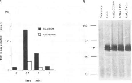
calmodulin-stimulated activity (Fig. 3A).Treatmentwith anti-IgM antibodies resulted in dramatic upregulation of both
autonomous and
Ca2+/calmodulin-dependent
activities (Fig.3A). Enzyme activation by surface 1gM signallingwashighest
at30 s after receptor engagement and rapidly declined there-after. The increased CaM kinase-Gr activity was not due to increased immunoprecipitation of CaM kinase-Gr following anti-IgM treatment, since equal amounts of the kinase were detected in immunoprecipitates of cells before orafter
cross-linking (Fig. 3B). The initialincrease andsubsequent declinein CaM kinase-Gr activity following ligation of surface IgM
molecules correlated with thepreviously reported timecourse for the rise in freeintracellular
Ca2+
concentrationsinLMP1-converted BJAB clones (25). Activation of CaM kinase-Gr following surface IgM cross-linking was partially blocked by chelation of extracellular
Ca2+
with EGTA, consistentwith a role for extracellularCa2+
in kinase activation following surface IgM cross-linking (data notshown).LMP2A has been reported to block Ca2+ mobilization following ligationof surfaceimmunoglobulins, CD19,ormajor
histocompatibility complex class II molecules in B
lympho-B
E -
us
EE' a .
o < < <
~t i
LA:
* Ca.2/CaM
o Autonomous
100
-67
-46
-3-
-31 f0tr
Time (min)
FIG. 3. Upregulation of CaM kinase-Gr catalyticactivity in
LMP1-transfected BJAB clonesby surface IgM signalling. (A) Activation of CaMkinase-Gr by signalling via surface IgM. Aliquots of LMPI clone 9-2 cells (2 x 10⁷ cells per time point) were treated with goat anti-human IgM antibodiesat20,ug/ml for the indicated time periods. At the end of the incubation periods, the cells were lysed in 0.5% Nonidet P-40 lysis buffer and CaM kinase-Gr immunoprecipitates wereprepared and assayed forCa2+/calmodulin-dependentactivityor autonomous (Ca2+/calmodulin-independent) kinase activity. The
as-says were run as described in Materials and Methods, and the data shown represent themeansof three replicatesrunforeach timepoint. Standard deviationswere less than 10% of themeans.Similar results were found in six other independent experiments. (B) Immunoblot analysis of CaM kinase-Gr immunoprecipitates isolated from LMP1 clone 9-2 cells either left unstimulated or stimulated with goat anti-human IgM antibodies (Anti p.) for the indicatedperiods. CaM kinase-Gr immunoprecipitates were resolved on a denaturing 8% acrylamide gel, transferred toPVDFmembranes, and immunobloted with anti-CaM kinase-Gr sera as described in Materials and Methods. Theprominent doublet represents CaM kinase-Gr isoforms(arrow), while the faint broad band is the rabbit IgGheavy chain. Thenumbers
onthe leftaremolecularsize markers in kilodaltons.
cytes. LMP1 partially reversestheinhibitory effect of LMP2A on Ca2+ signalling. LMP2Adoesnot appearto influence the capacity ofLMP1 to induce theexpressionofCaM kinase-Gr (Fig. 2). However, it could be demonstrated in preliminary
experiments that the magnitude of CaM kinase-Gr activation
following surface IgM cross-linking is reduced by more than
50% inLMP2A-superconvertedLMP1 BJABclonescompared
with LMPI clones. This would be consistent with the previ-ously proposed view that LMP2A may act to downmodulatc LMP1 effectsoncellgrowthor toantagonize inductionoflytic
EBVinfection following receptorligation (25).
Cloning and expression of a human cDNA that encodes CaM kinase-Gr.Since human CaM kinase-Gr cDNA hadnot been previously cloned, we isolated and sequenced cDNA
clones for the LMPI-induced kinase mRNA. A lambda gtlO
cDNAlibrary from EBV-positiveBLcelllineBL41/B95-8was
screened with a 454-bp fragment of human CaM kinase-Gr
cDNA(see Materials and Methods). Six positive cloneswere
identified (clones 3.1, 4.1, 4.3, 5.1, 5.2, and 5.3). All six
hybridized to probes from the middle or the 3' EcoRI
frag-ments of rat CaM kinase-Gr cDNA. Clones 4.1 and 5.2 also
hybridizedtoprobesfrom the 5' EcoRIfragmentofratkinase cDNA(bp +I to +752) (23). EcoRIdigestion ofphageDNA,
followed by sequencingofthe inserts, revealedthat clone 3.1
contained a 203-bp insert which corresponds to the middle
EcoRI fragment ofthe rat cDNA and a594-bp EcoRI insert whichcorrespondstothe 3' EcoRI fragmentof theratcDNA.
Clone 4.1 contained 869-, 203-, and 529-bp inserts that, by
J. VIROL.
on November 9, 2019 by guest
http://jvi.asm.org/
[image:4.612.323.554.74.219.2] [image:4.612.65.299.77.191.2]INDUCTION OF CaM KINASE-Gr EXPRESSION BY EBV LMP1
CCCGGACCCAGGTGCTGCCAGGGCGGCGGCTGGCGGCCGGCTTCTCGCTCGGGCAGCGCCGGCGGCGGCGGC 72
GGCGGCTTCCGGAGTCCCGCTGCGAAGATGCTCAAAGTCACGGTGCCCTCCTGCTCCGCCTCGTCCTGCTCT 144
METLeuLysValThrValProSerCysSerAlaSerSerCysSer 15
TCGGTCACCGCCAGTGCGGCCCCGGGGACCGCGAGCCTCGTCCCGGATTACTGGATCGACGGCTCCAACAGG 216
SerValThrAlaSerAlaAlaProGlyThrAlaSerLeuValProAspTyrTrpIleAspGlySerAsnArg 39
GATGCGCTGAGCGATTTCTTCGAGGTGGAGTCGGAGCTGGGACGGGGTGCTACATCCATTGTGTACAGATGC 288
AspAlaLeuSerAspPhePheGluValGluSerGluLeuGlyArgGlyAlaThrSerIleValTyrArgCys 63
AAACAGAAGGGGACCCAGAAGCCTTATGCTCTCAAAGTGTTAAAGAAAACAGTGGACAAAAAAATCGTAAGA 360
LysGlnLysGlyThrGlnLysProTyrAlaLeuLysValLeuLysLysThrValAspLysLysIleValArg 87
ACTGAGATAGGAGTTCTTCTTCGCCTCTCACATCCAAACATTATAAAACTTAAAGAGATATTTGAAACCCCT 432
ThrGluIleGlyValLeuLeuArgLeuSerHisProAsnIleIleLysLeuLysGluIlePheGluThrPro 111
ThrGluIleSerLeuValLeuGluLeuValThrGlyGlyGluLeuPheAspArgIleValGluLysGlyTyr 135
TACAGTGAGCGAGATGCTGCAGATGCCGTTAAACAAATCCTGGAGGCAGTTGCTTATCTACATGAAAATGGG 576
TyrSerGluArgAspAlaAlaAspAlaValLysGlnIleLeuGluAlaValAlaTyrLeuHisGluAsnGly 159
ATTGTCCATCGTGATCTCAAACCAGAGAATCTTCTTTATGCAACTCCAGCCCCAGATGCACCACTCAAAATC 648
IleValHisArgAspLeuLysProGluAsnLeuLeuTyrAlaThrProAlaProAspAlaProLeuLysIle 183
GCTGATTTTGGACTCTCTAAAATTGTGGAACATCAAGTGCTCATGAAGACAGTATGTGGAACCCCAGGGTAC 720
AlaAspPheGlyLeuSerLysIleValGluHisGlnValLeuMetLysThrValCysGlyThrProGlyTyr 207
TGCGCACCTGAAATTCTTAGAGGTTGTGCCTATGGACCTGAGGTGGACATGTGGTCTGTAGGAATAATCACC 792
CysAlaProGluIleLeuArgGlyCysAlaTyrGlyProGluValAspMetTrpSerValGlyIleIleThr 231
TACATCTTACTTTGTGGATTTGAACCATTCTATGATGAAAGAGGCGATCAGTTCATGTTCAGGAGAATTCTG 864
TyrIleLeuLeuCysGlyPheGluProPheTyrAspGluArgGlyAspGlnPheMetPheArgArgIleLeu 255
AATTGTGAATATTACTTTATCTCCCCCTGGTGGGATGAAGTATCTCTAAATGCCAAGGACTTGGTCAGAAAA 936
AsnCysGluTyrTyrPheIleSerProTrpTrpAspGluValSerLeuAsnAlaLysAspLeuValArgLys 279
TTAATTGTTTTGGATCCAAAGAAACGGCTGACTACATTTCAAGCTCTCCAGCATCCGTGGGTCACAGGTAAA 1008
LeuIleValLeuAspProLysLysArgLeuThrThrPheGlnAlaLeuGlnHisProTrpValThrGlyLys 303
AlaAlaAsnPheValHisMetAspThrAlaGlnLysLysLeuGlnGluPheAsnAAraLvsLeuLvs 327 GCAGCGGTGAAGGCTGTGGTGGCCTCTTCCCGCCTGGGAAGTGCCAGCAGCAGCCATGGCAGCATCCAGGAG 1152
AlaAlaVaLysAlaValValAlaSerSerArgLeuGlySerAlaSerSerSerHisGlySerIleGlnGlu 351
AGCCACAAGGCTAGCCGAGACCCTTCTCCAATCCAAGATGGCAACGAGGACATGAAAGCTATTCCAGAAGGA 1224
SerHisLysAlaSerArgAspProSerProIleGlnAspGlyAsnGluAspMetLysAlaIleProGluGly 375
GAGAAAATTCAAGGCGATGGGGCCCAAGCCGCAGTTAAGGGGGCACAGGCTGAGCTGATGAAGGTGCAAGCC 1296
GluLysIleGlnGlyAspGlyAlaGlnAlaAlaValLysGlyAlaGlnAlaGluLeuMetLysValGlnAla 399
TTAGAGAAAGTTAAAGGTGCAGATATAAATGCTGAAGAGGCCCCCAAAATGGTGCCCAAGGCAGTGGAGGAT 1368
LeuGluLysValLysGlyAlaAspIleAsnAlaGluGluAlaProLysMetValProLysAlaValGluAsp 423 GGGATAAAGGTGGCTGACCTGGAACTAGAGGAGGGCCTAGCAGAGGAGAAGCTGAAGACTGTGGAGGAGGCA 1440
GlyIleLysValAlaAspLeuGluLeuGluGluGlyLeuAlaGluGluLysLeuLysThrValGluGluAla 447
GCAGCTCCCAGAGAAGGGCAAGGAAGCTCTGCTGTGGGTTTTGAAGTTCCACAGCAAGATGTGATCCTGCCA 1512
AlaAlaProArgGluGlyGlnGlySerSerAlaValGlyPheGluValProGlnGlnAspValIleLeuPro 471
GAGTACTAAACAGCTTCCTTCAGATCTGGAAGCCAAACACCGGCATTTTATGTACTTTGTCCTTCAGCAAGA 1584
GluTyr** 473
[image:5.612.314.559.73.361.2]AAGGTGTGGAAGCATGATATGTACTATAGTGATTCTGTTTTTGAGGTGCAAAAAAC
FIG. 4. Sequenceof human CaM kinase-Gr cDNA. The DNA and deduced amino acid sequences of human CaM kinase-Gr are shown. The nucleotide sequence is shown on theupper line, and the deduced amino acid sequence is shown on the lower line with the three-letter nomenclature for amino acids. Glycine 53, the first residue of the
consensus ATP-binding site, is indicated by the underlying asterisk. The putative calmodulin-binding site (amino acids 322 to 335) is double underlined. The latter was identified on the basis of its homology with the calmodulin-binding site of rat
Ca2+/calmodulin-dependentprotein kinase typeIla(aminoacids296 to309) (31). Both peptides share structuralfeatures deemed important for calmodulin binding, includinganN-terminal stretch of basic amino acids followed by anotherstretch of hydrophobicresidues and an overall ox-helical secondary structure (100% for CaM kinase-Grpeptide amino acids 322to335),asdeterminedbythealgorithimsofGarnieretal.(8)and Chou and Fasman (2).
sequenceanalysis, correspondtothe5', middle, and 3'EcoRI fragments of the rat cDNA. The middle and 3' EcoRI
frag-ments of the human CaM kinase-Gr cDNA were fully
se-quenced from clone 3.1, and the5' EcoRIfragmentwasfully sequenced from clone 4.1. Clone 4.1 was used for PCR
amplification of the entire open reading frame of CaM ki-nase-Gr (see Materials and Methods). The PCR product of thisamplification wassubcloned into pSG5 and sequenced.
The nucleotide and deduced amino acid sequences of the human CaM kinase-Gr obtained from clones 3.1 and 4.1 are
shown inFig.4, andalignment of the amino acidsequencesof
thehuman,mouse,andratCaMkinase-Grenzymes areshown
in Fig. 5. The human enzyme is predicted to consist of 473
aminoacids,withacalculated Mr of51,925,andcontainsmany of the amino acid residues found to be conserved among
HGRPROT MLKVTVPSCSASSCSSVTASAAPGTASLVPDYWIDGSNRDALSDFFEVES 50
MGRPROT MLKVTVPSCPSSPCSSVTAS----TENLVPDYWIDGSNRDPLGDFFEVES 46
RGRPROT MLKVTVPSCPSSPCSSVTSS----TENLVPDYWIDGSKRDPLSDFFEVES 46
HGRPROT ELGRGATSIVYRCKQKGTQKPYALKVLKKTVDKKIVRTEIGVLLRLSHPN 100
MGRPROT ELGRGATSIVYRCKQKGTQKPYALKVLKKTVDKKIVRTEIGVLLRLSHPN 96 RGRPROT ELGRGATSIVYRCKQKGTQKPYALKVLKKTVDKKIVRTEIGVLLRLSHPN 96
HGRPROT IIKLKEIFETPTEISLVLELVTGGELFDRIVEKGYYSERDAADAVKQILE 150 MGRPROT IIKLKEIFETPTEISLVLELVTGGELFDRIVEKGYYSERDARDAVKQILE 146 RGRPROT IIKLKEIFETPTEISLVLELVTGGELFDRIVEKGYYSERDAADAVKQILE 146
HGRPROT AVAYLHENGIVHRDLKPENLLYATPAPDAPLKIADFGLSKIVEHQVLMKT 200
MGRPROT AVAYLHENGIVHRDLKPENLLYATPAPDAPLKIADFGLSKIVEHQVLMKT 196 RGRPROT AVAYLHENGIVHRDLKPENLLYATPAPDAPLKIADFGLSKIVEHQVLMKT 196
HGRPROT VCGTPGYCAPEILRGCAYGPEVDMWSVGIITYILLCGFEPFYDERGDQFM 250
MGRPROT VCGTPGYCAPEILRGCAYGPEVDMWSVGIITYILLCGFEPFYDERGDQFM 246
RGRPROT VCGTPGYCAPEILRGCAYGPEVDMWSVGIITYILLCGFEPFYDERGDQFM 246
HGRPROT FRRILNCEYYFISPWWDEVSLNAKDLVRKLIVLDPKKRLTTFQALQHPWV 300 MGRPROT FRRILNCEYYFISPWWDEVSLNAKDLVKKLIVLDPKKRLTTFQALQHPWV 296 RGRPROT FRRILNCEYYFISPWWDEVSLNAKDLVKKLIVLDPKKRLTTFQALQHPWV 296
HGRPROT TGKAANFVHMDTAQKKLQEFNARRKLKAAVKAVVASSRLGSASSSHGSIQ 350
MGRPROT TGKAANFVHMDTAQKKLQEFNARRKLKAAVKAVVASSRLGSASSSHTSIQ 346
RGRPROT TGKAANFVHMDTAQKKLQEFNARRKLKAAVKAVVASSRLGSASSSHTNIQ 346
HGRPROT ESHKASRDPSPIQDGNEDMKAIPEGEKIQG- --DGAQAAVKGAQAELMKV 397
MGRPROT ENHKASSDPPSTQDAKDSTDLL--GKKMQEEDQEEDQVEAEASADEMRKL 394
RGRPROT ESNKASSEAQPAQDGKDKTDPL--ENKIQAGDHEA----AKAAADETMKL 390
HGRPROT QALEKVKGADINAEE---APKMVPKAVEDGIKVADLELEEGLAEE 439
MGRPROT QSEEVEKDAGVKEEE---TSSMVPQDPEDELETDDPEMKRD-SEE 435
RGRPROT QSEEVEEEEGVKEEEEEEEEEEETSRMVPQEPEDRLETDDQEMKRN-SEE
...
...
439HGRPROT KLKTVEEAAAPREGQGSSAVGFEVP-QQDVILPEY 473 MGRPROT KLKSVEEEMDPMTEEEAPDAGLGVP-QQDAIQPEY 469
RGRPROT TLKSVEEEMDPKAEEEAAAVGLGVPPQQDAILPEY 474
FIG. 5. Amino acid alignment of mouse (MGRPROT), rat (RGRPROT), and human (HGRPROT) CaM kinase-Gr sequences. The mouse and rat CaM kinase-Gr sequences have been previously published (14, 23, 28). Amino acidsarerepresentedbythesingle-letter code. Identical amino acids are indicated by stars, and conserved amino acid substitutions are indicated by dotes. Gaps inserted to optimize the alignment are indicated by dashes.
serine-threonine-specific protein kinases (10). In common with other members of the family of Ca2+/calmodulin-dependent protein kinases (36), human CaM kinase-Gr was found to contain three major domains: an amino-terminal catalytic do-main,starting at the first residue of the consensusATP-binding site(glycine 53; subdomain I of Hanks et al. [10]) and ending at the conservedresidue arginine 288 (subdomain XI of Hanks et al. [10]), a regulatory domain containing the putative
calmodu-lin-bindingsite (alanine 322toalanine335), aswell as pseusubstrate sequences, and a carboxyl-terminal associative do-main that starts at alanine 342. Additionally, human CaM kinase-Grpossesses a fourth amino-terminal domain (methio-nine 1 to leucine 52) that is also found in rodent CaM kinase-Gr enzymes and is particularly rich in serine residues (22). This domain has been determined, in the case of the rat enzyme, to contain themajorsite of enzyme autophosphorylationand may thus function as a second regulatory domain that upregulates the activity of the enzyme upon autophosphorylation.
As seen inFig. 5, sequencecomparison of the human, mouse (14), and rat (23, 28) enzymes reveals virtual identity between thecatalyticandregulatory domains ofthe human enzymeand
their rodent counterparts. In contrast, significant sequence divergence between the human associative domain and its rodent counterparts was found. The carboxyl-terminal region of the human enzyme is significantly less acidic (the net
negative charge is 13) than the corresponding region of the mouse (net negative charge of31) andrat(net negative charge of32) enzymes.
VOL.68, 1994 1701
on November 9, 2019 by guest
http://jvi.asm.org/
1702 MOSIALOS ET AL.
X m N__
vz CI CJL CLC
=)7im -i
.J ca .J J ~J
Actin AAi
CaMkinase-Gr -- be*
FIG. 6. Analysis of CaM kinase-Gr transcripts in lymphoid cell populations. (A) Northern blot analysis of CaM kinase-Gr transcripts
inselectedB-andT-lymphoid celllines. Samples (5 pLg) of poly(A)+ mRNAsderived from BJAB, LMPI-expressing BJAB clone LMPI.2, lymphoblastoid cell lineLCL, and the Jurkat T-cell linewereapplied perlane and probed witharandomly labeled cDNA probespanning
the entirecoding region ofhumanCaM kinase-Gr. Prominent hybrid-izing mRNAspeciesare indicated by arrowheads, while the locations ofthe 18Sand 28S rRNAbandsareindicated byarrows.(B) Analysis of CaMkinase-Gr transcriptsinlymphocytecelllines byaradiolabeled PCRtechnique. Samples (2p,g)oftotalRNAfromJurkat, LCL, and
BJABcells and LMP2A-expressing BJAB cloneLMP2A.1and
LMP1-expressing BJAB clones LMP1.2 and LMPl.9A were primed with
random hexamers, reverse transcribed, and amplified by using CaM kinase-Gr-specific or,-actin-specific primers. PCRwascarriedoutas
detailedin Materials and Methods, andthe amplified productswere
separated byelectrophoresis on a 12%acrylamide gel.
Aninteresting feature of the enzyme's cDNA is thepresence inthe 5'-untranslated region ofastretch ofsevenCGGtriplet
repeats(bp 58to78). Size amplification of trinucleotiderepeats
in disease genes has been determined to be the underlying genetic defect in myotonic dystrophy, Kennedy disease, Hun-tington disease, and fragile X syndrome (17). Instability of CGGrepeatshasbeenspecifically associated with the fragile X syndrome,adisease in which the affectedgene,FMR], exhibits dramatic expansion of these repeats (7). It remains to be establishedwhetherthe CGGrepeats in theCaMkinase-Gr 5' region arestable orwhether theyvary insizebetween individ-ualsand, ifso,whether this is associated withadiseasestate(s).
Expression of CaM kinase-Gr mRNA was analyzed in human B and T lymphocytes by hybridization of poly(A)+ RNA understringent conditions with acDNA probe encom-passing the coding region of human CaM kinase-Gr. No hybridizing RNA specieswasdetected inBJABcells, whereas 2.3-,3.5-, 4.6-, and 4.8-kbhybridizing RNA specieswereclearly andreproducibly detected inanLMP1-converted BJAB clone,
inanEBV-transformed B-lymphoblastoid cell line, and in the
Jurkathumanleukemic T-cell line.Additionally, larger hybrid-izing RNA specieswere also apparent in all CaM kinase-Gr-expressing cell lines; however, their significance remains
un-known. The multiplicity of mRNA transcripts that hybridized
withaCaMkinase-Gr probemaybe duetoalternative splicing, thepresence ofseveraltranscription initiation sites, and/orthe presence ofseveral polyadenylation sites.
Expression of CaM kinase-Gr transcripts in lymphoid cell populationswasalsoanalyzed by usingasensitiveradiolabeled
PCR method. Even by this technique, no CaM kinase-Gr
transcripts could be detected in BJAB cells or in LMP2A-converted BJAB cells (Fig. 6B). CaM kinase-Gr transcripts
were readily detected in LMPI-converted BJAB clones, in
EBV-transformedlymphoblastoid celllines, and in the human T-cell line Jurkat. These results indicated that LMP1 induces CaM kinase-Grexpression at the mRNAlevel.
0
E -5
0
CL
B
E
'~ E -Z
L o <
100
.E .E
_ ur
67-4
46
-0 0.5 1 5 Time (min)
[image:6.612.66.302.72.184.2]31
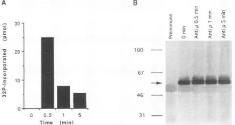
FIG. 7. Expression and activation of recombinant CaM kinase-Gr transcripts in BJAB cells. BJAB cellswere transfected withplasmid pHGR, containing the codingsequenceof human CaM kinase-Gr. At 72h later,aliquots of transfected BJAB cells (2.5 x 10⁷ cellspertime
point)weretreatedwithgoatanti-humanIgM antibodiesat20 p.g/ml for the periods indicated. The cells were then lysed, and CaM kinase-Grimmunoprecipitates wereprepared and immunoblotted or
assayed for Ca2+/calmodulin-dependent kinase activityasdescribed in Materialsand Methods.(A) Activation of wild-type recombinant CaM kinase-Gr molecules following treatment of transfected BJAB cells withanti-surface IgM antibodies (Anti pL). The enzymatic activity data inpanel Bare meansof three replicates done for each time point. The standarddeviationwasless than 10% of themean. Results similarto those reproduced herewerefound in twoother independent
experi-ments. (B) Immunoblots of recombinant CaM kinase-Gr molecules
immunoprecipitated from unstimulated and anti-IgM antibody-treated, transfected BJAB cells. CaM kinase-Grimmunoprecipitates
wereresolvedon adenaturing5to16% acrylamide gel, transferredto PVDF membranes, and immunoblotted with anti-CaM kinase-Grsera as described in Materials and Methods. The prominent doublet
represents CaM kinase-Gr isoforms (arrow), while the faint broad
band is the rabbit IgG heavy chain. The numbers on the left are
molecular size markersinkilodaltons.
Transfection of BJAB cells with the full-length CaM kinase-Gr cDNA under control of the simian virus 40promoterand enhancer in pSG5 resulted in the expression of two closely
migrating proteins which cross-reacted with anti-human CaM kinase-Gr serum and were identical in size to the enzyme
proteins expressed in the Jurkat and the EBV-transformed human lymphoblastoid cell lines (Fig. 7 and data notshown).
Since thecDNA hasonlyoneATG codon nearthe beginning of theopenreading frame and since plasmid expression of the cDNA in BJAB cells gives risetoboth isoforms of thehuman kinase, the cDNA is likely to comprise the complete coding
sequence and the isoforms are probably the product of post-translational processing. The recombinant kinase was func-tional, as evidenced by its enhanced activity following cross-linking of surface IgM molecules in transfected BJAB cells (Fig. 7). The timecourse ofrecombinant kinase activation in BJAB cells mirrored that noted for kinase molecules in LMP1-expressing BJAB clones: enzymeactivitywashighestat
30spoststimulation and declined thereafter (compare Fig. 7A and3A).
Since the antipeptide serum used for immunoblotting and immunoprecipitation of the human enzyme did not work in immunofluorescence experiments,weexamined the
intracellu-lar localization in transfected BJAB cells of a recombinant
human CaM kinase-Gr which had been tagged at its N terminus withtheFLAGepitope (see Materials and Methods). The FLAG epitope-tagged kinasewas similartoitswild-type counterpart in that it was also expressed as two isoforms (slightly shifted in molecular weight because of the additional
B A
N ..,
M- cm
-n J J =3
.
..
28S -*
185 - o
-A
J.VIROL.
on November 9, 2019 by guest
http://jvi.asm.org/
[image:6.612.319.559.76.204.2]FIG. 8. Recombinant CaM kinase-Gr exhibits both nuclear and
cytoplasmic localizations in transfected BJAB and COS cells. (a)
Immunofluoresenceanalysisof BJAB transfectants. BJAB cells(1.5 x
107) were transfected with plasmid pHGR-FLAG, which codes for FLAGepitope-taggedhuman CaM kinase-Gr. At 18 hlater,the cells were analyzed for CaM kinase-Gr expression with an anti-FLAG monoclonal antibody (Kodak) and fluorescein isothiocyanate-conju-gatedgoat F(ab')2 anti-mouse immunoglobulin, as detailed in
Mate-rialsand Methods.(b)Immunofluorescence of COS cell transfectants.
COS cells(1.5 x 10⁷)were transfected withplasmid pHGR-FLAG, andCaM kinase-Grexpressionwasanalyzed2days later,asdescribed
for panela.
tag) andwassimilarly activateduponsignalling via surfaceIgM
molecules (data not shown). The FLAG epitope-tagged
en-zyme appeared to associate, in part, with cytoplasmic struc-tures when expressed in BJAB cells (Fig. 8A). To better visualize the intracellular distribution of CaM kinase-Gr, we
expressed the FLAG epitope-tagged kinase in COS cells to
take advantageof thesuperiorresolution allowedbythelarger cytoplasmic compartment of these cells. The FLAG epitope-tagged kinase was associated with a fine cytoplasmicfibrillar
network which was accentuated around the nucleus. Nuclei
alsostained,consistent withpossiblenuclearlocalization of the
enzyme. However, the apparent nuclear stainingcould
alter-natively be due to the intensity of the perinuclear staining. Previous intracellular localization studies with rat neurons
have demonstrated the localization of therat enzyme toboth cytoplasmicand nuclear compartments (13).
DISCUSSION
The present study demonstrates that CaM kinase-Gr, a
Ca2/calmodulin-dependent protein, is inducible in human B lymphocytes byEBVtransforming protein LMP1 and that the
inducedprotein is fully functional, asevidencedbyits
respon-siveness toCa'+ signallingvia surfaceimmunoglobulin
mole-cules.Thisproteinkinase isnotnormally expressedin primary Blymphocytes; thus,its presence in EBV-transformedprimary
B lymphocytes suggestsspecific association with latent
infec-tion or the concomitant cell growth and transformation. The
specific induction ofthe kinase mRNA in BJABcellby LMP1 ties the kinase to the LMPI pathway and strengthens the
hypothesis that the kinase plays a role in the downstream
mediation ofLMP1 activating and/or transformingeffects.
The CaM kinase-Gr fromEBV-transformed B cells isnearly identical tothe rodentenzymes inthe first 351 aminoacids of the enzyme, which include the amino-terminal regulatory domain, the catalytic domain, and in the calmodulin-binding domain.The last 122amino acidsofthehuman,rat, andmouse enzymes, which comprise most of the carboxyl-terminal
asso-ciative domain, are significantly divergent. The carboxyl
ter-mini of the rodent enzymes are particularlyrich in glutamate
residues, a structural feature common to several nuclear
proteins which may allow these proteins to associate with chromatin (4). Indeed, the rodent enzymehas been shown to
localize, inpart, tothe nuclei ofneurons(13). The C-terminal
region of the human enzyme isless endowed with glutamate
residues and is consequently less acidic. Nevertheless, the human enzyme also localizes in part to nuclear structures of
BJABand COS cells.
LMP1 isanintegral membrane phosphoprotein which bears
no direct homology to other signal-transducing membrane proteins. LMP1 isindespensible for viral transformation and is
one of a restricted number of viral gene products that are expressed duringvirallatency (15, 38). LMP1 exertsprofound
effects on host cell growth. It transforms established rodent
fibroblastsand renders B cellsresistanttoapoptosis.The latter
effect maybe mediatedbybcl-2 andA20, whose expression is
inducedby LMP1 and whichcanprotect Bcellsfromapoptosis
(12). LMP1 also upregulates the expression of several cell surface adhesion moleculesin Bcells, includingLFA-1,LFA3, ICAM-1, andCD23 (39).
While the mechanisms by which this membrane-bound protein effects cellular activation and transformation are not
well delineated, LMP1 may act in part by activating the
function of nuclear transcription factors which, in turn, can
initiate the transcription of LMP1-inducible genes. Evidence forsuchamechanismhas beenprovidedinthecaseof the A20
zinc finger protein (18). This LMPI-inducible gene has been
demonstrated to be transcriptionally activated in LMPI-ex-pressingB cells through cis-acting sequencesfound within its promoter regionwhich recognize nuclear transcription factor
NF-KB. Inthe case of CaM kinase-Gr, it is likely that LMP1 upregulatesthe expression of this kinase by direct transcrip-tional activation rather than by a posttranscriptional
mecha-nism, since nokinase RNAcan be detected in BJAB cells by
Northern (RNA) blot analysis or by a very sensitive PCR
method. NF-KB could mediate the induction of CaM ki-nase-Gr expression byLMP1 aswith the A20 gene.
CaM kinase-Gris the first
Ca2+/calmodulin-dependent
en-zyme determined to be regulated by a viral gene product. Although the role CaM kinase-Gr plays in B-lymphocytetransformation by EBV is undefined, upregulated expression
ofCaMkinase-Gr maymediatesome of theactivating and/or growth-transforming effects of LMP1. The multifunctional
Ca2+/calmodulin-dependent
protein kinase type II, whose substrate specificity overlaps that of CaM kinase-Gr (3, 26),has beenrecently reportedtomediate cellcycleprogressionin fertilizedXenopuseggsbyinactivating M-phasepromotingand
cytostatic factors (20).Onecanspeculatethat CaM kinase-Gr
on November 9, 2019 by guest
http://jvi.asm.org/
[image:7.612.70.276.73.372.2]1704 MOSIALOS ET AL.
plays a similar role in facilitating cell cycle progression in EBV-transformed B lymphocytes. The apparent cytoskeletal and perinuclear-nuclear localization of CaM kinase-Gr is compatible with its being a modulator of cell shape and/or
motility and/or of the activity of a cytoskeleton-associated nuclearfactor.Itscapacitytoinfluence cell growth and motility is also supportedby the previously reported range of substrates of the rodent enzyme, which includes thep21ras-related RaplB
GTP-binding protein (33),histoneHi, myosin lightchain- and
microtubule-associated proteins MAP-2 and Tau (26), and transcriptional factors CREB (cyclicAMPresponse
element-binding protein) and SRF(serum responsefactor) (3). Addi-tional studies, includinggenetic approaches which target the
expression and/or activity of the kinase, are required to
delineate the role of CaM kinase-Gr in B-lymphocyte activa-tion and transformaactiva-tion.
Finally, Arole for CaM kinase-Gr in B-cell
lymphomagen-esis is further supported by the intriguing observation that while CaMkinase-Gr is absent from normal humanBcells and from humanpre-B-cell lines Reh andNalm-6, itwasfound to
be expressed in several of the EBV-negative BL cell lines examined (e.g., BL30, BL41, and Ramos), with the sole
exception of BJAB (our unpublished observations). Unlike BJAB, which demonstratesnochromosomaltranslocations,all oftheaforementionedBLcelllines bear thechromosome 8:14 translocations common to this malignancy (27, 32). Further studiesarerequiredtoexaminethe link betweenchromosomal translocations in B-cell lymphomas, including BLs, and the
expressionof CaM kinase-Gr. Italso remainstobe established whether LMP1 induces expression of CaM kinase-Gr in
epi-thelial cells, which are themselves a prime target of EBV
transformation.
ACKNOWLEDGMENTS
We thank Richard Longnecker, Cheryl Miller, BlakeTomkinson, and FredWangforprovidingcelllines,MarkBirkenbachforproviding the cDNAlibrary,andAnthonyMeansforcommunicatingdataprior topublication.
This researchwassupportedbyPublic Health Service research grant CA 47006from the National Cancer Institute(to E.K.) andNational Institutes of Health grant1R29AI30550-01fromthe National Institute ofAllergyandInfectious Diseases (toT.A.C.).
REFERENCES
1. Birkenbach,M.,K.Josefsen,R.Yalamanchili,G. Lenoir,andE.
Kieff. 1993. Epstein-Barr virus-induced genes: first lymphocyte-specific G protein-coupled peptide receptors. J. Virol. 67:2209-2220.
2. Chou, P. Y., and G. D. Fasman. 1974. Prediction of protein conformation. Biochemistry13:222-245.
3. Cruzalegui,F.H.,andA. R.Means.1993.Biochemical character-ization of themultifunctionalCa+2/calmodulin-dependentprotein kinase type IVexpressedininsect cells. J. Biol. Chem. 268:26171-26178.
4. Earnshaw,W. C.1987. Anionicregionsinnuclearproteins.J.Cell Biol. 105:1479-1482.
5. Frangakis,M.V.,T.Chatila,E.R.Wood,andN.Sahyoun. 1991. Expression of a neuronal Ca+2/calmodulin-dependent protein kinase,CaM kinase-Gr,inratthymus.J.Biol. Chem. 266:17592-17596.
6. Frangakis, M. V., C.-A. Ohmstede, and N. Sahyoun. 1991. A
brain-specific Ca+2/calmodulin-dependent protein kinase (CaM kinase-Gr) is regulated by autophosphorylation. J. Biol. Chem. 266:11309-11316.
7. Fu,Y.-H., D. P. A. Kuhl, A.Pizzuti,M. Pierti, J. S. Sutcliffe, S. Richards,A.J.M.H.Verkerk,J. J. A.Holden,R.G.Fenwick, S.T.
Warren, B. A. Oostra, D. L. Nelson, and C. T. Caskey. 1991. Variation of the CGG repeatatthefragileXsite results in genetic instability:resoultion of the Shermanparadox.Cell 67:1047-1058.
8. Garnier, J.,D.J.Osguthorpe,andB. Robson.1978.Analysisof the accuracy and implications ofsimple methods forpredicting the secondarystructureofglobular proteins.J. Mol.Biol. 120:97-120. 9. Hanissian, S.H., M.Frangakis,M. M. Bland, S.Jawahar, and T.A.Chatila. 1993.ExpressionofaCa+2/calmodulindependent
protein kinase,CaMkinase-Gr, in humanTlymphocytes. Regu-lationof kinaseactivity byTcell receptor signalling. J. Biol. Chem. 268:20055-20063.
10. Hanks, S. K., A. M. Quinn, and T. Hunter. 1988. The protein kinasefamily: conserved features and deduced phylogenyof the catalyticdomains. Science241:42-52.
11. Hanson,P.I.,and H.Schulman. 1992. Neuronal Ca+2/calmodu-lin-dependent proteinkinases. Annu. Rev. Biochem. 61:559-601. 12. Henderson, S., M. Rowe, C. Gregory, D. Croom-Carter, F.Wang, R.Longnecker,E.Kieff,and A.Rickinson. 1991. Induction ofbcl-2 expression by Epstein-Barr virus latent membrane protein 1 protects infected B cells from programmed cell death. Cell 65: 1107-1115.
13. Jensen,K.F., C.-A. Ohmstede,R.S.Fisher, and N.Sahyoun.1991. Nuclear and axonal localization ofCa+2/calmodulin-dependent protein kinase type Gr in rat cerebellar cortex. Proc. Natl. Acad. Sci. USA88:2850-2853.
14. Jones, D. A., J. Glod, D. Wilson-Shaw, W. E. Hahn, and J. M. Sikela. 1991. cDNA sequence and differential expression of the
mouse Ca2+/calmodulin dependent protein kinase IV gene. FEBS Lett. 289:105-109.
15. Kaye, K. M., K. M. Izumi, and E. Kieff. A membrane protein essential for Blymphocyte growth transformation by EBV. Proc. Natl. Acad.Sci.USA, in press.
16. Kieff, E., and D. Liebowitz. 1990. Epstein-Barr virus and its replication, p. 1889-1920. In B. N. Fields and D. M. Knipe(ed.), Virology. Raven Press, New York.
17. Kuhl, D. P. A., and C. T. Caskey. 1993. Trinucleotide repeats and genomevariation. Curr.Opin. Genet. Dev. 3:404-407.
18. Laherty, C. D., H.M.Hu, A. W.Opipari,F.Wang,and V. M.Dixit. 1992. The Epstein-Barr virus LMP1 gene product induces A20 expression byactivating nuclear factor KB. J. Biol. Chem. 267: 24157-24160.
19. Leibowitz, D., and E. Kieff. 1993. Epstein-Barr virus, p. 107-172. In B. Roisman, R.Whitley,and C. Lopez (ed.),The human herpes viruses. Raven Press, New York.
20. Lorca, T., F. H. Cruzalegui, D. Fesquet, J.-C. Cavadore, J. Mery, A. Means, and M. Doree. 1993. Calmodulin-dependent protein kinase II mediates inactivation of MPF and CSF upon fertilization ofXenopus eggs. Nature (London) 366:270-273.
21. Mann, K. P., D. Staunton, and D.A.Thorley-Lawson. 1985. An Epstein-Barr virus-encoded protein found in plasma membranes of transformed cells. J. Virol.55:710-720.
22. McDonald, 0. B., B. M. Merrill, M. Bland, L. C.E.Taylor,andN. Sahyoun. 1993.Site and consequences of theautophosphorylation ofCa+2/calmodulin-dependent protein kinase, type 'Gr', CaM kinase-Gr. J. Biol. Chem.268:10054-10059.
23. Means, A. R., F.Cruzalegui, B. LeMagueresse,D.S.Needleman, G. R. Slaughter, and T. Ono. 1991. A novel Ca2+/calmodulin-dependent protein kinase and a male germcell-specific calmodu-lin-binding protein are derived from the same gene. Mol. Cell. Biol. 11:3960-3971.
24. Menezes, J., W. Leibold, G. Klein, and G. Clements. 1975. Establishment and characterization of an Epstein-Barr virus (EBV)-negative lymphoblastoid B cell line (BJA-B) from an exceptional, EBV-genome negative African Burkitt'slymphoma. Biomedicine 22:276-284.
25. Miller, C. L.,R.Longnecker, and E. Kieff. 1993.Epstein-Barrvirus
latent membrane protein 2A blocks calcium mobilization in B lymphocytes.J. Virol. 67:3087-3094.
26. Miyano, O., I.Kameshita, and H. Fujisawa. 1992. Purification and characterization of a brain-specific multifunctional calmodulin-dependent protein kinase from rat cerebellum. J. Biol. Chem. 267:1198-1203.
27. Nilsson, K., and G. Klein. 1982. Phenotypic and cytogenetic characteristics of humanB-lymphoid cell lines and their relevance for theetiologyofBurkitt'slymphoma.Adv. CancerRes. 37:319-380.
J.VIROL.
on November 9, 2019 by guest
http://jvi.asm.org/
28. Ohmstede,C.-A., M. M. Bland, B. M. Merrill, and N.Sahyoun.
1991. Relationship ofgenes encoding
Ca+2/calmodulin-depen-dent protein kinase Gr andcalspermin:a genewithinagene.Proc.
Natl. Acad.Sci.USA88:5784-5788.
29. Ohmstede, C.-A., K. F. Jensen, and N. E. Sahyoun. 1989. Ca+2/ calmodulin-dependent protein kinase enrichedincerebellar gran-ulecells. J. Biol. Chem.264:5866-5875.
30. Ono, T., G. R. Slaughter, R. G. Cook, and R. Means. 1989. Molecular cloning and distribution of rat calspermin, a high affinity calmodulinbindingprotein. J. Biol. Chem. 246:2081-2087.
31. Payne, M. E., Y.-L. Fong, T. Ono, R. J.Colbran,B. E. Kemp,T. R.
Soderling,and A. R. Means.1988.Calcium/calmodulin-dependent protein kinaseII.Characterization of distinct calmodulin binding andinhibitory domains. J. Biol.Chem. 263:7190-7195.
32. Rowe, M.,C. M. Rooney, C. F. Edwards, G. M. Lenoir, and A. B.
Rickinson. 1986.Epstein-Barr virusstatusandtumorcell
pheno-typeinsporadic Burkitt's lymphoma. Int.J. Cancer37:367-373.
33. Sahyoun, N., 0. B. McDonald, F. Farrell, and E. G. Lapetina. 1991. Phosphorylation of a Ras-related GTP-binding protein,
Rap-lb, by a neuronal Ca+2/calmodulin-dependent protein ki-nase,CaM kinase Gr. J. Biol. Chem. 88:2643-2647.
34. Sancho,J.,L. B.Silverman,E.Castigli,D. Ahern, A. P. Laudano,
C.Terhorst, R. S. Geha, and T. A. Chatila. 1992. Developmental regulation of transmembrane signalling via the T cell antigen receptor/CD3 complex in human T lymphocytes. J. Immunol.
148:1315-1321.
35. Schulman,H.1993. The multifunctional Ca+ 2/calmodulin-depen-dent protein kinases. Curr.Opin.Cell Biol. 5:247-253.
36. Schulman, H.,and L. L. Lou. 1989. Multifunctional Ca+2/calm-odulin-dependent protein kinase: domain structure and regula-tion. TrendsBiochem.Sci. 14:62-66.
37. Spertini, F., T. Chatila, and R. S. Geha. 1992. Engagement of
MHC class I molecules induces cell adhesion via both LFA-1 dependent and LFA-1 independent pathways. J. Immunol. 148:
2045-2049.
38. Wang, D., D.Leibowitz, and E. Kieff. 1985. An EBV membrane
protein expressed inimmortalizedlymphocytestransforms
estab-lished rodent cells. Cell 43:831-840.
39. Wang, F., C. Gregory, C. Sample, M. Rowe, D. Liebowitz, R. Murray, A.Rickinson,and E. Kieff. 1990.Epstein-Barrvirus latent
membrane protein (LMPI) and nuclear proteins 2 and 3C are effectors of phenotypic changes in B lymphocytes: EBNA-2 and LMP1cooperatively induce CD23. J. Virol. 64:2309-2318.