GENITAL DERMATOSES IN A DERMATOLOGY
CLINIC OF A REFERRAL HOSPITAL
DISSERTATION
Submitted in partial fulfillment of university regulations for
M.D. DEGREE IN
DERMATOLOGY, VENEREOLOGY & LEPROSY
(BRANCH XX)
CHENGALPATTU MEDICAL COLLEGE,
CHENGALPATTU.
THE TAMILNADU DR.M.G.R.MEDICAL UNIVERSITY
CHENNAI
This is to certify that this dissertation entitled
“CLINICAL SPECTRUM OF NON VENEREAL GENITAL
DERMATOSES IN A DERMATOLOGY CLINIC OF A REFERRAL
HOSPITAL”is a bonafide work done by Dr.P.NITHYA, Postgraduate student
of Department of Dermatology,Venereology and Leprosy , Chengalpattu
Medical College, Chengalpattu – 603 001 during the academic year 2009 –
2012 for the award of degree of M.D. (Dermatology, Venereology and
Leprosy) – Branch XX. This work has not previously formed the basis for the
award of any Degree or Diploma.
Dr.V.Anandan,M.D.,
Head Of Department,
Department of Dermatology,Venereology& Leprosy,
Chengalpattu Medical College,
Chengalpattu– 603 001.
Prof.Dr.P.Ramakrishnan, M.D.,D.L.O.,
DEAN
Chengalpattu Medical College & Hospital
I sincerely thank Dr.P.Ramakrishnan,M.D.,D.L.O., Dean,
Chengalpattu Medical College and Government General Hospital,
Chengalpattu – 603 001 for granting me permission to use the resources of this
I sincerely thank Dr.V.Anandan, M.D.,Head of Department of
Dermatology, Venereology & Leprosy for his invaluable guidance and
encouragement for the successful completion of this study.
I render my sincere thanks to Dr.S.Venkateshwaran, M.D.,D.V.,
Associate Professor , Department of Venereology,for his help and support.
I express my heartfelt gratitude to Dr.G.Srinivasan, M.D.,D.V.,former
Head of Department of Dermatology, Venereology & Leprosy, who was
instrumental in the initiation of this project giving constant guidance
throughout my work.
I immensely thankDr.P.C.Chittambalam, M.D.,D.D.,former Head of
Department of Dermatology,Venereology & Leprosy for his invaluable
guidance in all stages of this study.
I earnestly thank Dr.V.Sivasubramaniam, M.D.,D.V., former
Associate Professor , Department of Venereology.
My sincere and heartfelt gratitude goes toDr.T.K.Anandhi, M.D.,(DVL)
Assistant Professor of Dermatology, for her valuable continuing guidance and
support.
I am thankful to Dr.S.Murugan, M.D.,(DVL)Assistant Professor,
Department of Venereology, for his immense help and encouragement
Department of Dermatology, for her help and support.
I also thank Dr.B.Vijayalakshmi, M.D.,(DVL) former Assistant
Professor Department of Dermatology, for her help and support.
I duly acknowledge the paramedical staff and my colleagues for their
help and favour.
I am deeply grateful to the patients involved in this study for their
S.NO. TITLE PAGE NO.
1 INTRODUCTION 1
2 REVIEW OF LITERATURE 4
3 AIMS OF THE STUDY 40
4 MATERIALS AND METHODS 41
5 OBSERVATIONS 43
6 DISCUSSION 68
7 CONCLUSION 72
8 BIBLIOGRAPHY
9 ANNEXURE
PROFORMA
MASTER CHART
INTRODUCTION
The diseases that affect the genitalia are unique. It is classified into
venereal and non venereal diseases. The term non venereal used to designate
large group of disorders involving the genitalia not transmitted sexually.
Because venereal and non-venereal dermatoses tend to be confused, the
occurrence of these dermatoses may be associated with mental distress and
guilt feelings in affected patients. However, careful dermatological history
taking, a complete cutaneous examination and sometimes a skin biopsy usually
allow accurate diagnosis and satisfactory medical and surgical management in
most cases.
A number of dermatoses and skin tumours affect the genitalia in an
unique (or) distinct manner that they warrant separate discussion. The normal
characteristics of common dermatoses are modified on genitals. For most of
them this may be the only one of the many sites involved while in others it may
be predominantly confined to the genitalia. The features are frequently
modified by moisture in local environment.
The genital area differs between the sexes being the good example of
regional human variation. There is a considerable variability in size, shape,
pigmentation and amount of hair distribution. Moreover, perineal area is
plentifully endowed with functional eccrine, non-functional apocrine sweat
glands and holocrine sebaceous glands usually in association with hair follicles
but also occurring as free glands(Tyson’s glands) around the coronal sulcus.
In males natal cleft, perianal skin, distal penile shaft, prepuce and glans
of dermatoses on the glans and corona. The pattern of keratinisation of the
epithelium also differs throughout the anogenital area. This is most marked at
the mucosal junctions, the prepuce, distal penile shaft and the glans. Rugosity
and thin skin of scrotum allows excellent penetration of topical agents.
In females vulva is the anterior portion of perineum within it are the clitoris,
urethra and vagina. Vulva itself is subdivided into mons pubis, labia majora
and labia minora. Medial aspect of labia majora is smooth and hairless with
numerous sebaceous glands. Vulvar vestibule which extends between clitoris
anteriorly to the posterior fourchette and laterally bounded by labia minora is
the major site for inflammatory disorders.
Sex hormones play a role in regional differentiation and maturation of
skin in this area and may undergo atrophy after menopause. Vulval dermotoses
may confer persistent discomfort in the form of chronic itch (eg. LSA, eczema)
and may be painful (eg. Erosive LP, Pemphigus), which may interfere with
normal activities of daily living. Sometimes it causes relationship problem due
to sexual dysfunction (eg. LSA) and some patients with chronic symptom may
have symptoms and signs of depression.
Since, genital disease caused by dermatoses, frequently resemble those
used by sexually transmitted diseases, it is important to be aware of this. It
causes extreme anxiety in patients, because venereal disease is often patient’s
primary concern.
In premalignant conditions it is important to recognize them at an
is established patient must be reassured. If it occurs in children, the question of
sexual abuse may arise.
This study highlights the conditions and features of non-venereal genital
dermatoses, of both the sexes. This study is to find the pattern of non-venereal
dermatoses in male and female external genitalia and their relative frequencies
REVIEW OF LITERATURE
Though there is no uniform classification for the non venereal genital
lesions, various authors have described them as follows:
Tomaz F has categorized skin diseases producing genital lesions into
four Groups(1)
1. Those caused by infective agents such as bacteria, fungus and viruses.
2. Benign tumors
3. Premalignant and malignant conditions.
4 Miscellaneous category that includes allergic conditions, atrophic
lesions and skin diseases of unknown etiology.
Cutaneous diseases of female genitalia have been described by Pinkus
HS as follows(2)
1. Vulvar manifestations of common diseases.
2. Unique vulvar diseases
3. Infections
4. Vulval neoplasia
5. Vulvar manifestations of systemic diseases
Johnson R.A. has classified cutaneous diseases of male genitalia as
follows:
1. Generalised cutaneous diseases
a. Papulosquamous eruptions
b. Dermatitis
d. Infestations
e. Drug eruptions
f. Mucosal disorders
2. Diseases related to specialized anatomy or function
3. Infections
a. Bacterial
b. Fungal
c. Viral
4. Neoplasia
a. Benign
b. Premalignant
c. Malignant
The available literature on the different diseases affecting the genitalia is
reviewed here. In both males and females it can be discussed under the
following conditions.
I. Congenital
II. Variation from normal conditions.
III. Infections and Infestations.
IV. Inflammatory disorders
V. Genital manifestations of cutaneous diseases
VI. Genital manifestations of systemic diseases.
VII. Adverse cutaneous drug reactions.
VIII. Bites and stings
IX. Mechanical injuries
X. Benign tumors
I. CONGENITAL
Males
Haemangioma of glans penis
Median raphe cyst
Melanocytic naevi
II. VARIATION FROM NORMAL CONDITIONS
Common to both sexes
Fox-Fordyce disease
Cutis anserina
Acrochordans
Males
Pearly penile papules
Phimosis
Paraphimosis
Hypospadias
Females
Vulvar vestibular papillomatosis
III.INFECTIONS AND INFESTATIONS
BACTERIAL
Common to both sexes
Staphylococcal aureus
Streptococcal infections
Ecthyma gangrenosum
Erythrasma
Mycobacterium tuberculosis
Leprosy and rarely others
Males
Fournier’s gangrene
Females
Streptococcal vulvovaginitis
VIRAL
Common to both sexes
Herpes Simplex
Varicella Zoster
Molluscum contagiosum
Human papilloma viral infections and others
FUNGAL
Common to both sexes
Dermatophytosis
Candidiasis
Pityriasis versicolor
Cutaneous cryptococcosis
INFESTATIONS
Common to both sexes
Scabies
Pediculosis
Lymphatic filariasis
Cutaneous larva migrans
Schistosomiasis
Amoebiasis and others
IV.GENITAL MANIFESTATIONS OF CUTANEOUS
DISEASES
Common to both sexes
Psoriasis
Lichen planus
Lichen nitidus
Pityriasis rosea
Seborrheic dermatitis
Vitiligo
Pemphigus vulgaris and its variants
Bullous and Cicatricial pemphigoid
Erythema multiforme and Steven Johnson syndrome
Lichen sclerosus et atrophicus
Primary amyloidosis of skin
Males
Balanitis xerotica obliterans
Plasma cell balanitis
V. INFLAMMATORY DISORDERS
Common to both sexes
Eczema
Allergic contact dermatitis
Irritant contact dermatitis
Contact urticaria
Atopic dermatitis
Hidradenitis suppurativa
Crohn’s disease
Toxic epidermal necrolysis
MALES: FEMALES:
Balanitis Vulval vestibulitis
Balanoposthitis Plasma cell vulvitis
Acute Scrotum Vulvar burning Syndrome
Aphthosis
Priapism
VI. GENITAL MANIFESTATIONs OF SYSTEMIC DISEASES
Common to both sexes
Reiter’s disease
Behcet’s syndrome
Phlebothrombosis and thrombosis
Collagen vascular disorders
Males
Sclerosing lymphangitis of penis
Scrotal/ Penile oedema
VII. ADVERSE CUTANEOUS DRUG REACTIONS
FIXED DRUG ERUPTION
1. Antibiotics-Cotrimoxazole, Tetracycline, Ampicillin
2. Foscarnet
3. Topical 5 Flurouracil Cream
4. Imiquimod Cream
5. Glucocorticoid induced atrophy
6. Papaverine induced ulcer
7. Isotretinoin
8. Warfarin necrosis
9. PUVA therapy
VIII. BITES & STINGS
IX. MECHANICAL INJURIES
Trauma
Foreign body
Zip fastner injuries in males
Cosmetic ring occlusion injuries
X. BENIGN TUMORS
Common to both sexes
Verruciform xanthoma
Angiokeratoma of Fordyce
Syringoma
Other tumors
Males
Idiopathic calcinosis of scrotum
Benign penile melanosis/ lentiginosis
Scrotal cyst
Dartoic leiomyoma
Leiomyosarcoma and Rhabdomyosarcoma of Penis and Scrotum
Females
Hidradenoma papilliferum
XI.PREMALIGNANT AND MALIGNANT TUMORS
PREMALIGNANT
Erythroplasia of Queyrat
Bowenoid papulosis
Extramammary paget’s disease [EMPD]
Males
Balanitis xerotica obliterans
Cutaneous horn
Pseudoepitheliomatous,Micaceous and Keratotic Balanitis of
Civatte.
Females
Lichen sclerosus et atrophicus.
Malignant
Squamous cell carcinoma
Basal cell carcinoma
Malignant melanoma
Bowen’s disease of the vulva
Connective tissue tumors [ sarcomas]
CONGENITAL GENITAL LESIONS IN MALES
Haemangioma of glans penis
Haemangiomas of the glans penis are a rare but distinctive disorder. The
origin of these vascular lesions is controversial(3). It has been hypothesized that
they may represent either a maldevelopment of cavernous tissue or a congenital
herniation of cavernous tissue. Kopf and Bart have suggested that they
represent venous ectasias similar in pathogenesis to the venous lakes of the
oral lips(4).
Clinically, penile haemangiomas are asymptomatic vascular lesions
usually first noted during childhood or adolescence. The primary lesions are
solitary or multiple and reddish-purple to blue haemangiomas that empty on
detected on palpation. Microscopic examination demonstrates multiple, dilated,
blood-filled vascular spaces lined by endothelium. A distinct pseudocapsule of
fibrous tissue is absent. The vessels are thin-walled and do not demonstrate
significant smooth muscle. Occasional vascular thrombi may be observed.
Penile haemangiomas are benign and rarely of clinical importance except that
they might be confused with Kaposi’s sarcoma.
Median raphe cyst
It is an uncommon developmental defect noticed within the first three
years of life. It denotes defects in the embryologic development of the male
genitalia(5). It has been proposed that they develop secondary to incomplete
closure of the urethral or genital folds or they arise from out growths of
embryologic epithelium after primary closure of folds. The presence of
serotonin storing cells in the lining of three of four cysts suggests their origin
from the endodermal portion of the urethra which contains similar endocrine
cells(7).
Clinically, they present as translucent cysts that most commonly occur
along the ventral aspect of the penis but may also occur anywhere along the
midline from the urethral meatus to the anus. Puncture of the cyst wall
characteristically yields clear watery fluid. Occasionally infections like
gonorrhoea may develop(8). The cyst may be histologically confused with
apocrine cystadenoma of the penis. It does not communicate with either the
Fox fordyce disease
It involves the mons pubis and labia and is extremely pruritic. The pink
follicular papules may be obscured by secondary infections or lichenification.
The itching distinguishes this condition from syringomas, but apocrine or
miliarial retention cysts may cause difficulty in diagnosis.
Melanocytic naevi
Congenital and acquired melanocytic naevi are common. Divided or
kissing naevus has been reported, with one component located on the glans and
the other on the distal penile shaft or prepuce, separated by uninvolved skin
across the coronal sulcus(9).
Pearly penile papules
They are common normal anatomic structures located on the proximal
glans penis. Clinically, they appear as asymptomatic skin coloured 1 to 2 mm,
discrete domed papules, evenly distributed circumferentially around the corona
& extending proximally on each side of the frenulum. Microscopic studies
suggested that these represent angiofibromas(10).
Phimosis
A narrowing of the prepuce due to lack of expansion of the preputial
ring or of Ehrmann’s dorsal fibre bundle , results from a congenital stenosis (or
from a later secondary balanitis.) with lack of retraction of the prepuce, whose
opening has been severely narrowed. The concomitant impaired urinary
Paraphimosis
In this the prepuce is retracted behind the glans and cannot be brought
back without manual help, thus causing severe oedema in the area of the glans
and surrounding tissue. Such condition has been observed with congenital or
acquired phimosis & in various forms of balanoposthitis & infections.
Hypospadias
In the male it signifies that the urethra is located on the underside of the
penis. This can lead to difficulties in sexual act and insemination.
BACTERIAL INFECTIONS
Cellulitis of the scrotum and penis
This is an uncommon condition probably because of the rich vasculature
of the tissue. In the immunocompetent host, aggressive pathogens such as
Group A Streptococci, Group B Streptococci, or Staphylococcus aureus can
enter through a break in the epithelium. Group B Streptococci is the most
common bacterial pathogen in the neonatal period and can cause cellulitis
following circumcision. In the immunocompromised host pathogen such as
Pseudomonas aeroginosa can cause soft tissue infections such as ecthyma
gangrenosum. Symptomatically, early infection is associated with local pain
and fever. Clinically, the genital skin is red, warm and tender and may be
associated with an obvious portal of entry.
Staphylococcus infections
These organism causes primary infections in the anogenital region
infections of dermatoses (atopic dermatitis, lichen simplex chronicus and at
times, psoriasis) or superinfections (gential herpes, chancre, candidal
intertrigo).
Trichomycosis pubis
Cornybacterium species causes asymptomatic yellow, red (or) black
micronodules around hair shaft(12) .
Erythrasma
It is caused by diphtheroid gram positive cornybacterium minutissimum.
They are present mostly in the groin and sometimes also in the axillae and the
interdigital spaces of the toes. They appear as uniformly coloured pink to tan,
well demarcated, mildly scaly plaques over the upper thighs with bilateral
symmetrical distribution. It is more common in diabetics and under wood’s
light they show a coral red fluorescence.
Fournier’s gangrene (idiopathic scrotal
gangrene)
It is a necrotising soft tissue infections of the genital and anorectal
region characterised by tissue necrosis and rapid progression and lack of
suppuration with severe systemic toxicity(13). The infection is usually
polymicrobial with urinary extravasation, indwelling catheter after trauma(14)
intra venous drug abuse into the dorsal vein of the penis and infiltration of the
urethra from a bladder cancer. The infection is limited to skin and
subcutaneous tissue and extends to the base of the scrotum(15). The testis, glans
Mycobacterium tuberculosis infections
Acute tuberculous ulcers on the penis are small erythematous nodules,
rapidly breaking down to form painful shallow ulcers with undermined bluish
border of size less than 2 cm(16). If successive crops of lesions occur, the
eventual scarring leads to remarkable worm eaten appearance.
Papulonecrotic tuberculide of the glans penis and lupus vulgaris of the
vulva has also been reported(17).
Leprosy
Involvement of the genitals in leprosy can be seen in all varieties and is
manifested as nodules, infiltration, ulceration, shrinkage of testicles, thinning
of pubic hair and vitiligo like depigmentation. In the early classic observation
of this disease by Danielson and Boeck, involvement of the penis, prepuce and
coronary sulcus was reported in about 20 % of cases.
Pathologically, according to G. Klingmuller (18) we can distinguish
chiefly four different conditions 1) Atrophy of the testicles 2) Distinct
thickening of the tunica vaginalis 3) Thickening with hyalinization of the basal
membrane, ending in complete replacement of the renal tubules by hyalinised
fibrous tissue 4) Hypertrophy of the Leydig cells with clumping is seen. Four
hundred and sixty seven male patients with leprosy were screened for genital
involvement. Genital lesions were observed in 6.6% of all male cases of
leprosy(19). They were seen most frequently in lepromatous leprosy (25.8%)
followed by borderline lepromatous (13.3%) and borderline tuberculoid (1.4%)
leprosy. Genitals are involved in Histoid leprosy, which is a multibacillary
VIRAL INFECTIONS
Herpes simplex
Occasionally, genital herpes simplex may be acquired non-sexually(21)
(eg. during contact sports such as rugby foot ball).
Herpes zoster
Herpes Zoster infections of the second,third or fourth sacral nerves
involves penis, scrotum and perineal skin. Clinically, zoster is characterized by
grouped vesicles in a dermatomal distribution often associated with varying
degrees of neuritic pain and may be associated with disturbances of defaecation
and urination(22).
Molluscum contagiosum
Molluscum contagiosum is seen commonly on the scrotal, perineal skin
of children and young adults(23). This is a pox virus infection characterised by
clustered 1-5 mm dome shaped papules with central umblication, commonly it
varies from this classic description following trauma or spontaneous involution,
becoming hyperkeratotic and/ or inflamed, such that the skin coloured,
centrally umblicated domed becomes a red scaly papule.
Human papilloma virus [HPV]
Non sexual acquisition of anogenital warts in adults is assumed to be
possible. HPV 1 and 2 may occur in genital warts(24). The sensitivity of PCR
analysis has shown that HPV – DNA may be present on innerwear and fingers
of patients with genital warts suggesting that transmission could occur by
FUNGAL INFECTION
Genital candidiasis
Skin of glans penis in uncircumcised men, may sometimes be colonized
by candida asymptomatically(27). This is common in uncontrolled diabetes,
immunosuppressed and severe debilitating illness. Candidal intertrigo in males
usually represents over growth of endogenous candida albicans with recurrent
balanoposthitis, the source is exogenous from the sexual partner. It may
present with soreness, fissuring, irritation, transient tiny papules or pustules on
the glans which ruptures leaving a peeling edge. In females, pregnancy,
contraceptive use, IUD have been associated with elevated carrier states,
presenting with itching, soreness, dusky erythema of vaginal mucosa and
vulvar skin with thick creamy white discharge.
Dermatophytosis
This commonly involves the inguinal area i.e. Tinea cruris, but rarely
caused superficial infection of the scrotum or penis,scaling is minimal and
inflammation is inconspicuous against a background that is normally rugose
and erythematous(29). Chronic scratching may induce an eczematous or lichen
simplex chronicus on the scrotum or less commonly on the penis. The
causative agents are Trichophyton rubrum or Epidermophyton floccosum.
Pityriasis versicolor
This occurs uncommonly as an asymptomatic scaling hypo or
hyperpigmented macular eruption on the shaft of the penis. This superficial
fungal infection may occur only on the penis, but is usually present on the
Deep fungal infections
Rarely genital involvement of histoplasmosis, blastomycosis,
paracoccidioidomycosis have been reported
PARASITIC INFECTIONS
Scabies
Scabies presents with pruritus associated with small serpiginous tunnels
on the penis and/or scabetic nodules on the scrotum and penis(31).
They are intensely itchy and may persist for weeks or months after the
effective treatment of scabies(32). Histology of the lesion may simulate
pseudolymphoma(33). Eczematous dermatitis occurs secondary to scratching.
Hyperkeratotic scabies occurs in the immunocompromised individuals,
presenting with hyperkeratotic and crusted lesions of the penis.
Phthiriasis pubis
It is most commonly manifested in the pubic hair. Clinical findings
include adult lice, appearing as 1 to 2 mm brownish grey specks in the pubic,
scrotal and inguinal hairy sites(34). Nits are attached to the hair. Papular
urticaria, secondary changes such as lichenification, excoriation, impetiginized
excoriation and maculae caeruleae – slate grey or bluish grey macules 0.5 to
1cm in diameter on the lower abdomen, buttocks, and upper thighs may be
present.
Lymphatic filariasis
This is caused by filarial worm Wuchereria bancrofti, Brugia malayi and
individuals in tropical clinics(35). In many endemic regions upto 25% of adult
male population have lymphatic filariasis with thickened scrotal skin and
hydrocoele. Adult worms lodge in lymphatic vessels, resulting in a chronic
inflammatory lymphatic obstruction and chronic lympheodema. Clinically
early signs of infection include swelling, erythema and tenderness of
scrotum(36). Long standing disease may result in orchitis, hydrocoele,
thickening of scrotal skin, scrotal elephantiasis, secondary bacterial cellulitis or
lymphangitis and a verrucous epidermal hypertrophy.
Cutaneous larva migrans
This is caused by nematodes and as they wander, a serpiginous track is
created from the sites of penetration, i.e. the groin or buttocks.
Schistosomiasis
Rarely genital lesions occur as ova shed by schistosoma haematobium
enter the perineal vessels(37). The papules and nodules may be skin coloured,
pink or brown, scattered or grouped affecting the penis and scrotum and may
rarely ulcerate. In females vulval lesions are chronic, scarring, granulomatous
and may ulcerate and calcify(38).
Amoebiasis
Genital and perianal ulceration occur either at the site of penetration of
amoeba, Entaemoba histolytica, most commonly on the penis of homosexual
males or as a consequence of enteric amoebiasis(39). Serpiginous ulcer with
distinct raised,thickened often undermined edges with an erythematous rim of
mucopurulent exudates and necrotic slough. Vulval amoebiasis is usually
secondary to intestinal amoebiasis(40).
GENITAL MANIFESTATIONS OF COMMON DERMATOSES
Psoriasis
This is the most common non infectious dermatosis occurring on the
penis. In circumcised males, appears as a well demarcated erythematous
plaque with varying degrees of scaling. In uncircumcised males, plaques occur
on both the glans and the inner aspect of the foreskin and lack scaling (inverse
psoriasis).
In females, plaques are often found on the labia majora (or) mons
pubis(41). Genital psoriasis is frequently accompanied by asymptomatic,
unrecognized intertriginous psoriasis, perianally and in the intergluteal cleft,
which appears as an elongated, well-demarcated erythematous plaques. The
frequency with which the genital area alone is involved appears to be low, but
this area is not uncommonly involved together with other areas(42).
Lichen planus (LP)
Lichen planus of penis may be the sole manifestation of the condition,
but it is most often part of a more wide spread eruption. Clinically, violaceous
flat topped papules with a lacy white surface pattern is seen. It occurs most
commonly on the glans penis and also on the penile shaft, prepuce. Older
scrotal lesions may have a greyish hue associated with melanin incontinence
into the dermis. Annular lesions occur on the glans and penile shaft. Erosive
cases, penile lichen planus undergoes spontaneous remission in due course with
residual post inflammatory hyperpigmentation.
Squamous cell carcinoma is a rare complication of chronic lichen
planus(43). Lichen planus female genitalia are fairly common. The clinical
presentation may be subtle, fine reticulate papules to severe erosive disease
which is painful and is accompanied by scarring and loss of normal vulvar
architecture(44). Association of erosive lichen planus of the vulva and vagina
with desquamative gingivitis has been termed the Vulvo vaginal – gingival
syndrome(45). In older females the association of coexisting vulvar and
lichenoid oral lesions has been described(46).
Lichen nitidus [LN]
An uncommon asymptomatic cutaneous disorder characterized by the
appearance of small, discrete, skin coloured papules occurring most commonly
on the penis, abdomen and arms(47). Clinically shiny 1-2 mm, well demarcated
domed, skin coloured papules are seen on the shaft of the penis.The course of
penile lichen nitidusis chronic and extending over years.
Pityriasis rosea
The first manifestation called Herald patch which is large and more
conspicuous than the later eruption, rarely may be seen on the penis(48).
Clinically, sharply defined bright red, round or oval plaque covered by a fine
scale.
Seborrheic dermatitis
Seborrheic dermatitis may occasionally involve the groin, scrotum,
scales, as well as the more diffuse nature of the process and the presence of
seborrheic dermatitis in other areas, may help distinguish this condition from
psoriasis.
Vitiligo
Vitiligo is an acquired pigmentary disorder characterized by loss of
melanocytes resulting in depigmentation . Approximately 0.1 percent to 4
percent of people worldwide are affected by vitiligo. Indian studies report 0.46
percent to 8.8 percent prevalence of vitiligo. Amelanotic macules in vitiligo are
found in areas that are normally hyperpigmented as in genitalia(49).
Both vitiligo and occupational leukoderma may involve the scrotum(50).
BULLOUS DERMATOSES
Pemphigus Vulgaris
It may first develop on the penis with flaccid bullae rupturing readily to
form erosions and crusts. In females,presents as erosions. Cervix, urethra and
vulva may be involved,erosion extending peripherally with shedding of
epithelium(51) and chronic lesions in folds, including vulva and groin often
become vegetating.
Pemphigus Vegetans
It is exceedingly rare.
Bullous Pemphigoid
It is more common in males. In females, it is a rare entity characterized
by recurrent blistering confined to the vulva of young girls, which does not
result in scarring(52). The bullae are tense and older lesions may be
Pemphigoid gestationis and Dermatitis herpetiformis
They have predilection for the genital area(53). Vesicles, bullae and
erosions with intense itching . scabies should be excluded before confirming
diagnosis with biopsy and immunofluorescence.
Benign mucosal Pemphigoid
It is rare in males, but may involve the corona (or) glans and scarring
may produce meatal strictures. Genitals are involved in half of female patients
with vaginal soreness, blisters and erosions of vulva(54). Scarring leading to
obliteration of vulvar architecture with labial fusion, introital shrinkage and end
stage scarring resemble lichen sclerosus(55).
Chronic Familial Benign Pemphigus
Flaccid vesiculopustules, crusted erosions or expanding circinate
plaques appear in perineum and groin. Hypertrophic vulvar lesions are
seen(57).Localized perineal papules and plaques with an acantholytic histology
and without other features has been reported.
Linear IgA Dermatoses
Involvement of perineum and vagina leads to scarring(57). Tense bullae
are present in the vulval and pubic areas.
Erythema Multiforme
Bullae are commonly seen over the penis.
Epidermolysis Bullosa
Lichen sclerosus et atrophicus [LSA]
This is a chronic idiopathic asymptomatic dermatosis characterized by
white papules or plaques often occurring on the anogenital skin. Penile LSA is
diagnosed most commonly in middle age. Symptomatic individuals
reportitching, burning with urination, painful erections, diminished sensation of
the glans, or diminution in the caliber and force of the urinary stream in
uncircumcised males, a sclerotic, constricting band forms 1 to 2 cm from the
distal end of the prepuce sometimes causing phimosis and urinary obstruction.
Clinically ivory white macules and plaques are noted in all patients(57).
It occurs most commonly on the glans and inner aspect of the prepuce,
in some individuals, it may occur circumferentially around the urethral meatus.
Untreated sclerotic lesions progress to BXO.
In females mean age of onset is 5th or 6th decade and is more common
than men at the ratio of 6 : 1 to even 10: 1.Anogenital LS is characterized by
porcelain white atrophic plaques that may become confluent extending around
vulval and perianal skin in a figure-of-eight configuration. The resulting
atrophic plaque may have a cellophane-paper-like texture, wrinkled and fragile
surface associated with telangiectasia, purpura, erosions, fissuring or ulceration
Atrophy can lead to loss of labia minora, burying of the clitoris,
obstruction of urinary outflow, or other architectural changes. Vagina is never
involved in LSA.
Avitaminosis
Deficiency of Vitamin A, C, Riboflavin and Nictotinamide produces
blackish coarsely lamellar scaling or hyperkeratotic erythemas that form a
rhagadiform pleated sheet. Erythema with seborrheic scaling may be seen.
In males scrotal dry dermatitis occurs.
Balanitis xerotica obliterans (BXO)
This is the end stage of some cases of chronic balanoposthitis. The most
common disorder associated with BXO is lichen sclerosus(58). Clinically
prepuce is thickened, contracted, fissured and fixed over glans penis
andphimosis may result. Ivory white macules, papules or plaques are seen on
the glans penis and prepuce. Risk of carcinoma is about 4-6%.
Plasma cell balanitis [PCB]
PCB or balanitis cicumscripta plasma cellularis is a benign, idiopathic
condition.This presents as a solitary, smooth, shiny red orange persistent
plaque on the glans of uncircumcised, middle aged to older men.
The etiology and pathogenesis are unknown. Clinically an erythematous,
shiny, moist and glistening macular to slightly raised plaque on the glans
penis(59). Coronal sulcus and inner prepuce may be involved. The color of the
lesion is usually bright red due to microhaemorrhage resembling cayenne
pepper appearance usually solitary but multiple and erosive, vegetative types
have been reported(60).
Histologically, thinned epidermis with diamond shaped lozenge
keratinocytes with uniform intercellular spaces termed watery spongiosis seen.
Dense band like plasma cell infiltrate in dermis with vascular proliferation is
Plasma cell vulvitis
It is the female counterpart of plasma cell balanitis. It is also known as
Zoon’s vulvitis or vulvitis circumscripta plasma cellularis. It may be pruritic
with burning and dyspareunia. Clinically present as shiny, glazed,
erythematous patches on the vulva, orange discolouration with multiple
purpuric spots. Painful erosions may occur. Histologically similar to plasma
cell balanitis.
INFLAMMATORY DISORDERS
Allergic Contact Dermatitis [ACD]
It is by contact induced type IV cell mediated hypersensitivity after prior
sensitisation to the agent concerned(61). The challenges are to consider the
diagnosis and identify the allergen and its likely source(62). Patch testing is
required. Clinically, genital ACD presents with erythema and marked oedema
and, in time, with microvesiculation and exudation seen on the penis. On the
vulva, exudative, painful plaques occur.
Allergens of relevance to anogenital contact dermatitis(63)
1. Methyldibromoglutaronitrile
2. Kathon CG
3. Lignocaine and other topical anaesthetics
4. Neomycin
5. Nystatin
6. Steroid moieties
7. Rubber
8. Latex condoms
Irritant contact dermatitis
Genital skin is more susceptible to topical irritants with high
transepidermal water loss, predisposing the area to irritant and allergic contact
dermatitis. This is commonly caused by condoms, douching agents, applied
medicaments soaps, home remedies and underwear. Clinically characterized by
erythema and oedema on the scrotum and penis rather than vesiculation. In
chronic cases lichenification, fissured dermatitis with or without
papulovesiculation occurs. In females poorly demarcated erythema and
hyperpigmentation which later on becomes lichenified.
Atopic dermatitis
Individuals with atopic dermatitis frequently have involvement of the
penis or scrotum as part of either flexural or generalized dermatitis. Atopic
patients can have the dermatitis confined to single area of lichen simplex
chronicus on the scrotum for year or decades.
Hidradenitis suppurativa
Hidradenitis suppurativa, sometimes called apocrinitis, acne inversa
generally is manifested by painful genital skin involvement with fibrous
bridges, comedones, folliculitis, furunculosis, deep discharging sinuses,
nodules, cysts, and scars in the groins. Perineum is most commonly affected in
men. One of the manifestation of hidradenitis suppurativa is that of chronic
ulceration as inflamed cysts break down. This can occur on keratinized, hair
bearing areas of the vulva but also can occur on the modified mucous
membranes(64).Ulcerations are especially likely to occur in the setting of
Crohn’s disease
Crohn’s disease of the penis is rare. Metastatic cutaneous ulceration of
the penile shaft, multiple scrotal urinary fistulae and destruction of the
proximal urethra have been reported(66). In females vulval oedema(67) which is
firm and often associated with fissures and sinus formation.
Balanitis & Balanoposthitis
Balanitis is an inflammatory condition of the glans penis, posthitis is an
inflammation of the mucosal surface of the prepuce. Balanoposthitis is an
inflammation of the contiguous & opposing mucosa of the glans penis and
prepuce. It occurs in uncircumcised males. It may occur due to infection,
trauma & irritants such as retained smegma & soaps(68). Incidence is higher in
2to 5 years of age, where it is characterized by erythema, swelling, discharge,
dysuria, bleeding & ulceration of the glans. In adult uncircumcised males, it
commonly occurs as an intertrigo with no specific etiologic agent identifiable.
Diabetes & glycosuria is a common predisposing condition(69).
Peyronie’s disease
This is an idiopathic disorder of the penis that leads to distortion or
angulation of the erect penis. Onset is usually in the middle age.
Symptomatically, erection may be associated with pain, caused by fibrosis of
the tunica albuginea, the covering sheaths of the corpora cavernosa(70). The
inflammatory plaque begins in the dorsal midline connective tissue near the
Vulvodynia and vestibulitis
This includes vulvodynia and vestibulitis, where patients complaints of
chronic sensation of burning or redness of the vulval skin(71), in the former
condition, and triad of dyspareunia, vestibular tenderness to light touch and
erythema of the vestibular epithelium in the later condition(72).
GENITAL MANIFESTATIONS OF SYSTEMIC DISEASES
Reiter’s Disease
This is characterized by an episode of peripheral arthritis and urethritis
and frequently is accompanied by circinate balanitis (CB), conjunctivitis,
stomatitis, and keratoderma blenorrhagica. In uncircumcised males with CB,
superficially erosive plaques with ragged margins occur around the corona,
with smaller satellite lesions on the glans and prepuce(73). In circumcised males,
balanitis circinata sicca is seen associated with urethral discharge and
periurethral erythema.
Behcet’s syndrome
It is the association of recurrent aphthous stomatitis with genital
ulceration, uveitis and skin lesions(74). Large, painful, deep aphthous- type
ulceration occur commonly on the scrotum and penis. In females the genital
ulcers are very painful.
Phlebothrombosis and thrombophlebitis
Severe Raynaud’s phenomenon associated with progressive systemic
sclerosis can reduce penile arterial blood flow and cause impotence. In
dysfunctions and rarely, infarction/gangrene of the penis. Penile gangrene can
be associated with urolithiasis, urinary tract infections, infected piles, anaemia
and penile calciphylaxis, occurring in diabetes mellitus with renal failure(75).
Clinically, they present as a subcutaneous cord that is usually nontender
and lacks any signs of inflammation.
Collagen vascular disorders
Genital involvement in lupus erythematoses and dermatomyositis do
occur, but they seem to be non specific(76).
Non venereal sclerosing lymphangitis of the penis
It is a rare condition due to thrombosed or sclerosed distal lymphatic
vessel of penis(77). It’s etiology is unknown, but often follows vigorous sexual
activity. It has been reported to be frequently associated with trauma to this
area and has a minimum inflammatory component. Clinically, a painless, firm,
at times nodular translucent cord appears suddenly, usually parallel to the
corona or the glans. It is a self limiting condition.
Scrotal/Penile Oedema
This is characteristically painless, non tender and non erythematous and
may be acute or chronic and occurs as a result of local or distant disorders of
lymphatic vessel inflammation, fibrosis, or obstruction. Because of the loose
connective tissue support and the abundant vasculature of the genitalia,
lympheodema is often confined to the penis and scrotum and does not involve
the abdominal wall. The causes are contact dermatitis, angioedema, parenteral
ADVERSE CUTANEOUS DRUG REACTIONS
Fixed Drug Eruption [FDE]
They follow ingestion of a sensitizing drug, occurring most commonly
on the glans and distal shaft(78). The common drugs implicated are(79) ,
1. Antibiotics – Cotrimoxazole, Tetracycline, Ampicillin.
2. Foscarnet
3. Topical 5 Flurouracil cream
4. Imiquimod cream
5. Glucocorticoid induced atrophy
6. Papaverine induced ulcer
7. Isotretinoin
8. Warfarin necrosis
9. PUVA therapy
10. Intravenous drug users
Patients often give a history of having identical lesions occurring at the
same site. Clinically they occur as inflammatory plaque of 2 to 3 cm in
diameter that become bullous in some cases. Patients with previous fixed drug
eruption often have macular, violaceous to brown hyperpigmentation.
In females sometimes erosions occur on the vulva, mostly located in the
vestibule (or) on the modified mucous membrane of labia minora (or) medial
aspects of labia majora. Erosions are irregular with shaggy border. The
deepening hyperpigmentation as in keratinized skin is usually absent. The
NSAIDS, tetracycline, penicillin, sulfonamides,oral contraceptive pills and
frusemide.
BITES AND STINGS
The clinical picture following Latrodectus bites is called latrodectism,
being located most often on the genitals or buttocks as a result of being bitten
while seated in a lavatory.
MECHANICAL INJURIES
Traumatic urethral diverticula may be present as soft, compressible,
nodulocystic lesions at the site of penile shaft(80). Penile nodule due to self
insertion of glass beads may be mobile & inert(81) .Sexual aids can result in
abrasions, eczema and ulceration. Fracture of the penis can occur, during
sexual act(82) . Zip fastner injuries to the penis and strangulation by condom
rings(83), rubber bands, string, nuts, bushes may occur.
BENIGN EPITHELIAL AND APPENDAGEAL TUMORS
Verruciform Xanthoma
This condition was first described in scrotal skin in 1981(84). It seems to
be more pedunculated and common in the Japanese. The etiology and
pathogenesis are unknown.. No evidence of an infective cause has yet been
demonstrated and are rare over vulva(85). Clinically, presents as a painless
yellow, brown or red verrucous , sessile or papillary plaque in the anogenital
Angiokeratoma of Fordyce
Angiokeratoma of scrotum & penis are characterized by ectasia of
superficial dermal blood vessels and hyperkeratosis, pathogenesis is
unknown(87) , but proposed etiologies include vascular ectasia, neoplasia and
venous obstruction(88). A defect of elastic fibres of scrotum has also been
incriminated as the cause of ectasia.
It most commonly occurs in the scrotum as soft and compressible
purple, 1- 5 , usually multiple with upto 50 – 100 caviar like scrotal papules
and often line up along small veins. A corresponding lesion of the female
genitalia has also been occasionally reported(89). The lesions may be solitary or
multiple may cause bleeding in pregnancy.
Syringomas
Benign tumors of the intraepidermal eccrine sweat duct, occur on the
penis, presenting as discrete, skin-colored, dome-shaped papules, 1 to 3 mm in
size located on the dorsal and lateral aspects of the shaft. In females it is seen
around the time of puberty. Vulvar syringomas are relatively a rare occurrence.
Scrotal Cysts
A common occurrence, whereas those arising on the penis are
infrequent. Epidermal inclusion cysts may arise in scars.
Idiopathic Calcinosis of the Scrotum
It is a benign idiopathic, common condition presenting as rock hard,
smooth white papules or nodules on the scrotum. It is much rarer on vulva(90). It
Hidradenoma Papilliferum
These appear as skin coloured papules or nodules on the vulva,
showing apocrine type differentiation histologically.
Other Tumors
Seborrheic keratosis of the male genitalia may be mistaken for viral
warts(91). Rarely, Juvenile xanthogranuloma, Fabry’s disease, Hansen’s disease,
glomangioma, pyogenic granuloma, epitheloid haemangioma(92),
lymphangioma circumsciptum(93), neurofibroma, granular cell myoblastoma(94)
have been described.
PREMALIGNANT AND MALIGNANT TUMORS
Buschke – Lowenstein Tumor
This is a slowly growing squamous cell carcinoma accounting for upto
25% of penile cancers, caused by human papilloma virus infection(95). It is also
known as giant condyloma. It occurs most frequently on the glans, prepuce
and less often on the scrotum & perineal region, appearing as a cluster of
genital warts. They are locally extremely aggressive and tend to recur
repeatedly even after apparently adequate surgery(96). Because of low grade
aggressive nature, the prognosis is usually excellent.
Squamous Cell Carcinoma in Situ (Erythroplasia of Queyrat)
It is a premalignant condition most commonly occurring in
uncircumcised males, also in chronic inflammatory dermatoses such as lichen
sclerosus, lichen planus, BXO, and HPV infections(97). The penile lesion is
situated on the glans, beginning under the foreskin as a red glazed, barely
has a Lacquered appearance(98). It is soft and supple and erosions may occur
later. Invasive squamous cell carcinoma, usually presents as warty, exophytic
papule (or) nodule, erythematous and indurated. SCC of the scrotum is less
common than on the penis.
SCC of the vulva presents with firm, indurated papules and nodules
which may ulcerate. Metastasis to regional lymphnodes is common in both
penile and vulval carcinoma and has been reported in about 60%(99)and 30% of
cases respectively(100).
Extra mammary Paget’s disease [EMPD]
In males, it can occur anywhere in the anogenital area, including the
glans penis, may be multicentric(101) and presents as irritating, itchy, burning,
red scaly patches or plaques. In females vulva is the most common involved
site, which is subdivided in to primary and secondary disease. The two most
common tumors associated with secondary vulval EMPD are anorectal adeno
carcinoma, and urothelial carcinoma of the bladder or urethra, and other
associated tumors reported include cervix, endometrium, and ovary(102).
Bowenoid Papulosis
It is charaterised by multiple, brown, small 2-10 mm, slightly elevated
papules. The lesions are asymptomatic, usually seen over penile shaft and glans
in men and over perineal area and vulva in women. The histopathological
features are identical to erythroplasia of Queyrat and Bowen’s disease, but the
cytological atypia is less severe(103).
Invasive carcinoma is extremely rare(104) and spontaneous regression has
Pseudo epitheliomatous, micaceous, keratotic balanitis of Civatte
[PEMKB]
PEMKB is a rare penile condition, which presents as thick scaly
micaceous patches on glans penis in older uncircumcised men(106). Some
consider this as a variant of lichen sclerosus or a form of locally invasive
verrucous carcinoma. Metastatic spread has not occurred except where there
was a penile horn(107).
Basal cell carcinoma (BCC) of the penis and scrotum
BCC, the most common malignancy in geographic regions populated by
fair-skinned individuals, rarely arises in the penis or scrotum. It is a locally
invasive neoplasm of the pilosebaceous apparatus.Ultraviolet radiation
exposure, which is a major etiologic factor in BCC at other sites, is an
uncommon factor in the pathogenesis of anogenital lesions. As with other skin
cancers, BCC of the genital skin is much more common in fair-skinned than
heavily melanised males. Clinically, the most common presentation of penile
and scrotal BCCs is pearly papule or nodule with surface telangiectasia but
ulcerative, cicatricial, and superficial multicentric variants do occur.
Bowen’s disease of the vulva
This is best regarded as an analogue of Erythroplasia of Queyrat(108). It is
etiologically related to previous HPV infection(109), and is characterized by
intractable, severe itching. There are multiple lesions which are flat, red (or)
pigmented, velvetty (or) granular plaques, with well demarcated
hyperpigmented margins(110). Anterior vulva and especially labia minora are the
main sites involved. History of bleeding (or) a palpable mass suggests invasive
Malignant Melanoma
Melanoma of the penis and vulva are rare and comprise about 1% and
4-10%(112) of all malignancies at these sites respectively. They present as a
pigmented or amelanotic papule or nodule which may ulcerate or bleed. In
male it is seen over the glans. Forty to fifty per cent of patients have lymphatic
or metastatic dissemination.
Sarcomas
AIMS OF THE STUDY
1. To study the clinical profile of common non-venereal dermatological
disorders involving the genitalia.
2. To study the age and sex relationship of the non-venereal genital
dermatoses.
3. Clinical presentation of genital dermatoses and confirmation by relevant
MATERIALS AND METHODS
Materials
The study was conducted from September 2009 – October 2010. All
new patients presenting to the Department of Dermatology, Chengalpattu
Medical College Hospital, Chengalpattu, during this period were screened, and
those found to have genital lesions were included in this study. A total of
hundred and eight cases with different forms of genital lesions formed the
subject of study.
All age groups and both sexes were included. A proforma was filled
with demographic data, history, clinical features, provisional diagnosis,
laboratory investigation and final diagnosis.
Methodology
A detailed history of presenting complaints was taken after which the
patients were classified into one of the following groups.
i). Those who presented with lesion only on the genitals.
ii). Those who presented with generalised skin lesions but on
examination were found to have genital lesion.
iii). Those who have history of exposure to the risk of sexually
transmitted diseases, who had genital lesion and associated
Only groups (i) & (ii), which are not transmitted by sexual route formed
the subject of study. Certain specific and relevant history like history of trauma,
drug intake, topical irritant application, urethral symptoms, circumcision and
history of recurrence were elicited from patients in relevant cases. Menstrual
and parturition history were elicited in female patients.
Thorough examination of genital lesion was done with special relevance
to the morphology, number, tenderness, regional lymph node involvement etc.
Complete physical and systemic examination was done. Associated skin lesions
were noted.
Investigations like complete haemogram, urine examination was done in
all cases. Other tests like LFT, RFT, serum cholesterol, CXR, ECG were done
in selected cases. Special tests like Gram’s stain, Tzanck smear, urine culture
and sensitivity, biopsy are tailored to the needs of individual cases. In
suspected cases, VDRL, HIV test were done to rule out STD. The data obtained
OBSERVATION
Age –sex distribution analysis is shown in
Table 1
Age Male Female Total Percentage
0-10 7 4 11 10.1
11-20 5 3 8 7.4
21-30 15 2 17 15.7
31-40 16 4 20 18.5
41-50 10 13 23 21.2
51-60 6 8 14 12.9
61-70 8 4 12 11.1
71-80 2 0 2 1.8
Total number of males - 69(63.88%)
Total number of females - 39(36.12%)
Total number of adults - 89(82.4%)
Total number of children - 9(17.5%)
Male-female ratio - 1.7:1
Peak incidence in age group - 41-50 years(21.2%)
Sex distribution is shown in
Table 2
Sex No.of patients Percentage
Male 69 63.88%
Female 39 36.12%
Total 108 100%
108 patients with genital lesions were classified into four major groups
and are shown in chart 1 and table 3.
1. General cutaneous diseases
2. Infections and infestations
3. Tumors
NON VENEREAL GENITAL LESIONS
Table 3
Diagnosis No. Of patients Percentage of the total
General cutaneous diseases Total No.
Of patients = 53
49.07 %
Vitiligo 24 22.22
Lichen sclerosus et
atrophicus
9 8.3
Lichen planus 5 4.6
Lichen nitidus 3 2.7
Psoriasis 3 2.7
Bullous disorders 3 2.7
Plasma cell balanitis 2 1.8
Lichen simplex chronicus 2 1.8
Infections & Infestations Total No.
Of patients = 24
22.22 %
Fungal 13 4.6
Bacterial 6 12.03
Parasitic 5 5.5
Tumours Total No.
Of patients = 15 13.88 %
Benign 10 11.1
Premalignant & Malignant 5 4.6
Miscellaneous Total No.
Of patients = 16 14.81%
Pearly penile papules 6 5.5
Fixed drug eruption 3 2.7
Angiokeratoma of Fordyce 3 2.7
Epidermal naevi 2 1.8
Paraphimosis 1 0.9
Fox Fordyce spots 1 0.9
Distribution of diagnosis in non-venereal genital lesions
Chart 1
General cutaneous diseases(49.07%)
Infections&infestations(22.2 2%)
Tumours(13.88%)
Distribution of diagnosis in adults and children are given in Table 4.
Diagnosis
Adults total No.& % 59 + 30 = 89
Children total No. & % 13 + 6 = 19
Total
Male Female Male Female
Vitiligo 7 (11.86) 11 (36.66) 3 (23.07) 3 (50) 24
Lichen sclerosis et atrophicus
3
(5.08)
6
(20) _ _ 9
Lichen planus 3
(5.08)
2
(6.66) _ _ 5
Psoriasis 3
(5.08) _ _ _ 3
Pemphigus
vulgaris _
1
(3.33) _ _ 1
Pemphigus foliaceus
2
(3.39) _ _ _ 2
Scabies 2 (3.39) _ 2 (15.38) 1 (16.66) 5 Candidiasis 4 (6.78) 3
(10) _ _ 7
Tinea genitalis 2
(3.39)
1
(3.33)
3
(23.07) _ 6
Sebaceous cyst 3
(5.08)
_ _ _
3
Lichen nitidus _ _ 3
(23.07)
_
Eczema 2 (3.39) _ _ _ 2 Squamous cell carcinoma 3 (5.08) _ _ _ 3 Pearly penile papules 6 (10.16) _ _ _ 6 Lymphangioma 2 (3.39) _ _ _ 2
Haemangioma _ _ 1
(7.69)
1
(16.66) 2
Epidermal naevi 1
(1.69)
_ 1
(7.69)
_
2
Neurofibroma _ 1
(3.33) _ _ 1 Paraphimosis 1 (1.69) _ _ _ 1 Fixed drug eruption 3 (5.08) _ _ _ 3
Fox Fordyce spots 1
(1.69) _ _ _ 1 Erythroplasia of Queyrat 2 (3.39) _ _ _ 1 Lichen simplex chronicus _ 2 (6.66) _ _ 1
Tuberculosis _ 2
(6.66)
_ _
Hansen’s disease 1 (1.69) _ _ _ 1 Folliculitis 2 (3.39) _ _ _ 2
Vulvovaginitis _ _ _ 1
(16.66) 1 Angiokeratoma of Fordyce 3 (5.08) _ _ _ 3
Syringoma _ 1
(3.33)
_ _
1
Calcinosis cutis 1
Results about the frequency of each condition is analysed here.
VITILIGO: This condition formed the largest group in this study with
[image:57.612.156.470.199.524.2]twenty four patients (22.22%) Age- sex distribution is given in Table - 5.
Table 5
Age Male Female
0-10 2 3
11-20 1 2
21-30 1
-31-40 3 1
41-50 - 5
51-60 - 2
61-70 2 2
71-80 -
CLINICAL PRESENTATION
Table 6
Complaints No . of Patients Percentage
Skin & genitalia 6 25%
Genitalia only 18 75%
SITE OF LESION
Table 7
Site No . of Patients Percentage
Prepuce 9 100%
Glans penis 5 55.55%
Labia majora 10 66.66%
THE PREPUCE AND GLANS PENIS
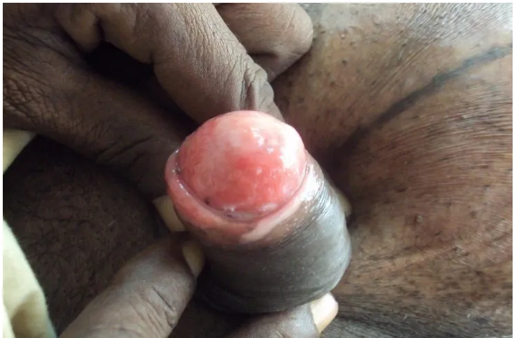
Fig 4: CANDIDIASIS -WHITISH PLAQUES AND FISSURES
[image:60.612.124.514.397.617.2]CANDIDAL HYPHAE
Fig 6 : TINEA GENITALIS – HYPERPIGMENTED SCALY PLAQUE WITH RAISED EDGES SEEN OVER MONS PUBIS, LABIA MAJORA
[image:61.612.124.510.404.641.2]PERIPHERAL RAISED EDGES AND CENTRAL CLEARING PRESENT OVER THE SCROTUM
Fig 8: LICHEN SCLEROSUS ET ATROPHICUS OVER THE VULVAL
[image:62.612.127.509.402.614.2]THE PENILE SHAFT, PREPUCE AND SCROTUM
Fig 10 : H&E- LICHEN PLANUS - HYPERKERATOSIS, IRREGULAR ACANTHOSIS, BAND DENSE LYMPHOCYTIC
[image:63.612.139.497.389.592.2]ROUND,FLAT-TOPPED SKIN COLOURED PAPULES
Fig 12: PEMPHIGUS VULGARIS
-MULTIPLE EROSIONS AND FEW FLACCID BLISTERS WERE
[image:64.612.133.488.375.613.2]SUPRABASAL BULLAE WITH ROW OF TOMB STONES APPEARANCE
[image:65.612.133.503.86.323.2] [image:65.612.131.496.404.615.2]MOIST, SHINY, PLAQUE OVER THE GLANS PENIS AND PREPUCE
Fig 16 :SCABETIC PAPULE,CRUSTING,EROSION OVER THE MALE
[image:66.612.128.502.394.603.2]SCROTUM
Fig 18: HISTOID LEPROSY - H&E SHOWING CLEAR UNNA BAND
WITH GRANULOMA AND SPINDLE SHAPED HISTIOCYTES. WADE
FITE STAIN SHOWING NUMEROUS LARGER BACILLI.
AROUND THE CORONAL SULCUS
[image:68.612.126.511.402.638.2]RED PAPULES OVER THE PENILE SHAFT AND SCROTUM
Fig 22: H & E SECTION IN ANGIOKERATOMA OF FORDYCE
SHOWING NUMEROUS, DILATED, THIN WALLED, CONGESTED
[image:69.612.124.511.111.325.2] [image:69.612.126.511.408.619.2]OVER THE PREPUCE
Fig 24 : LINEAR EPIDERMAL NAEVI OVER PREPUCE AND
[image:70.612.128.505.406.622.2]CAULIFLOWER LIKE ULCERATIVE PLAQUES OVER THE PENILE
[image:71.612.131.502.94.306.2]SHAFT
Fig 26: SEBACEOUS CYST OF SCROTUM - MULTIPLE DISCRETE
YELLOWISH WAXY PAPULES AND NODULES OVER THE
[image:71.612.130.502.392.602.2]Fig 28: LYMPHANGIOMA CIRCUMSCRIPTUM- H&E SHOWING
NUMEROUS DILATED LYMPHATIC SPACES IN THE PAPILLARY
[image:72.612.133.502.367.598.2]PAPULES OVER THE LABIA MAJORA WITH HAIRS EMERGING
FROM FEW PAPULES
Fig 30 : H& E SECTION OF SYRINGOMA – NUMEROUS TUBULAR
STRUCTURES, SOME DUCT POSSESS COMA LIKE TAIL –
[image:73.612.129.505.110.313.2] [image:73.612.150.487.419.633.2]FUNGAL INFECTIONS
Candidiasis and dermatophytic infections formed the 2nd largest group
with 13(12.03%) patients. All the patients had itching as the predominant
symptom.
AGE – SEX DISTRIBUTION
Table 8a
Age Male Female
0 – 10 1
-11 - 20 2 1
21 - 30 1 1
31 - 40 2 1
41 - 50 1 2
51 - 60 -
-61 - 70 1
-71 - 80 -
-Total 8 5
Table 8b
Dermatoses No. Of patients Percentage
Candidiasis 7 53.84%
Tinea genitalis 6 46.16%
CANDIDA INFECTION
Seven patients (53.84%) had candidial infection. All patients were
symptomatic with itching and erythema or both. In males fissuring of the
preuce and balanitis present. All the male patients had candidal balanoposthitis.
Duration of symptoms ranges from 4 days to 5 months. Four patients had prior
therapy. 10% KOH examination was done in all the patients which showed
candidal hyphae, confirming the diagnosis.
CLINICAL PRESENTATION
Table 9
Morphology No. of Patients Percentage
Fissures 4 57.14%
Erythematous plaques 3 42.85%
Whitish plaques 3 42.85%
Table 10
Site No. Of Patients Percentage
Prepuce 4 57.14 %
Glans penis 3 42.85 %
Labia majora 3 42.85 %
TINEA INFECTIONS
Five male patients (46.15%) had Tinea genitalis with involvement of
scrotum and penile shaft. One female patient had involvement of labia majora
and mons pubis.10 % KOH examination was positive in these patients. Two
patients had prior topical treatment.
Table 11
Site No. Of Patients Percentage
Penile shaft 4 66.66 %
Prepuce 2 33.33 %
Scrotum 5 83.33 %
Labia majora & mons pubis 1 16.66 %
LICHEN SCLEROSUS ET ATROPHICUS (LSA)
Genital LSA forms the 3rd largest group with 9 (8.3%) patients. Of this
6 (66.66%) were females and 3 (33.33%) were males. All patients presented
with atrophic hypopigmented plaques. Two female and one male patient had
diabetes mellitus. Three patients complained of burning micturition and
itching.
All male patients had phimotic prepuce. No patient had skin lesions.
Two patient had prior topical therapy. Biopsy was done in three female patients
AGE – SEX DISTRIBUTION
Table 12
Age Male Female
0 – 10 -
-11 – 20 -
-21 – 30 -
-31 – 40 -
-41 – 50 2 2
51 – 60 1 2
61 – 70 - 2
71 – 80 -
-Total 3 6
LICHEN PLANUS
Five patients (4.6 %) of which three are males (60%) and two are
females (40 %). Patients were in the age group of 11 – 50 years. All patients
had itching. Three had skin lesions also. Biopsy was done and was consistent
AGE – SEX DISTRIBUTION
Table 13
Age Male Female
0 – 10 -
-11 – 20 - 1
21 – 30 2
-31 – 40 1
-41 – 50 - 1
51 – 60 -
-61 – 70 -
-71 – 80 -
-Total 3 2
CLINICAL PRESENTATION
Table 14a
On examination No. Of Patients Percentage
Skin & Genital lesion 3 60%
Table 14b
Site No. Of Patients Percentage
Prepuce 2 66.66%
Penile shaft 1 33.33%
Glans penis 2 66.66%
Scrotum 1 33.33%
Labia majora 1 50%
Labia minora 2 100%
Table 14c
Morphology No. Of Patients Percentage
Papules 3 60%
Plaques 2 40%
PSORIASIS
Three patients(2.7 %) in the age group of 31 – 60 years presented with
scaly plaques covered with silvery scales. Plaques were present over the penile
shaft and scrotum. Two patients had skin lesions also. Biopsy was consistent
AGE – SEX DISTRIBUTION
Table 15
Age Male Female
0 -10 -
-11 – 20 -
-21 – 30 -
-31 – 40 1
-41 -50 -
-51 – 60 2
-61 – 70 -
-71 – 80 -
-Total 3
-CLINICAL PRESENTATION
Table 16
Site No of Patients Percentage
Penile shaft 3 100%