Copyright © 2000, American Society for Microbiology. All Rights Reserved.
Circular Double-Stranded Forms of TT Virus DNA in the Liver
HIROAKI OKAMOTO,
1MASATO UKITA,
1TSUTOMU NISHIZAWA,
1JUNICHI KISHIMOTO,
2YUJI HOSHI,
3HITOSHI MIZUO,
4TAKESHI TANAKA,
3YUZO MIYAKAWA,
5ANDMAKOTO MAYUMI
1*
Immunology Division and Division of Molecular Virology, Jichi Medical School, Tochigi-Ken 329-0498,
1Institute of Immunology, Tokyo 112-0004,
2Japanese Red Cross Saitama Blood Center,
Saitama-Ken 338-0001,
3Department of Internal Medicine, Kin-ikyo Chuo Hospital,
Hokkaido 007-0870,
4and Miyakawa Memorial Research Foundation,
Tokyo 107-0062,
5Japan
Received 8 December 1999/Accepted 13 March 2000
TT virus
(TTV) is an unenveloped, circular, and single-stranded DNA virus commonly infecting human
beings worldwide. TTV DNAs in paired serum and liver tissues from three viremic individuals were separated
by gel electrophoresis and characterized biophysically. TTV DNAs in sera migrated in sizes ranging from 2.0
to 2.5 kb. TTV DNAs in liver tissues, however, migrated at 2.0 to 2.5 kb as well as at 3.5 to 6.1 kb. Both
faster-and slower-migrating forms of TTV DNAs in the liver were found to be circular faster-and of the full genomic length
of 3.8 kb. TTV DNAs migrating at 2.0 to 2.5 kb, from either serum or liver tissues, were sensitive to S1 nuclease
but resistant to restriction endonucleases, and therefore, they were single-stranded. By contrast, TTV DNAs in
liver tissues that migrated at 3.5 to 6.1 kb were resistant to S1 nuclease. They migrated at 3.7 to 4.0 kb after
digestion with
EcoRI, which suggests that they represent circular, double-stranded replicative intermediates of
TTV. When TTV DNAs were subjected to strand-specific primer extension and then amplified by PCR with
internal primers, those in serum were found to be minus-stranded DNAs while those in liver tissues were found
to be a mixture of plus- and minus-stranded DNAs. These results suggest that TTV replicates in the liver via
a circular double-stranded DNA.
TT virus
(TTV) is an unenveloped, single-stranded, and
cir-cular-DNA virus (12, 13, 22). It was originally recovered from
a patient with posttransfusion hepatitis of unknown etiology
(16, 21) and is very common in every country examined (3, 6,
14, 16, 21, 27, 33). TTV most closely resembles members of the
Circoviridae
family (9). It is transmitted parenterally through
transfusions and blood products and can establish a persistent
infection in hosts (16, 21, 27). It is excreted from the liver via
the bile duct into feces with a buoyant density comparable to
that in the circulation (19, 31), resulting in a possible fecal-oral
transmission route. The presumed dual mode of transmission
may help TTV penetrate deeply and broadly into the
commu-nity. Species-specific TTVs are reported in high frequencies for
chimpanzees and the other nonhuman primates (1, 8, 20, 32),
as well as farm animals (8). Hence, TTV is a ubiquitous virus,
being highly prevalent in many species of animals.
There has been limited information on the virological
char-acteristics of TTV. It is not known in what organs TTV
repli-cates, although TTV DNA is detected in liver tissues in levels
10 to 100 times higher than those in sera from the same
infected individuals (21). TTV DNAs in paired serum and liver
specimens from three infected individuals were fractionated by
electrophoresis on agarose gel. Distinct molecular forms of
TTV DNAs in serum and liver tissues were amplified by
long-distance PCR for amplification of the full-length TTV DNA of
3.8 kb and by inverted PCR for demonstration of a circular
genomic structure, and then they were examined for their
polarity and single or double strandedness.
MATERIALS AND METHODS
Extraction of nucleic acids from serum and liver tissues.Paired serum and liver specimens were obtained from three patients who were infected with TTV (Table 1). Serum (1 ml) was diluted twofold with Tris-HCl buffer (50 mM, pH 8.0) containing 150 mM NaCl and 1 mM EDTA and centrifuged in a TLA 100.3 rotor (Beckman Instruments, Inc., Palo Alto, Calif.) at 190,583⫻gfor 1 h. The pellet was suspended in Tris-HCl buffer (10 mM, pH 8.0) containing 5 mM EDTA and supplemented with 0.5 mg of proteinase K per ml as well as 0.5% (wt/vol) sodium dodecyl sulfate and incubated at 65°C for 2 h as described previously (25). Then, nucleic acids were extracted with phenol-chloroform, precipitated with ethanol, and dissolved in 100l of Tris-HCl buffer (10 mM, pH 8.0) containing 1 mM EDTA (TE buffer).
Liver tissues (10 or 100 mg) were homogenized and incubated in the presence of proteinase K-sodium dodecyl sulfate at 37°C for 14 h. Then, nucleic acids were extracted from them with phenol-chloroform and precipitated with ethanol. DNA species of chromosomal origin, which emerged as a cloudy precipitate immediately after addition of ethanol, were removed. The remaining nucleic acids were collected by centrifugation and dissolved in 200l of TE buffer.
Semiquantitation of TTV DNA.Nucleic acids extracted from serum and liver were serially diluted 10-fold in TE buffer containing 20g of glycogen (Boehr-inger Mannheim, Mannheim, Germany) per ml, and the highest dilution in which TTV DNA was detectable by PCR with seminested primers was determined (19, 23). Based on the results, quantifications of TTV DNA in 10ncopies per 10l in
serum or per 10 mg of liver tissue were obtained.
Agarose gel electrophoresis and amplification of TTV DNA by PCR.The concentration of TTV in plasma is estimated to be 50 to 50,000 (geometric mean, 620) copies per ml (27). Since sera and liver tissues from infected individuals contained TTV DNA in titers too low to be detected by Southern hybridization, the length and strandedness of the TTV genome were determined by the fol-lowing procedure.
DNAs (25l) extracted from sera and liver tissues from the three viremic patients (patients 1 to 3) were subjected to electrophoresis on 1% (wt/vol) agarose gel (SeaKem GTG agarose; FMC BioProducts, Rockland, Maine) in DNase-free electrophoresis buffer (pH 8.3) containing 40 mM Tris-acetate and 1 mM EDTA (1:10 dilution of 10⫻TAE buffer; Gibco-BRL, Grand Island, N.Y.). They were run horizontally for 115 mm in parallel with a size marker (500-bp DNA ladder; TaKaRa Shuzo Co. Ltd., Shiga, Japan) and 1 pg of the cloned linear and double-stranded DNA of hepatitis B virus (HBV) (24), which served as a control (Fig. 1A). DNAs (25l) from liver tissues from patients 1 to 3 were subjected to electrophoresis with or without prior digestion with 360 U of
EcoRI (TaKaRa Shuzo) at 37°C for 12 h (Fig. 1B). After electrophoresis, the gel was stained with ethidium bromide.
First, the full-length gel, spreading over 115 mm from the baseline to the bottom, was cut into 23 slices at 5-mm intervals with a razor, and DNAs were
* Corresponding author. Mailing address: Minamikawachi-Machi,
Tochigi-Ken 329-0498, Japan. Phone: 81-285-58-7404. Fax:
81-285-44-1557. E-mail: [email protected].
5161
on November 9, 2019 by guest
http://jvi.asm.org/
recovered from each gel slice in a volume of approximately 80 l with a GenElute agarose spin column (Sigma Chemical Co., St. Louis, Mo.). A 4- to 15-l portion thereof was subjected to amplification by PCR with the primers NG061 and NG063 (Table 2) for 35 cycles as described previously (23) to generate a fragment of 271 bp. The screening revealed TTV DNAs from patients 1 to 3 to be exclusively in the area of 1.7 to 6.8 kb in size. Based on these findings, the gel corresponding to this size was cut into 20 slices at 2.5-mm intervals. Thereafter, DNAs were recovered from each gel slice and subjected to amplifi-cation by PCR with NG061 and NG063 as described above.
The amplification was performed with or without digestion of the recovered DNAs from patients 1 to 3 by the restriction endonucleaseNdeI (TaKaRa Shuzo) under conditions described previously (21). In parallel, the effects of prior digestion with 10 U of S1 nuclease (TaKaRa Shuzo) at 37°C for 15 min were evaluated. The products were electrophoresed on a 2% NuSieve 3:1 aga-rose gel (FMC BioProducts), stained with ethidium bromide, and photographed under UV light. The strandedness of TTV DNAs was evaluated over an addi-tional three genomic sequences. They spanned, respectively, nucleotides (nt) 91 to 233, amplifiable by PCR with NG133 and NG147 and having anAccII site; nt 621 to 729, amplifiable by PCR with NG057 and NG058 and carrying anNcoI site; and nt 3547 to 3709, amplifiable by PCR with NG017 and NG021 and bearing anSplI orAfaI site. HBV DNA was amplified by PCR by the method reported elsewhere (7), which generated a fragment of 233 bp.
Long-distance PCR for amplification of the full-length TTV genome.The full-length genomic DNA of TTV was amplified by a long-distance PCR with nested primers having their 5⬘ends back to back in the circular genomic DNA (Fig. 2A).
On a template of DNAs extracted from gel slices from electrophoresis, for paired serum and liver tissues from patients 1 to 3, the first-round PCR was performed with NG212 and NG215 for 35 cycles (94°C for 45 s with an additional 3 min in the first cycle, 65°C for 45 s, and 72°C for 4 min with an additional 7 min in the last cycle) in the presence of TaKaRa LATaqwith GC buffer (TaKaRa Shuzo). On the products of the first-round PCR (5l), the second round was performed for 25 cycles under the same conditions as in the first round. PCR products were run by electrophoresis on 1% (wt/vol) SeaKem GTG agarose gel (FMC BioProducts) and examined for a band at 3.8 kb.
Inverted PCR for confirming the circular nature of TTV DNA.An inverted PCR was performed, with the primers indicated in Fig. 2A and Table 2, for demonstrating the circular nature of TTV DNA by the connection between nt 210 and 211 in the target genome. On a template of DNAs extracted from gel slices, for paired serum and liver tissues, and in the presence of Perkin-Elmer AmpliTaq Gold (Roche Molecular Systems, Inc., Branchburg, N.J.), PCR was performed for 35 cycles with NG133 and NG147. The antisense primer NG147 had a sequence complementary to the sense primer NG212, which was used in the full-length PCR and possessed nt 211 at its 5⬘end.
Sequence analysis of TTV DNA.DNAs were extracted from the gel slices containing slower-migrating forms of TTV DNA in the liver (slice 13, corre-sponding to 3.5 to 3.7 kb, and slice 17, correcorre-sponding to 4.8 to 5.1 kb). Using the DNAs as templates, PCR was performed with four sets of primers (NG061 and NG063, NG133 and NG147, NG057 and NG058, and NG017 and NG021 [Table 2]). The products were subjected to reaction with a BigDye Terminator Cycle Sequencing Ready Reaction Kit (PE Applied Biosystems, Foster City, Calif.) and then analyzed in an automatic DNA sequencer (ABI PRISM310 Genetic Ana-lyzer; PE Applied Biosystems). The products obtained with the long-distance PCR for amplification of a full-length TTV genome were inserted into pT7Blue T-Vector (Novagen Inc., Madison, Wis.) to transformEscherichia coli. Nucleo-tide sequences representing TTV DNA in obtained clones were determined as indicated above.
Polarity of TTV DNA.On a template of DNAs (5l) extracted from serum or liver tissues from three infected individuals, strand-specific primer extension was performed with either the sense or the antisense primer in the presence of Perkin-Elmer AmpliTaq Gold (Roche Molecular Systems) for 50 cycles (95°C for 30 s with an additional 9 min in the first cycle, 55°C for 30 s, and 72°C for 45 s with an additional 7 min in the last cycle) in a reaction volume of 50l. The extension was carried out for 50 additional cycles, with addition of the supple-mental primer and enzyme in equal amounts to 50l of the buffer that was provided with the enzyme kit. From 100l of the extension products, 3l was
separated and amplified with a pair of primers internal to the sense and antisense primers used for extension.
As controls, TTV DNA of 3,254 bp (nt 12 to 3265) was excised from the pTV-TRM1-1 clone (DDBJ/EMBL/GenBank accession no. AB038340 [23]) and inserted into the M13 phage vectors mp18 and mp19 (New England Biolabs, Inc., Beverly, Mass.), and single-stranded DNAs having a plus- or minus-stranded sequence of TTV [designated M13-TTV(⫹)-ssDNA and M13-TTV(⫺)-ssDNA] were obtained.
The polarity of two sequences was evaluated (see regionsaandbin Fig. 3B). One sequence, spanning nt 601 to 759, was extended with the sense primer NG055 or the antisense primer NG056, and the products were amplified with a primer pair (NG057 and NG058), which gave rise to a fragment of 109 bp. The other sequence, stretching from nt 1862 to 2277, was extended with the sense primer NG001 or the antisense primer RD038, and the products were amplified with another primer pair (NG061 and NG063) to generate a fragment of 271 bp. The extension was also performed without primers to obtain mock products. An intensification of PCR signals, with prior extension by the used primer, verified the extension; it was attributed to the nature of the target strand that was complementary to the primer.
Nucleotide sequence accession numbers.The nucleotide sequence data in this report have been deposited in the DDBJ/EMBL/GenBank nucleotide sequence databases under accession no. AB040776 to AB040788.
RESULTS
Strandedness of TTV DNA in paired serum and liver
tis-sues.
DNAs recovered from paired serum and liver tissues
from the three viremic individuals (patients 1 to 3) were
sub-jected to electrophoresis (Fig. 1A). TTV DNAs in serum
mi-grated at sizes ranging from 2.0 to 2.5 kb, while those in liver
tissues migrated at two positions of different sizes. One of them
migrated at 2.0 to 2.5 kb, and the other migrated at 3.5 to 6.1
kb. DNA of HBV run in parallel, as a control, migrated at a
peak size range of 3.1 to 3.3 kb, in agreement with the genome
size of HBV at 3.2 kb.
TTV DNAs from liver tissues of patients 1 to 3 were
di-gested with
Eco
RI. The positions of TTV DNAs, migrating at
2.0 to 2.5 kb, did not change after they had been digested with
Eco
RI. By contrast, TTV DNAs from patient 1 migrating at 3.5
to 6.1 kb, including those at 3.5 to 4.3 kb and 4.8 to 6.1 kb,
shifted to the position of 3.7 to 4.3 kb after digestion with
Eco
RI (Fig. 1B); the same results were obtained for TTV
DNAs recovered from patients 2 and 3. Hence, these
slower-migrating forms of TTV DNAs in liver tissues are in
double-stranded, replicative intermediate forms. They were linearized
to have a genomic size of 3.8 kb after digestion with
Eco
RI;
there is only one restriction site for this enzyme in the TTV
DNA of genotype 1a or 1b (4, 13, 22).
[image:2.612.54.553.92.163.2]The strandedness of TTV DNA in serum or liver tissues was
determined in four different regions by digestion with S1
nu-clease and four restriction endonunu-cleases, followed by
detec-tion of TTV DNA by PCR with primers designed to amplify
each of the four regions. Examples are shown for a target
sequence (nt 1915 to 2185) bearing the
Nde
I site, amplifiable
with primers NG061 and NG063 (Fig. 1C). When TTV DNAs
were digested with S1 nuclease, those migrating at 2.1 to 2.3
kb, recovered from either serum or liver tissues, were no longer
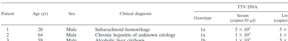
TABLE 1. Characteristics of three patients with TTV infection whose paired serum and liver tissues were studied
for characterization of TTV DNA
Patient Age (yr) Sex Clinical diagnosis
TTV DNA
Genotype (copies/10Seruml) (copies/10 mg)Livera
1
20
Male
Subarachnoid hemorrhage
1a
5
⫻
10
35
⫻
10
42
64
Male
Chronic hepatitis of unknown etiology
1a
1
⫻
10
31
⫻
10
53
59
Male
Alcoholic liver cirrhosis
1b
1
⫻
10
35
⫻
10
4aLiver tissues were obtained at necropsy from patient 1 and by needle biopsy from patients 2 and 3.
on November 9, 2019 by guest
http://jvi.asm.org/
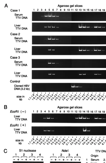
FIG. 1. (A) Separation of TTV DNAs extracted from serum or liver tissues by gel electrophoresis. TTV DNA samples extracted from paired serum and liver tissues from three viremic patients (Cases 1 to 3) along with a molecular size marker (500-bp DNA ladder [TaKaRa Shuzo]) were subjected to electrophoresis, and areas corresponding to 1.7 to 6.8 kb were cut into 20 gel slices. HBV DNA run in parallel served as a control. DNA was recovered from each slice and amplified by PCR for TTV DNA or HBV DNA. The size of DNA was estimated by reference to slice numbers. (B) Separation of TTV DNAs extracted from liver tissues with or without prior digestion withEcoRI. TTV DNAs from liver tissues of patient 1 were digested withEcoRI and electrophoresed on agarose gel. (C) Susceptibility of TTV DNAs from serum or liver tissues of patient 1 to S1 nuclease or restriction endonucleaseNdeI. DNAs extracted from agarose gel slices were digested with S1 nuclease orNdeI and then amplified by PCR for a sequence bearing theNdeI cutting site. PCR was also performed without digestion with S1 nuclease orNdeI. The products were subjected to electrophoresis, and signals were compared for intensity.
on November 9, 2019 by guest
http://jvi.asm.org/
[image:3.612.120.472.91.632.2]amplified while those that migrated at 3.5 to 3.7 kb and 4.8 to
5.1 kb stayed amplifiable. When digested with
Nde
I, however,
TTV DNA molecules that migrated at 2.1 to 2.3 kb, recovered
from either serum or liver tissues, remained amplifiable. By
contrast, TTV DNAs migrating at 3.5 to 3.7 kb or 4.8 to 5.1 kb
were no longer amplified after digestion with
Nde
I. The
sus-ceptibility of TTV DNAs of distinct migration positions to
treatment with S1 nuclease and restriction endonucleases was
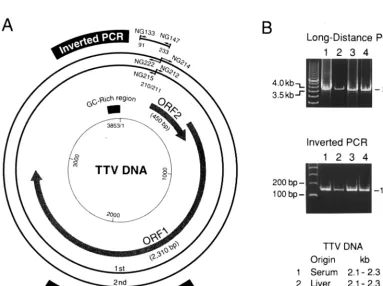
FIG. 2. (A) Organization of the TTV genome and primers for long-distance or inverted PCR. The genomic structure of the prototype TTV isolate (TA278) (21, 22) is shown in the center. The products of long-distance PCR with nested primers for amplification of the entire genome and those of inverted PCR are shown in the periphery. The first round of long-distance PCR was performed with the primer pair NG212-NG215, and the second round of PCR was performed with NG214-NG222. Sense primers (NG212 and NG214) were designed to have their 5⬘ends at nt 211, and antisense primers (NG215 and NG222) were designed to have theirs at nt 210. The inverted PCR was carried out with the primer pair NG133-NG147 to amplify a sequence covering both ends of the products of long-distance PCR. Primer NG147 for inverted PCR had a sequence complementary to that of NG212 for long-distance PCR and had its 3⬘end at nt 211. ORF, open reading frame. (B; upper panel) Agarose gel electrophoresis of the amplification products of the long-distance PCR. TTV DNAs of various migrating sizes recovered from paired serum and liver tissues from patient 1 (Fig. 1A) were used as templates. The products of 3.8 kb (6l) were electrophoresed vertically on 1% (wt/vol) SeaKem GTG agarose gel (FMC BioProducts). The 500-bp DNA ladder (TaKaRa Shuzo) on the left served as a size marker. (Lower panel) TTV DNAs amplified by inverted PCR. The products (6
[image:4.612.54.553.82.248.2]l) of inverted PCR, on templates of TTV DNAs from serum or liver tissues of patient 1, were electrophoresed on 3% NuSieve 3:1 agarose gel (FMC BioProducts) for generating products of 143 bp. The 100-bp DNA ladder (Gibco-BRL) on the left was used as a size marker.
TABLE 2. Positions and nucleotide sequences of oligonucleotide primers used for PCR amplification and primer extension
Primer Polarity Nt positiona Nucleotide sequenceb
NG001
Sense
1862–1881
5⬘-
CAC CAG GAG CAT ATA CAG AC
-3⬘
NG017
Sense
3547–3566
5⬘-
ATG GTG GAC AAC ATC TTC CG
-3⬘
NG021
Antisense
3690–3709
5⬘-
AAA GAG GAA GGA AGT CAG CC
-3⬘
NG055
Sense
601–623
5⬘-
TGG TGG CGC CGA AGG AGA AGA CG
-3⬘
NG056
Antisense
736–759
5⬘-
TCT CCT ATA TCT CCT CCT CCA CCT
-3⬘
NG057
Sense
621–642
5⬘-
ACG GTG GCG CAG GTG GAG ACG C
-3⬘
NG058
Antisense
706–729
5⬘-
TCT GCG GCG TCT CCT TAC GTT TCT
-3⬘
NG061
Sense
1915–1938
5⬘-
GGC AAC ATG YTR TGG ATA GAC TGG
-3⬘
NG063
Antisense
2161–2185
5⬘-
CTG GCA TTT TAC CAT TTC CAA AGT T
-3⬘
NG133
Sense
91–115
5⬘-
GTA AGT GCA CTT CCG AAT GGC TGA G
-3⬘
NG147
Antisense
211–233
5⬘-
GCC AGT CCC GAG CCC GAA TTG CC
-3⬘
NG212
Sense
211–233
5⬘-
GGC AAT TCG GGC TCG GGA CTG GC
-3⬘
NG214
Sense
211–238
5⬘-
GGC AAT TCG GGC TCG GGA CTG GCC GGG C
-3⬘
NG215
Antisense
185–210
5⬘-
CCT TGA CTG CGG TGT GTA AAC TCA CC
-3⬘
NG222
Antisense
179–210
5⬘-
CCT TGA CTG CGG TGT GTA AAC TCA CCT HCG GC
-3⬘
RD038
Antisense
2258–2277
5⬘-
TGA CTG TGC TAA AGC CTC TA
-3⬘
aNucleotides were numbered on the coding strand of TTV DNA, in accord with the numbering of TA278 isolate of 3,853 nt (21, 22), since the virion DNA was found
to be minus stranded (see Results).
bY denotes T or C; R denotes A or G; and H denotes A, T, or C.
on November 9, 2019 by guest
http://jvi.asm.org/
[image:4.612.112.496.350.636.2]evaluated with three other sequences of TTV DNA with
re-producible results. They were a target sequence of PCR (nt 91
to 233) with an
Acc
II site, a sequence (nt 621 to 729) with an
Nco
I site, and a sequence (nt 3547 to 3709) with an
Spl
I site
(data not shown). The results obtained with TTV DNAs from
patient 1 were confirmed with those recovered from patients 2
and 3.
Slower-migrating DNAs recovered from liver tissues were
confirmed to have the nucleotide sequences specific for TTV
by the following procedure. Slower-migrating TTV DNAs in
liver tissues from patients 1 to 3 were recovered from gel slices
(slice 13, corresponding to 3.5 to 3.7 kb, and slice 17,
corre-sponding to 4.8 to 5.1 kb). Using the DNAs as templates, PCR
was performed with four primer sets (NG061-NG063,
NG133-NG147, NG057-NG058, and NG017-NG021) as described
above, and the products were sequenced. The slower-migrating
DNA from patients 1 and 2 possessed sequences 96.8 to 100%
similar to that of a TTV isolate of genotype 1a (TA278 [22]),
while that from patient 3 had a sequence 97.6 to 98.9% similar
to that of a TTV isolate of genotype 1b (JA20 [4]).
On the basis of these results, TTV DNAs in serum were
single stranded while those in liver tissues existed in both
single- and double-stranded forms. The double-stranded TTV
DNAs in liver tissues represent replicative intermediates.
These results were in agreement with the previous observation
that TTV DNA in plasma is digested with mung bean nuclease
that has a specificity for the single-stranded DNA (21).
Circular structure of TTV DNA in serum and liver tissues.
A two-stage, long-distance PCR method was performed on
TTV DNAs from paired serum and liver tissues from patient 1.
The PCR was designed to amplify the full-length TTV genome,
using sense primers with the 5
⬘
ends at nt 211 and antisense
primers with the 5
⬘
ends at nt 210 (Fig. 2A). A product of 3.8
kb in size, compatible with the full-length genome, was
ampli-fied on single-stranded TTV DNAs from either serum or liver
tissues that migrated at 2.1 to 2.3 kb, as well as on
double-stranded TTV DNAs from liver tissues that migrated at 3.5 to
3.7 kb or 4.8 to 5.1 kb (Fig. 2B). The products of 3.8 kb (Fig.
2B, lanes 3 and 4) were sequenced within the 5
⬘
-terminal 549
nt (nt 211 to 759) and the 3
⬘
-terminal 530 nt (nt 3534 to 3853
and nt 1 to 210). The sequences were 99.6% similar to that of
the TA278 isolate, thereby confirming that the product of
long-distance PCR contained TTV sequences.
The circular nature of TTV DNAs from serum and liver
tissues of patient 1 was confirmed by an inverted PCR with a
primer pair (NG133-NG147) designed to amplify a sequence
of 143 bp that includes nt 210 and 211; these primers
repre-sented both ends of the product of full-length PCR (Fig. 2B).
The connection between nt 210 and 211 was confirmed by
determination of the 143-bp sequence. These results were
con-firmed by similar analyses of TTV DNAs from sera and liver
tissues of patients 2 and 3. Together, they indicate that a
circular full-length TTV DNA of 3.8 kb was present both in
serum and liver tissues.
Polarity of TTV DNA in serum and liver tissues.
The
polar-ity of TTV DNA was evaluated under the assumption that the
detection of TTV DNA by PCR would be enhanced by prior
extension with primers having a complementary sequence. For
verifying this notion, two single-stranded TTV DNAs of plus
or minus polarity [M13-TTV(
⫹
)-ssDNA and M13-TTV(
⫺
)-ssDNA] were extended over two regions (
a
and
b
) for 100
cycles with appropriate sense or antisense primers and then the
products were tested by PCR with the primers that were
in-ternal to both primers (Fig. 3A). With M13-TTV(
⫹
)-ssDNA
as a template, the amplification signal was intensified only
when it had been extended by the antisense primer; the reverse
was the case for M13-TTV(
⫺
)-ssDNA. The same results were
obtained over two distinct regions (
a
and
b
) of the TTV
ge-nome. A weak signal was detected by PCR with region
b
primers in lanes with no primers. Hence, weak signals observed
for the extension products with the sense primer on the
single-and minus-strsingle-anded TTV DNA, as well as those for the
exten-sion products observed with the antisense primers on the
sin-gle- and plus-stranded TTV DNA, were attributed to the
am-plification of these regions on the template. The lower
intensities of signals for region
a
than region
b
were due to the
sizes of products of region
a
being less than half those of region
b
. The findings with single-stranded TTV DNA of plus or
minus polarity attested to the credibility of extension
proce-dures used to determine the polarity.
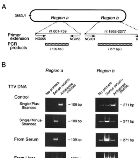
When TTV DNAs from the serum of patient 1 were
ex-tended by these procedures, the amplification signal was
inten-sified remarkably by prior extension with sense, but not
anti-sense, primers for the two regions examined (Fig. 3B). The
extension was performed with TTV DNAs from sera of
pa-tients 2 and 3, and the same types of intensification were
obtained. Based on these results, the TTV DNA in circulating
virions was deduced to have a sequence complementary to the
two sense primers and, therefore, to be minus stranded.
By contrast, the amplification of TTV DNAs in the liver
tissues of patient 1 was enhanced markedly by prior extension
FIG. 3. (A) Primer extension of TTV DNA sequence. Using DNAs extracted from serum or liver tissues as templates, strand-specific primer extension was performed on two genomic regions (aandb) with sense or antisense primers specific for TTV. Nucleotides were numbered from position 1 (nt 1) of the coding strand of the prototype TTV isolate of genotype 1a (TA278) (21, 22), complementary to the viral strand, because TTV was found to have a minus-stranded DNA in the circulating virion. Single-minus-stranded DNAs from recombi-nant M13 phage, either plus-stranded DNA [M13-TTV(⫹)-ssDNA] or minus-stranded DNA [M13-TTV(⫺)-ssDNA], served as controls. (B) Amplification of extension products by PCR. Using extension products as templates, PCR was performed with primer pairs internal to those used for primer extension. The extension was performed without primers to obtain mock products. The ampli-fication products were subjected to electrophoresis, and signals were compared between the products with and without prior extension.
on November 9, 2019 by guest
http://jvi.asm.org/
[image:5.612.55.289.71.338.2]with either sense primers or antisense primers for the two
regions (Fig. 3B). The same results were obtained with TTV
DNAs in the liver tissues of patients 2 and 3. On the basis of
these results, there were two kinds of TTV DNA species in the
liver, one of which was minus stranded and the other of which
was plus stranded.
DISCUSSION
Although TTV DNA is detected in liver tissues (21, 26, 34),
it has not yet been characterized. In the present study, a
cir-cular nature was demonstrated for TTV DNAs from the paired
serum and liver tissues of three infected individuals (patients 1
to 3). Since TTV is a DNA virus, it should replicate in the
nuclei of hepatocytes. The origin of TTV DNA in liver tissues
has not been identified, however; it is not known whether it is
in the episomes, in the nucleus, or both.
As an unenveloped, single-stranded, and circular DNA
vi-rus, TTV would be classified as belonging in the
Circoviridae
family (9, 12). There are, however, some characteristics of
TTV distinct from those of known animal circoviruses. First,
TTV does not share sequence similarity with any of the three
animal circoviruses reported, i.e., chicken anemia virus, beak
and feather disease virus of parrots, and porcine circovirus.
Second, the genomic size of TTV is approximately twice as
large as those of these three animal circoviruses, which possess
1,758 to 2,319 bases (11, 15, 17). Third, animal and plant
circoviruses conserve stem-loop structures, with a 9-nt motif in
the loop that is essential for the replication of viral DNA (2,
10). This motif, however, has not been identified in TTV.
TTV DNA of genotype 1 is detected in liver tissues in levels
10 to 100 times higher than those in corresponding sera (21).
This was confirmed for three patients with TTV of genotype 1a
or 1b in the present study (Table 1). A 10- to 100-fold
differ-ence in TTV DNA titers was observed also in individuals
infected with TTV of genotype 2 or 3 (unpublished
observa-tions). Hence, the present results obtained with TTV of
geno-type 1 can be extrapolated to many other genogeno-types that have
accumulated up to the present (13, 14, 16, 21, 23, 28, 29). TTV
DNA titers in both serum and liver tissues, however, are too
low to allow analysis by Southern hybridization. Therefore,
TTV DNAs were separated by electrophoresis on agarose gel,
and then those in gel slices were amplified by PCR to
deter-mine their sizes.
Viruses with a circular single-stranded DNA genome
de-pend on a double-stranded DNA for the correct transcription
and replication of the virus DNA (17). In the present study,
double-stranded DNA of TTV was isolated from liver tissues
from three infected patients. TTV DNAs from serum showed
only one form, which migrated at 2.0 to 2.5 kb, while TTV
DNAs from the liver showed, in addition, two slower-migrating
forms at 3.5 to 4.3 kb and 4.8 to 6.1 kb; they both generated a
product of 3.8 kb, in accord with the size of the full-length
genome, by long-distance PCR. Slower-migrating TTV DNAs
in liver tissues, when digested with
Eco
RI, generated
frag-ments migrating at 3.7 to 4.0 kb, sizes compatible with the size
of the full-length genome. Hence, size differences in the two
species of slower-migrating TTV DNA were generated by
con-formation and not by differences in the genomic length; both
are circular molecules. These findings suggest that two forms
of circular, double-stranded species of TTV DNA exist in the
liver. Of slower-migrating TTV DNAs, those that migrated at
3.5 to 4.3 kb represent a closed-circular duplex and those that
migrated at 4.8 to 6.1 kb represent a relaxed- or open-circular
duplex. Two such species of double-stranded viral DNAs have
also been reported for cells infected with chicken anemia virus
(11, 30). The sizes of TTV DNAs in paired serum and liver
tissues were in accord among the three studied patients who
were infected.
Circoviruses possess up to seven open reading frames,
in-cluding the one coding for Rep protein, which is involved in
rolling-circle replication (2, 15). The sequences corresponding
to two of the four conserved Rep protein motifs are identified
in the product of open reading frame 1 of TTV isolates (13),
suggesting that TTV might replicate via circular
double-stranded DNA intermediates, possibly by rolling-circle
repli-cation. However, the Rep protein motifs are not conserved in
the majority of TTV sequences reported so far (4, 5).
By means of primer extension analysis, the polarity of TTV
DNA in serum was determined to be negative in the present
study. A minus-strandedness has been postulated for TTV
DNA extracted from human plasma by a
hybridization-nucle-ase protection assay (13). The minus polarity of TTV is
con-sistent with that of chicken anemia virus and at variance with
that of beak and feather disease virus of parrots and porcine
circovirus, which have ambisense genomes (15). Circular
dou-ble-stranded TTV DNAs in liver tissues most likely represent
replicative intermediate forms of the TTV genome. The exact
location for replication of TTV needs to be determined by
PCR in situ hybridization, which allows for detection of limited
viral copies (18), as well as immunological detection of
virus-encoded proteins in the liver.
REFERENCES
1.Abe, K., T. Inami, K. Ishikawa, S. Nakamura, and S. Goto.2000. TT virus infection in nonhuman primates and characterization of the viral genome: identification of simian TT virus isolates. J. Virol.74:1549–1553. 2.Bassami, M. R., D. Berryman, G. E. Wilcox, and S. R. Raidal. 1998.
Psittacine beak and feather disease virus nucleotide sequence analysis and its relationship to porcine circovirus, plant circoviruses, and chicken anaemia virus. Virology249:453–459.
3.Charlton, M., P. Adjei, J. Poterucha, N. Zein, B. Moore, T. Therneau, R. Krom, and R. Wiesner.1998. TT-virus infection in North American blood donors, patients with fulminant hepatic failure, and cryptogenic cirrhosis. Hepatology28:839–842.
4.Erker, J. C., T. P. Leary, S. M. Desai, M. L. Chalmers, and I. K. Mushahwar.
1999. Analyses of TT virus full-length genomic sequences. J. Gen. Virol.
80:1743–1750.
5.Hijikata, M., K. Takahashi, and S. Mishiro.1999. Complete circular DNA genome of a TT virus variant (isolate name SANBAN) and 44 partial ORF2 sequences implicating a great degree of diversity beyond genotypes. Virology
260:17–22.
6.Hohne, M., T. Berg, A. R. Muller, and E. Schreier.1998. Detection of sequences of TT virus, a novel DNA virus, in German patients. J. Gen. Virol.
79:2761–2764.
7.Iizuka, H., K. Ohmura, A. Ishijima, K. Satoh, T. Tanaka, F. Tsuda, H. Okamoto, Y. Miyakawa, and M. Mayumi.1992. Correlation between anti-HBc titers and HBV DNA in blood units without detectable HBsAg. Vox Sang.63:107–111.
8.Leary, T. P., J. C. Erker, M. L. Chalmers, S. M. Desai, and I. K. Mushahwar.
1999. Improved detection systems for TT virus reveal high prevalence in humans, non-human primates and farm animals. J. Gen. Virol.80:2115– 2120.
9.Lukert, P. D., G. F. de Boer, J. L. Dale, P. Keese, M. S. McNulty, J. W. Randers, and I. Tisher.1995. FamilyCircoviridae, p. 166–168. InF. A. Murphy, C. M. Fauquet, D. H. L. Bishop, S. A. Ghabrial, A. W. Jarvis, G. P. Martelli, M. A. Mayo, and M. D. Summers (ed.), Virus taxonomy. Classifi-cation and nomenclature of viruses. Sixth report of the International Com-mittee on Taxonomy of Viruses. Springer-Verlag, New York, N.Y. 10. Mankertz, A., J. Mankertz, K. Wolf, and H. J. Buhk.1998. Identification of
a protein essential for replication of porcine circovirus. J. Gen. Virol.79:
381–384.
11. Meehan, B. M., D. Todd, J. L. Creelan, J. A. P. Earle, E. M. Hoey, and M. S. McNulty.1992. Characterization of viral DNAs from cells infected with chicken anaemia agent: sequence analysis of the cloned replicative form and transfection capabilities of cloned genome fragments. Arch. Virol.124:301– 319.
12. Miyata, H., H. Tsunoda, A. Kazi, A. Yamada, M. A. Khan, J. Murakami, T. Kamahora, K. Shiraki, and S. Hino.1999. Identification of a novel GC-rich 113-nucleotide region to complete the circular, single-stranded DNA ge-nome of TT virus, the first human circovirus. J. Virol.73:3582–3586. 13. Mushahwar, I. K., J. C. Erker, A. S. Muerhoff, T. P. Leary, J. N. Simons,
on November 9, 2019 by guest
http://jvi.asm.org/
L. G. Birkenmeyer, M. L. Chalmers, T. J. Pilot-Matias, and S. M. Dexai.
1999. Molecular and biophysical characterization of TT virus: evidence for a new virus family infecting humans. Proc. Natl. Acad. Sci. USA96:3177–3182. 14.Naoumov, N. V., E. P. Petrova, M. G. Thomas, and R. Williams.1998. Presence of a newly described human DNA virus (TTV) in patients with liver disease. Lancet352:195–197.
15.Niagro, F. D., A. N. Forsthoefel, R. P. Lawther, L. Kamalanathan, B. W. Ritchie, K. S. Latimer, and P. D. Lukert.1998. Beak and feather disease virus and porcine circovirus genomes: intermediates between the gemini-viruses and plant circogemini-viruses. Arch. Virol.143:1723–1744.
16. Nishizawa, T., H. Okamoto, K. Konishi, H. Yoshizawa, Y. Miyakawa, and M. Mayumi.1997. A novel DNA virus (TTV) associated with elevated transam-inase levels in posttransfusion hepatitis of unknown etiology. Biochem. Bio-phys. Res. Commun.241:92–97.
17. Noteborn, M. H., G. F. de Boer, D. J. van Roozelaar, C. Karreman, O. Kranenburg, J. G. Vos, S. H. Jeurissen, R. C. Hoeben, A. Zantema, G. Koch, H. van Ormondt, and A. J. van der Eb.1991. Characterization of cloned chicken anemia virus DNA that contains all elements for the infectious replication cycle. J. Virol.65:3131–3139.
18. Nuovo, G. J.1994. PCRin situhybridization. Methods Mol. Biol.33:223–241. 19. Okamoto, H., Y. Akahane, M. Ukita, M. Fukuda, F. Tsuda, Y. Miyakawa, and M. Mayumi.1998. Fecal excretion of a nonenveloped DNA virus (TTV) associated with posttransfusion non-A-G hepatitis. J. Med. Virol.56:128– 132.
20. Okamoto, H., M. Fukuda, A. Tawara, T. Nishizawa, Y. Itoh, I. Hayasaka, F. Tsuda, T. Tanaka, Y. Miyakawa, and M. Mayumi.2000. Species-specific TT viruses and cross-species infection in nonhuman primates. J. Virol.74:1132– 1139.
21. Okamoto, H., T. Nishizawa, N. Kato, M. Ukita, H. Ikeda, H. Iizuka, Y. Miyakawa, and M. Mayumi.1998. Molecular cloning and characterization of a novel DNA virus (TTV) associated with posttransfusion hepatitis of un-known etiology. Hepatol. Res.10:1–16.
22. Okamoto, H., T. Nishizawa, M. Ukita, M. Takahashi, M. Fukuda, H. Iizuka, Y. Miyakawa, and M. Mayumi.1999. The entire nucleotide sequence of a TT virus isolate from the United States (TUS01): comparison with reported isolates and phylogenetic analysis. Virology259:437–448.
23. Okamoto, H., M. Takahashi, T. Nishizawa, M. Ukita, M. Fukuda, F. Tsuda, Y. Miyakawa, and M. Mayumi.1999. Marked genomic heterogeneity and frequent mixed infection of TT virus demonstrated by PCR with primers
from coding and noncoding regions. Virology259:428–436.
24. Okamoto, H., F. Tsuda, H. Sakugawa, R. I. Sastrosoewignjo, M. Imai, Y. Miyakawa, and M. Mayumi.1988. Typing hepatitis B virus by homology in nucleotide sequence: comparison of surface antigen subtypes. J. Gen. Virol.
69:2575–2583.
25. Okamoto, H., S. Yotsumoto, Y. Akahane, T. Yamanaka, Y. Miyazaki, Y. Sugai, F. Tsuda, T. Tanaka, Y. Miyakawa, and M. Mayumi.1990. Hepatitis B viruses with precore region defects prevail in persistently infected hosts along with seroconversion to the antibody against e antigen. J. Virol.64:
1298–1303.
26. Okamura, A., M. Yoshioka, M. Kubota, H. Kikuta, H. Ishiko, and K. Koba-yashi.1999. Detection of a novel DNA virus (TTV) sequence in peripheral blood mononuclear cells. J. Med. Virol.58:174–177.
27. Simmonds, P., F. Davidson, C. Lycett, L. E. Prescott, D. M. MacDonald, J. Ellender, P. L. Yap, C. A. Ludlam, G. H. Haydon, J. Gillon, and L. M. Jarvis.
1998. Detection of a novel DNA virus (TTV) in blood donors and blood products. Lancet352:191–195.
28. Takayama, S., S. Yamazaki, S. Matsuo, and S. Sugii.1999. Multiple infec-tion of TT virus (TTV) with different genotypes in Japanese hemophiliacs. Biochem. Biophys. Res. Commun.256:208–211.
29. Tanaka, Y., M. Mizokami, E. Orito, T. Ohno, T. Nakano, T. Kato, H. Kato, M. Mukaide, Y. M. Park, B. S. Kim, and R. Ueda.1998. New genotypes of TT virus (TTV) and a genotyping assay based on restriction fragment length polymorphism. FEBS Lett.437:201–206.
30. Todd, D., J. L. Creelan, B. M. Meehan, and M. S. McNulty.1996. Investi-gation of the transfection capability of cloned tandemly-repeated chicken anaemia virus DNA fragments. Arch. Virol.141:1523–1534.
31. Ukita, M., H. Okamoto, N. Kato, Y. Miyakawa, and M. Mayumi.1999. Excretion into bile of a novel unenveloped DNA virus (TT virus) associated with acute and chronic non-A-G hepatitis. J. Infect. Dis.179:1245–1248. 32. Verschoor, E. J., S. Langenhuijzen, and J. L. Heeney.1999. TT viruses
(TTV) of non-human primates and their relationship to the human TTV genotypes. J. Gen. Virol.80:2491–2499.
33. Woodfield, D. G., E. Gane, and H. Okamoto.1998. Hepatitis TT virus is present in New Zealand. N. Z. J. Med.111:195–196.
34. Yamamoto, T., K. Kajino, M. Ogawa, I. Gotoh, S. Matsuoka, K. Suzuki, M. Moriyama, H. Okubo, M. Kudo, Y. Arakawa, and O. Hino.1998. Hepato-cellular carcinomas infected with the novel TT DNA virus lack viral inte-gration. Biochem. Biophys. Res. Commun.251:339–343.
on November 9, 2019 by guest
http://jvi.asm.org/