0022-538X/96/$04.0010
Copyrightq1996, American Society for Microbiology
Tracking Hepatitis C Virus Quasispecies Major and Minor
Variants in Symptomatic and Asymptomatic Liver
Transplant Recipients
DAVID R. GRETCH,1* STEPHEN J. POLYAK,1JEFFREY J. WILSON,1
ROBERT L. CARITHERS, JR.,2JAMES D. PERKINS,3
ANDLAWRENCE COREY1,2,4
Departments of Laboratory Medicine,1Medicine,2Surgery,3and Microbiology,4
University of Washington Medical Center, Seattle, Washington
Received 31 January 1996/Accepted 25 July 1996
To evaluate the possibility that distinct viral quasispecies play a role in the pathogenesis of progressive hepatitis C virus (HCV) infection, we performed a detailed evaluation of HCV quasispecies before and after liver transplantation in five patients infected with HCV genotype 1, three of whom developed severe recurrent hepatitis C and two of whom developed asymptomatic posttransplant infections with high-titered viremia. HCV quasispecies were characterized by using a combination of nucleotide sequencing plus heteroduplex tracking assay of the second envelope gene hypervariable region (HVR). An average of 30 HVR clones were analyzed per specimen; an average of five specimens were analyzed per patient over a 6- to 24-month study period. The complexity of HCV quasispecies in pretransplant serum varied, ranging from one to nine genetically distinct variants for the five patients. However, in all five cases, relatively homogenous quasispecies variants emerged after liver transplantation. In the three patients who developed recurrent hepatitis, quasispecies major variants present in pretransplant serum were efficiently propagated immediately after liver transplantation and were propagated throughout the course of acute and chronic hepatitis. In contrast, in the two asymptom-atic cases, we observed rapid depletion of pretransplant quasispecies major variants from posttransplant serum, followed by emergence of new quasispecies variants by posttransplant day 30. Genetic analysis sug-gested that in these cases, the new quasispecies variants were derived from minor variants present at relatively low clonal frequency (less than 5% of HVR clones) within the pretransplant quasispecies populations. These data demonstrate that quasispecies tracking patterns are associated with the rapidity and severity of HCV-associated liver disease after liver transplantation. Further characterization of HCV quasispecies in animal model systems is warranted.
Hepatitis C virus (HCV) is an important cause of chronic liver disease leading to cirrhosis and end-stage liver disease in humans, and chronic hepatitis C is now recognized as a leading indication for orthotopic liver transplantation in the United States. Nearly 100% of HCV-infected patients who undergo liver transplantation develop recurrent viremia after the pro-cedure (1, 5, 6, 8, 14, 24). However, only about 50% of such patients develop recurrent hepatitis in their liver allografts during the first few years posttransplantation, despite the high levels of viremia in most patients (8, 24).
HCV exists as a viral quasispecies in infected humans, char-acterized by extensive genetic mutation within the second en-velope gene (E2) hypervariable region (HVR) (10, 21). The role of HCV quasispecies mutation in the pathogenesis of liver disease is uncertain. In the nonimmunosuppressed human host, HCV quasispecies mutations have been associated with viral persistence via antibody escape mechanisms (4, 12, 13, 19–22). Furthermore, one preliminary study has suggested that the degree of HCV genetic diversity within HVR is directly proportional to the extent of liver disease in the nonimmuno-suppressed host (11), although a second study found no such correlation (16). Because of the lack of a suitable animal model or tissue culture system for HCV propagation, there have been no data to suggest that different HCV quasispecies
variants might differ in either cytopathic potential or the ability to cause persistent infection without overt liver disease.
Transplantation of an uninfected liver allograft into an in-fected person offers the opportunity to evaluate HCV quasi-species infection in a new tissue host. The present study de-scribes tracking of HCV quasispecies major and minor variants before and after liver transplantation in five individuals, three of whom developed severe recurrence of chronic hepatitis, whereas two had only asymptomatic posttransplant infections. The five patients were selected because they were all infected with the same HCV genotype (genotype 1), and they all de-veloped recurrence of high-titered HCV viremia within 30 days posttransplantation. In addition, at least five serial liver biop-sies were available in each case to evaluate the histopathologic course of posttransplant infection. HCV quasispecies were characterized and tracked over time, using the techniques of nucleotide sequencing plus heteroduplex tracking assay (HTA) as originally described by Delwart et al. (3) for characterization of human immunodeficiency virus quasispecies and modified by our laboratory (23) for characterization of HCV quasispe-cies.
MATERIALS AND METHODS
Patients and clinical monitoring.The five patients were selected from a larger cohort of HCV-infected patients who underwent orthotopic liver transplantation at the University of Washington Medical Center as described in detail elsewhere (8). In brief, all five patients had previous HCV infection as demonstrated by HCV antibody positivity and HCV viremia. They were monitored by a common protocol in which serum specimens were obtained immediately prior to liver
* Corresponding author. Mailing address: Department of Labora-tory Medicine, Box 357110, University of Washington, Seattle, WA 98195. Phone: (206) 621-4169. Fax: (206) 621-4178.
7622
on November 9, 2019 by guest
http://jvi.asm.org/
transplantation and at regularly scheduled posttransplant intervals, including multiple days during the first posttransplant month, were separated from whole blood within 2 h of venipuncture, aliquoted, and immediately stored at2708C until further virological testing.
Patients 1, 2, and 3 were selected because they developed biochemical and histologic evidence of chronic active hepatitis in liver allografts within the first 3 months after liver transplantation, which appeared unusually severe. In each case, either bridging fibrosis or bridging necrosis developed in the allograft within the first year after transplantation. Patients 4 and 5 were selected because they were infected with the same HCV genotype as the first three patients (HCV genotype 1) and had very similar patterns of high-titered HCV viremia after the first month posttransplantation (Table 1), but they had asymptomatic HCV infections for up to 2 years after transplantation. Asymptomatic HCV infection was defined as the absence of histologic evidence of liver injury on five consec-utive protocol liver biopsies performed at 10 days, 21 days, 3 months, 6 months, and 12 months after liver transplantation and by normal values of alanine ami-notransferase (ALT), aspartate amiami-notransferase, and other indices of hepatic function, which were monitored by routine posttransplant protocol as previously described (8).
HCV RNA quantitation and genotype analysis.Absolute levels of HCV RNA (molecules per milliliter of serum) were determined by quantitative competitive reverse transcription-PCR (RT-PCR) as previously described (7–9). Comparison of PCR products generated from internal control RNA (315 bp in size) and native HCV RNA (255 bp) allowed determination of native HCV RNA copy number. Two independent determinations were performed for each specimen. Quantitative competitive RT-PCR provided linear measurement of HCV RNA levels over a range of 3 to 10 log units (7, 9). HCV genotype was determined as described previously (8) and classified phylogenetically as proposed by Sim-monds and colleagues (18).
Cloning and nucleotide sequencing of the E2 HVR.Total RNA was extracted from patient sera or liver biopsy specimens by the single-step guanidinium method (2), and HVR sequences were amplified by nested-set RT-PCR as described previously (23), using sense and antisense E2 HVR primers originally described by Weiner et al. (21). The size of the first-round PCR product was 244 bp, while the second PCR reaction yielded a 176-bp amplified product. Ampli-fication products were purified (QIAQuick columns; Qiagen, Chatsworth, Cal-if.), and 6 to 8 ng was ligated into 50 ng of PCRII vector (TA cloning kit; Invitrogen, San Diego, Calif.) according to the manufacturer’s protocol. Plasmid DNA was purified (QIAwell plasmid preparation system; Qiagen) and se-quenced, using an Applied Biosystems (Foster City, Calif.) model 373A auto-mated sequencer and M13 universal primers. When multiple parallel RNA extractions and RT-PCRs were performed on the same specimen, we observed highly similar or identical frequencies of major variants within quasispecies populations, indicating that minimal bias was occurring by our methods. Cloning of HCV quasispecies after 100-fold dilution of the HCV RNA also gave a consistent distribution of quasispecies variants, indicating the quasispecies pat-tern was not a function of HCV RNA concentration in these cases (data not shown). A quasispecies major variant was defined as any clone which was repro-ducibly detected at a frequency of greater than 10% of clones, while minor variants invariably comprised less than 5% of clones on repetitive sampling.
Heteroduplex tracking assay (HTA).HVR inserts were amplified by PCR from plasmid clones, and 20-ng aliquots of column-purified PCR product were end labeled with T4 polynucleotide kinase (Gibco BRL) plus 100mCi of [32P] ATP (Amersham, Arlington Heights, Ill.) to generate probes. Probes (2 to 4 fmol; approximately 2.53104cpm/fmol) were then hybridized to a 5,000-fold molar excess of unlabeled HVR PCR products (10 to 20 pmol) derived either by direct amplification from patient serum or liver (heterogeneous HVR sequences) or from recombinant HVR-containing plasmids (clonal HVR sequences) pre-pared as described previously (23). Hybridization reaction mixtures consisted of unlabeled driver HVR sequences plus labeled HVR probe, 2 M NaCl, 100 mM
Tris (pH 8.0), and 20 mM EDTA. Reaction mixtures were denatured at 958C for 5 min and annealed at 558C for 2 h before the entire reaction volume was loaded on 1-mm-thick 6% polyacrylamide MDE gel (Baker) and electrophoresed for 18 to 20 h at 400 V. The gel was vacuum dried at 808C on filter paper and exposed to X-ray film. Probe hybridized to itself (unlabeled) served as a marker for identification of homoduplexes. Hybrids with nucleotide mismatches showed aberrant migration and were identified as heteroduplexes.
RESULTS
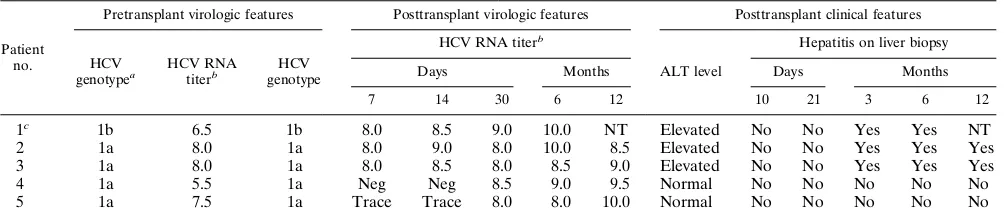
Clinical and virological features of HCV infection after liver transplantation.Table 1 summarizes the clinical and virolog-ical features of HCV infection before and after liver transplan-tation for the five patients. All five patients had active HCV infections with HCV genotype 1 both before and after trans-plantation; four patients were infected with HCV subgenotype 1a, while one patient was infected with HCV subgenotype 1b. Mixed genotype infections were excluded by nested PCR anal-ysis of the HCV core gene with type-specific primers (17). Pa-tients 1 to 3 each developed biochemical and histologic evi-dence of hepatitis in liver allografts by 3 months after liver transplantation. In all three patients liver biopsies performed at 10 days and 21 days posttransplantation showed no evidence of hepatitis. Biopsies performed at 2 to 3 months posttrans-plantation showed acute hepatitis in all three patients, and by 6 months posttransplantation, liver biopsies indicated severe hepatitis in all three. Patient 1 expired 7 months after liver transplantation as a result of an infectious pulmonary compli-cation. Patients 2 and 3 had histologic findings of bridging fibrosis and bridging necrosis, respectively, in their liver allo-grafts by 12 months posttransplantation. In patients four and five, liver biopsies performed at 10 days, 21 days, 3 months, 6 months, and 12 months after liver transplantation showed no evidence of hepatitis (Table 1), and serum ALT values re-mained normal.
The only difference in HCV viremia patterns between asymptomatic patients 4 and 5 and diseased patients 1 to 3 was observed during the first 2 weeks after liver transplantation. The diseased patients remained viremic on posttransplant days 7 and 14, with high viral RNA titers (greater than 7 log units/ ml). In contrast, the asymptomatic patients had significant re-ductions in HCV viremia on posttransplant days 7 and 14, with HCV RNA undetectable by RT-PCR in patient 4 and detect-able at only trace levels (less than 500 RNA molecules per ml) by RT-PCR in patient 5 (Table 1). However, by the end of the first posttransplant month, all five patients had developed high-titered HCV viremia (greater than 7 log units/ml) which per-sisted throughout the course of study in all cases (Table 1).
[image:2.612.58.560.82.190.2]Characterization of HCV quasispecies variants. Figure 1
TABLE 1. Clinical and virological features of HCV infection after orthotopic liver transplantation
Patient no.
Pretransplant virologic features Posttransplant virologic features Posttransplant clinical features
HCV genotypea
HCV RNA titerb
HCV genotype
HCV RNA titerb
ALT level
Hepatitis on liver biopsy
Days Months Days Months
7 14 30 6 12 10 21 3 6 12
1c 1b 6.5 1b 8.0 8.5 9.0 10.0 NT Elevated No No Yes Yes NT
2 1a 8.0 1a 8.0 9.0 8.0 10.0 8.5 Elevated No No Yes Yes Yes
3 1a 8.0 1a 8.0 8.5 8.0 8.5 9.0 Elevated No No Yes Yes Yes
4 1a 5.5 1a Neg Neg 8.5 9.0 9.5 Normal No No No No No
5 1a 7.5 1a Trace Trace 8.0 8.0 10.0 Normal No No No No No
aSimmonds nomenclature (18).
bViral RNA levels are expressed as log transformed molecules per milliliter determined by quantitative competitive PCR (8). Trace, HCV RNA positive but less
than 3 logs/ml, below the range of quantitation. Neg, HCV RNA negative by reverse transcription PCR. NT, not tested. cPatient 1 expired at 7 months posttransplantation (see text for details).
VOL. 70, 1996 HCV QUASISPECIES TRACKING 7623
on November 9, 2019 by guest
http://jvi.asm.org/
illustrates characterization of HCV quasispecies variants at multiple time points before and after liver transplantation in patient 1. A total of 73 independent HVR clones from six different time points were analyzed by nucleotide sequencing (Fig. 1A). In pretransplant sera, 9 of 17 (53%) of the HVR clones had identical amino acid sequences, while the remaining 8 clones each had a single unique amino acid change relative to the consensus pretransplant sequence. Thus, patient 1 had a single quasispecies major variant in pretransplant serum, des-ignated MV-1, plus numerous minor variants which were all
closely related to MV-1 by sequencing. At 5 days and 1 month posttransplantation, 16 of 18 (89%) and 9 of 10 (90%), respec-tively, of the HVR clones had the consensus MV-1 amino acid sequence. At 6 months posttransplantation, 9 of 15 (60%) of the serum-derived clones and 10 of 13 (77%) of the liver-derived clones had the consensus MV-1 sequence. Therefore, after liver transplantation, the consensus quasispecies major variant (MV-1) remained unchanged, and none of the minor variants identified in pretransplant serum showed evidence of emergence or propagation after liver transplantation. Figure 1B illustrates the gel shift profile of 10 representative HVR clones isolated from patient 1 serum at 6 months posttrans-plantation: although very similar gel shift patterns were ob-FIG. 1. Analysis of a simple HCV quasispecies before and after liver trans-plantation in patient 1 (diseased). The E2 HVR was amplified from sequential clinical specimens by RT-PCR and analyzed by either nucleotide sequencing (A) or HTA (B) as described in Materials and Methods. (A) Deduced amino acid sequences of 73 HVR clones from pretransplant serum (Pre-Tx) or from post-transplant sera (post-Tx) obtained at 5 days (5D), 1 month (1M), and 6 months (6M) posttransplantation or from a liver biopsy specimen obtained at 6 months posttransplantation. The consensus amino acid sequence of MV-1 is in boldface 1 (see text for details). (B) HTA analysis of 10 representative HVR clones isolated from patient 1 serum at 6 months posttransplantation, using radiola-beled MV-1 sequences as the probe. PCR amplification products were prepared from HVR clones and hybridized to a32
P-labeled probe derived from a consen-sus clone (MV-1), and the hybrids were analyzed by gel electrophoresis and autoradiography as described in Materials and Methods. Lane 7 contains the homoduplex shift control. (C) Bar graph comparing percentage of HVR clones identical to the consensus clone (MV-1) by either sequence analysis (black bars) or HTA (gray bars) at various time points before and after liver transplantation.
on November 9, 2019 by guest
http://jvi.asm.org/
[image:3.612.321.551.99.430.2]served for all 10 clones relative to the MV-1 probe, slight shifts in mobility were detected for the clones in lanes 4, 6, 9, and 11, suggesting minor differences from the MV-1 probe (Fig. 1B, lane 7).
The bar graph in Fig. 1C summarizes the strong correlation observed between predicted amino acid sequence and HTA for assessing the relatedness of HCV quasispecies variants over time. Over the course of study of patient 1, a total of 167 individual HVR clones were analyzed by the HTA technique: while the majority of HVR clones were identical in mobility to the probe homoduplex control, a subset of quasispecies vari-ants were distinguishable from MV-1 by HTA. The percentage of quasispecies variants which were slightly different from MV-1 fluctuated over time. For example, gel shift patterns ap-pearing identical to that for MV-1 were observed for approx-imately 90% of HVR clones obtained from four independent RT-PCRs at either 5 days or 1 months posttransplantation. Amino acid sequence patterns were also 90% homologous at these time points (Fig. 1A). In contrast, identical-appearing HTA and amino acid patterns accounted for 54 to 58% of pretransplant HVR clones and 68 to 80% of HVR clones isolated from liver and serum at 6 months posttransplantation (Fig. 1C).
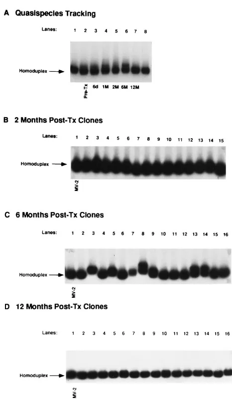
Propagation of quasispecies major variants after liver transplantation in symptomatic patients.Persistence of qua-sispecies major variants after liver transplantation was also seen in patients 2 and 3, both of whom developed severe hepatitis in their liver allografts within the first 3 months after liver transplantation. In patient 2, nucleotide sequencing of 46 HVR1 clones isolated from pre- and posttransplant sera also indicated a single quasispecies major variant in this case, des-ignated MV-2 (data not shown). Figure 2A illustrates a quasi-species tracking experiment using MV-2 as a probe, after hy-bridization with heterogeneous HVR sequences amplified from pretransplant serum (lane 2) and posttransplant serum obtained at 6 days (lane 3), 1 month (lane 4), 2 months (lane 5), 6 months (lane 6), and 12 months (lane 7) after surgery. Mobility of the heterogeneous HVR sequences appeared highly similar or identical to that of the MV-2 probe control at all time points except 6 months posttransplantation, when a small but reproducible gel shift pattern was evident. Clinically, patient 2 had a sharp peak in serum ALT values (357 IU/liter) at the 6-month time point, with findings of extensive inflam-mation and hepatocellular necrosis on liver biopsy. At this time, 13 of 30 (43%) of the HVR1 clones showed detectable shifts by HTA (e.g., Fig. 2C, lanes 3, 5, 7, 8, 9, 13, and 14), compared with only 1 of 30 (3%) HVR clones obtained at 2 months posttransplantation (Fig. 2B). The minor variants ob-served at 6 months posttransplantation were not obob-served at 12 months posttransplantation by HTA using heterogeneous HVR sequences (Fig. 2A, lane 7) or by either HTA or se-quence analysis of 39 different HVR clones generated from two independent RT-PCRs (Fig. 2D).
These data confirm that patient 2 was also infected with a relatively homogeneous HCV quasispecies both before and after liver transplantation and that severe hepatitis occurred during the time period of quasispecies homogeneity. We ob-served a transient increase in quasispecies minor variants dur-ing the peak of hepatitis, as reflected by high ALT values and liver biopsy findings; however, these minor variants were not propagated.
Analysis of pretransplant serum in patient 3 revealed a com-plex HCV quasispecies pattern consisting of three major se-quence variants, designated MV-3a, MV-3b, and MV-3c, in roughly equal proportions (Fig. 3A, lanes 1 to 3). The doublet shift pattern observed in lanes 2 and 3 of Fig. 3A resulted from
[image:4.612.317.550.62.463.2]hybridization of both strands of the radiolabeled DNA probe to target DNA strands and was highly reproducible and char-acteristic of a particular quasispecies variant. Figure 3D com-pares the consensus amino acid sequences of the three major variants in pretransplant serum: MV-3a differed from MV-3b by five amino acids and 10 nucleotides; MV-3a differed from MV-3c by seven amino acids and 11 nucleotides; and MV-3b differed from MV-3c by six amino acids and 8 nucleotides. The HTA patterns were consistent with the differences in nucleo-tide sequences between the three quasispecies major variants, regardless of which major variant was used as the probe (data not shown).
FIG. 2. Analysis of a simple HCV quasispecies over time in patient 2 (dis-eased). HVR sequences were amplified from sequential serum specimens ob-tained before and after liver transplantation, and HTA was performed as de-scribed in the legend to Fig. 1. (A) HCV quasispecies tracking experiment. HVR sequences were amplified from pretransplant serum (Pre-Tx; lane 2) and post-transplant sera at 6 days (lane 3), 1 month (lane 4), 2 months (lane 5), 6 months (lane 6), and 1 year (lane 7) after liver transplantation. Heterogeneous HVR amplification products were hybridized to a32P-labeled probe derived from the pretransplant consensus clone (MV-2), and hybrids were analyzed by polyacryl-amide gel electrophoresis and autoradiography. Lanes 1 and 8 contain the homoduplex shift control. (B to D) Clonal frequency analysis of 45 individual HVR clones isolated from patient 2 serum at 2 (B), 6 (C), and 12 (D) months posttransplantation (Post-Tx), after hybridization with MV-2 probe. Lane 1 contains the probe homoduplex control.
VOL. 70, 1996 HCV QUASISPECIES TRACKING 7625
on November 9, 2019 by guest
http://jvi.asm.org/
Tracking of this relatively complex HCV quasispecies before and after liver transplantation by using the HTA technique is illustrated in Fig. 3A, lanes 4 to 8. Lane 4 illustrates HTA of heterogeneous HVR sequences amplified directly from pre-transplant serum: the three gel shift patterns representing
MV-3a, MV-3b, and MV-3c are clearly evident. Lanes 5 to 8 illus-trate tracking of the three quasispecies major variants after liver transplantation. MV-3a was detectable in all posttrans-plant serum specimens and was the only quasispecies major variant detectable at 13 months posttransplantation (lane 8). MV-3b was detected at 8 days, 1 month, and 7 months after liver transplantation (lanes 5 to 7) but was no longer detected at 13 months (lane 8). MV-3c was not detectable in any of the posttransplant serum specimens by the HTA tracking tech-nique (lanes 5 to 8).
The clonal frequency of the three major variants was de-tected both before and after liver transplantation, as illustrated in Fig. 3B and C. On the basis of HTA of 59 HVR clones isolated from pretransplant serum, MV-3c was the quasispe-cies major variant of highest clonal frequency before transplan-tation (50% of HVR clones), followed by MV-3a (28% of clones) and MV-3b (22% of clones). At 8 days posttransplan-tation, MV-3a represented approximately 66% of HVR clones, MV-3b comprised approximately 30% of clones, and MV-3c represented less than 3% of clones. At 1 and 7 months post-transplantation, MV-3b became the most prevalent quasispe-cies major variant in serum, comprising 52 and 67%, respec-tively, of clones (Fig. 3B). At 13 months posttransplantation, MV-3b was not detected in any of the 30 HVR clones ana-lyzed, and MV-3a was the only quasispecies major variant identified (Fig. 3C).
Emergence of quasispecies minor variants after liver trans-plantation in asymptomatic patients.Characterization of HCV quasispecies before and after liver transplantation in the two asymptomatic patients (patients 4 and 5) is summarized in Fig. 4. HTA analysis of 30 HVR clones isolated from pretransplant sera in patient 4 indicated this patient had a relatively simple quasispecies pattern, with a single major variant designated MV-4prerepresenting greater than 95% of clones (Fig. 4A, left
panel). In contrast, analysis of 40 HVR clones from pretrans-plant serum in patient 5 revealed a more complex quasispecies pattern (Fig. 4A, right panel): two distinct quasispecies major variants, designated MV-5a (45% of clones; represented by lanes 4, 10, 11, and 12) and MV-5b (18% of clones; lanes 2 and 6) were identified. For this experiment, an MV-5b-derived HVR clone was randomly picked as a probe (lane 1). Numer-ous minor variants were also detected in the pretransplant quasispecies of patient 5, each at a frequency of less than 3% of pretransplant clones (e.g., lanes 3, 5, 8, and 9).
Figure 4B illustrates quasispecies tracking by the HTA tech-nique after liver transplantation in patients 4 and 5. In patient 4, MV-4prewas used as a probe, and in patient 5, MV-5b was
used as a probe. In both experiments, the posttransplant HTA patterns were clearly different from the pretransplant HTA patterns, indicating emergence of new quasispecies which ap-peared homogeneous over time. Figure 4C illustrates HTA of multiple HVR clones isolated from posttransplant sera of pa-tients 4 and 5, using the same probes as used for Fig. 4A and B. Analysis of more than 90 HVR clones isolated from four different posttransplant time points yielded indistinguishable HTA patterns in 99% of clones in both cases (Fig. 4C), con-firming that the posttransplant HCV quasispecies were re-markably homogeneous at the clonal level with respect to HVR sequences. These patterns were not due to sampling or selection bias, because (i) the same patterns were observed at multiple time points over a 1- to 2-year period and (ii) the same patterns were obtained with diluted and undiluted spec-imens (data not shown). Importantly, the pretransplant quasi-species major variants identified in patients 4 and 5 were not found in any of the posttransplant sera tested in either case.
[image:5.612.66.293.59.506.2]We next performed a series of experiments to determine the FIG. 3. Analysis of a complex HCV quasispecies before and after liver
trans-plantation in patient 3 (diseased). (A) Heteroduplex tracking assay of the HCV quasispecies. Lanes 1 to 3 show individual HTA profiles of the three quasispecies major variants (MV-3a, MV-3b, and MV-3c, respectively) after amplification and cloning from pretransplant serum. Radiolabeled MV-3a was used as a probe. Lanes 4 through 8 illustrate a HCV quasispecies tracking experiment. HVR se-quences were amplified from pretransplant serum (Pre-Tx; lane 4) and post-transplant sera at 8 days (lane 5), 1 month (lane 6), 7 months (lane 7), and 13 months (lane 8) after liver transplantation. Heterogeneous HVR amplification products were hybridized to a32
P-labeled probe derived from MV-3a, and hy-brids were analyzed by polyacrylamide gel electrophoresis and autoradiography. (B) Analysis of 15 HVR clones isolated from patient 3 serum at 7 months post-transplantation (Post-Tx), using radiolabeled MV-3a as the probe. Lane 1 con-tains the homoduplex shift control. Nine of 15 clones gave gel shift patterns identical that of to MV-3b, while 6 of 15 clones gave patterns indistinguishable from that of the MV-3a homoduplex. (C) Analysis of 15 HVR clones isolated from patient 3 serum at 13 months posttransplantation. All 15 clones gave gel shift patterns that were indistinguishable from that of the MV-3a probe control. (D) Predicted amino acid sequences of the quasispecies major variants MV-3a, MV-3b, and MV-3c.
on November 9, 2019 by guest
http://jvi.asm.org/
relationships between the pre- and posttransplant quasispecies in patients 4 and 5. First, a representative posttransplant HVR probe was prepared for both patients; the probes were desig-nated MV-4postfor patient 4 (Fig. 5A) and MV-5postfor
pa-tient 5 (Fig. 5B). The posttransplant probes were hybridized with large panels of either pretransplant or posttransplant HVR clones and analyzed by the HTA technique. In patient 4, all posttransplant clones tested gave HTA shift patterns which appeared identical to that of the MV-4posthomoduplex control
(Fig. 5A, lanes 8 to 10, for example) but gave different HTA shift patterns for all 87 pretransplant HVR clones tested (Fig. 5A, lanes 2 to 4, for example). Although we found no pretrans-plant clones identical in mobility to MV-4post, we did identify
three pretransplant minor variants (clones 25, 61, and 87) which gave HTA shift patterns which were intermediate be-tween those of MV-4preand MV-4post(Fig. 5A, lanes 5 to 7).
The right panel of Fig. 5A compares the consensus amino acid sequences of the pretransplant quasispecies major variant (lanes 2 to 4), the pretransplant minor variants (lanes 5 to 7), and the posttransplant quasispecies major variant. Relative to the pretransplant major variant (MV-4pre), three amino acid
changes were observed in 100% of posttransplant HVR clones: an Arg to Gly at position 384, a Leu to Phe at position 403, and an Ile to Val at position 414. Furthermore, the predicted amino acid sequences of two pretransplant minor variants (clones 61 and 87) both contained the Arg-to-Gly mutation at position 384 but no change at position 403 or 414 relative to MV-4pre. Clones 61 and 87 differed from each other by three
nucleotides, which accounted for the differences in HTA shift patterns. The nucleotide sequences suggest that MV-4postmay
[image:6.612.59.552.65.458.2]have been derived from or related to pretransplant minor variant clone 87 (data not shown).
FIG. 4. Analysis of HCV quasispecies before and after liver transplantation in the asymptomatic liver transplant recipients 4 and 5. (A) Heteroduplex tracking assay of pretransplant quasispecies. The left panel illustrates HTA profiles for 11 HVR clones isolated from patient 4 pretransplant serum, after hybridization with patient 4 pretransplant probe MV-4pre. Lane 12 contains the homoduplex shift control. The right panel illustrates HTA profiles for 11 HVR clones isolated from patient 5 pretransplant serum, after hybridization with the patient 5 pretransplant probe MV-5b. Lane 1 contains the homoduplex shift control. (B) HCV quasispecies tracking before and after liver transplantation in patients 4 and 5. HVR sequences were amplified from pretransplant serum (Pre-Tx) and posttransplant sera at 1 month (1M), 2 months (2M), 6 months (6M), 12 months (12M), and 24 months (24M) after liver transplantation. Heterogeneous HVR amplification products were hybridized to 32
P-labeled probes derived from pretransplant HVR clone MV-4pre(patient 4) or pretransplant HVR clone MV-5b (patient 5). Homoduplex gel shift controls are present in lanes 1 and 7 for both experiments. (C) HTA profiles of representative HVR clones isolated from patient 4 serum at 1 month posttransplantation, after hybridization with pretransplant probe MV-4pre, and from patient 5 serum at 12 months posttransplantation, after hybridization with pretransplant probe MV-5b. Homoduplex shift controls are indicated.
VOL. 70, 1996 HCV QUASISPECIES TRACKING 7627
on November 9, 2019 by guest
http://jvi.asm.org/
FIG. 5. Relationship between pre-and posttransplant quasispecies in patients 4 and 5 (asymptomatic patients). (A) HTA profiles and amino acid sequences of HCV quasispecies variants isolated from patient 4 serum before and after liver transplantation. A consensus posttransplant HVR clone (MV-4 post ) was radiolabeled with 32 P and hybridized to HVR sequences isolated from three pretransplant (Pre-Tx) major variant clones (lanes 2 to 4), three pretransplant minor variant clones (lanes 5 to 7), and three posttransplant (Post-Tx) major variant clones (lanes 8 to 10). Lanes 1 and 11 contain homoduplex gel shift controls; deduced amino acid sequences of the HVR clones are summarized on the right. (B) HTA profiles of 20 HVR clones isolated from patient 5 pretransplant serum, using a consensus posttransplant HVR clone as the probe (MV-5 post ). The rightmost lane contains the homoduplex shift control. Pretransplant clone p 21 gave a homoduplex shift pattern. Amino acid sequences of HCV quasispecies variants in patient 5, including the consensus pretransplant quasispecies major variants MV-5a and MV-5b, the consensus posttransplant quasispecies major variant MV-5 post (clones 1, 2, and 3), and pretransplant clone p 21, are shown on the right. The positions and identities of amino acids are shown relative to the MV-5a sequence.
on November 9, 2019 by guest
http://jvi.asm.org/
[image:7.612.89.283.86.335.2]In patient 5, the posttransplant probe (MV-5post) also gave
homologous HTA patterns with all posttransplant HVR clones tested (data not shown) but gave clearly different HTA shift patterns for 28 of 30 pretransplant clones (Fig. 5B). However, MV-5postdid give homologous-appearing HTA shift patterns
for two pretransplant HVR clones (e.g., clone 21 in Fig. 5B). The right panel of Fig. 5B compares the consensus amino acid sequences of the pretransplant quasispecies major variants in this patient (MV-5a and MV-5b), the pretransplant minor ant (clone 21), and the posttransplant quasispecies major vari-ants (MV-5postclones 1, 2, and 3). The amino acid sequence of
pretransplant minor variant 21 was identical to that of the posttransplant quasispecies major variant, implying that the posttransplant HCV quasispecies was derived from a pretrans-plant minor variant in patient 5.
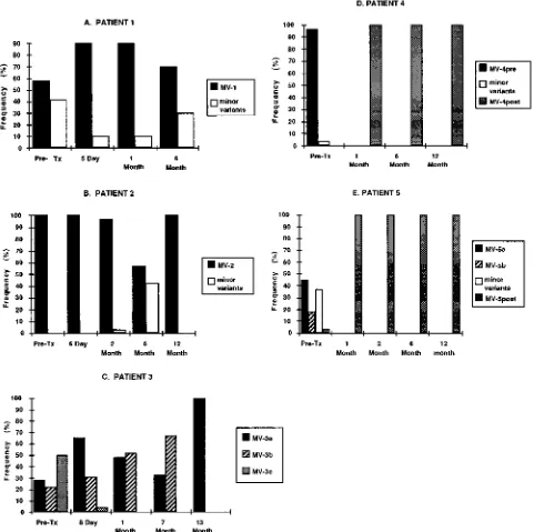
In summary, these experiments demonstrate that in asymp-tomatic patients 4 and 5, the pretransplant HCV quasispecies major variants were genetically different from the posttrans-plant quasispecies major variants. Analysis of the HVR se-quences in the asymptomatic patients suggested that the post-transplant quasispecies were closely related to or derived from pretransplant minor variants. The HCV quasispecies patterns in these five patients are summarized by bar graphs in Fig. 6.
DISCUSSION
This study used the techniques of HTA and nucleotide se-quencing to investigate patterns of HCV quasispecies before and after liver transplantation in five patients who were in-fected with HCV genotype 1. Three of the patients developed severe recurrence of liver disease during the first few months after liver transplantation, while the other two patients devel-oped high-titered yet asymptomatic posttransplant HCV in-fections. In the three diseased patients (patients 1 to 3), qua-sispecies major variants present in pretransplant serum were efficiently propagated after liver transplantation (Fig. 6). In the two asymptomatic patients (patients 4 and 5), consensus sequences of the quasispecies major variants present in pre-transplant serum were not detectable at any time after liver transplantation, and by 1 month posttransplantation, new qua-sispecies major variants of the same genotype had emerged in both cases; these quasispecies appeared to be more closely related to pretransplant quasispecies minor variants, as judged from HTA patterns and sequence analysis.
This study shows that quasispecies major variants were effi-ciently propagated immediately after liver transplantation in the three diseased patients but not in the asymptomatic pa-tients. We selected our patients so that other virological char-acteristics associated with disease progression (genotype and level of viremia) were similar. These data may suggest that in some instances, certain HCV quasispecies have a higher patho-genic potential than others, although further testing of this concept in animal models is needed. It is conceivable that the quasispecies described in the current study differ in regions of the HCV genome other than the HVR, a possibility which is currently under investigation.
It was of interest to characterize HCV quasispecies propa-gation before and during the acute and chronic phases of hepatitis in our diseased patients. The pretransplant HCV quasispecies were either relatively homogeneous, with a single major variant (patients 1, 2, and 4) or complex, with 2 or more major variants (patients 3 and 5), and the complexity of HCV quasispecies in pretransplant serum did not correlate with the posttransplant disease course. Similarly to Martell et al. (15), we observed remarkably homogeneous propagation of HCV quasispecies major variants in patients 1 and 2, indicating that
quasispecies genetic diversity posttransplant is not directly pro-portional to disease activity per se. In patient 1, the pretrans-plant HCV quasispecies was 54% homogeneous at the amino acid sequence level immediately prior to liver transplantation and became approximately 90% homogeneous during the first week posttransplantation, which might be explained by a burst of new viral replication in the transplanted liver allograft, as would be expected from a highly hepatotropic quasispecies variant. Over time, the quasispecies accumulated minor muta-tions, although the major variant remained unchanged. In pa-tient 2, we observed a transient increase in HVR heterogeneity due to an accumulation of quasispecies minor variants at 6 months posttransplantation, but at 12 months posttransplanta-tion, we observed reversion to a homogeneous pattern resem-bling the pretransplant quasispecies major variant (Fig. 6B).
The two patients who developed asymptomatic posttrans-plant infections (patients 4 and 5) had dramatic reductions in HCV viremia during the initial 2 weeks after surgery, followed by emergence of new quasispecies major variants by 1 month after liver transplantation. In both cases, the emergent quasi-species major variants were most likely derived from quasispe-cies minor variants present in pretransplant serum. It is of considerable interest that in these two patients, the emergent quasispecies variants did not cause apparent liver disease, even though they were replicating at high titer, and both repre-sented HCV genotype 1, which is considered relatively patho-genic. Perhaps the HCV quasispecies variants were attenuated as a result of mutation, being efficient at replication but inef-ficient with regard to the induction of hepatitis. A second possibility is that the emergent quasispecies variants have a reduced hepatotropism. Additional quantitative in situ exper-iments are under way to determine the relative contributions of hepatic and extrahepatic replication in patients with and without disease. Another possibility is that the major HVR sequences identified in pretransplant serum represented de-fective (noninfectious) virus derived from the explanted liver. It will be important to establish the relative infectivity of these isolates in tissue culture and small animal models once devel-oped.
It is likely that the differences in viral quasispecies pheno-types and disease patterns are strongly influenced by host fac-tors in these patients. For example, patients 1 to 3, HCV viremia remained high during the initial 2 weeks immediately after liver transplantation, raising two possibilities: (i) highly efficient extrahepatic replication was occurring at the time of liver transplantation in these cases and (ii) the quasispecies major variants had a high affinity for hepatocyte receptor rec-ognition on the transplanted allograft, or the allograft may have been highly susceptible to these major variants for un-known reasons. Regardless, high-titered viremia during the first weeks posttransplantation has been identified as a marker for early recurrence of liver disease in these and other HCV-infected patients whom we have studied (8). In asymptomatic patients 4 and 5, HCV viremia was greatly reduced during the first 2 weeks after liver transplantation, which may suggest that either extrahepatic replication was not highly active at the time of liver transplantation or the HCV RNA in the pretransplant serum was not indicative of highly infectious virus, or perhaps the liver allograft was not susceptible to the pretransplant quasispecies in these cases. Finally, quasispecies selection by immunological mechanisms at the time of liver transplantation is also a factor. All liver transplant recipients receive intense induction immunosuppressive therapy at the time of transplan-tation. Whether selective differences in humoral or cellular immune responses are associated with the different patterns of
VOL. 70, 1996 HCV QUASISPECIES TRACKING 7629
on November 9, 2019 by guest
http://jvi.asm.org/
quasispecies variation that we encountered remains to be de-fined.
This study demonstrates the utility of the HTA technique for characterizing and tracking HCV quasispecies major and minor variants, since hundreds of HVR sequences can be screened for mutation within a relatively short period of time. This technique may be particularly useful for identifying
[image:9.612.67.548.73.552.2]im-mune escape variants during vaccine studies, for characterizing persistent HCV quasispecies during interferon therapy in hu-mans, or for tracking HCV quasispecies variants during animal challenge experiments. The technique is also potentially valu-able for rapidly assessing other regions of the HCV genome. When combined with careful clinical studies, such an approach may ultimately lead to identification of new and important FIG. 6. Bar graphs comparing percentage of HVR clones related to the quasispecies major variants in patients 1 through 5. The frequency of quasispecies variants was determined either by clonal frequency analysis using the HTA technique, as described for Fig. 1 and 2, or by amino acid sequence analysis, using serum obtained prior to transplantation (Pre-Tx) or at sequential times after liver transplantation. Patients 1 and 2 were similar in that the vast majority of HVR clones isolated either before and after liver transplantation appeared identical to a consensus quasispecies major variant present in pretransplant serum; this major variant was designated MV-1 in patient 1 and MV-2 in patient 2. In patient 3, the pretransplant quasispecies consisted of three distinct major variants: MV-3a, MV-3b, and MV-3c. MV-3a and MV-3b were both propagated after liver transplantation, whereas MV-3c was not. In asymptomatic patients 4 and 5, the quasispecies major variants present in pretransplant serum, MV-4preand MV-5pre, were not propagated after liver transplantation. Instead, new quasispecies major variants designated MV-4postand MV-5postemerged after liver transplantation. The predicted amino acid sequence of MV-5postwas identical to that of a minor variant identified in pretransplant serum. See text for details.
on November 9, 2019 by guest
http://jvi.asm.org/
genetic markers of HCV persistence, virulence, and resistance to therapy.
ACKNOWLEDGMENTS
We gratefully acknowledge Amy Weiner and Michael Houghton for providing E2 primer sequences and helpful advice; James Mullins for assistance with the gel shift technique and for critical review of the manuscript; Greg Faulkner, Timothy Day, Corazon dela Rosa, and Minjun Chung excellent technical support; and Colleen Lasley for assistance in preparation of the manuscript.
REFERENCES
1. Chazouilleres, O., M. Kim, C. Combs, L. Ferrell, P. Bacchetti, J. Roberts,
N. L. Ascher, P. Neuwald, J. Wilber, and M. Urdea.1994. Quantitation of hepatitis C virus RNA in liver transplant recipients. Gastroenterology 106: 994–999.
2. Chomczynski, P., and N. Sacchi. 1987. Single-step method of RNA isolation by acid guanidinium thiocyanate-phenol-chloroform extraction. Anal. Bio-chem. 162:156–159.
3. Delwart, E. L., E. G. Shpaer, J. Louwagie, F. E. McCutchan, M. Grez, W. H.
Rubsamen, and J. I. Mullins.1993. Genetic relationships determined by a DNA heteroduplex mobility assay: analysis of HIV-1 env genes. Science 262: 1257–1261.
4. Farci, P., H. J. Alter, D. C. Wong, R. H. Miller, S. Govindarajan, R. Engle,
M. Shapiro, and R. H. Purcell.1994. Prevention of hepatitis C virus infection in chimpanzees after antibody-mediated in vitro neutralization. Proc. Natl. Acad. Sci. USA 91:7792–7796.
5. F’eray, C., M. Gigou, D. Samuel, V. Paradis, J. Wilber, M. F. David, M.
Urdea, M. Reynes, C. Bre´chot, and H. Bismuth.1994. The course of hepatitis C virus infection after liver transplantation. Hepatology 20:1137–1143. 6. F’eray, C., D. Samuel, V. Thiers, M. Gigou, F. Pichon, A. Bismuth, M.
Reynes, P. Maisonneuve, H. Bismuth, and C. Bre´chot.1992. Reinfection of liver graft by hepatitis C virus after liver transplantation. J. Clin. Invest.
89:1361–1365.
7. Gretch, D., L. Corey, J. Wilson, C. dela Rosa, R. Willson, R. Carithers, Jr.,
M. Busch, J. Hart, M. Sayers, and J. Han.1994. Assessment of hepatitis C virus RNA levels by quantitative competitive RNA polymerase chain reac-tion: high-titer viremia correlates with advanced stage of disease. J. Infect. Dis. 169:1219–1225.
8. Gretch, D. R., C. E. Bacchi, L. Corey, C. D. Rosa, R. R. Lesniewski, K.
Kowdley, A. Gown, I. Frank, J. D. Perkins, and J. R. L. Carithers.1995. Persistent hepatitis C virus infection following liver transplantation: clinical and virologic features. Hepatology 22:1–9.
9. Gretch, D. R., C. dela Rosa, R. L. Carithers, R. A. Willson, B. Williams, and
L. Corey.1995. Assessment of hepatitis C viremia using molecular amplifi-cation technologies: correlations and clinical impliamplifi-cations. Ann. Intern. Med.
123:321–329.
10. Hijikata, M., N. Kato, Y. Ootsuyama, M. Nakagawa, S. Ohkoshi, and K.
Shimotohno.1991. Hypervariable regions in the putative glycoprotein of hepatitis C virus. Biochem. Biophys. Res. Commun. 175:220–228. 11. Honda, M., S. Kaneko, A. Sakai, M. Unoura, S. Murakami, and K.
Koba-yashi.1994. Degree of diversity of hepatitis C virus quasispecies and pro-gression of liver disease. Hepatology 20:1144–1151.
12. Kato, N., T. Nakazawa, Y. Ootsuyama, K. Sugiyama, S. Ohkoshi, and K.
Shimotohno.1994. Virus isolate-specific antibodies against hypervariable region 1 of the hepatitis C virus second envelope protein, gp70. Jpn. J. Cancer Res. 85:987–991.
13. Kato, N., H. Sekiya, Y. Ootsuyama, T. Nakazawa, M. Hijikata, S. Ohkoshi,
and K. Shimotohno.1993. Humoral immune response to hypervariable re-gion 1 of the putative envelope glycoprotein (gp70) of hepatitis C virus. J. Virol. 67:3923–3930.
14. Konig, V., J. Bauditz, H. Lobeck, R. Lusebrink, P. Neuhaus, G. Blumhardt,
W. O. Bechstein, R. Neuhaus, R. Steffen, and U. Hopf.1992. Hepatitis C virus reinfection in allografts after orthotopic liver transplantation. Hepa-tology 16:1137–1143.
15. Martell, M., J. I. Esteban, J. Quer, V. Vargas, R. Esteban, J. Guardia, and
J. Go´mez.1994. Dynamic behavior of hepatitis C virus quasispecies in pa-tients undergoing orthotopic liver transplantation. J. Virol. 68:3425–3436. 16. Naito, M., N. Hayashi, T. Moribe, H. Hagiwara, E. Mita, Y. Kanazawa, A.
Kasahara, H. Fusamoto, and T. Kamada.1995. Hepatitis C viral quasispe-cies in hepatitis C virus carriers with normal liver enzymes and patients with type C chronic liver disease. Hepatology 22:407–412.
17. Okamoto, H., Y. Sugiyama, S. Okada, K. Kurai, Y. Akahane, Y. Sugai, T.
Tanaka, K. Sato, F. Tsuda, and Y. Miyakawa.1992. Typing hepatitis C virus by polymerase chain reaction with type-specific primers: application to clin-ical surveys and tracing infectious sources. J. Gen. Virol. 73:673–679. 18. Simmonds, P., E. C. Holmes, T. A. Cha, S. W. Chan, F. McOmish, B. Irvine,
E. Beall, P. L. Yap, J. Kolberg, and M. S. Urdea.1993. Classification of hepatitis C virus into six major genotypes and a series of subtypes by phy-logenetic analysis of the NS-5 region. J. Gen. Virol. 74:2391–2399. 19. Taniguchi, S., H. Okamoto, M. Sakamoto, M. Kojima, F. Tsuda, T. Tanaka,
E. Munekata, E. E. Muchmore, D. A. Peterson, and S. Mishiro.1993. A structurally flexible and antigenically variable N-terminal domain of the hepatitis C virus E2/NS1 protein: implication for an escape from antibody. Virology 195:297–301.
20. van Doorn, L. J., I. Capriles, G. Maertens, R. DeLeys, K. Murray, T. Kos, H.
Schellekens, and W. Quint.1995. Sequence evolution of the hypervariable region in the putative envelope region E2/NS1 of hepatitis C virus is corre-lated with specific humoral immune responses. J. Virol. 69:773–778. 21. Weiner, A. J., M. J. Brauer, J. Rosenblatt, K. H. Richman, J. Tung, K.
Crawford, F. Bonino, G. Saracco, Q. L. Choo, and M. Houghton.1991. Variable and hypervariable domains are found in the regions of HCV cor-responding to the flavivirus envelope and NS1 proteins and the pestivirus envelope glycoproteins. Virology 180:842–848.
22. Weiner, A. J., H. M. Geysen, C. Christopherson, J. E. Hall, T. J. Mason, G.
Saracco, F. Bonino, K. Crawford, C. D. Marion, K. A. Crawford, M. Bru-netto, P. J. Barr, T. Miyamura, J. McHutchinson, and M. Houghton.1992. Evidence for immune selection of hepatitis C virus (HCV) putative envelope glycoprotein variants: potential role in chronic HCV infections. Proc. Natl. Acad. Sci. USA 89:3468–3472.
23. Wilson, J. J., S. J. Polyak, T. D. Day, and D. R. Gretch. 1995. Characteriza-tion of simple and complex hepatitis C virus quasispecies by heteroduplex gel shift analysis: correlation with nucleotide sequencing. J. Gen. Virol. 76:1763– 1771.
24. Wright, T. L., E. Donegan, H. H. Hsu, L. Ferrell, J. R. Lake, M. Kim, C.
Combs, S. Fennessy, J. P. Roberts, and N. L. Ascher.1992. Recurrent and acquired hepatitis C viral infection in liver transplant recipients. Gastroen-terology 103:317–322.
VOL. 70, 1996 HCV QUASISPECIES TRACKING 7631