0022-538X/05/$08.00⫹0 doi:10.1128/JVI.79.4.2221–2229.2005
Copyright © 2005, American Society for Microbiology. All Rights Reserved.
RNA Recombination of Hepatitis Delta Virus in Natural
Mixed-Genotype Infection and Transfected
Cultured Cells
Tzu-Chi Wang and Mei Chao*
Department of Microbiology and Immunology, College of Medicine, Chang Gung University, Tao-yang, Taiwan
Received 25 March 2004/Accepted 4 October 2004
Most RNA viruses encode their own RNA polymerases for genome replication, and increasing numbers of them appear to be capable of undergoing RNA recombination. Here, we provide the first report of intergeno-typic recombination in hepatitis delta virus (HDV), the only animal RNA virus that requires host RNA polymerase(s) for viral replication. In vivo, we analyzed RNA species derived from the serum of a patient with mixed genotype I and genotype IIb HDV infection by using multiple restriction fragment length polymorphism (RFLP) assays and sequence analysis of cloned reverse transcription (RT)-PCR products. Six HDV recombi-nants were isolated from 101 tested clones, and HDV recombination in this patient was further confirmed by RT-PCR with genotype-specific primer pairs. Analysis of the recombination junctions suggested that the HDV genome rearrangement occurred through faithful homologous recombination. We then used an RNA cotrans-fection cell culture system to investigate HDV RNA recombination in vitro. We found that HDV recombinants could indeed be detected in the transfected cells; some of these possessed recombination junctions identical to those identified in vivo. Furthermore, using a PCR-independent RNase protection assay, we were able to readily identify the recombined HDV RNA species in cultured cells. Taken together, our results demonstrate that HDV RNA recombination occurs in both natural HDV infections and cultured cells, thereby presenting a novel mechanism for HDV evolution.
Hepatitis delta virus (HDV) is a subviral agent of the hep-atitis B virus that requires hephep-atitis B surface antigen for its assembly and transmission (48, 49). HDV is unique among animal viruses in that it resembles some plant pathogens, such as viroids, in having a circular RNA with an unbranched rod-like structure (25, 59), and a replication strategy that utilizes host RNA polymerases (Pols) (37, 41). Upon entering a cell, genomic HDV RNA is transcribed into complementary sub-genomic mRNA for the delta antigen (HDAg) (15) and mul-timeric antigenomic RNAs (31, 57). These then undergo self-cleavage and ligation to generate unit-length circular RNA (27, 46, 53, 63). Similarly, the circular antigenomic RNA serves as a template for production of additional genomic RNA.
In both virions and cells, the HDV RNA forms a ribonucle-oprotein complex with HDAg (9, 35). There are two forms of HDAg, small and large, which play distinct biological roles. The small HDAg (195 amino acids) is required for HDV RNA replication (8, 29). The large HDAg is produced through spe-cific RNA editing by the small form of the cellular enzyme ADAR1 (20, 44, 62), which converts the UAG stop codon for the small HDAg into a UGG tryptophan codon (36). This process leads to translation of an additional 19 amino acids and creates the large HDAg (214 amino acids), which in turns inhibits genome replication and is required for packaging (6, 8).
Viral hepatitis due to HDV infection has been found world-wide, and the complete nucleotide (nt) sequences of HDV genomes have been reported from many locations (5, 10, 11, 16, 28, 32, 38, 51, 59). Comparison of these by Casey et al. (5) indicated that there are at least three HDV genotypes: I, II, and III. HDV genotype III has been isolated only in northern South America (5) and is associated with severe acute hepati-tis, whereas the milder HDV genotype II is found primarily in Asia (64). In contrast, HDV isolates of genotype I have been reported in every part of the world, and the pathogenesis of genotype I infections varies from fulminant hepatitis to asymp-tomatic chronic liver disease (10, 11, 16, 32, 38, 51, 59). Phy-logenetic analysis of additional HDV sequences further sug-gested that HDV genotype IIb (isolated from Taiwan and the Miyako Islands, Okinawa, Japan) and genotype IIb-M (iso-lated from the Miyako Islands) form genetic subgroups (50, 60, 61, 65). It is worth noting that most of the above-mentioned sequences were isolated from non-African countries. Recently, characterization of 22 African sequences revealed that 15 of them form three new lineages and that the other 7 are scat-tered within genotype I (45). These analyses also suggested that genotype IIb sequences form a distinct clade instead of being a subtype of genotype II. Therefore, recent work has indicated that the various HDV genotypes fall into at least seven clades (45). Interestingly, HDV genotype I is the only genotype detected in some locations, including Europe and North America (5, 61), whereas multiple genotypes have been detected in Africa (45) and in Asian locations, including Japan, Russia, and Taiwan (16, 17, 33, 60, 61, 65). Furthermore, mixed infections of genotypes I and II or II and IIb have been re-ported in Taiwan (66). However, the roles of these mixed HDV
* Corresponding author. Mailing address: Department of Microbi-ology and ImmunMicrobi-ology, College of Medicine, Chang Gung University, 259, Wen-Hwa 1st Rd., Kwei-Shan, Tao-yang 333, Taiwan. Phone: 886-3-211-8800, ext. 5134. Fax: 886-3-211-8956. E-mail: pa0728@mail .cgu.edu.tw.
2221
on November 8, 2019 by guest
http://jvi.asm.org/
infections in viral biology and evolution remain to be eluci-dated.
It has been reported that specific RNA editing as well as random mutations occurring during RNA replication lead to sequence heterogeneity in the HDV genome (5, 12, 16, 32, 36, 42, 55, 59). Genetic recombination is an important process for eliminating detrimental mutations and ensuring greater ge-netic diversity. Recombination occurs in mixed infections of some closely related RNA viruses that encode their own RNA polymerases (30) and in viroids that are dependent on the cellular enzymatic activities for replication (1, 4, 14, 23, 24, 43, 47, 52, 54). However, although phylogenetic analysis suggests that genetic recombination can occur both between and within HDV genotypes (67), HDV recombinants have never been isolated from HDV mixed-genotype infections (66) either in nature or in the laboratory. In this report, we demonstrate that intergenotypic recombination of HDV RNA occurs both in a patient with a mixed genotype I and IIb infection and also in a RNA cotransfection cultured cell system.
MATERIALS AND METHODS
RNA extraction and RT-PCR.HDV-related sequences were characterized from serum sample HDM2-18, which was derived from a 39-year-old chronic hepatitis patient (patient HDM-2) who was positive for serum hepatitis B surface antigen and antibodies to HDV. Total RNA was extracted from the serum using the TRI REAGENT LS for liquid samples (Molecular Research Center, Inc.) as described previously (60). In vitro-transcribed HDV RNAs from pG4B-D1I containing a full-length genotype I cDNA clone obtained from the woodchuck-adapted HDV genome of an Italian patient (28, 60), pG4B-D1IIb (containing a full-length cDNA of the HDV genome derived from the Taiwan-IIb-1 clone of genotype IIb HDV) (60), and pG4B-D1II-1 (containing a full-length cDNA of the HDV genome derived from the Taiwan-3 clone of genotype II) (34) were synthesized as described previously (34, 60) and used as controls for the subse-quent genotyping and recombination analyses. Reverse transcription (RT)-PCR amplification was performed using a Titan One Tube RT-PCR system (Boehr-inger Mannheim). The reaction conditions were as reported elsewhere (60), except that a longer elongation time (2 min) and fewer cycles (25 to 30 cycles) were employed to suppress in vitro recombination between related PCR tem-plate sequences, as suggested in a previous report (21). The sequences of the primers used are summarized in Table 1. The nucleotide numbering system used in this report is in accordance with that of Wang et al. (60). Primers 18 and 55⬘
were consensus primers used to amplify sequences of all HDV genotypes. The resulting fragment 1855⬘represents a region (nt 889 to 1310) that is generally used for the classification of HDV genotypes (5). A second region (nt 940 to 1271) was produced using genotype-specific primer pairs (I5⬘-1–IIb3⬘-1 and IIb5⬘-1–I3⬘-2) for specific amplification of 5⬘-I–IIb-3⬘and 5⬘-IIb–I-3⬘ recombi-nants, respectively. Total RNA was also extracted from cultured cells cotrans-fected with genotype I and IIb RNAs and then subjected to RT-PCR using primer pair 18-55⬘for HDV genotyping analysis, as well as genotype-specific primer pairs I5⬘-1–IIb3⬘-1 and IIb5⬘-1–I3⬘-1 for the examination of recombina-tion juncrecombina-tions.
HDV genotyping and analysis of HDV RNA recombinants.XhoI restriction fragment length polymorphism (RFLP) analysis, previously shown to effectively
differentiate between HDV genotypes within PCR fragment 1855⬘(nt 889 to 1310) (64), was used to screen mixed infections of HDV. Genotypes I, IIb, II, and III yielded different XhoI-digested PCR products, so mixed HDV infections could be defined by RFLP patterns showing combinations of the variously sized bands. PCR products showing an XhoI RFLP pattern with multiple bands were then cloned into a T vector (TOPO TA cloning vector; Invitrogen). Plasmids were extracted from 101 colonies and subjected to double digestion with EcoRI (to release the HDV cDNA inserts) and XhoI (to differentiate the HDV geno-types). To further investigate the sequence heterogeneity of the HDV-related sequences, clones were also subjected to multiple RFLP assays, including double digestion of clones with EcoRI plus SacII and EcoRI plus SphI. Finally, 10 clones each of genotype I and IIb candidates and clones with unique RFLP patterns (clones 8, 23, 59, 60, 83, and 85) were subjected to sequence analysis using an ABI 377 DNA sequencing system (Perkin-Elmer/Applied Biosystems). For anal-ysis of genotype-specific amplicons (nt 940 to 1271), the PCR products were cloned into the TOPO TA cloning vector (Invitrogen), and 10 clones of each recombination type were subjected to a XhoI RFLP assay and sequence analysis.
RNA transfection and posttransfection analysis of HDV RNA recombinants.
COS7-SmT1 cells stably expressing small HDAg from an integrated genotype I small-HDAg-encoding plasmid under the control of the simian virus 40 late promoter (34) were selected and cultured as described previously (60), with the addition of 100g of gentamicin/ml. One day before transfection, COS7-SmT1 cells (5⫻105) were seeded in six-well (35-mm-diameter) plates. Cells
were transfected with HDV RNA monomers for initiation of HDV genome replication, as reported previously (13). Transfections were carried out with RNA mixtures containing 1g each of in vitro-transcribed 1.7-kb genotype I (28, 60) and genotype IIb (60) RNAs by using 2l of SuperFect transfection reagent (QIAGEN), according to the manufacturer’s instructions. The RNAs were transcribed in vitro from linearized plasmids D1I and pG4B-D1IIb as previously described (60). After in vitro transcription, the template DNA was removed with RNase-free DNase I (Promega), and the RNA transcripts were purified by using an RNeasy mini kit (QIAGEN). Six days posttransfection, RNA was extracted and subjected to RT-PCR and XhoI RFLP genotyping. In addition, HDV RNA recombinants were amplified by RT-PCR using genotype-specific primers as described above, inserted into the TOPO TA vector, and sequenced.
HDV RNA recombinants with recombination junctions mapping to nt 1159 to 1207 were further confirmed by an RPA III RNase protection assay, according to the manufacturer’s instructions (Ambion). Briefly, a [32P]UTP-labeled probe was
transcribed with T7 RNA polymerase from BamHI-linearized TOPO-R6 plas-mids (containing a 5⬘-I–IIb-3⬘recombinant with a crossover region located be-tween nt 1159 and 1207), to produce HDV RNA in the antigenomic orientation. The HDV insert in the TOPO-R6 plasmid was also subcloned into another T vector (YT&A; Yeastern Biotech). The resulting plasmid, YT&A-R6, was di-gested by BamHI and transcribed with T7 RNA polymerase to produce a genomic RNA containing a 5⬘-I–IIb-3⬘recombinant with the crossover region located between nt 1159 and 1207. Three genomic HDV RNAs transcribed in vitro from linearized YT&A-R6, pG4B-D1I, and pG4B-D1IIb (60) were also hybridized with the probe and served as controls for RNase protection. Approx-imately 106
cpm of labeled probe was hybridized with 10g of total RNA extracted from transfected cells. After hybridization, the sample was digested by RNase A and T1, precipitated, resuspended in gel loading buffer, and subjected to gel electrophoresis in a 5% acrylamide–8 M urea gel. The protected bands were finally visualized by autoradiography.
[image:2.585.44.541.80.174.2]Nucleotide sequence accession numbers.The sequences discussed in this work can be found in the GenBank database under accession numbers M21012, AF209859, AY452981, AY526577, AY546081, and AY546082.
TABLE 1. List of primers used for PCR amplification of the HDV genome
Primer type Primer name Sequence (5⬘-3⬘) Location (nt)
Consensus 18 ATGCCATGCCGACCCGAAGAGGAA 889–912
55⬘ GGAGAGACGGGATCACCGAAGAAGGAAGGC 1310–1281
Genotype specific I5⬘-1 TGGAAACCCGCTTTATTCAC 940–959
I3⬘-1 CAAGAGGAAGCAGCTATCGG 1271–1252
I3⬘-2 CAAGAGGAAGCAGCTATCGT 1271–1252
IIb5⬘-1 TGGAACCCTCGATCCTTTAT 940–959
IIb3⬘-1 CAAGAAGAAGCAGCTCTCCT 1271–1252
on November 8, 2019 by guest
http://jvi.asm.org/
RESULTS
Discovery of a complex XhoI RFLP pattern in an HDV PCR
product from patient serum.Analysis of HDV isolates from
around the world has revealed multiple phylogenetically dis-tinct genotypes (5, 16, 17, 33, 45, 60, 61, 65), and researchers have extensively studied the genetic heterogeneity of HDV sequences in various single-genotype infections and transfec-tions, of both animals and cultured cells (5, 12, 16, 26, 32, 36, 42, 55, 59). Although mixed-genotype infections of HDV have been reported (66), there has been little previous work inves-tigating the genetic diversity in mixed-genotype HDV infec-tion. To address this issue, we examined the HDV-related RNA species present in mixed-genotype HDV infections. HDV genotyping based on the XhoI RFLP pattern of reverse-transcribed PCR products covering nt 889 to 1310 was used to screen HDV samples as previously described (64). Within the amplified fragment, genotypes I and IIb each have a single XhoI site, at nt 1274 and 971, respectively (Fig. 1A), genotype II contains both of these XhoI sites, and genotype III does not contain any XhoI site. Consequently, after XhoI digestion of the PCR products, the larger bands of genotypes I, IIb, and II were 387 bp (Fig. 1B, lanes 2 and 9), 340 bp (lanes 3 and 8), and 304 bp (lane 7), respectively, in size. Mixed infections of HDV were thereby defined by RFLP patterns showing combi-nations of the above-described bands.
To confirm that this RFLP assay was able to detect various viral genotypes in the same patient, and to rule out the possi-bility that the detected bands were artifacts generated during RT-PCR, equal amounts of in vitro-transcribed HDV geno-type I and IIb RNAs were mixed together for a positive control analysis. As expected, only two bands corresponding to the
sizes of the input HDV RNAs were observed (Fig. 1B, lane 4). Similar results were obtained when a mixture of genotype I and genotype IIb RNAs extracted from separately transfected cells was subjected to RT-PCR and XhoI RFLP analysis (lane 5). It has been reported that strand mixing during PCR could result in formation of heteroduplex products containing a restriction site on one strand but not the other, thus leading to digestion resistance (21). Under the reaction conditions used in this study, the XhoI-undigested band was extremely faint or unde-tectable (Fig. 1B, lanes 4 and 5), suggesting that this was not a problem in our experiments. Artificial recombination is known to occur between related sequences during PCR, leading to the generation of different RFLP patterns, but this may be sup-pressed by using PCR programs with increased elongation times and a minimal number of cycles (21). Using these PCR conditions, we performed RT-PCR on the premixed RNAs and inserted the resulting fragment into a T vector with two EcoRI cutting sites flanking the insertion site. Plasmid DNAs extracted from 100 colonies were then subjected to double digestion with EcoRI (leading to the release of HDV cDNA inserts) and XhoI (allowing differentiation of the HDV geno-types). As expected, no recombinants were detected under these conditions.
In marked contrast, when RNA extracted from the serum of a mixed-infection HDV patient was analyzed in this way, we observed complex RFLP patterns, including four bands (Fig. 1B, lane 6). The electrophoretic mobilities of three of these bands matched genotypes I (lane 9), IIb (lane 8), and II (lane 7), with the fourth band corresponding to the size of the un-cleaved PCR product (lane 10). Although genotype III has no XhoI cutting site within the amplified region, it has been found only in northern South America and is associated with severe acute hepatitis (5). Thus, it is unlikely that this patient would bear genotype III. However, it is theoretically possible that this patient’s serum contained unidentified novel HDV sequences that were not recognized by our PCR-RFLP strategy. Overall, our finding of a variety of HDV-related RNA species in the same patient suggests that genetic diversity of clinical HDV is much more complicated than previously thought.
Identification of HDV recombinants in a mixed-genotype
HDV infection.To further investigate the HDV RNA species
[image:3.585.55.273.71.218.2]present in this case, we next cloned the PCR products into T vectors, extracted plasmid DNA from 101 colonies, and sub-jected the samples to EcoRI-XhoI double digestion. HDV genotypes I and IIb were identified in 70 and 25 clones, re-spectively, representing a ratio of 3:1. This ratio was consistent with the relative intensities of the bands corresponding to HDV genotypes I and IIb in the RT-PCR–XhoI RFLP assay (Fig. 1B, lane 6). As summarized in Table 2, clones 8, 23, and 85 showed the genotype II XhoI RFLP pattern (containing two XhoI sites), and clones 59, 60, and 83 were unable to be cleaved by XhoI. According to a previous report, SacII sites were present in the PCR products of most genotype I and II Taiwan isolates (64). By sequence analysis of the published data, we found that a SphI (nt 1091) site was highly conserved in genotype IIb Taiwan clones, while such a conserved site was not observed in genotype II Taiwan clones and was present only in some genotype I sequences. Furthermore, SacII and SphI restriction sites were not found in the PCR products of genotype III HDV. Therefore, the genome organization of 10
FIG. 1. XhoI RFLP analysis for detection of different HDV geno-types in a mixed infection. (A) XhoI restriction sites in the amplified sequences of different genotypes. The PCR products of genotypes I and IIb each have a single XhoI site, at nt 1274 and 971, respectively. Genotype II PCR products contain both of these XhoI sites, whereas genotype III contains no XhoI site in this region. (B) The XhoI-cleaved PCR products of HDV genomes were electrophoresed on a 3% agarose gel and stained with ethidium bromide. Lanes: M, 100-bp-ladder molecular size markers; 1 and 10, undigested PCR products; 2 and 9, digested genotype I PCR products; 3 and 8, digested genotype IIb PCR products; 4, digested PCR products of mixed in vitro-tran-scribed genotype I and IIb RNAs; 5, digested PCR products of mixed total cellular RNAs extracted from cells transfected separately with genotype I and IIb RNAs; 6, a complex RFLP pattern obtained from the HDV patient; 7, digested genotype II PCR products.
on November 8, 2019 by guest
http://jvi.asm.org/
clones of each genotype I and IIb candidates, three clones of a potential genotype II candidate, and three XhoI-undigested plasmids were further evaluated by SacII and SphI RFLP anal-ysis.
As summarized in Table 2, all of the tested genotype I clones were sensitive to SphI and resistant to SacII, while all tested genotype IIb clones were sensitive to both enzymes. Surpris-ingly, the SacII- and SphI-RFLP patterns for one potential genotype II clone (clone 23), but not the other two (clones 8 and 85), were identical to the RFLP patterns of a control genotype II T3 clone (33, 34). The SacII and SphI RFLP patterns for the three XhoI-undigested clones (clones 59, 60, and 83) were not identical to those expected for the published genotype III clones and not consistent with one another. Therefore, a significant degree of sequence heterogeneity was present in the populations with or without two XhoI sites.
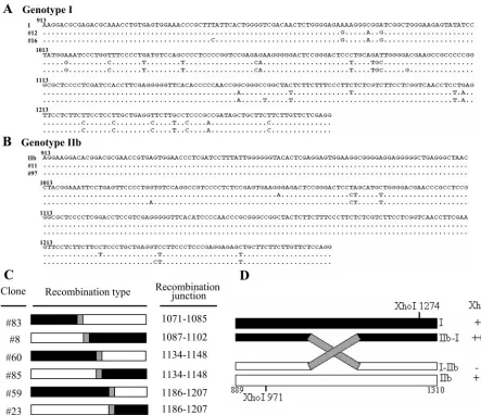
To evaluate genomic diversity, all clones analyzed by SacII and SphI RFLPs were then subjected to sequence analysis. For the analyzed clones of genotypes I and IIb, the genotyping results obtained by multiple RFLP assays were in complete agreement with those from sequencing analysis. Two represen-tative sequences for each genotype are shown in Fig. 2A and B. Interestingly, the sequencing data for the six clones with or without two XhoI sites revealed that they were recombinants between parental genotype I and IIb sequences (Fig. 2C), instead of being genotype II, genotype III, or new variants. Based on the assumption that only one recombination event occurred between two XhoI sites, the 5⬘-I–IIb-3⬘recombinant should not contain a XhoI site (Fig. 2D). Similarly, sequences containing two XhoI sites could be detected in 5⬘-IIb–I-3⬘ recombinants (Fig. 2D). Therefore, the genome organization obtained from the XhoI RFLP assays was consistent with that from our sequence analysis, except that the latter identified the recombination junctions (Fig. 2C and Table 3). Four crossover regions were identified: nt 1071 to 1085, 1087 to 1102, 1134 to 1148, and 1186 to 1207. The last two could be detected in the reciprocal recombinants, 5⬘-I–IIb-3⬘and 5⬘-IIb–I-3⬘. Mixed in-fections of HDV genotypes I and II and of genotypes II and IIb have been reported previously (66), but this is the first report of a mixed infection of genotypes I and IIb in a human patient. Furthermore, the identification of recombinants in this patient indicates for the first time that HDV RNA recombination occurs during natural mixed-genotype infection.
Detection of HDV recombinants using genotype-specific
RT-PCR primers.As shown above, we observed RT-PCR products
corresponding to recombinant RNAs from mixed-genotype in-fection of HDV. However, large-scale RFLP and sequencing
of cloned PCR products is time-consuming and labor-inten-sive. Therefore, we next studied HDV RNA by using genotype-specific RT-PCR primer pairs to genotype-specifically amplify 5⬘-I–IIb-3⬘ and 5⬘-IIb–I-3⬘recombinants (covering nt 940 to 1271). This method had the benefit that the genotype-specific primers were able to suppress potential artificial recombination during PCR, as described previously (19, 56). cDNAs were produced from primers specific to either genotype I or genotype IIb; thus, the use of the recombinant-specific primers in the subse-quent PCRs should amplify only the recombinant sequences. When in vitro-transcribed genotype I and IIb RNAs were mixed and then subjected to RT-PCR using the recombinant-specific primer pairs, no signals were observed (Fig. 3, lanes 1 and 2), suggesting that recombination did not occur during RT-PCR amplification. However, bands corresponding to the potential 5⬘-I–IIb-3⬘(lane 3) and 5⬘-IIb–I-3⬘(lane 6) recombi-nants were observed in samples amplified from the patient’s serum. As expected, 5⬘-I–IIb-3⬘recombinants were resistant to XhoI digestion (lane 4), while 5⬘-IIb–I-3⬘ recombinants were sensitive to XhoI digestion (lane 5).
To investigate the recombination junctions, PCR products were cloned into T vectors, and 10 clones each of the 5⬘-I– IIb-3⬘and 5⬘-IIb–I-3⬘recombinants were subjected to sequenc-ing analysis. As summarized in Table 3 and Fig. 4, the cross-over regions were mapped to nt 1087 to 1102, 1110 to 1125, 1134 to 1148, 1172 to 1184, 1186 to 1207, and 1211 to 1222. Three of these (nt 1087 to 1102, 1134 to 1148, and 1186 to 1207) were also detected in recombinants when the consensus primers were used during PCR. Detection of the same recom-bination junctions by two independent analyses further proved that the observed HDV RNA recombination occurred in the patient rather than being a random event. Sequence analysis of the recombinants clearly illustrated that the observed HDV recombination was likely a homologous process, since all cross-overs occurred at homologous regions between the two paren-tal HDV RNAs, and no insertions, deletions, or mismatches were observed (Fig. 4). No consensus sequence was identified around the HDV recombination junctions (Fig. 4).
Although the two XhoI sites span nt 971 to 1274, the ob-served HDV recombination sites were clustered from nt 1071 to 1222 (Fig. 4). However, this is explained by the fact that nt 971 to 1070 overlaps the highly divergent C terminus of the large HDAgs, which would not favor homologous recombina-tion. The observation that two pairs of crossover regions (nt 1071 to 1085 and 1087 to 1102, and nt 1172 to 1184 and 1186 to 1207) were separated from each other by only a single nucleotide further implicates the nonrandom distribution of crossover regions for HDV recombination.
HDV RNA recombination in cultured cells. We recently
[image:4.585.43.284.90.175.2]showed that a cloned HDV genotype IIb (Taiwan-IIb-1) could replicate in cultured cells (60). To mimic the natural mixed HDV infection, we cotransfected in vitro-transcribed genomic genotype I (28, 60) and IIb (60) RNAs into COS7 cells stably expressing small HDAg. Six days after transfection, RNA was extracted from the cells and subjected to RT-PCR with con-sensus primer pair 18–55⬘. Similar to our results with the nat-ural infection, multiple bands were observed after XhoI diges-tion of the PCR products (Fig. 5A, lane 4). Similarly, multiple bands were detected after XhoI RFLP analysis of PCR prod-ucts from cells cotransfected with antigenomic genotype I and
TABLE 2. Summary of multiple RFLP patterns observed in clones carrying HDV genomes obtained from patient serum
Restriction site (nt)
Cleavage ina:
Genotype I clones
Genotype IIb clones
Clone no. Genotype II
T3 clones 8 23 85 59 60 83
XhoI (971) ⫺ ⫹ ⫹ ⫹ ⫹ ⫺ ⫺ ⫺ ⫹
XhoI (1274) ⫹ ⫺ ⫹ ⫹ ⫹ ⫺ ⫺ ⫺ ⫹
SacII (1160) ⫺ ⫹ ⫺ ⫹ ⫺ ⫺ ⫹ ⫹ ⫹
SphI (1091) ⫹ ⫹ ⫹ ⫺ ⫹ ⫹ ⫹ ⫹ ⫺
aCleavage was present (⫹) or absent (⫺) in the indicated clone(s).
on November 8, 2019 by guest
http://jvi.asm.org/
IIb RNAs (data not shown). The sizes of the two major bands (lane 4) were undistinguishable from those of parental geno-types I and IIb (lanes 2 and 3), while the sizes of the two faint bands (lane 4) were consistent with those of uncleaved (lane 1) and double-cleaved bands. These data imply that HDV RNA recombination also occurred in cultured cells.
[image:5.585.66.509.70.452.2]To further investigate the molecular nature of the HDV recombinants in cultured cells, genotype-specific primers were used for RT-PCR, and the recombination junctions of the cloned PCR products were analyzed. The crossover regions of 10 clones each of the 5⬘-I–IIb-3⬘(designated TOPO-R1⬃R10) and 5⬘-IIb–I-3⬘ (designated TOPO-R11⬃R20) recombinants were analyzed (Fig. 5B). The results indicated five crossover regions, mapping to nt 1050 to 1054, 1110 to 1125, 1134 to 1148, 1159 to 1207, and 1212 to 1229. Again, all crossovers
FIG. 2. HDV-related sequences detected in the serum of a patient with mixed genotype I and IIb infection. (A and B) Alignment of nucleotide sequences (nt 913 to 1280) from representative HDV isolates of genotypes I (A) and IIb (B), and sequences identified in the mixed infection of HDV. Dots indicate conserved nucleotides. Sources of the representative isolates of different genotypes are as follows: I, HDV genotype I isolated from Italy (GenBank accession number M21012) (28); IIb, genotype IIb from the Taiwan-IIb-1 clone (GenBank accession number AF209859) (60). The sequences obtained from two clones each of HDV genotypes I (clones 12 and 16) and IIb (clones 11 and 97) from the mixed infection are shown. (C) Schematic demonstration of HDV recombinants identified by sequence analysis. The recombination junctions are shaded in gray and summarized on the right. The genotype I and IIb sequences are indicated by closed and open bars, respectively. (D) Restriction maps of the potential clones carrying recombinant HDV genomes. The XhoI RFLP profiles and the predicted genome organization of recombinants 5⬘-I–IIb-3⬘ and 5⬘-IIb–I-3⬘are summarized on the right.
TABLE 3. Categories of recombinant genomes and localization of recombination junctions
Localization of recombination junctions (nt)
No. of recombinant genomes identified by PCR using:
Consensus primers Genotype-specific primers
I-IIb IIb-I I-IIb IIb-I
1071–1085 1 0 0 0
1087–1102 0 1 2 0
1110–1125 0 0 1 1
1134–1148 1 1 1 2
1172–1184 0 0 1 1
1186–1207 1 1 3 4
1211–1222 0 0 2 2
on November 8, 2019 by guest
http://jvi.asm.org/
[image:5.585.302.541.594.725.2]occurred at homologous regions shared between the two pa-rental HDV RNAs (Fig. 5B). Although the transfected se-quences were different from those identified in the mixed-genotype infection (Fig. 2A and 2B), four crossover regions (nt 1110 to 1125, 1134 to 1148, 1159 to 1207, and 1212 to 1229)
detected in the cotransfected cultured cells were identical to or overlapped with those observed in the patient’s serum (Fig. 4 and 5B). Therefore, this work shows the establishment of a cotransfected cell culture system that could serve as an exper-imental system for future study of HDV RNA recombination.
Detection of HDV recombinants using RNase protection
assay.As shown in Fig. 5B, the recombination junction of 10 of
20 tested clones from the cotransfected cultured cells mapped to nt 1159 to 1207. This finding prompted us to directly detect the HDV RNA recombinants by an RNase protection assay using a 5⬘-I–IIb-3⬘ probe (in vitro transcribed from the TOPO-R6 plasmid) with the recombination junction located at nt 1159 to 1207. This method avoided the use of PCR ampli-fication, thus guarding against the possibility that the detected recombinants were artifacts generated during RT-PCR. To avoid detection of HDAg mRNA, cells were cotransfected with antigenomic genotype I and IIb RNAs and the extracted RNA was hybridized with an antigenomic RNase protection probe specific to the HDV genomic RNA, which should be present only after HDV replication. As demonstrated in Fig. 6, a TOPO-R6 plasmid containing a recombinant HDV sequence (recombination junction nt 1159 to 1207) was transcribed in vitro to produce a 446-base RNA probe. After hybridization and RNase treatment, the protected bands for genotypes I and IIb were 267 and 113 bases, respectively. In contrast, genomic 5⬘-I-(1159 to 1207)–IIb-3⬘ recombinant sequences yielded a 332-base protected band. The probe alone was sensitive to RNase digestion (data not shown), whereas it was resistant to RNase treatment after hybridization with an in
[image:6.585.303.540.68.248.2]vitro-tran-FIG. 3. Detection of HDV recombinants by use of genotype-spe-cific primers. cDNAs generated with genotype I- or IIb-spegenotype-spe-cific prim-ers served as the templates for subsequent PCRs with recombinant-specific primer pairs. The PCR products were cleaved with XhoI, separated on 3% agarose gels, and stained with ethidium bromide. The observed 5⬘-IIb–I-3⬘ recombinant contains one XhoI site (nt 971), whereas the 5⬘-I–IIb-3⬘ recombinant lacks a XhoI site. Lanes: M, 100-bp-ladder molecular size markers (the dominant band is 500 bp in size); 1 and 2, undigested PCR products of mixture of in vitro-tran-scribed genotype I and IIb RNA by use of primer pairs I5⬘-1–IIb3⬘-1 and IIb5⬘-1–I3⬘-1, respectively; 3 and 6, undigested PCR products obtained from the HDV patient with primer pairs I5⬘-1–IIb3⬘-1 and IIb5⬘-1–I3⬘-2, respectively; 4 and 5, digested PCR products obtained from the HDV patient with primer pairs I5⬘-1–IIb3⬘-1 and IIb5⬘-1– I3⬘-2, respectively.
[image:6.585.89.240.68.220.2]FIG. 4. Primary structure of the crossover regions of intergenotypic HDV recombinants identified in a natural mixed-genotype HDV in-fection. Genomic sequences (nt 952 to 1280) of HDV genotypes I and IIb are given. Short lines depict the homologous bases between two genotypes. The crossover regions identified using genotype-specific primers are indicated by gray shading, while those obtained using consensus primers are indicated by rectangles.
FIG. 5. HDV recombination in transfected cultured cells. (A) XhoI RFLP analysis of PCR products amplified from total RNA extracted from cotransfected cultured cells by use of consensus primers. Lanes: M, 100-bp-ladder molecular size markers (the dominant band is 500 bp in size); 1, undigested PCR products; 2, digested genotype I PCR products; 3, digested genotype IIb PCR products; 4, a complex RFLP pattern obtained from transfected cultured cells. (B) Primary structure of the crossover regions of intergenotypic HDV recombinants identi-fied in transfected cultured cells by use of genotype-specific primers. The nucleotide sequences of the genomic segments (nt 960 to 1251) of HDV genotypes I (Italian clone) and IIb (Taiwan-IIb-1 clone) are given. Short lines depict the homologous bases between the two geno-types. The crossover regions identified are indicated by gray shading. Numbers given in parentheses and square brackets indicate the num-ber of 5⬘-I–IIb-3⬘and 5⬘-IIb–I-3⬘recombinant clones, respectively.
on November 8, 2019 by guest
http://jvi.asm.org/
[image:6.585.79.253.462.659.2]scribed complementary recombinant sequence (Fig. 6, lane 2). When RNA samples extracted from cotransfected cells were subjected to the RNase protection assay, we observed two dominant bands corresponding to genotypes I and IIb (lane 3). The gel was then subjected to extended film exposure (lanes 7 to 12) to detect the less common HDV RNA recombinants. As expected, a 332-base band identical to that of the protected HDV RNA recombinant of interest (lane 9) appeared follow-ing the longer exposure. Furthermore, when a mixture of in vitro-transcribed genotype I and IIb HDV RNAs was sub-jected to this RNase protection assay, only two bands with the expected sizes (267 and 113 bp) were observed (lanes 5 and 11). Similarly, when genotype I and IIb RNAs obtained from cells separately transfected with genotype I and IIb RNAs were mixed with the probe and subjected to the RNase protection assay, no recombinant bands were observed (lanes 6 and 12). No band was found when RNA extracted from untransfected cells was subjected to the RNase protection assay (lanes 4 and 10). This finding confirmed the recombination junction in an RT-PCR-independent fashion, showing that the result was not a PCR-based artifact. Taken together, these data suggest that HDV RNA recombination did occur at the junction mapped to nt 1159 to 1207 in cultured cells expressing two replicating HDV clones. The identification of recombinant HDV RNAs by an RNase protection assay provided direct evidence for HDV RNA recombination in cultured cells.
DISCUSSION
Although phylogenetic analysis has suggested that genetic recombination can occur in HDV (67), HDV RNA recombi-nants have not been previously identified from natural
mixed-genotype infections (66). Here, we provide the first identifica-tion of HDV RNA recombinants in natural mixed-genotype infections and cotransfected cultured cells.
In this study, PCR methods were used to investigate HDV RNA recombination. Several lines of evidence showed that the HDV recombination observed here was not the result of arti-facts caused by reannealing and extension of incompletely syn-thesized strands during PCR. First, control RT-PCR amplifi-cations using template mixtures of two RNAs (either produced from in vitro transcription or extracted from single-transfec-tion cells) showed no evidence of recombinasingle-transfec-tion. In contrast, HDV recombinants were readily detectable by RT-PCR from infected or cotransfected samples by using either consensus primers or genotype-specific primers. Second, instead of being randomly distributed, the crossover regions identified in this study all mapped to homologous regions between two RNA species. Furthermore, identical recombination junctions were observed in reciprocal 5⬘-I–IIb-3⬘ and 5⬘-IIb–I-3⬘ recombi-nants. Third, some crossover regions detected in the cultured cells were identical to or overlapped those observed in the patient’s serum, even though the transfected sequences dif-fered from those identified in the mixed-genotype infection. This nonrandom HDV RNA recombination suggests that the crossovers were guided by a mechanism that was more specific than premature termination and reinitiation during RT-PCR amplification. And, last, HDV RNA recombination at junc-tions identified from RT-PCR amplification could be detected in transfected cultured cells by an RNase protection assay (Fig. 6), which avoided PCR amplification. Taken together, these observations and controls clearly illustrate that true HDV RNA recombination was identified in this study.
HDV mixed infections of genotypes II and IIb and geno-types I and II have been reported, but RNA recombinants have not been identified in the individuals with these infections (67). It has been reported that the frequency of homologous recom-bination between more distantly related viruses is significantly lower (22, 39, 40, 58). However, the sequence homologies between genotypes I and II and genotypes II and IIb are 76 and 78%, respectively, and that between genotypes I and IIb is 75%. Therefore, sequence homology alone does not appear to explain the absence of HDV recombination in the previously reported mixed infections with genotypes I and II and geno-types II and IIb (66). Based on the fact that the minor strain of HDV always constituted less than 10% of the total colony population in the previous report (66), it is likely that the replication levels of each individual HDV strain in the mixed infections played an important role in determining the recom-bination frequency. In addition, genotype II HDV was de-tected in all of the mixed-genotype HDV infections in the previous report (66); recently, the replication level of a Tai-wanese genotype II clone was shown to be 100-fold lower than that of a genotype I prototype (34). These observations sup-port a hypothesis in which the HDV replication level is an important factor in dictating the HDV recombination rate. Thus, we propose that HDV RNA recombination is performed via a replication-dependent mechanism.
The template switching mechanism is a well-accepted model of recombination for RNA viruses encoding their own RNA polymerases. The idea that HDV RNA recombination is a replication-dependent event suggests that HDV
recombina-FIG. 6. Detection of HDV RNA recombinants with an RNase pro-tection assay. Probes specific to the HDV RNA recombinant with the crossover region located at nt 1159 to 1207 were synthesized in vitro, hybridized with various RNA samples, and subjected to RNase diges-tion for detecdiges-tion of the HDV RNA recombinants. Lanes 7 to 12 represent the results of a longer exposure of the data shown in lanes 1 to 6. Lanes: 1 and 7, no-target, no-RNase control lanes corresponding to the full-length probe; 2 and 8, probe hybridized with a complemen-tary in vitro-transcribed HDV RNA; 3 and 9, probe hybridized to RNA samples extracted from cotransfected cultured cells; 4 and 10, probe hybridized to RNA samples extracted from untransfected cells; 5 and 11, probe hybridized to mixtures of in vitro-transcribed genotype I and IIb RNAs; 6 and 12, probe hybridized to mixtures of total RNA extracted from cells independently transfected with genotype I and IIb HDV RNAs. The probe and protected bands are schematically de-picted on the right. The genotype I, genotype IIb, and vector sequences are indicated by a closed box, open box, and thin line, respectively.
on November 8, 2019 by guest
http://jvi.asm.org/
[image:7.585.50.278.69.202.2]tion might also occur via a template switch mechanism. Re-cently, Pol II was implicated in intramolecular template switch-ing, which is a process that can occur with precision or that can involve deletions and even nontemplated insertions (7). We propose that host polymerases might also have the ability to undergo precise intermolecular template switching during HDV replication to generate recombinants. In the conven-tional template switch mechanism, the polymerase pauses dur-ing RNA recombination, perhaps in response to RNA second-ary structures around the recombination site, and then jumps from the original donor template to the acceptor template (18, 30). During HDV RNA replication, polymerase locally un-winds the duplex region of the HDV rod-like structure; the HDV genome is GC-rich, making it likely to form local sec-ondary structures, such as hairpins, where the polymerase might pause and initiate a crossover. About 70% of the HDV genome is base paired to form the rod-like structure; however, only 53% of the bases are paired between nt 1182 to 1196 and the side opposite (nt 405 to 412) on the predicted HDV rod-like genome, perhaps suggesting that there is another mecha-nism causing the polymerase complex to dissociate from the template during HDV replication. The role of RNA secondary structures in facilitating HDV RNA recombination remains to be investigated.
HDV is the only animal virus that utilizes host polymerases to replicate the viral genome. From our data, we suggest that host RNA polymerases might have the ability to undergo pre-cise intermolecular template switching during HDV replica-tion to generate recombinants. According to this hypothesis, RNA recombination might also occur between cellular and viral RNAs. It has been proposed that HDV might have evolved from a viroid-like RNA via capture of a cellular tran-script (2, 3). Indeed, a cellular homolog of HDAg has been claimed (3). Therefore, the original incorporation of the HDAg coding sequence and its complementary sequence (to maintain the rod-like structure) into the HDV viroid-like do-main might have occurred through RNA recombination.
In this study, we found that in the region analyzed, the recombination frequency between genotypes I and IIb in a natural HDV infection was 6% (the recombination frequency was determined by dividing the yield of recombinants by the sum of the yields of the parental viruses). Considering the typically high reversion rate and the occurrence of intrageno-typic recombination, this value might underestimate the HDV recombination frequency. Also, if two recombination events occurred in the analyzed region, the RFLP pattern might be undistinguishable from that of the parental RNAs. In addition, the HDV recombinants observed here were isolated from pa-tient serum rather than from infected liver, thus representing the virion RNA that has been packaged and released into the bloodstream. The recombinant population detected in the se-rum could thereby represent a selective growth advantage in the infected liver cells and/or greater resistance against the various selective pressures in the host. Thus, the data reported here may not completely reflect the actual recombination fre-quency and mechanism. We are currently using the RNA co-transfection cell culture system established in this report to investigate recombination across the entire HDV genome. Characterization of the HDV recombinants present in the cells
instead of in the released viral particles should provide more insights into the mechanism of HDV recombination.
HDV has been reported to acquire highly modified genomes via both incorporation errors during RNA replication and RNA editing (5, 12, 16, 32, 36, 42, 55, 59). Our results further suggest that RNA recombination occurs during HDV replica-tion and thereby represents another mechanism for HDV ge-nome heterogeneity. The fact that crossover sites could be detected in the HDAg-coding region suggests that recombina-tion might yield viral pathogens with new biological properties. Therefore, such genetic heterogeneity will make the preven-tion and control of viral hepatitis even more difficult. Under-standing the mechanism and the results of genetic heteroge-neity of HDV will allow us to further comprehend the biological roles of HDV in human liver diseases.
ACKNOWLEDGMENTS
We thank Y.-F. Liaw (Liver Research Unit, Chang Gung Memorial Hospital, Tao-yang, Taiwan), who generously provided the serum sam-ples used to screen the HDV RNA recombinants.
This work was supported by grants from the National Science Coun-cil of Taiwan (NSC 90-2320-B-182-042 and NSC 92-2320-B-182-053) and the Chang Gung Memorial Hospital (CMRP 955).
REFERENCES
1.Ambros, S., J. C. Desvignes, G. Llacer, and R. Flores.1995. Pear blister canker viroid: sequence variability and causal role in pear blister canker disease. J. Gen. Virol.76:2625–2629.
2.Branch, A. D., B. J. Benenfeld, B. M. Baroudy, F. V. Wells, J. L. Gerin, and H. D. Robertson.1989. An ultraviolet-sensitive RNA structural element in a viroid-like domain of the hepatitis delta virus. Science243:649–652. 3.Brazas, R., and D. Ganem. 1996. A cellular homolog of hepatitis delta
antigen: implications for viral replication and evolution. Science274:90–94. 4.Cascone, P. J., C. D. Carpenter, X. H. Li, and A. E. Simon.1990. Recom-bination between satellite RNAs of turnip crinkle virus. EMBO J.9:1709– 1715.
5.Casey, J. L., T. L. Brown, E. J. Colan, F. S. Wignall, and J. L. Gerin.1993. A genotype of hepatitis D virus that occurs in northern South America. Proc. Natl. Acad. Sci. USA90:9016–9020.
6.Chang, F. L., P. J. Chen, S. J. Tu, C. J. Wang, and D. S. Chen.1991. The large form of hepatitis delta antigen is crucial for assembly of hepatitis delta virus. Proc. Natl. Acad. Sci. USA88:8490–8494.
7.Chang, J., and J. Taylor.2002. In vivo RNA-directed transcription, with template switching, by a mammalian RNA polymerase. EMBO J.21:157– 164.
8.Chao, M., S. Y. Hsieh, and J. Taylor.1990. Role of two forms of hepatitis delta virus antigen: evidence for a mechanism of self-limiting genome rep-lication. J. Virol.64:5066–5069.
9.Chao, M., S. Y. Hsieh, and J. Taylor.1991. The antigen of hepatitis delta virus: examination of in vitro RNA-binding specificity. J. Virol.65:4057– 4062.
10.Chao, Y. C., M. F. Chang, I. Gust, and M. M. Lai.1990. Sequence conser-vation and divergence of hepatitis delta virus RNA. Virology178:384–392. 11.Chao, Y. C., C. M. Lee, H. S. Tang, S. Govindarajan, and M. M. Lai.1991.
Molecular cloning and characterization of an isolate of hepatitis delta virus from Taiwan. Hepatology13:345–352.
12.Ciccaglione, A. R., M. Rapicetta, A. Fabiano, C. Argentini, M. Silvestro, R. Giuseppetti, F. Varano, N. D’Urso, L. Dinolfo, A. Morgando, and.1993. Chronic infection in woodchucks infected by a cloned hepatitis delta virus. Arch. Virol. Suppl.8:15–21.
13.Glenn, J. S., J. M. Taylor, and J. M. White.1990. In vitro-synthesized hepatitis delta virus RNA initiates genome replication in cultured cells. J. Virol.64:3104–3107.
14.Hammond, R., D. R. Smith, and T. O. Diener.1989. Nucleotide sequence and proposed secondary structure of Columnea latent viroid: a natural mo-saic of viroid sequences. Nucleic Acids Res.17:10083–10094.
15.Hsieh, S. Y., M. Chao, L. Coates, and J. Taylor.1990. Hepatitis delta virus genome replication: a polyadenylated mRNA for delta antigen. J. Virol.
64:3192–3198.
16.Imazeki, F., M. Omata, and M. Ohto.1991. Complete nucleotide sequence of hepatitis delta virus RNA in Japan. Nucleic Acids Res.19:5439. 17.Ivaniushina, V., N. Radjef, M. Alexeeva, E. Gault, S. Semenov, M. Salhi, O.
Kiselev, and P. Deny.2001. Hepatitis delta virus genotypes I and II cocir-culate in an endemic area of Yakutia, Russia. J. Gen. Virol.82:2709–2718.
on November 8, 2019 by guest
http://jvi.asm.org/
18.Jarvis, T. C., and K. Kirkegaard.1991. The polymerase in its labyrinth: mechanisms and implications of RNA recombination. Trends Genet.7:186– 191.
19.Jarvis, T. C., and K. Kirkegaard.1992. Poliovirus RNA recombination: mechanistic studies in the absence of selection. EMBO J.11:3135–3145. 20.Jayan, G. C., and J. L. Casey.2002. Inhibition of hepatitis delta virus RNA
editing by short inhibitory RNA-mediated knockdown of ADAR1 but not ADAR2 expression. J. Virol.76:12399–12404.
21.Judo, M. S., A. B. Wedel, and C. Wilson.1998. Stimulation and suppression of PCR-mediated recombination. Nucleic Acids Res.26:1819–1825. 22.Kirkegaard, K., and D. Baltimore.1986. The mechanism of RNA
recombi-nation in poliovirus. Cell47:433–443.
23.Kofalvi, S. A., J. F. Marcos, M. C. Canizares, V. Pallas, and T. Candresse.
1997. Hop stunt viroid (HSVd) sequence variants from Prunus species: evidence for recombination between HSVd isolates. J. Gen. Virol.78:3177– 3186.
24.Koltunow, A. M., and M. A. Rezaian.1989. Grapevine viroid 1B, a new member of the apple scar skin viroid group contains the left terminal region of tomato planta macho viroid. Virology170:575–578.
25.Kos, A., R. Dijkema, A. C. Arnberg, P. H. van der Meide, and H. Schellekens.
1986. The hepatitis delta (delta) virus possesses a circular RNA. Nature
323:558–560.
26.Kos, T., A. Molijn, L. J. van Doorn, A. van Belkum, M. Dubbeld, and H. Schellekens.1991. Hepatitis delta virus cDNA sequence from an acutely HBV-infected chimpanzee: sequence conservation in experimental animals. J. Med. Virol.34:268–279.
27.Kuo, M. Y., L. Sharmeen, G. Dinter-Gottlieb, and J. Taylor.1988. Charac-terization of self-cleaving RNA sequences on the genome and antigenome of human hepatitis delta virus. J. Virol.62:4439–4444.
28.Kuo, M. Y., J. Goldberg, L. Coates, W. Mason, J. Gerin, and J. Taylor.1988. Molecular cloning of hepatitis delta virus RNA from an infected woodchuck liver: sequence, structure, and applications. J. Virol.62:1855–1861. 29.Kuo, M. Y., M. Chao, and J. Taylor.1989. Initiation of replication of the
human hepatitis delta virus genome from cloned DNA: role of delta antigen. J. Virol.63:1945–1950.
30.Lai, M. M.1992. Genetic recombination in RNA viruses. Curr. Top. Micro-biol. Immunol.176:21–32.
31.Lai, M. M.1995. The molecular biology of hepatitis delta virus. Annu. Rev. Biochem.64:259–286.
32.Lee, C. M., F. Y. Bih, Y. C. Chao, S. Govindarajan, and M. M. Lai.1992. Evolution of hepatitis delta virus RNA during chronic infection. Virology
188:265–273.
33.Lee, C. M., C. S. Changchien, J. C. Chung, and Y. F. Liaw.1996. Charac-terization of a new genotype II hepatitis delta virus from Taiwan. J. Med. Virol.49:145–154.
34.Lin, F. M., C. M. Lee, T. C. Wang, and M. Chao.2003. Initiation of RNA replication of cloned Taiwan-3 isolate of hepatitis delta virus genotype II in cultured cells. Biochem. Biophys. Res. Commun.306:966–972.
35.Lin, J. H., M. F. Chang, S. C. Baker, S. Govindarajan, and M. M. Lai.1990. Characterization of hepatitis delta antigen: specific binding to hepatitis delta virus RNA. J. Virol.64:4051–4058.
36.Luo, G. X., M. Chao, S. Y. Hsieh, C. Sureau, K. Nishikura, and J. Taylor.
1990. A specific base transition occurs on replicating hepatitis delta virus RNA. J. Virol.64:1021–1027.
37.Macnaughton, T. B., S. T. Shi, L. E. Modahl, and M. M. Lai.2002. Rolling circle replication of hepatitis delta virus RNA is carried out by two different cellular RNA polymerases. J. Virol.76:3920–3927.
38.Makino, S., M. F. Chang, C. K. Shieh, T. Kamahora, D. M. Vannier, S. Govindarajan, and M. M. Lai.1987. Molecular cloning and sequencing of a human hepatitis delta (delta) virus RNA. Nature329:343–346.
39.McCahon, D., W. R. Slade, R. A. Priston, and J. R. Lake.1977. An extended genetic recombination map for foot-and-mouth diseases virus. J. Gen. Virol.
35:555–565.
40.McCahon, D., A. M. King, D. S. Roe, W. R. Slade, J. W. Newman, and A. M. Cleary.1985. Isolation and biochemical characterization of intertypic recom-binants of foot-and-mouth disease virus. Virus Res.3:87–100.
41.Moraleda, G., and J. Taylor.2001. Host RNA polymerase requirements for transcription of the human hepatitis delta virus genome. J. Virol.75:10161– 10169.
42.Netter, H. J., T. T. Wu, M. Bockol, A. Cywinski, W. S. Ryu, B. C. Tennant, and J. M. Taylor. 1995. Nucleotide sequence stability of the genome of hepatitis delta virus. J. Virol.69:1687–1692.
43.Owens, R. A., G. Yang, D. Gundersen-Rindal, R. W. Hammond, T. Can-dresse, and M. Bar-Joseph.2000. Both point mutation and RNA recombi-nation contribute to the sequence diversity of citrus viroid III. Virus Genes
20:243–252.
44.Polson, A. G., B. L. Bass, and J. L. Casey.1996. RNA editing of hepatitis
delta virus antigenome by dsRNA-adenosine deaminase. Nature380:454– 456.
45.Radjef, N., E. Gordien, V. Ivaniushina, E. Gault, P. Anais, T. Drugan, J. C. Trinchet, D. Roulot, M. Tamby, M. C. Milinkovitch, and P. Deny.2004. Molecular phylogenetic analyses indicate a wide and ancient radiation of African hepatitis delta virus, suggesting a deltavirus genus of at least seven major clades. J. Virol.78:2537–2544.
46.Reid, C. E., and D. W. Lazinski.2000. A host-specific function is required for ligation of a wide variety of ribozyme-processed RNAs. Proc. Natl. Acad. Sci. USA97:424–429.
47.Rezaian, M. A.1990. Australian grapevine viroid—evidence for extensive recombination between viroids. Nucleic Acids Res.18:1813–1818. 48.Rizzetto, M., M. G. Canese, J. L. Gerin, W. T. London, D. L. Sly, and R. H.
Purcell.1980. Transmission of the hepatitis B virus-associated delta antigen to chimpanzees. J. Infect. Dis.141:590–602.
49.Rizzetto, M., B. Hoyer, M. G. Canese, J. W. Shih, R. H. Purcell, and J. L. Gerin.1980. delta Agent: association of delta antigen with hepatitis B sur-face antigen and RNA in serum of delta-infected chimpanzees. Proc. Natl. Acad. Sci. USA77:6124–6128.
50.Sakugawa, H., H. Nakasone, T. Nakayoshi, Y. Kawakami, S. Miyazato, F. Kinjo, A. Saito, S. P. Ma, H. Hotta, and M. Kinoshita.1999. Hepatitis delta virus genotype IIb predominates in an endemic area, Okinawa, Japan. J. Med. Virol.58:366–372.
51.Saldanha, J. A., H. C. Thomas, and J. P. Monjardino.1990. Cloning and sequencing of RNA of hepatitis delta virus isolated from human serum. J. Gen. Virol.71:1603–1606.
52.Sano, T., and A. Ishiguro.1998. Viability and pathogenicity of intersubgroup viroid chimeras suggest possible involvement of the terminal right region in replication. Virology240:238–244.
53.Sharmeen, L., M. Y. Kuo, and J. Taylor.1989. Self-ligating RNA sequences on the antigenome of human hepatitis delta virus. J. Virol.63:1428–1430. 54.Spieker, R. L.1996. The molecular structure of Iresine viroid, a new viroid
species from Iresine herbstii (⬘beefsteak plant’). J. Gen. Virol.77:2631–2635. 55.Sureau, C., J. Taylor, M. Chao, J. W. Eichberg, and R. E. Lanford.1989. Cloned hepatitis delta virus cDNA is infectious in the chimpanzee. J. Virol.
63:4292–4297.
56.Tanabe, K., N. Sakihama, A. Farnert, I. Rooth, A. Bjorkman, D. Walliker, and L. Ranford-Cartwright.2002. In vitro recombination during PCR of Plasmodium falciparum DNA: a potential pitfall in molecular population genetic analysis. Mol. Biochem. Parasitol.122:211–216.
57.Taylor, J. M.1990. Hepatitis delta virus: cis and trans functions required for replication. Cell61:371–373.
58.Tolskaya, E. A., L. I. Romanova, V. M. Blinov, E. G. Viktorova, A. N. Sinyakov, M. S. Kolesnikova, and V. I. Agol.1987. Studies on the recombi-nation between RNA genomes of poliovirus: the primary structure and nonrandom distribution of crossover regions in the genomes of intertypic poliovirus recombinants. Virology161:54–61.
59.Wang, K. S., Q. L. Choo, A. J. Weiner, J. H. Ou, R. C. Najarian, R. M. Thayer, G. T. Mullenbach, K. J. Denniston, J. L. Gerin, and M. Houghton.
1986. Structure, sequence and expression of the hepatitis delta (delta) viral genome. Nature323:508–514.
60.Wang, T. C., and M. Chao.2003. Molecular cloning and expression of the hepatitis delta virus genotype IIb genome. Biochem. Biophys. Res. Commun.
303:357–363.
61.Watanabe, H., K. Nagayama, N. Enomoto, R. Chinzei, T. Yamashiro, N. Izumi, H. Yatsuhashi, T. Nakano, B. H. Robertson, H. Nakasone, H. Saku-gawa, and M. Watanabe.2003. Chronic hepatitis delta virus infection with genotype IIb variant is correlated with progressive liver disease. J. Gen. Virol.84:3275–3289.
62.Wong, S. K., and D. W. Lazinski.2002. Replicating hepatitis delta virus RNA is edited in the nucleus by the small form of ADAR1. Proc. Natl. Acad. Sci. USA99:15118–15123.
63.Wu, H. N., Y. J. Lin, F. P. Lin, S. Makino, M. F. Chang, and M. M. Lai.1989. Human hepatitis delta virus RNA subfragments contain an autocleavage activity. Proc. Natl. Acad. Sci. USA86:1831–1835.
64.Wu, J. C., K. B. Choo, C. M. Chen, T. Z. Chen, T. I. Huo, and S. D. Lee.1995. Genotyping of hepatitis D virus by restriction-fragment length polymorphism and relation to outcome of hepatitis D. Lancet346:939–941.
65.Wu, J. C., T. Y. Chiang, and I. J. Sheen.1998. Characterization and phylo-genetic analysis of a novel hepatitis D virus strain discovered by restriction fragment length polymorphism analysis. J. Gen. Virol.79:1105–1113. 66.Wu, J. C., I. A. Huang, Y. H. Huang, J. Y. Chen, and I. J. Sheen.1999. Mixed
genotypes infection with hepatitis D virus. J. Med. Virol.57:64–67. 67.Wu, J. C., T. Y. Chiang, W. K. Shiue, S. Y. Wang, I. J. Sheen, Y. H. Huang,
and W. J. Syu.1999. Recombination of hepatitis D virus RNA sequences and its implications. Mol. Biol. Evol.16:1622–1632.