COMPARISON OF FRACTURE RESISTANCE OF TEETH
OBTURATED WITH DIFFERENT ROOT CANAL SEALERS –AN
INVITRO STUDY
Dissertation submitted to
THE TAMILNADU Dr. M.G.R. MEDICAL UNIVERSITY
In partial fulfilment for the Degree of MASTER OF DENTAL SURGERY
BRANCH IV
CONSERVATIVE DENTISTRY AND ENDODONTICS
CERTIFICATE
This is to certify that this dissertation titled “COMPARISON OF FRACTURE
RESISTANCE OF TEETH OBTURATED WITH DIFFERENT ROOT CANAL
SEALERS –AN INVITRO STUDY” is a bonafide record of work done by Dr. REMYA VARGHESE under my guidance and to my satisfaction during her
postgraduate study period, 2014 – 2017. This dissertation is submitted to
THE TAMILNADU Dr. M.G.R. MEDICAL UNIVERSITY, in partial fulfilment for the award of the degree of Master of Dental Surgery in Conservative Dentistry and Endodontics, Branch IV. It has not been submitted (partially or fully) for the award of any other degree or diploma.
________________________ ________________________
Dr. Minu Koshy, MDS, Dr. Subha Anirudhan, MDS,
__________________________
Dr. V. Prabhakar, MDS,
Date:
Place: Coimbatore
Principal, Professor and HOD
Sri Ramakrishna Dental College and Hospital Coimbatore
Guide, Professor,
Department of Conservative Dentistry and Endodontics,
Sri Ramakrishna Dental College and Hospital. Coimbatore.
oimbatore
Co-Guide, Reader,
Department of Conservative Dentistry and Endodontics,
Sri Ramakrishna Dental College and Hospital. Coimbatore.
ACKNOWLEDGEMENT
This thesis is the result of work done with immense support from many people and it
is with immense pleasure that I express my heartfelt gratitude to all of them.
I devote my heartfelt thanks to Dr. V. Prabhakar,MDS, Principal & Head of
Department, whose discipline and skills that run deep under his authoritative yet natural care
during my post graduate period which enabled me to successfully conclude my thesis.
I would like to thank and acknowledge Dr. Minu Koshy, MDS,Professor,my Guide
has always been a source of support and encouragement at any moment, in and out of the
department. I am grateful to her for her innovative ideas, constructive suggestions, valuable
criticism and constant encouragement.
I am indebted to my Co-Guide Dr. Subha Anirudhan, MDS, Reader,for her
valuable guidance that enabled me to comprehend this dissertation and reach its successful
culmination. I am grateful to her for sparing her valuable time in guiding me through this
thesis.
I take this opportunity to express my sincere gratitude to Dr. S. Sudhakar,MDS,
Reader, Dr.Sriman Narayanan, MDS, Senior Lecturer, Dr.Gayathri Velusamy, MDS,
Senior Lecturer, Dr.MohanKumar.S, MDS, Senior Lecturer who supported me at every
juncture throughout my postgraduate curriculum.
I thank the management for allowing me to use the facilities in the college and all the
staffs in the college who were concerned in my study.
This study wouldn’t have come to existence without the effort and time of the
Technology, Nilambur, and Coimbatore. I sincerely acknowledge Mr.Selvakumar,
M.Tech, Assistant Professor of PSG Institute of Advanced Studies and
Mr.Muthukumar M.Tech, Project Manager, for their sincere efforts and constant help
during the fracture resistance testing of tooth samples.
I express my sincere thanks to Dr.Vipin Jain, MDS, Department of Community
Dentistry, KLE DENTAL COLLEGE, BANGALORE for his guidance in the statistical
analysis of this study.
I am thankful to my seniors, my colleagues and my juniors, who have been together
as friends and of great support throughout my period of study here. I am thankful to all other
department staff members, my fellow colleagues in other departments, all UG staff members
and non-clinical staffs of my department for their great support and encouragement.
I express my dearest gratitude to my husband Dr.George. J.Manayath,my kids and
my parents, for their innovative supporttowards my study and this dissertation.
Last but not the least, I am greatly indebted to God the Almighty, for blessing me
with all the good things in my life and guiding me throughout.
ABSTRACT
Introduction: The aim of this study was to evaluate the fracture resistance of teeth
filled with 4 different endodontic sealers.
Methods: Hundred single rooted extracted mandibular premolars were decoronated
to a length of 11 mm. The teeth were randomly divided into 6 groups (n = 20 for each
group). In group1A, the teeth were left unprepared and unfilled (negative control),
and in group 1B, the teeth were left unobturated (positive control). The rest of the
roots were prepared by using the ProTaper System up to a master apical file size of
F3.In group 2, Epoxy resin based sealer (AHPlus) + gutta-percha; In group 3, mineral
trioxide aggregate–based sealer (MTAFill apex) + guttapercha; In group 4, Calcium
phosphate cement based sealer (Chitra-CPC)+guttapercha and in group 5,Bioceramic
based sealer(Endosequence BC ) +guttapercha .All root specimens were stored for 2
weeks at 100% humidity to allow the complete setting of the sealers. Each specimen
was then subjected to fracture testing by using a universal testing machine at a
crosshead speed of 1.0 mm/min until the root fractured. The force required to fracture
each specimen was recorded, and the data were analysed statistically.
Results:The fracture values of groups 4 and 5 (Chitra-CPC and Endosequence BC
Sealer) were significantly higher than those of group 2 and 3(P < .05). There was no
significant difference between groups 4 and 5 (P > .05).
Conclusions: In contrast to MTA Fill apex and AH Plus,Chitra-CPC and
Endosequence BC increased the force to fracture in root-filled single-rooted premolar
CONTENTS
TITLE
PAGE NO
1. Introduction
1
2. Aim and Objective
6
3. Review of Literature
7
4. Materials and Methods
21
5. Results
35
6. Discussion
43
7. Summary and Conclusion
50
8. Bibliography
53
Introduction
1
An endodontically treated tooth is weaker and more prone to fracture than
vital teeth1. 11%– 13% of extracted teeth with endodontic treatment are associated
with vertical root fractures rendering it the second most frequent identifiable reason
for loss of root-filled teeth 2, 3. There are several factors that affect the strength of
endodontically treated tooth including loss of tooth structure because of caries or
trauma, access cavity preparation, dehydration of dentin, overzealous instrumentation
and irrigation of the root canal, excess pressure during root obturation, and
preparation of intra-radicular post space 4, 5. These factors interact cumulatively to
influence tooth loading and distribution of stresses, ultimately increasing the
possibility of catastrophic failure.
The most commonly used root canal filling material is gutta-percha in
combination with sealer but the low elastic modulus of gutta-percha presents little or
no capacity to reinforce roots after treatment 6, 7. The ability of the present day sealer
to bond to radicular dentin is advantageous in maintaining the integrity of the
sealer-dentin interface during mechanical stresses, thus increasing resistance to fracture. The
sealers used had shortcomings in that a fluid‑tight seal along the dentinal walls was
not routinely achieved and the adhesive strength between endodontic sealers, dentin,
and Guttapercha was shown to be very weak 8, 9. Therefore, the use of a root canal
sealer possessing an additional quality of strengthening the root against fracture
would be of obvious value10. New root canal obturation materials and sealers have
Introduction
2
Growing interest in reinforcing the root canal system has led to the
development of adhesive root canal sealers. It is thought that adhesion and mechanical
interlocking between the material and root canal dentin will strengthen the remaining
tooth structure, and thus reduce fracture risk11. The accepted technique is to obturate
the root canal space using a solid or semi-solid material along with a sealer to obtain a fluid tight seal, occupying the interstitial spaces, foraminae, as well as accessory and
lateral canals 12, 13, 14. Guttapercha has been the standard obturation material used in
root canal therapy15. One of the disadvantages of Guttapercha as a root canal
obturation material is that it does not bond or adhere to the dentinal walls of the root
canal resulting in an incomplete obliteration of root canal space 15, 16. Differences in
the adhesive properties of sealers to dentin may be expected for several reasons,
including differences of root dentin between specimens, or even in different sites of
the same root, the presence or absence of smear layer, and the sealer’s chemical
composition and interaction with dentin 17, 18.
Bio-ceramic materials have been seen as the dawn of a new era in dentistry.
Although used mainly for dental implants and coatings for implants, their introduction
into endodontics as mineralising materials has brought about enormous productive
changes. The applications vary from their use for Pulp Capping, to apexogenesis,
apexification, and furcation repair19. Bio-ceramics are biocompatible ceramic
materials. They include alumina and zirconia, bioactive glass, glass ceramics, calcium
silicates, hydroxyapatite and resorbable calcium phosphates, and radiotherapy glasses.
The physical properties associated with bio-ceramics are very attractive to
dentistry;absolute biocompatibility, osseo conductivity, ability to achieve excellent
Introduction
3
tissue fluids, good radio-opacity and easy handling characteristics have led to the
widespread use of these materials in the area of endodontic science19.
A new Bio Ceramic sealer Endosequence BC sealer (Brasseler, USA), has
recently been introduced to the market. It is a premixed bioceramic endodontic sealer According to the manufacturer’s description, it is a convenient, ready-to-use
injectable white hydraulic cement paste developed for permanent root canal filling
and sealing applications. Also, it is an insoluble, radiopaque, and aluminium-free
material which requires the presence of water to set and harden20.
Epoxyresin-based dental materials (AH Plus) have been proposed to be
excellent agents to reinforce an endodontically treated tooth through the use of
adhesive sealers in the root canal system.11 However, despite several advantages
exhibited by bonding agents and resins studied to date, they had problems in working
properties (hydrophobic nature), radio-opacity and lack of re-treatability when used
for endodontic purposes.21, 22
MTA Fillapex is the first MTA based salicylate resin sealer. It is a bioceramic
type of sealer that can readily set in presence of moisture and is able to cause
cementogenesis and thus helps in repair of apical tissue.23 As it is known that MTA
does not bond to dentin, the presence of resins in Fillapex sealer increases the flow
properties, and the presence of MTA would cause interfacial deposition of
hydroxyapatite, which would increase the frictional resistance of the obturating
material.24 However, MTA has certain drawbacks like difficulty in handling,
Introduction
4
Calcium phosphate based Bioceramic sealers are emerging as promising
candidates in endodontics because of their superior biocompatibility features. They
also satisfy most of the requirements for an ideal sealer.12,14 These materials are
modified forms of self-setting calcium phosphate cements (CPC) that contain
inorganic calcium and phosphate minerals, which upon wetting with an aqueous
solution get converted to hydroxyapatite. Biomedical Technology Wing of Sree
Chitra Tirunal Institute for Medical Sciences and Technology, Thiruvananthapuram
have introduced a new calcium phosphate cement based root canal filling material
(Chitra –CPC) which is supplied in the form of powder and liquid. The optimum
wetting ratio is 0.8 ml of liquid per gram powder.26
It can be used for inducing hard tissue formation, pulp capping, apical barrier
formation, and apexification and as regenerative scaffold. 27, 28. Calcium phosphate
based sealers have been found to be less cytotoxic than AH Plus29, AH 26 and Zinc
Oxide Eugenol (ZOE) sealers and have the potential to promote bone regeneration.30
Various studies have showed that the bonding of endodontic sealers to inter-radicular
dentin after obturation enhance the resistance to fracture of endodontically treated
teeth.31Hence, the concept of bonded sealers used in conjunction with core filling
material has been established to improve the fracture resistance.
Many root canal obturating systems are available to clinicians, yet no
consensus exists regarding the superiority of any one in root canal obturation. Hence,
the present study was undertaken with the objectives to evaluate fracture resistance of
Introduction
5
like Endosequence BC sealer (Brasseler USA), MTA based-MTA Fillapex (Angelus)
[image:12.595.90.545.201.731.2]and Epoxy Resin-based sealer AH plus (DENTSPLY ).
TABLE 1
Endodontic sealer Composition Manufacturer
Endosequence BC
Zirconium oxide, calcium silicates, calcium phosphate monobasic, calcium hydroxide, filler and thickening
agents.
Brasseler USA (Savannah, GA)
Chitra-CPC
Powder: tetra- calcium phosphate (TTCP) and dicalcium phosphate dihydrate (DCPD) in equimolar ratio
Liquid: solution of disodium hydrogen phosphate in distilled water (Na2HPO4, in 0.2M concentration).
SCTIMST, Trivandrum
MTA Fillapex MTA, salicylate resin, natural resin, bismuth oxide and silica
Angelus
AH Plus
Paste A: bisphenol- A and F as epoxy resin, calcium tungstate, zirconium oxide, silica and iron oxide pigments.
Paste B: amine paste contains dibenzyldiamine,aminoadmantace, tricyclodecane – diamine, calcium tungstate, zirconium oxide, silica and silicone oil.
Aim and Objective
6
The purpose of this study was
To assess the fracture resistance of root canals obturated with
single-cone gutta-percha using AH Plus, MTA Fillapex, BC Sealer and CPC
Review of Literatures
7
Kirsten et al (2012)32 investigated the mutagenicity of resin‑based
endodontic sealer(1 epoxy resin–based endodontic sealer(AH Plus Jet) and 2
methacrylate-based endodontic sealers (EndoRez and Real Seal) and Calcicur,
a Ca(OH)2-based sealer by evaluating their potential to induce DNA double‑strand
breaks (DSBs) on extrusion into the periapical tissue. The gH2AX immuno
fluorescence assay was used to microscopically detect DNA DSBs. They found that
there were no indications for increased risk of genotoxicity of resin‑based root canal
sealers caused by the induction of DNA DSBs.
Velugu et al (2016) 33 evaluated the fracture resistance of endodontically
treated teeth obturated using lateral compaction technique with AH plus/Gutta‑
percha, Resilon/RealSeal self‑etch (SE), and Endofill/Gutta‑percha using universal
testing machine.Their study demonstrated higher fracture resistance values for
Resilon/RealSeal SE than AH plus/Gutta‑percha, followed by Endofill/ Gutta‑percha.
Kaplan et al (1999)34 investigated the antimicrobial effects of endodontic
sealers ( Apexit Vivadent), Endion(voco Germany), AH‑26(Dentsply), AH‑Plus
(Dentsply), Procosol (Star dental,USA), and Ketac Endo( Espe Germany) at 2,20, and
40 days interval against Candida albicans, Staphylococcus aureus, Streptococcus
mutans on agar plates and colony forming units were counted. They found out that
AH Plus produced slight inhibition on streptococcus mutans at 20 days and on
Actinomyces Israeli at every time interval but no effect was found on Candida
Review of Literatures
8
Wadhwani and Gurung et al (2000)35 evaluated the fracture resistance of
root canals filled with Resilon and Epiphany( Pentron Clinical Technologies LLC,
Wallingford), gutta-percha and AH plus( Dentsply DeTrey, Konstanz, Germany),
gutta-percha with Endomethasone sealer using Instron Machine .They concluded that
all materials significantly increased the fracture toughness of the instrumented roots
after obturation
Pecora et al (2001)36 compared the effect of Er:YAG laser(KaVo Key laser
II, Warthausen, Germany at 2.25 W potency; 11 mm focal distance; 4 Hz frequency;
200mJ energy; 62 J total energy; 313 mean impulse) application and EDTAC on the
adhesion of epoxy resin-based endodontic sealers -AH Plus(De Trey-Dentsply,
Konstanz, Germany) ,Topseal (Dentsply-Maillefer) , Sealer 26 (Dentsply,Petrópolis,
RJ, Brazil), AH 26 (Dentsply, Konstanz, Germany), and Sealer Plus (Dentsply,
Petrópolis, RJ, Brazil) to human dentin. The adhesion was measured with a Universal
testing machine. The results showed that the dentin treated with Er:YAG laser showed
an adhesion of 4 MPa for AH Plus to dentin than EDTAC .
Ungor et al (2006)37 compared the pushout bond strength of the resin-based
Epiphany–Resilon root canal filling system, and AH Plus, gutta-percha using
universal testing machine. They revealed that (Epiphany + gutta-percha) had
significantly greater bonding strength than all the other groups. (AH Plus +
Review of Literatures
9
Emel , Uzunoglu et al (2015)38 evaluated the effect of temperatures(220 C
and 37oC) of QMix (Dentsply Tulsa Dental, Tulsa, OK, USA) and EDTA on the
bond-strength of AH Plus (Dentsply DeTrey, Konstanz, Germany) . The QMix and
17% EDTA solutions that were at room temperature were heated by using a heating
cup that had a digital temperature display (Oushiba, OB-009 280 mL, Guangdong,
China).The specimens from each group were observed under scanning electron
microscopic (QuantaTM 450 FEG, FEI,Oregon, USA) to evaluate smear layer
removal after final irrigation procedures. Remaining roots were obturated and
prepared for a push-out test using Instron Universal Testing Machine.They found that
temperature of the final irrigant does affect the bond strength values of AH plus to
root dentin irrigated with EDTA. Bond strength of AH Plus sealer to root canal dentin
may improve with QMix.
Girish et al (2013)39 compared the sealing ability of polymethylmethacrylate
(PMMA) bone cement and Chitra Calcium phosphate cement (CPC-Chitra) with
MTA when used as root end filling material using Rhodamine B dye and confocal
laser scanning microscope .The study showed that PMMA bone cement was a better
material than CPC-Chitra as root end filling material to prevent apical microleakage
and MTA still continued to be a gold standard root end filling material showing
minimum microleakage.
Jacob et al (2014)29 histopathologically evaluated the periapical tissue
Review of Literatures
10
sealer, AH Plus (Dentsply) at 1 month and 3 months interval .They found out that in
the 1-month time period, CPC showed mild to moderate periapical tissue reaction but
in the 3-month time period, the slides of CPC showed an absence of inflammation to mild inflammatory reaction in the periapical area than AH Plus.
Ratnakumari and Thomas B (2012)40evaluated the efficacy of Chitra-CPC
as a pulpotomy agent in comparison with formocresol, through histopathologic
responses of pulpal tissues of human deciduous teeth. The results did not reveal
statistically significant difference between the two groups. But Chitra-CPC gave more
favourable results, in respect of pulpal inflammation, dentin bridge formation, quality
of dentin bridge and connective tissue in dentin bridge.
Nanjappa et al (2015) 41 compared the sealing ability of mineral trioxide
aggregate (MTA), Biodentine, and Chitra-calcium phosphate cement (CPC) as
root-end filling material using confocal laser scanning microscope and Rhodamine B dye.
They evaluated the effect of ultrasonic retro prep tip and an erbium: yttrium
aluminium garnet (Er:YAG) laser on the integrity of three different root-end filling
materials and the result showed Root-end cavities prepared with Er:YAG laser and
restored with Biodentine showed superior sealing ability compared to those prepared
Review of Literatures
11
Abad et al (2010)42 compared the sealing ability of bone cement, mineral
trioxide aggregate and calcium phosphate cement(CPC – Chitra) as furcation
perforation repair material using stereomicroscope on extracted mandibular molars.
They observed MTA showed minimum microleakage (mean 54.5%), calcium
phosphate cement showed maximum microleakage (100%), and bone cement showed
moderate microleakage (87.8%).
Gomes‑Filho et al(2011)43 evaluated the rat subcutaneous tissue reaction to
implanted polyethylene tubes filled with MTA Fillapex , MTA‑Angelus and Sealapex
including their ability to stimulate mineralization at 7th 15th , 30th and 90th day.
They found that all materials caused moderate reactions after 7 days, which decreased
with time. The reactions were moderate and similar to that evoked by the control and
Sealapex on the 15th day. MTA Fillapex and Angelus MTA caused mild reactions
beginning after 15 days and concluded that MTA Fillapex was biocompatible and
stimulated mineralization.
Sagsen et al( 2011)44 compared the push-out bond strength of an epoxy-based
root canal sealer AH Plus (Dentsply DeTrey GmbH, Konstanz, Germany), with two
new calcium silicate-based root canal sealers, I Root SP and MTA Fillapex, to root
canal dentine of extracted teeth using Universal testing machine. They observed that
IRoot SP and AH Plus had significantly higher bond strength values than the MTA
Review of Literatures
12
Mandava et al (2014)24 assessed the influence of AH plus (Dentsply,
Germany), MetaSEAL (Parkell, USA) and MTA Fillapex (Angeles, Brazil) sealers on
the fracture resistance of endodontically treated teeth using universal testing machine.
They concluded that MTA Fillapex as a root canal sealer was not able to reinforce the
tooth against fracture.
Morgental et al.(2011)45 evaluated the effect of two MTA-based root canal
sealers (Endo CPM Sealer and MTA Fillapex) against E. faecalis by two different
methods: the Agar Diffusion Test and the Direct Contact Test before and after setting, respectively. White MTA and Endofill were used as references for comparison. The
pH values were also recorded and correlated to the antibacterial activity results. They concluded that MTA Fillapex and Endofill had an antibacterial effect against E.
faecalis before setting, but none of the sealers maintained antibacterial activity after
setting, despite the high pH of the MTA-based materials.
Bin et al (2012)46 studied the cytotoxicity and genotoxicity of MTA canal
sealer (Fillapex) compared with white MTA cement ((MTA Branco;Angelus) and
AH Plus(Dentsply) , and found that white MTA group was the less cytotoxic material
in this study. The Cytotoxicity and genotoxicity was evaluated by
methol-thiazol-diphenyl tetrazolium assay in spectrophotometer and the micronucleus formation
assay respectively. Both AH Plus and Fillapex MTA sealer showed the lowest cell
Review of Literatures
13
Hatibovic‑Kofman et al(2008)47 studied the effect of two endodontic
materials; Calcium hydroxide (Ultradent–UltraCal XS, South Jordan, UT, USA) and
ProRoot MTA system (Dentsply, Woodbridge, ON, Canada) on the fracture strength of root dentin after apexification treatment for different length of time( 2 weeks, 2
months, and 1 year) using Instron Universal testing machine. They also histologically
evaluated the degradation of dentin organic matrix at different time period and
concluded that MTA treated teeth after the initial decrease in fracture strengths
reverse the process, and the strength increased between 2 months and 1 year as MTA
induced the expression of TIMP-2 in the dentin matrix.
Nikhil, Jha, and Suri (2016) 48 studied in vitro, the apical sealing ability of
MTA combined with either distilled water or 2% chlorhexidine solution, in simulated
immature teeth, using glucose penetration, fluid filtration, and dye penetration
methods. They found that MTA mixed with chlorhexidine showed superior sealing as
compared to MTA mixed with distilled water with exception of glucose penetration
test, in which MTA mixed with distilled water showed better results.
Mestieri et al (2015) 49in an invitro study evaluated the biocompatibility and
bioactivity of MTA Plus (Avalon Biomed Inc., USA) and MTA Fillapex (Angelus
Industry Dental Products S/A, Londrina, PR, Brazil) in primary culture of human
dental pulp cells (hDPCs).They observed that MTAP showed more biocompatibility
and bioactivity in the primary culture of cells from human dental pulp but MTAF
Review of Literatures
14
Kuga et al (2013)50 evaluated pH , calcium release and antibacterial activity
of MTA Fillapex sealer(Angelus, Brazil) compared to AH Plus(Dentsply De Trey,
Konstanz, Germany) and Sealapex (Kerr and Sybron .USA) sealers. The pH and
calcium release by endodontic sealers evaluated after 24 hours, 14 and 28 days by
using pH Metre and atomic absorption spectrophotometer (AA6800, Shimadzu,
Tokyo, Japan) respectively. The sealers antibacterial activity was evaluated against
Enterococcus faecalis and Staphylococcus aureus by means of agar diffusion test.
They concluded that pH values and calcium release provided by MTA Fillapex were
lower than provided by Sealapex and higher than provided by AH Plus and its
antibacterial action was similar to other endodontic sealers.
Mirhadi H et al. (2016) 51 in an invitro study evaluated and compared the
effect of alkaline pH on the sealing ability of calcium-enriched mixture (CEM
(BioniqueDent; Tehran, Iran) and mineral trioxide aggregate (MTA (Angelus;
Londrina, Paraná, Brazil) apical plugs. The leakage was assessed by using the fluid
filtration technique at 1, 7, 14, 30 days intervals. They observed that alkaline pH had
no adverse effect on the sealing ability of MTA and CEM cement used as apical plugs
and CEM cement had better sealing ability in alkaline pH.
Pawar , Pujar and Makandar (2014)52 compared and evaluated the apical
sealing ability of Endosequence BC Sealer (Brasseler, Savannah, USA)and two
commonly used sealers - AH plus( Dentsply, De Trey Konstanz, Germany)and
Epiphany sealer_ Real Seal SE (SybronEndo, Korea) on extracted human single
rooted permanent teeth . The microleakage was examined using dye penetration
Review of Literatures
15
from the apex. They suggested that Endodontic-BC sealer and Epiphany sealer
sealed the root canal better compared to AH plus Sealer.
Arora et al (2015) 53 in an invitro study compared the fracture resistance of
roots obturated with three hydrophilic systems - novel CPoint system, Resilon/
Epiphany system, and EndoSequence BC sealer; and one hydrophobic gold standard
gutta-percha/AHPlus system using universal testing machine . They concluded that
hydrophilic systems showed higher fracture resistance than hydrophobic systems;
among the hydrophilic systems C Point system and EndoSequence BC sealer had the
highest fracture resistance.
Zhang et al (2009)54 studied the antibacterial activity of 7 endodontic sealers
AH Plus (Dentsply International Inc, York, PA), Apexit Plus (Vivadent Schaan,
Liechtenstein) iRoot SP, Tubli Seal (SybronEndo Corporation, Orange, CA), Seal
apex (Sybron Endo Corporation, Orange, CA), Epiphany SE (Pentron Clinical
Technologies LLC, Wallingford, CT), and EndoRez (Ultradent, South Jordan, UT)
against Enterococcus faecalis using Direct Contact Test 20 minutes after mixing
(fresh samples) and 1, 3, and 7 days after mixing (set samples). They concluded that
fresh iRoot SP, AH Plus, and EndoRez killed E. faecalis effectively.IRoot SP and
EndoRez continued to be effective for 3 and 7 days after mixing. Sealapex and
Review of Literatures
16
Loushine et al (2011)55 investigated the setting time and micohardness of a
premixed calcium phosphate silicate–based sealer (EndoSequence BC Sealer;
Brasseler USA, Savannah, GA) in the presence of different moisture contents (0–9
wt%) and also evaluated the in vitro cytotoxicity of the sealer with an epoxy resin–
based sealer.They observed BC Sealer required at least168 hours to reach the final
setting using the Gilmore needle method, and its microhardeness significantly
declined when water was included in the sealer.The cytotoxicity of AH Plus gradually
decreased and became noncytotoxic, whereas BC Sealer remained moderately
cytotoxic over the 6-week period. Further studies are required to evaluate the
correlation between the length of setting time of BC Sealer and its degree of
cytotoxicity.
Hess et al( 2011)56 evaluated the efficacy of solvent and rotary
instrumentation in the removal of Bioceramic sealer when used in combination with
gutta-percha (GP) as compared with AH Plus sealer (Dentsply, Tulsa, OK). Canals
were retreated using heat, chloroform, rotary instruments, and hand files. The ability
to regain the WL and patency were evaluated as well as the time required to remove
obturation material via scanning electron microscopy. The result showed that
conventional retreatment techniques were not able to fully remove Bioceramic sealer.
Candeiro et al (2012)57 compared the physicochemical properties(
Radiopacity, pH, release of calcium ions (Ca2+), and flow) of Endosequence BC
Sealer with AH Plus cement. The radiopacity value was determined according to
radiographic density (mm Al). The flow test was performed using a digital caliper.
Review of Literatures
17
hours with spectrophotometer and pH meter, respectively. They observed the
bioceramic endodontic cement showed radiopacity (3.84 mm Al) significantly lower
than that of AH Plus (6.90 mm Al). The pH analysis showed that Endosequence BC
Sealer showed pH and release of Ca2+ greater than those of AH Plus (P < .05) during
the experimental periods. The flow test revealed that BC Sealer and AH Plus
presented flow of 26.96 mm and 21.17 mm, respectively (P < .05) and they concluded
that Endosequence –BC sealer have exhibited favourable values for a root canal
sealer.
Tuncel , Nagas , Cehreli , Uyanik , Vallittu ,and Lassila ( 2015)58 in an in
vitro study evaluated the effect of 17%Ethylenediamine tetra acetic acid (EDTA)
(Pulp dent Corporation, Watertown, MA ), 9% etidronic acid (Zschimmer & Schwarz
Mohsdorf GmbH & Co. KG, Burgstädt, Germany), and 1% peracetic acid (PAA)
(Sigma-Aldrich, Steinheim, Germany ) chelating solutions on the bond strength of
iRoot SP((Innovative BioCeramix Inc. Vancouver, Canada) and a resin-based root
canal sealer (AH Plus(Dentsply DeTrey GmbH, Konstanz,Germany ) to radicular
dentin. The canal openings were sealed with Cavit™-G (3M ESPE, GmbH, Seefeld,
Germany) and the push out bond strength was tested by using Universal Testing
machine. They concluded that the tested chelating solutions do not improve the bond
strength of AH Plus and iRoot SP to the radicular dentin.
Gade et al ( 2015) 59 evaluated the push-out bond strength of Endosequence
BC sealer(Brasseler USA, Savannah, GA) with lateral condensation and
thermoplasticized technique (Calamus obturating delivery system (DENTSPLY Tulsa
Review of Literatures
18
(Dentsply DeTrey GmbH, Konstanz, Germany) and Endomethasone N sealer
(Septodont). The shear bond strength was then tested with micro push-out technique
by using universal testing machine (Star testing System, 248).They concluded that
AH Plus sealer along with cold lateral condensation showed the highest bond strength
than Endosequence BC sealer (P<0.05) but the push-out bond strength of
Endosequence sealer was higher than AH Plus when thermoplasticized technique
was used. (P < 0.05).
Madhuri et al. (2016)60 compared the bond strength of four different
endodontic sealers to root dentin, that is, Bioceramic sealer (Endosequence Brasseler,
Savannah, GA, USA), MTA-based sealer (MTA Fill apex ,Angelus, Londrina, Brazil)
epoxy resin-based sealer (MM-Seal ,Micro Mega, France) ), and dual cure resin-based
sealer (Hybrid Root Sealer, Mitsui Chemicals, New Delhi, India) ) using universal
testing machine at a speed of 0.5 mm/ min until deboning occurred. They concluded
that the push-out bond strength of Bioceramic sealer was highest followed by
resin-based sealer and lowest bond strength was observed in MTA-resin-based sealer.
Kumar et al. (2016)61 compared the push-out bond strength of the smart seal
C-point obturating system (EndoTechnologies, LLC, Shrewsbury, MA, USA) with
epiphany resilon obturation system (Pentron Clinical Technologies, Wallingford, CT)
and the gold standard gutta-percha/AH plus system using universal testing machine
.They concluded that C-point/bioceramic sealer showed the highest pushout bond
Review of Literatures
19
Shokouhinejad et al. (2012)62 compared push-out bond strength of
EndoSequence BC sealer (Brasseler USA, Savannah, GA), used with gutta-percha in
the presence or absence of phosphate-buffered saline solution (PBS) within the root
canals for 7 days and 2 months. The push- out bond strength was evaluated using
universal testing machine. In this in vitro study, they found that the presence of PBS
within the root canals increased the bond strength of gutta-percha in combination with
the EndoSequence BC sealer at 1 week. However, no difference was found between
the bond strength of this obturation material in the presence and absence of PBS in the
root canals at 2 months.
Zhang et al (2009)84 compared the apical sealing ability of root canals of
human anterior single rooted teeth that were prepared by ProTpaer files and obturated
with two different sealers AH plus with continuous wave condensation technique and
iRootSP sealer with either continuous wave or single cone technique. Evaluation was
done by fluid filtration method at 24 hours, 1,4 and 8 weeks and apical leakage was
qualitatively assessed by Scanning Electron Microscopy (SEM). It was concluded that
there was no significant difference between the groups and iRootSP was equivalent to
AH plus sealer in apical sealing ability thus suggesting that iRootSP could be a
suitable cement paste for use in single-cone filling technique.
Borges et al (2011)85 compared the changes in the surface structure and
elemental distribution, as well as the percentage of ion release, of four calcium
silicatecontaining endodontic materials : iRootSP, MTA Fillapex, Sealapex and MTA
Angelus with a well-established epoxy resin based sealer AH plus, submitted to a
Review of Literatures
20
was found that AH Plus and MTA-A were in accordance with ANSI/ADA’s
requirements regarding solubility whilst iRoot SP, MTA Fillapex and Sealapex did not fulfil ANSI/ADA’s protocols. High levels of Ca2+ ion release were observed in
all materials except AH Plus.
Materials and Methods
21
MATERIALS:
SAMPLES:
Freshly extracted mandibular premolars were stored in physiological normal
saline.
IRRIGANTS:
Sodium Hypochlorite 3% (Novo Dental Products PVT LTD, Mumbai)
Ethylene diamine tetra acetic acid - EDTA 17% (DenoR , DenSMEAR,
Red Gold Mines Bangalore)
Saline (0.9% NS- 5OO ml, Claris Otsuka, Ahamedabad)
OBTURATION:
AH Plus. (Dentsply,Switzerland)
Endosequence BC sealer. (Brasseler,Savannah, USA).
Chitra-CPC root filling material. (Sree Chitra Tirunal Institute for Medical
Sciences and Technology, Thiruvananthapuram)
MTA-FILLAPEX. (Angelus)
ProTaper Gutta Percha cones – Size F3 (Dentsply Maillefer; Ballaigues,
Switzerland)
Materials and Methods
22
ARMAMENTARIUM:
Ultrasonic Scaler (Satelec, Acteon Groups, U.K)
Diamond Saw
Diamond Round Burs (Mani, Japan)
Size 10, 15 – K Files (Mani, Japan)
5 ml Disposable syringe (Dispovan, Hindusthan Syringes and Medical
Device Faridabad, India)
ProTaper rotary files sizes - SX, S1, S2, F1, F2, F3 (Dentsply Maillefer;
Ballaigues, Switzerland)
X Smart EndoMotor (DENTSPLY Maillefer; Ballaigues, Switzerland)
ProTaper paper points (Dentsply Maillefer; Ballaigues Switzerland)
Aluminium foil
Lentulo Spirals (Dentsply Maillefer; Ballaigues, Switzerland)
Contra-angled Micromotor Hand Piece (NSK, Japan)
Tweezers (GDC)
Mixing pad, Spatula
Hand Pluggers (Dispodent, Chennai, India)
Materials and Methods
23
METHODS:
Teeth Selection
Hundred freshly extracted human single canal mandibular premolar with a
root length of at least 11 mm were collected from the Oral and Maxillofacial Surgery
Department of Sri Ramakrishna Dental College and Hospital, Coimbatore and stored
in physiological saline. Soft tissue remnants and calculus in the teeth were removed.
They were confirmed by digital radiograph (RVG, Gendex) from buccal, lingual and
proximal views to ensure that they had single canals. Teeth with immature apices,
those that had undergone root canal treatment, or those that had root caries or
restorations, two root canals, fractures, resorption and calcified canals were excluded
from the study.
TOOTH PREPARATION:
All the teeth specimens were decoronated using a double sided diamond
coated disc, to adjust the remaining root length to a standardized length of 11 mm.
The bucco-lingual and mesio distal diameter of the coronal planes were measured
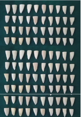
with the help of Vernier callipers and standardized to 5-7 mm. (Fig: 1) In all the
specimens, access openings were prepared using #4 round bur and working length
was determined by placing a No 10 K file (Mani) in to the root canal, until it was just
visible at the apical foramen. The length of the instrument was measured and one
millimetre subtracted from it to establish the working length (11 mm). Ninety teeth
Materials and Methods
24
ProTaper rotary instruments by using a 16:1 reduction hand piece with a torque
controlled and speed controlled electric motor (X Smart DENTSPLY, Maillefer,
Ballagigues, Switzerland)).The speed and torque values were set as recommended by
the manufacturer.
Copious root canal irrigation using 5ml of 3 % sodium hypochlorite solution
using a syringe and 27 gauge needle was performed after each instrumentation. Final
flush with 5ml of 17% EDTA was done in order to remove the smear layer for 1-2
minutes. This was followed by a final irrigation with 5ml of 0.9% Normal saline.
Each of the root canal specimens were dried with sterile Protaper paper points.
Ten teeth were randomly selected to serve as a negative control (Group 1A)
and ten teeth were selected to serve as positive control (group 1B). The remaining
eighty teeth were then obturated with sealer by using the matched single-cone
technique.
All the 100 specimens were randomly allocated into 6 experimental groups
Group 1 A: Negative control group - Roots were neither instrumented nor obturated.
(n=10)
Group 1B: Positive control group – Roots were not obturated. (n=10)
Group 2: Roots were obturated using gutta-percha and AHplus sealer (n=20).
Materials and Methods
25
Group 4: Roots were obturated using gutta-percha and Chitra - CPC root filling
material (n=20).
Group 5: Roots were obturated using gutta-percha and Endosequence BC Sealer
(n=20).
OBTURATION OF ROOT CANALS:
In Groups 2 and 3, sealers were mixed according to the manufactures instructions
and coated in the root canals using a lentulo spiral (DENTSPLY Maillefer) placed in
low speed hand piece. Lentulo spiral was introduced into the root canal to a location 2
to 3 mm short of the working length and then slowly withdrawn from the canal, with
continuous rotation. For standardization, lentulospiral was used for ten seconds only
in all the canals. Obturation was completed by placing sealer-coated single cone
gutta-percha points (ProTaper -F3) (Dentsply Maillefer).
In Group 4. Sealer was mixed according to the manufacturer instruction to get an
injectable paste form which was loaded in the syringe. The syringe was then placed in
to the canal 3mm short of the working length .The cone was coated with the sealer
and was placed in to the canal.(Fig:6)
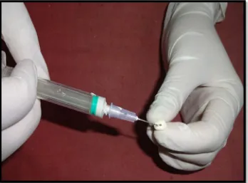
In Group 5: BC sealer packaged in a pre-loaded syringe with disposable intra canal
tips were placed in the root canal and it was deposited by compressing the plunger of
the syringe (Fig: 8) Sealer was coated with master guttapercha and slowly introduced
into the canal there by carrying sufficient sealer to the apex .The roots of this group
Materials and Methods
26
For all the experimental root samples, post-obturation radiographs were taken in both
labio-lingual and mesio-distal directions to ensure homogeneous adequate root filling
without voids. After root filling, the coronal 1 mm of the filling materials was
removed, and the spaces were filled with a temporary filling material (Cavit; 3M
ESPE, Seefeld, Germany). The teeth were stored at 370C in 100% humidity for 14
days to allow the sealers to set. (Fig: 9)
MECHANICAL TESTING:
The root surface of the samples were wrapped around by an aluminium foil to
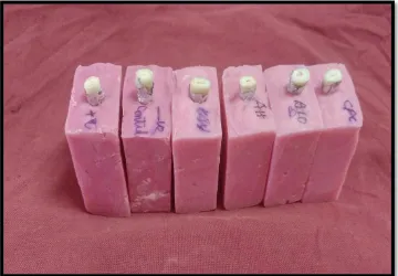
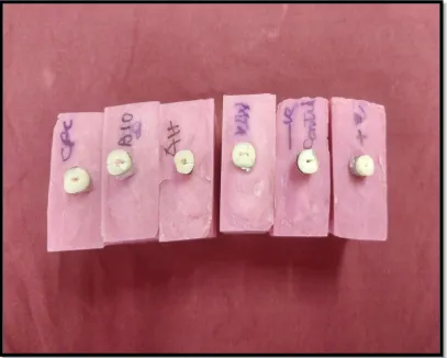
simulate the periodontal ligament 63. All the roots were then mounted vertically in
copper blocks (4cm height, 3cm length and 2 cm width ) and filled with self-curing
acrylic resin (Imicryl, Konya, Turkey), exposing 7 mm of the coronal parts of the
roots.(Fig :10) As soon as the acrylic hardened, blocks were removed from the copper
blocks . A universal testing machine (Instron Corp, Canton, MA) was used for the
strength test (Fig: 11). The acrylic blocks were placed on the lower plate of the
machine. The upper plate consisted of a spherical steel tip with a diameter of 3mm.
The tip was centred over the canal orifice, and slowly increasing vertical force was
exerted (1 mm/min) until fracture occurred. (FIG: 12) The fracture moment was
determined when a sudden drop in force occurred that was observed on the testing
machine display. The maximum force required to fracture each specimen was
recorded in Newtons. The data thus obtained was recorded, tabulated and subject to
statistical evaluation. Analysis of variance was used to analyse the difference between
various test groups. It was seen that there was a statistically significant difference
within the groups (P = 0.001). Hence the further analysis was done using Tukey’s
Materials and Methods
27
[image:37.595.155.480.226.694.2]
Fig: 1 MANDIBULAR PREMOLARS AFTER DECORONATION AT
CEMENTOENAMEL JUNCTION
Materials and Methods
[image:38.595.109.549.229.568.2]28
FIG: 2 MATERIALS USED FOR THE STUDY
Materials and Methods
29
FIG: 3 AH PLUS SEALER WITH BASE AND CATALYST PASTE AND
MIXING PAD
[image:39.595.154.517.485.726.2]
FIG: 4 MTA-FILLAPEX SEALER WITH BASE AND CATALYST PASTE
AND MIXING PAD
Materials and Methods
[image:40.595.145.485.152.397.2]30
FIG 5: CHITRA-CPC SEALER POWDER AND LIQUID ALONG WITH
MIXING PAD, SPATULA AND INJECTION TIP
[image:40.595.141.489.463.720.2]Materials and Methods
[image:41.595.136.493.153.392.2]31
FIG: 7 ENDOSEQUENCE BC SEALER IN PREMIXED SYRINGE ALONG
WITH INJECTING TIP
FIG: 8 ENDOSEQUENCE BC SEALER BEING INJECTED INTO THE
CANAL
[image:41.595.132.487.477.725.2]
Materials and Methods
[image:42.595.146.506.152.400.2]32
FIG: 9 SAMPLES PLACED IN HUMIDITY CHAMBER FOR A PERIOD OF
1 WEEK TO ALLOW COMPLETE SETTING OF THE SEALERS
FIG: 10 TEETH MOUNTED IN ACRYLIC BLOCK FOR FRACTURE
[image:42.595.147.507.480.730.2]Materials and Methods
33
[image:43.595.165.475.230.600.2]
FIG: 11 TEETH SAMPLE MOUNTED ON UNIVERSAL TESTING
MACHINE FOR FRACTURE TESTING
Materials and Methods
[image:44.595.122.529.227.553.2]34
FIG: 12TEETH SPECIMENS SHOWING FRACTURE IN THE LABIL
LLINGUAL DIRECTION
Results
35
The mean values and their respective standard deviations of the force required
to fracture the roots are presented in Table2. The strongest mean force required to
fracture the roots was seen in the negative control group (teeth left unprepared)
whereas the weakest force required was seen in the positive control group (teeth
prepared and unobturated).
In the present study, the mean fracture resistance using Universal testing
machine was found to be highest in the negative control group (428.44+/-151.70),
which was comparable to the mean fracture resistance of Chitra-CPC
(391.60+/-77.19) and Endosequence-BC Sealer (361.84+/-73.04). However, the MTA Fill apex
(287+/- 68.99) and AH Plus (299.93+/-63.27) showed lower fracture resistance.
In Comparison to the positive control, Chitra-CPC showed the highest fracture
resistance followed by Endosequence-BC sealer, AH plus and MTA Fill apex. When
compared to the positive control all groups were showing highest fracture which was
highly significant. (P value 0.001). However MTA Fill apex showed the least fracture
resistance among all the groups. The other groups were marginally stronger than the
positive control.
In Comparison to the negative control, there was no statistically significant
difference between Chitra-CPC and Endosequence –BC sealer as far as the fracture
resistance is concerned. However MTA Fill apex and AH Plus were significantly
weaker compared to the negative control, and the positive control being the weakest.
Results
36
On comparing the fracture resistance between the 4 groups of sealants, the
variability in the mean difference of fracture resistance was not statistically
significant. Though Chitra –CPC showed a higher difference in fracture resistance
compared to MTA Fill apex which was statistically significant. (Table 4)
To summarise,
Chitra-CPC and Endosequence-BC sealer have comparable fracture
resistance to negative control.
Chitra-CPC had significantly high fracture resistance compared to
positive control.
AH Plus sealer showed higher fracture resistance than MTA Fillapex
but lower than Chitra-CPC and Enosequence BC Sealer.
MTA Fill apex showed the least fracture resistance and had statistically
significant lower fracture resistance compared to negative control and
Results
[image:48.595.81.531.221.487.2]37
TABLE: 2 MEAN, STANDARD DEVIATION, MINIMUM AND MAXIMUM
VALUES OF EACH GROUP.
GROUPS n MEAN STD.
DEVIATION
MINIMUM N
MAXIMUM N
NEGATIVE
CONTROL 10 428.44 151.70 230.42 600.84
POSITIVECONTROL 10 243.29 55.08 185.83 317.09
Chitra-CPC 20 391.60 77.14 299.60 549.34
Endosequence BC 20 361.84 73.04 229.52 462.83
MTA Fillapex 20 287.63 68.99 206.07 424.29
Results
[image:49.595.105.513.203.465.2]38
TABLE: 3 DIFFERENCE BETWEEN THE STUDY GROUPS USING ANOVA TEST
GROUPS N MEAN df F P value
NEGATIVE CONTROL 10 428.44
5 4.936 0.001
POSITIVECONTROL 10 243.29
Chitra-CPC 20 391.60
Endosequence BC 20 361.84
MTA Fillapex 20 287.63
AH Plus 20 299.93
P<0.05 – Statistically Significant
Analysis of variance was used to analyse the difference between various test groups.
It was seen that there was a statistically significant difference within the groups (P =
0.001). Hence the further analysis was done using Tukey’s Post-Hoc Test. (Tukey’s
Post-Hoc test to analyse the difference between the groups.) (TABLE 4)
Results
39
TABLE 4: MULTIPLE COMPARISONS
GROUPS COMPARISION MEAN
DIFFERENCE P VALUE
NEGATIVE CONTROL
POSITIVECON
TROL 185.15
*
.009
CPC 36.84 .959
BIO 66.60 .659
MTA 140.81* .029
AH+ 128.51* .048
GROUPS COMPARISION MEAN
DIFFERENCE P VALUE
POSITIVE CONTROL
CPC -148.31* .019
BIO -118.55 .097
MTA -44.34 .913
AH+ -56.64 .791
GROUPS COMPARISION MEAN
DIFFERENCE P VALUE
CPC
BIO 29.75 .961
MTA 103.96 .042
AH+ 91.66 .132
GROUPS COMPARISION MEAN
DIFFERENCE P VALUE
BIO
MTA 74.20 .325
AH+ 61.90 .526
GROUPS COMPARISION MEAN
DIFFERENCE P VALUE
MTA AH+ 12.30 .999
Results
40
Tukey’s post hoc analyses shows that there is statistically significant difference
between the values of negative control and positive control, MTA and AH+ with p
values 0.009, 0.029 and 0.048 respectively.
Apart from that there was also statistically significant difference between positive
Results
[image:52.595.123.515.207.537.2]41
FIG: 13 DISTRIBUTION OF MEAN VALUES OF DIFFERENT GROUPS.
428.44
243.29
391.6
361.84
287.63 299.93
0 50 100 150 200 250 300 350 400 450
VA
LU
ES
GROUPS
Results
[image:53.595.119.515.216.511.2]42
FIG:14 MINIMUM AND MAXIMUM VALUES IN EACH GROUPS
Discussion
43
Root canal instrumentation is an essential stage in endodontic treatment.
There is a cognizance that endodontic treatment weakens the tooth structure and
predisposes teeth to fracture. Zandbiglari et al and Schafer et al demonstrated that „Enlarged but unfilled roots are significantly weaker than filled roots, thus more
susceptible to fracture.‟67
Reinforcement of the remaining tooth structure after endodontic procedures is
an important extension of root canal therapy 11. Most root canal filling methods use a
root canal sealer as a complementary part of the obturation technique. The root canal
sealer fills the gaps between gutta-percha cones and the walls of the root canal and
also fills the voids between individual gutta-percha cones applied during obturation of
the root canal system 64. Some studies have claimed the ability of different root canal
filling materials to significantly strengthen the roots65, where as in other reports these
materials did not increase the fracture resistance of root filled teeth 66. However,
recent studies have suggested that sealers can adhere to the root canal dentin surface
and strengthen the remaining tooth structure, thereby contributing to the long-term
success of an endodontically treated tooth 67, 68.
Previous researchers showed that epoxy resin–based sealers (AH plus sealer)
had higher mechanical adhesion to root canal dentin and deeper penetration into
dentinal tubules than zinc oxide-eugenol–based and glass ionomer–based sealers 69, 70.
As a result of the benefits of epoxy resin–based sealers, resistance to fracture would
increase. Cobankara et al reported that sealers exhibiting chemical bonding
Discussion
44
Recent researchers have considered Endosequence BC sealer (Brasseler, USA)
and AH plus sealer (epoxy resin based sealer) as “Gold Standard” for sealers because
of their potential to adhere to the dentin. According to Topcuoglu et al teeth
obturated with chemically bonding Endosequence BC sealer (Brasseler, USA) by
using single cone technique showed significantly higher fracture resistance than AH
plus sealer.72
In the present study, we intended to find out the fracture resistance of roots
sealed with indigenously prepared Chitra-CPC root filling material in comparison
with AH Plus sealer, Endosequence-BC Sealer and MTA Fill apex, by using single
cone technique.
AH Plus is an epoxy based endodontic sealer that is used with gutta percha. It
has good adhesion to dentin and to gutta percha. Neto and Mamootil et al in their
study showed that epoxy resin–based sealers had higher adhesion to root canal dentin
and deeper penetration into dentinal tubules than zinc oxide-eugenol–based and glass
ionomer–based sealers 69, 70.
Bioceramic-based materials have been recently introduced in endodontics,
mainly as repair cement and as root canal sealer. Studies have showed that
bioceramics have enhanced biocompatibility, result in the increased strength of the
root after obturation, have a high pH during the setting process (which is strongly
antibacterial pH12), are easy to use, (particle size is so small it can be used in a
Discussion
45
exhibit chemical bonding to root canal dentin walls. Therefore, EndoSequence BC
Sealer (Bioceramic Sealer), which is based on a calcium silicate composition, has the
potential to adhere chemically to dentin decreasing the marginal leakage and gaps
and increased fracture resistance of teeth.20
MTA Fillapex is the first MTA based salicylate resin sealer. It has suitable
physiochemical properties such as good radiopacity, flow and alkaline pH. It has a
working time of 35 minutes.23 It is a bioceramic type of sealer that is compatible with
moisture and tissue fluids. It can readily set in presence of moisture and is able to
cause cementogenesis and thus helps in repair of apical tissue and is biocompatible.73
Calcium phosphate cement (CPC) based sealers are emerging as promising
candidates in endodontics because of their superior biocompatibility features. It is an
example for bioresorbable material (that upon placement within the human body starts
to dissolve (resorb) and slowly be replaced by advancing tissue (such as bone). 74They
also satisfy most of the requirements for an ideal sealer.12, 13 The past two decades
saw numerous attempts to manufacture a calcium phosphate sealer suited for its use in
endodontic therapy and tested with focus on the development of a non-mutagenic,
non-carcinogenic, and an overall tissue-friendly material.
In this study we have used a novel indigenous formulation of CPC: „Chitra-CPC‟ which has been developed at the Biomedical Wing of Sree Chitra Tirunal
Institute for Medical Sciences and Technology (SCTIMST), Thiruvananthapuram.
Discussion
46
that contain inorganic calcium and phosphate minerals (tetra- calcium phosphate
(TTCP) and dicalcium phosphate dihydrate (DCPD) in equimolar ratio) which upon
wetting with an aqueous solution (disodium hydrogen phosphate in distilled water)
get converted to hydroxyapatite28.
The cement has been tested for safety and efficacy and approved for human
clinical use by the Institutional Ethics Committee of Sree Chitra Tirunal Institute for
Medical Sciences and Technology (SCTIMST), Thiruvananthapuram75. Compared to
conventional CPC it has enhanced viscous and cohesive properties. Chitra-CPC could
be mixed in varying consistencies, from moldable putty to injectable paste. This
flexibility provides immense advantage in clinical application as a bone and dentine
substitute and root filling material 28, 75, 26. Besides this, it has a neutral pH during
setting, is highly adaptable and adheres to the root canal surface. 76It is also
dimensionally stable, easy to handle and has the property of osteotransductivity (i.e.,
active resorption at bony sites, facilitating bone remodelling).77
In the present in vitro study, results revealed that the negative control group
(group1A) has highest fracture resistance values (600.84 N) and the positive control
group (group 1B), the lowest (317.09N). Statistically significant higher fracture
resistance was offered by the indigenously prepared CPC formulation Chitra – CPC
(549 N) and Endosequence BC sealer (462 N), which was comparable to the negative
control and highly significant than the positive control, followed by AH Plus sealer
(424 N). MTA Fill apex showed the least fracture resistance (361 N) in comparison
Discussion
47
The mean fracture resistance of Chitra-CPC in the current study was higher
(549 N) than the mean fracture resistance of Endosequence BC sealer (457.61 N) as
reported by Topcuoglu et al72,AH Plus (248.36 N) as reported by Khanet al78 and
MTA Fillapex (261.47 N) as reported by Mandava et al24. Therefore the mean
fracture resistance with Chitra-CPC in our study was favourable and higher compared
to mean fracture resistance of various other sealants.
Singh et al 79 tested the performance of Chitra –CPC as a repair material and
evaluated the microleakage by using dye penetration method and showed favourable
result for this material. The high fracture resistance offered by Chitra-CPC could be
due to the formation of submicron-sized particles of hydroxyapatite, inter-grown to
form a homogeneous mass during cement setting as stated by Komath et
al26.However no studies have been done so far to compare the fracture resistance of
this material, longevity in the root canal and sealer penetration in to the dentinal
tubules.
In contrast to our study, Celikten et al 80 and Jainaen et al81 demonstrated
low fracture resistance for teeth obturated with Endosequence BC sealer, and AH Plus
sealer respectively. According to Celikten et al, 80 the reduced fracture resistance
offered by Endosequence BC sealer could be due to the reduced moisture in the
dentinal tubule required for the setting of sealer. Jainaen et al81 stated that reduced
fracture resistance of AH Plus was due to the reduced compressive and tensile
Discussion
48
Hatibovic and Kofman et al47assessed the fracture resistance of root dentine
in open apex cases for longer duration and they demonstrated an increase in fracture
resistance; however in closed apex cases MTA showed a reduction in fracture
resistance 26 which was similar to our study. Various Studies have showed that
fracture resistance of teeth obturated with MTA Fill apex was low probably due to
lack of bonding of MTA to the dentin.15 These results are similar to our study result
with MTA Fill apex
Researchers have analysed and concluded that single circular canals have
lower and more uniform stress distribution than oval canals in which greater stresses
are present at the labial and lingual canal extensions and at the cervical and middle
thirds. 9 In all of the premolar samples used in the present study, had a circular
cross-section, which would have resulted in uniform distribution of load and also simulated
the clinical situation where chewing forces are maximum.
Some studies have suggested that lateral condensation creates stresses in the
root during obturation, which could lead to subsequent fracture. Single cone
Obturation technique is a simple and time efficient technique which has become
popular after the advent of NiTi rotary instruments. Ersev et al 82 reported that the
group in which AH Plus was used with the matched taper single-cone technique
showed significantly higher fracture resistance than the instrumented but not
obturated roots. The major advantage of using single cone obturation technique is that
it forms a uniform mass in combination with endodontic sealers thereby preventing
failures observed among multiple cones as in cold lateral condensation technique63. In
Discussion
49
both the excessive dentin removal required to facilitate the plugger‟s insertion during
vertical compaction and the wedging forces of the spreaders during lateral
compaction. In the present studyaluminium foil and acrylic resin blocks were used to
simulate the periodontal ligament and alveolar bone and a single load to fracture was
applied vertically as in many other studies that evaluated the effect of root canal
sealers on the fracture resistance of root filled teeth.
In summary, the results from the present invitro study shows that both
indigenously prepared, cost effective Chitra-CPC and Endosequence –BC sealer have
improved the fracture resistance of endodontic ally treated teeth. However, there are
limited independent publications about the properties and applications of Chitra- CPC
root canal sealers in endodontics. Further invivo studies will throw light into the


