INFECTIONS AND IMMUNOMODULATION ON LIVER
TRANSPLANT PATIENTS
DISSERTATION SUBMITTED TO
THE TAMILNADU DR MGR MEDICAL UNIVERSITY
In partial fulfillment of the requirements
for the award of degree of
M.D. (MICROBIOLOGY)
BRANCH - IV
GOVT. STANLEY MEDICAL COLLEGE & HOSPITAL
THE TAMIL NADU DR.M.G.R MEDICAL UNIVERSITY
CHENNAI – 600 001
CERTIFICATE
This is to certify that this dissertation entitled “INFECTIONS AND
IMMUNOMODULATION ON LIVER TRANSPLANT PATIENTS” is the
bonafide orginal work done by Dr.M. M.EENA, Post graduate student, in the
department of Microbiology, Stanley Medical College, Chennai, in partial
fulfillment of the regulations of the Tamil Nadu Dr. MGR Medical University
for the award of M.D Degree in Microbiology (Branch IV).
GUIDE
Dr. Rosy Vennila M.D.,
Former HOD,Microbiology,Stanley Medical College, Director, Institute of Microbiology,
Madras Medical College, Chennai
Prof. Dr. S.Ponnambala Namasivayam, Dr. R.Selvi M.D.,
M.D., D.A., D.N.B., PROFESSOR &HOD,
DEAN Department of Microbiology
Govt. Stanley Medical College Govt. Stanley Medical College
Chennai-1 Chennai-1.
DECLARATION
I, Dr.M.MEENA, solemnly declare that this dissertation entitled,
“INFECTIONS AND IMMUNOMODULATION IN LIVER
TRANSPLANT PATIENTS” is the bonafide work done by me during my post
graduate course in MD Microbiology at the Department of Microbiology, Govt.
Stanley Medical College and Hospital, Chennai, during 2015–2018 under the
guidance and supervision of Dr. ROSY VENNILA, M.D., Director of
Department of Microbiology, Madras Medical College, Chennai. The
dissertation is submitted to The Tamilnadu Dr. M.G.R. Medical University,
Chennai in partial fulfillment of the University regulations for the award of
degree of M.D. Microbiology, Branch IV, examination to be held in April 2018.
Place: Chennai Dr.M.Meena
ACKNOWLEDGEMENT
My sincere and heartful thanks to Dr.S.Ponnambala
Namasivayam, M.D., D.A., D.N.B., Dean, Stanley Medical College and
Hospital, Chennai, for giving me permission for the study and use the
resources of this institution.
I owe my sincere and profound gratitude to Prof. Dr. ROSY
VENNILA. M.D, Director of the Department of Microbiology, Govt. Madras
Medical College Hospital, Chennai, for her able guidance and encouragement
during the study. I would like to acknowledge her constant support which
provided throughout the study. My sincere thanks to Prof . Dr. S.
JESWANTH, M.S.M.ch. Professor of the Department of Surgical
Gastroenterology for his support, encouragement and valuable inputs for the
study.
I extend my thanks my thanks and gratitude to Dr.R.Secunda M.D,
Senior Assistant Professor who guided me from the beginning. I thank all our
Assistant Professors, Dr. B. Shanthi M.D, Dr. P. Ponnammal M.D, Dr. V.
Sheeba M.D, Dr. A. Madhumathi M.D, Dr. P. Sivagamisundari M.D, of
Department of Microbiology, Stanley Medical College, Chennai for their
I extend my sincere thanks to Mr. Venkatesan, statistician for his work. I
express my sincere thanks to all the technical staff of the Department of
Microbiology and Surgical gastroenterology for their cooperation and kind help
during the study.
I acknowledge the immense faith of the volunteers and patients who have
participated in this study and express my gratitude for their cooperation. Lastly I
express my thanks to my husband J. Ramakrishnan, my son Ram. Niranjan and
LIST OF ABBREVIATIONS
LT – Liver Transplantation
HSV – Herpes simplex virus
CMV – Cytomegalo virus
EBV – Epstein Barr Virus
PTLD – Post Transplant Lymphoproliferative disease
PCR – Polymerase chain reaction
CNI – Calcineurin Inhibitors
MMF – Mycophenolate Mofetil
HAV – Hepatitis A virus
HBV – Hepatitis B virus
HCV – Hepatitis C virus
PBS – Primary Biliary Cirrhosis
BCS – Budd Chiari Syndrome
ALD – Alcoholic liver disease
DCLD – Decompensated Liver Disease
WD – Wilsons disease
CF – Cystic Fibrosis
TT 1 – Tyrosinemia 1
HCC – Hepatocellular Carcinoma
CCA – Cholangiocarcinoma
ALT – Alanine transaminase
AST – Aspatate transaminase
ALP – Alkaline phosphatase
GGT – Gamma glutamyl transferase
MIC – Minimum Inhibitory concentration
DNA – Deoxy Ribonucleic acid
RNA – Ribonucleic acid
SSI – Surgical Site Infections
UTI – Urinary tract Infections
PT – Prothrombin Time
ESBL – Extended Spectrum Beta lactamases
MRSA – Methicillin Resistant Staphylococcus aureus
VRE – Vancomycin Resistant Enterobacteriaceae
CV TIP – Central venous tip
DT Fluid – Drainage tube fluid
E.coli – Escherichia coli
CONTENTS
Chapter No.
Title Page No.
1. INTRODUCTION 1
2. AIM AND OBJECTIVES 5
3. REVIEW OF LITERATURE 6
4. MATERIALS AND METHODS 43
5. RESULTS 53
6. DISCUSSION 73
7. SUMMARY 83
8. CONCLUSION 85
9. BIBLIOGRAPHY
10. ANNEXURES
PROFORMA
CONSENT FORM
INFECTIONS AND IMMUNOMODULATION IN LIVER
TRANSPLANT PATIENTS
INTRODUCTION:
Liver Transplantation is the surgical replacement of diseased liver with the
healthy liver from another person (allograft). It is a standard life saving
procedure for many end stage liver diseases and acute liver diseases. According
to the United Network for Organ Sharing (UNOS) in the United States, a total
of 6331 liver transplantations were performed during 2008-2009, with a
survival rate of 85% at one year. Survival rate after liver transplantation has
improved over the years, due to advances in surgical techniques and a reduction
in allograft rejection produced by new potent immunosuppressive agents.
Inspite of use of protective barriers, antimicrobial prophylaxis, and vaccination,
infections still represent a major cause of morbidity and mortality after liver
transplantation. The opportunistic infections are the leading cause of death
during the first three years after liver transplantation. The diagnosis of these
infections are delayed because of potent immunosuppressive therapy that
diminishes inflammatory responses and the clinical signs of infection may be
absent or blunted, leading to delayed diagnosis and treatment.1 The risk of
overall level of immunosuppression. The infections after liver transplantation
may be acquired, reactivation of latent infection in the recipient, donor
transmitted diseases and nosocomial.
The incidence of infection is found to be 80% in liver transplant
recipients and among them, the most common infection is bacterial (70%),
followed by viral (20%) and fungal (8%) infection.2 The bacterial infection
occurs with the highest incidence during the first month after transplantation
and most commonly involve the surgical site, the abdominal cavity, blood
stream, urinary tract and the respiratory system. The common bacterial
infections are Enterococcus, Viridans Streptococci, Staphylococcus aureus and
members of the Enterobacteriaceae family and there is an increasing trend
towards antimicrobial resistance patterns among bacteria.2
Among the opportunistic viral infections, the most common are the
members of herpes virus group, of which Cytomegalovirus infection is the most
common infection followed by Epstein-barr virus infection, Herpes simplex
virus (HSV) 1 & 2 and Varicella Zoster infection. The fungal infections are
most commonly caused by the Candida species followed by Aspergillus species.
Cryptococcus neoformans occur less commonly in the form of meningitis, lung
disease and cellulitis. Endemic mycoses due to Histoplasma capsulatum,
Coccidiodes immitis and Blastomyces dermatidis may occur in liver transplant
occurs, primarily as diffuse bilateral pneumonitis and the use of trimethoprim-
sulfamethoxazole prophylaxis has remarkably reduced its incidence after liver
transplantation.1
Immunosuppressive agents are required in solid organ transplantation for
induction and maintenance of immunosuppression or for the treatment of organ
rejection. Usually, a corticosteroid in combination with a calcineurin inhibitors
(CNI), alone or with an antimetabolite, mycophenolate mofetil (MMF) is started
early to maintain immunosuppression to prevent transplant rejection.
Corticosteroids have been used for induction of immunosuppression since the
first successful cases of solid organ transplantation. Intravenous injection of
corticosteroid is administered in high doses during the first few days after
transplantation (usually 3 days) in combination with at least one
immunosuppressant agent. It is then rapidly tapered over the first week to
relatively low doses, 10 to 20 mg daily and are usually maintained in
immunosuppression for atleast, 3 to 6 months after transplantation.3 The
introduction of the two CNIs, Cyclosporine A (in 1970s and early 1980s) and
tacrolimus (in 1990s) as immunosuppressant agents, greatly improved the
outcome of Liver transplantation. Tacrolimus is superior to cyclosporine A in
increasing patient and graft survival. Acute graft rejection and steroid resistant
rejection episodes are less common with tacrolimus use during the first year
monitoring of tacrolimus is done for dose adjustments to prevent toxicity. The
major advantage in using, Mycophenolate mofetil is their lack of renal toxicity.
In patients with pre-existing renal disease, they have been used in conjunction
with low dose CNIs with promising results.4
More recently, antibody therapies have been combined with
corticosteroids or used to facilitate “steroid-free” regimens. The use of
antibodies that specifically inhibit or deplete recipient T-cells has been reported
to decrease acute rejection episodes in the liver allograft. It also provides an
opportunity to decrease the dose of other immunosuppressive agents such as
corticosteroids and calcineurin inhibitors. This “steroid-free” protocol may be
beneficial for patients with hepatitis C patients and for those with diabetes and
hypertension. Antibody induction along with delayed CNI introduction can be
used to reduce renal dysfunction in those with impairment. There was no
significant increase in adverse side effects in solid organ transplant recipients
AIM & OBJECTIVES:
AIM:
To determine the common organisms that cause infections in liver transplant
patients and the role of immunosuppressants in immunomodulation in liver
transplant patients.
OBJECTIVES:
1) To compare the incidence of alcoholic cirrhosis with non- alcoholic
cirrhosis indicated for liver transplantation.
2) To identify the common organisms that cause infections in liver
transplant patients.
3) The antibiotic sensitivity pattern of the pathogen isolated.
4) To evaluate the role of immunosuppressants in immumomodulation in
REVIEW OF LITERATURE
HISTORY:
Liver transplantation is the treatment of choice for patients with cirrhosis,
decompensated liver disease, acute liver failure and hepatocellular carcinoma.
In 1963 March 1st, Thomas Starzl in Denver did the first liver transplantation in
the world on a 3yr old boy with biliary atresia and the boy died during the
surgery because of coagulation disorder and uncontrolled bleeding. The first
successful human liver transplant was performed by Thomas Starzl in Denver in
1967 on an 18 month old child with unresectable hepatoblastoma, at the
university of Colorado, who survived more than 1 year.
In 1979, Calne used cyclosporine in two patients who had undergone
liver transplantation for the first time, launching a new step in history of liver
transplantation. In 1990, Starzl reported the first use of tacrolimus in patients
submitted to liver transplantation, who suffered a rejection even after
conventional immunosuppressive treatment. Currently, more than 10,00,000
liver transplantations have been performed in the world so far.7 The first
successful deceased liver transplantation in India was done in 1998 followed by
first successful living donor transplantation in November 1998 both done by
Dr.Rajasekar.8
(A) Acute liver failure:
1) Viral hepatitis A / hepatitis C
2) Intoxication - Paracetamol poisoning,
3) Wilsons disease,
4) Budd chiari syndrome.
(B) Chronic liver failure (Non-cholestatic cirrhosis):
1) Hepatitis B / hepatitis C
2) Autoimmune hepatitis,
3) Alcohol induced cirrhosis
(C) Chronic liver failure (Cholestatic cirrhosis):
1) Primary biliary cirrhosis (PBC)
2) Primary sclerosing cholangitis (PSC)
3) Secondary biliary cirrhosis
(D) Chronic liver failure (Metabolic):
1) Wilson’s disease
2) Hemochromatosis
3) α-1 Antitrypsin deficiency
4) Amyloidosis
5) Cystic fibrosis
6) Tyrosinemia
(E) Chronic liver failure (Vascular):
(F) Other Indications:
1) Primary oxalosis
2) Gycogen storage diseases
3) Hyperlipidemia
4) Polycystic liver disease
(G) Malignant diseases:
1) Hepatocellular carcinoma (HCC, within Milan criteria)
2) Fibrolamellar carcinoma (FLC)
3) Hepatoblastoma
4) Epitheloid hemangioendothelioma
5) Cholangiocellular adenocarcinoma
6) Neuroendocrine liver metastases
(H) Benign liver tumors:
1) Adenomatosis
(I) Liver transplantation in pediatric patients:
1) Biliary atresia
2) Byler’s disease
3) Alagille’s syndrome
4) Neonatal hepatitis/neonatal viral hepatitis
5) Autoimmune hepatitis
6) Hepatoblastoma.
(A) Absolute contraindications:
1) Active alcohol abuse
2) Uncontrolled systemic infections
3) Uncontrolled extrahepatic malignancy
4) Uncontrolled / limiting medical conditions
(B) Relative contraindications:
1) Psychosocial conditions
2) Advanced age
3) Severe hepato-pulmonary or severe hepato-renal syndrome
4) Severe obesity/malnutrition
INDICATIONS FOR LIVER TRANSPLANTATION:
1) Viral Hepatitis:
Hepatitis viruses A, B and C cause the majority of viral hepatitis.
(A) Hepatitis A Virus (HAV):
HAV belongs to the Picornaviridae family, is an RNA virus of 7.5kb
size and a diameter of 27 nm. It has one serotype but multiple genotypes. The
virus spreads from person to person most commonly via the faecal-oral route.
The incubation period of is 15-45 days (average, 4 weeks). The virus is excreted
in stool during the first few weeks of infection, before the onset of symptoms.
Young children are usually asymptomatic. Acute hepatitis A is a more severe
infection are fever, malaise, anorexia, nausea, vomiting, hepatomegaly and
elevated aminotransferase levels. Jaundice develops in more severe cases. The
detection of immunoglobulin M (IgM) for hepatitis A, is the standard for
diagnosing acute infection. The infection is usually self-limited and provides
lifelong immunity. It does not lead to chronic infection and less than 1% of
cases result in fulminant hepatic failure (FHF).10
(B) Hepatitis B Virus (HBV):
HBV, belong to the Hepadnaviridae family, is a partially
doubled-stranded DNA virus and 3.2 kb size. It has 8 genotype (A-H). The virus consists
of a nucleocapsid, HBcAg, which surrounds HBV DNA and DNA polymerase.
The nucleocapsid is coated with HBsAg and the intact HBV virion is known as
the Dane particle. The HBcAg is not detected in the circulation.10 HBV is
transmitted by both parenterally by transfusion of blood or blood products,
intravenous drug abuse with shared needles, hemodialysis, and needlestick
injury in healthcare workers and also sexually. The incubation period is 30-180
days (average 12 weeks). In acute infection, the symptoms are fever, malaise,
nausea, vomiting, jaundice and right upper quadrant pain. About 5% of adult
patients, 30-50% of infected children develop chronic infection.10 In chronic
infection, fatigue is the most common symptom. Patient have abnormal liver
function test and the detection of Ig M for hepatitis B core antigen (HBcAg) in
serum is required for the diagnosis of acute infection. Hepatitis B surface
chronic carriers (> 6 months). At present, the antiviral treatment is given for the
inhibition of viral replication and to prevent or delay the progression of chronic
hepatitis to cirrhosis or hepatocellular carcinoma. The drugs currently used for
chronic infection include pegylated interferon α-2a and the nucleoside
analogues, lamivudine and adenofovir. Globally, an estimated 30% of cases of
cirrhosis and 45% of cases of HCC are attributed to HBV infection.11
(C) Hepatitis C Virus (HCV):
HCV, belong to the Flaviviridae family, is a RNA virus with 9.4 kb size
and 55 nm diameter. It has one serotype, but six major genotypes and more than
80 subtypes. The virus is transmitted through transfusion of infected blood or
blood products, transplantation of organs from infected donors and sharing of
contaminated needles among IV drug users.11,13 The incubation period is 15- 45
days (8 weeks ). Acute infections are usually asymptomatic and donot develop
jaundice. About 55-85% of infected patients, remain viremic and may develop
chronic liver disease.13 In chronic hepatitis C, patients may or may not be
symptomatic with fatigue being the predominant symptom and
aminotransferases may be elevated. The infection can be confirmed by serologic
assays to detect antibody to HCV (anti-HCV) or with molecular tests
(Qualitative PCR) for the presence of viral particles. For patients with chronic
HCV infection, combination therapy with pegylated interferon and the antiviral
drug, ribavirin are given for patients with moderate or severe inflammation or
cirrhosis13 and this may take decades. Patients with HCV induced cirrhosis are
also at increased risk for the development of hepatocellular carcinoma
especially with HBV co-infection.
2) Alcoholic liver disease: (ALD)
It is caused by of increased alcohol consumption, leading to fatty liver,
alcoholic hepatitis leading to fibrosis and cirrhosis. Alcohol is metabolized by
alcohol dehydrogenase into acetaldehyde, then further metabolized by aldehyde
dehydrogenase into acetic acid, which is finally oxidized into carbon dioxide
and water. This process generates increased NADH that induces fatty acid
synthesis and a decreased NAD level that results in decreased fatty acid
oxidation. Subsequently, the increased fatty acids combine with glycerol to
form triglycerides and accumulate, resulting in fatty liver. Alcoholic hepatitis is
characterized by the inflammation of hepatocytes called as alcoholic steato-
hepatitis and predispose to liver fibrosis. Cirrhosis is a late stage of liver disease
characterised by inflammation, fibrosis and ending in scarring and necrosis.14
About 10% to 20% of heavy alcohol drinkers will develop cirrhosis of the liver.
Symptoms include jaundice, hepatomegaly and right upper quadrant pain. Late
complications of cirrhosis include portal hypertension, coagulation disorders,
ascites, encephalopathy and the hepato-renal syndrome. Fatty change and
alcoholic hepatitis are reversible by abstinence and the later stages of fibrosis
for cirrhosis and the patient should be abstinent from alcohol consumption for 6
months prior to transplantation.15
3) Primary Biliary Cirrhosis: (PBS)
It is a chronic cholestatic liver disease. It is characterised by destruction
of small intrahepatic bile ducts, which results in fibrosis and cirrhosis .The
serological marker of primary biliary cirrhosis is the anti-mitochondrial
antibody (AMA) and is identified in 95% of patients with primary biliary
cirrhosis. These patients usually have fatigue and pruritus. Other
auto-antibodies such as antinuclear antibody are also identified. Anti-Sp100 and
anti-gp210 have a high specificity for primary biliary cirrhosis and is helpful, when
AMA is negative and its presence is associated with clinically more aggressive
disease. Liver biopsy is helpful, when AMA is absent and the biochemical
profile shows a mixed cholestatic and hepatocellular pattern. Ursodeoxycholic
acid (UDCA) plays a major role in the treatment. The Liver transplantation is a
life-saving surgery for those with decompensated cirrhosis with excellent
outcome.17
4) Budd-Chiari Syndrome: (BCS)
It is a very rare condition caused by thrombotic occlusion of the hepatic
vein. It may be primary or secondary depending on the origin of the obstructive
lesion. It is caused by blockage of two or more major hepatic veins that increase
the sinusoidal pressure and reduces sinusoidal blood flow leading to sinusoidal
necrosis of liver is found in nearly 70% of cases and reperfusion injury may
lead to hepatocyte damage. Progressive fibrosis, nodular regenerative
hyperplasia and cirrhosis develop during the course of disease. The acute form
presents with jaundice, hepatomegaly, and upper abdominal pain with elevated
liver enzymes. Portal hypertension and ascites are usually seen in chronic form.
Liver biopsy shows congestion, liver cell loss and fibrosis predominantly in the
centrilobular area. It is diagnosed by doppler ultrasonography showing ״spider
web״ appearance of collateral hepatic venous circulation.18 Milder forms may be
treated with surgical shunts and liver transplantation is the treatment of choice
for patients with fulminant liver failure, failure of shunts or progression to
cirrhosis.19
5) METABOLIC DISORDERS OF LIVER:20
(a)Wilsons Disease (WD):
It is also known as ‘Hepato-lenticular degeneration’, is an autosomal
recessive disease caused by mutation of the WD gene (on the long arm of
chromosome 13). It causes an abnormal copper deposition in multiple organs
including liver, brain, kidney and cornea. Clinical features are usually
asymptomatic leading to chronic hepatitis, cirrhosis and fulminant hepatic
failure. Neurological symptoms like dysarthria, dysphagia, apraxia and tremor
predominantly seen in adults. The diagnosis is by the presence of
Kayser-Fleischer ring around the cornea, low serum ceruloplasmin level, increased
therapy with d- penicillamine, trientine, or ammonium tetrathiomolybdate has
been shown to be effective in preventing progression of Wilson’s disease.
When medical treatment fails or patients present with fulminant failure, liver
transplantation is the treatment of choice.
(b) Hereditary Haemochromatosis:
It is an autosomal recessive disorder characterised by deposition of iron in
tissues and organs. It is caused by homozygous C282Y mutation of the HFE
gene, located on chromosome 6 and it is assosciated with changes in hepcidin
which is the key regulator of iron homeostasis. Infants present acutely with liver
failure in the perinatal period and demonstrate massively elevated ferritin levels,
elevated iron saturation (98%), hyperbilirubinemia, hypoalbuminemia,
hypoglycemia, and coagulopathy. Treatment of choice is phlebotomy with
concomitant follow up of haematocrit. Liver transplantation is indicated for end
stage liver disease.
(c) α1-antitrypsin deficiency:
It is an autosomal recessive disease due to defective production of α1
antitrypsin leading to decreased activity of α-1 antitrypsin in the blood and
lungs. Severe deficiency causes, chronic obstructive pulmonary disease or
panacinar emphysema and liver injury in adults. In children, it may present as
neonatal jaundice, hepatomegaly or acute liver failure. Diagnosis is by
measurement of α1 antitrypsin in serum and liver biopsy (periodic acid Schiff
preparation of α1 antitrypsin with α1 antitrypsin < 11µmol/L and COPD. Liver
transplantation is the treatment of choice in children and adults for end stage
liver disease.
(d) Cystic Fibrosis (CF):
It is a multisystem disease caused by mutation in the cystic fibrosis
transmembrane conductance regulator (CFTR) gene. The lack of CFTR alters
ductular chloride secretion which causes viscous biliary secretions with
subsequent biliary obstruction that leads to focal biliary fibrosis and ultimately
cirrhosis. Depending on the report, 20%-50% of CF patients develop liver
disease, which ranges from asymptomatic derangement of liver function tests to
focal biliary fibrosis to cirrhosis with portal hypertension and chronic liver
failure. Treatment with ursodeoxycholic acid (UCDA) has demonstrated
improved bile flow and aminotransferases but does not stop the progression of
fibrosis. Liver transplantation is indicated for end stage liver disease.
(e) Tyrosinemia:
TT 1 is an autosomal recessive disorder due to a defect in fumaryl
acetoacetate hydrolase enzyme in the tyrosine catabolism pathway, which
results in accumulation of metabolites such as fumaryl acetoacetate and malelyl
acetoacetate. It causes apoptosis of hepatocytes and renal tubular epithelial
cells. Clinical features include acute liver failure, renal tubular dysfunction,
chronic liver disease and hepatocellular carcinoma. Fumaryl acetoacetate and
results in a predisposition to hepatocellular carcinoma. Treatment with
Cyclohexendiome blocks tyrosine degradation and preventing formation of the
alkylating metabolites. Liver transplantation is indicated for failure of medical
treatment.
6) Hepatocellular Carcinoma (HCC):
Hepatocellular carcinoma (HCC) is the sixth most common malignancy
and is the leading cause of mortality in patients with cirrhosis. Cirrhosis is the
most important risk factor for developing HCC in 80% to 90% of individuals
and the annual incidence is 1% to 6 %. It occurs in chronic inflammatory
condition of liver and is most closely linked to chronic viral hepatitis infection
(hepatitis B or C) or exposure to toxins such as alcohol or aflatoxin. Certain
metabolic diseases such as hemochromatosis and alpha 1-antitrypsin deficiency
and non-alcoholic steato-hepatitis (NASH) markedly increase the risk of
developing HCC.21 The clinical features are jaundice, abdominal distension,
loss of appetite, weight loss, abdominal pain, nausea, vomiting, and malaise.
The diagnosis is by liver biopsy, blood levels of α- fetoprotein (AFP),
Ultrasound, CT scan and MRI. On CT and MRI, it has three distinct patterns of
growth, a single large tumour or multiple tumours or a poorly defined tumour
with an infiltrative growth pattern. The most common sites of metastasis are the
lung, abdominal lymph nodes and bone. Radio frequency ablation (RFA) is
patients who are not eligible for resection, especially those within Milano
criteria (solitary tumour ≤ 5 cm and up to three nodules ≤ 3 cm).22
7) Cholangiocarcinoma: (CCA)
It is a primary neoplasm of the biliary system that arise from malignant
transformation of cholangiocytes, the epithelial cells that line the biliary tree.
The risk factors are chronic biliary inflammation, cholestasis, primary
sclerosing cholangitis, choledochal cyst, caroli’s disease and chronic parasitic
infection with liverfluke, Clonorchis sinensis. Morethan 90% of them are
adenocarcinoma. The clinical features are obstructive jaundice, pruritus,
malaise, abdominal pain, weight loss and loss of appetite. Laboratory tests
demonstrate increased total and direct bilirubin, alkaline phosphatase (ALP) and
gamma glutamyl transferase (GGT). Ulrasonography shows dilatation of
intrahepatic bileducts and the presence of intrahepatic metastasis. Endoscopic
retrograde cholangio pancreatography (ERCP), shows the biliary strictures and
allows brush cytology and biopsy of the bile ducts. CT scan (Computerised
tomography) and MRI (Magnetic Resonance Imaging) help in the evaluation of
resectability and CA 19-9 is elevated in most cases. Surgical resection and
neoadjuvant chemotherapy with liver transplantation is the treatment of choice
in stage I and II. In patients with advanced disease, palliative chemotherapy
MELD SCORE (MODEL FOR END – STAGE LIVER DISEASE):23
Model for End – Stage Liver Disease or MELD is a scoring system for
assessing the severity of chronic liver disease and prioritizing for of Liver
transplantation. It was initially used to predict the mortality of trans-jugular
intrahepatic porto-systemic shunt (TIPS) surgery.
This score is now used by the United Network for Organ Sharing
(UNOS) and Euro-transplant for prioritizing of liver transplants. It uses the
patients value of
1) Serum bilirubin,
2) Serum creatinine,
3) International normalised ratio (INR) for prothrombin time.
MELD = 3.78x log e [serum bilirubin (mg/dl)] + 11.2xlog e [INR] +
9.57xlog e [serum creatinine (mg/dl)] + 6.43
MELD scores are reported as whole numbers, so the result of the above
equation is rounded. Patient with decompensated liver disease and cirrhosis
with Child pugh B/C or MELD score ≥24 are considered for liver
Immunology of Transplantation:
Compared with other solid organ transplants, liver allografts have long
been considered to be immunologically privileged manifested by an absence of
hyper-acute rejection and a low incidence of graft loss due to chronic rejection.
The hepatocytes have the potential to regenerate after tissue injury. However,
acute liver rejection occurs in approximately 50% to 75% of liver transplant
recipients and it is readily reversed with immunosuppressive drugs.
Immunological Basis of Allograft Rejection:
In liver transplantation, grafts originate mainly from different members
of the human species. The process of rejection is very complicated and has been
shown to be caused by transplantation antigens including major
histocompatibility antigens, minor histocompatibility antigens and other
alloantigens. The major histocompatibility complex (MHC), encodes the
dominant transplantation antigens and were shown to be similar to human
leucocyte antigens (HLA) and are responsible for the self-restriction of
immunological responses to conventional antigens.25
Classification and Effector mechanisms of Allograft Rejection:
Allograft rejection mainly involves host-versus-graft reaction in liver
transplantation, which is the rejection of the transplant by the recipient's body.
damage or destruction of the graft cells. The graft rejection has been divided
into three groups namely hyper-acute rejection, acute rejection and chronic
rejection.
Type of rejection Time taken Cause
Hyper-acute Minutes-hours Pre-existing anti-donor antibodies &
complement activation.
Acute Weeks - months Primary activation of T cells
Chronic Months – years Causes unclear: antibodies, slow
cellular reactions, immune
complexes, recurrence of disease.
Hyper-acute Rejection:
It often occurs within minutes to hours, after the host blood vessels are
anastomosed to graft vessels. The rejection is mediated by preformed antibodies
specific to the graft antigens (including ABO blood type antigens and HLA
antigens) that can activate the complement of the host leading to damage to the
endothelial cells. Studies have reported that the process is often accompanied
with platelets activation and results in thrombotic occlusion of the graft
vasculature causing ischemia, denaturation and necrosis. This rejection is
Acute Rejection:
It occurs within days and up to three months after transplantation (80-90%
of cases occur within one month). The rejection occurs due to donor HLA
interaction with the host T cells, creating a cascade of immune responses. The
mechanisms involve humoral and/or cellular mechanisms. Antibodies can injure
the graft by activating complement and mononuclear cells with Fc receptors that
recognise allo-antigens on the endothelial cells resulting in vasculitis. Cytotoxic
T cells (CD8+) will recognise allo-antigens on antigen presenting cells (APC)
by direct presentation on the donor tissue and endothelial cells, which promotes
the apoptosis of transplanted tissue. It has been shown that CD8+ cells alone are
sufficient for the mediation of acute allograft rejection, but with the help of
CD4+ cytokines secretion such as IL-2, the expression and clonal expansion of
cytotoxic attack molecules will be upregulated.26 The Fas/Fas ligand (FasL)
pathway is another pathway which cause activation of CD8+ cells. The
pathological features of acute rejection are acute vasculitis and parenchymal cell
necrosis, along with the infiltration of lymphocytes and macrophages. The acute
rejection that occurs after liver transplantation is rare.
Chronic Rejection:
It occurs, months or years after acute rejection reactions have subsided. It
is an indolent but progressive form of allograft injury that is usually irreversible
and results in allograft failure. It affects only 4-8% of patients after 5 years of
compared to other solid organ transplants and has been mainly attributed to its
unique immunological privilege and its regenerative capacity. Liver biopsy
shows decreased number of bile ducts, known as "vanishing bile duct
syndrome".27 Chronic rejection is characterised by vasculopathy, fibrosis and a
progressive loss of organ function. The persistent viral infections which induce
cellular immune responses may synergise with donor-specific alloreactive T
cells within the allograft. It may also reflect chronic ischemia secondary to the
injury of blood vessels by antibody or cell-mediated mechanisms.
Prevention and Treatment of Allograft Rejection:
Treatment of allograft rejection refers to immunosuppressive therapy,
involving an immunosuppressive drugs selection and regimen.
IMMUNOSUPPRESSANTS:28
Survival of both patient and graft following Liver transplantation is made
possible through immunosuppression, using immunosuppressive drugs. Studies
have shown that standard therapy with steroids and calcineurin inhibitors are
highly effective in maintaining immunosuppression.
1) Corticosteroids:
Corticosteroids have been used for induction of immunosuppression since
the first successful cases of solid organ transplantation. Almost all patients
receive corticosteroids following transplantation. It has potent
lymphocytes, causing rapid decrease in peripheral blood lymphocyte count.
They bind to receptors inside cells and regulate the transcription of numerous
other genes. Additionally, they inhibit activation of NF-kB, which increase
apoptosis of activated cells. The pro-inflammatory cytokines such as IL-1, IL-2
and IL-6 are downregulated. T cells activation and proliferation are inhibited.
Corticosteroid are administered in high doses, as intravenous injections during
the first few days after transplantation in combination with at least one
immunosuppressant agent. The typical dosage is 500mg or 1000 mg of
methylprednisolone. They are rapidly tapered over the first week to relatively
low doses of 10 to 20 mg daily (oral prednisolone) and are usually maintained
in immunosuppression regimen for atleast, the first 3 to 6 months after
transplant. The use of steroid is associated with many adverse effects like
hypertension, cushingoid appearance, weight gain, dyslipidemia, osteoporosis,
diabetes, cataracts, and increased risk of infections. There is also concern that
higher doses of steroids, increase the risk of disease recurrence in LT patients
with chronic viral hepatitis and the risk of organ rejection may increase
following early corticosteroid dose reduction or withdrawal.
2) Cyclosporine A:
It is a cyclic polypeptide derived from the fungus, Beauvaria nivea. It acts
by binding to cyclophilin, a cytoplasmic protein receptor and the complex binds
dephosphorylation of the cytoplasmic component of nuclear factor activated T
cells (NFAT) and inhibit interleukin 2 (IL-2) production. It is the most effective
drug for prevention and treatment of graft rejection by suppressing cell
mediated immunity. It is metabolised in liver by CYP3A and its metabolites are
excreted through bile into the faeces, with some (6%) excretion in urine. The
plasma t½ is biphasic 4-6hrs and 12-18hrs. It is given according to blood levels
and renal function. The drug is started at 1-2 mg/kg/day in two divided doses
and increased as tolerated, but the maintenance dosage ranges from 1-10
mg/kg/day. Generally, the 2-hour post dose level is measured and is believed to
reflect immunosuppression. The major adverse effect is nephrotoxicity, due to
intra-renal vasoconstriction and occurs in 40-70% of patients, manifested by
acute elevations in blood urea nitrogen and creatinine level which is usually
reversible with reductions in dosage. Other adverse effects include
hyperkalemia, hypertension, venous thrombosis, tremor, headache, gout,
gingival hyperplasia, seizures and hyperlipidemia.
3) Tacrolimus (FK506):
It is a macrolide antibiotic produced by Streptomyces tsukubaensis. Like
cyclosporine, tacrolimus also inhibits calcineurin that causes decreased IL-2
production and thereby inhibit T-cell activation and proliferation. The drug acts
by binding to intracellular protein, FK 506 binding protein-12 (FKBP-12) and is
complex of tacrolimus-FKBP-12, calmodulin and calcineurin then forms and
inhibit calcineurin phosphatase activity which prevents dephosphorylation and
nuclear translocation of NFAT. This, inhibit T- cell activation and proliferation.
It is metabolized in the liver by cytochrome P-450 system. The plasma is 12
hours. The usual oral dosage is 0.1-0.15 mg/kg/day and was adjusted according
to liver function and tacrolimus trough concentration. It requires blood level
monitoring for dose adjustment and the ideal serum trough level is 5-10 ng/ml.
The adverse effects of tacrolimus are diabetes, neurotoxicity, gastrointestinal
disturbances, hypertension and hyperkalemia.
4) Sirolimus:
It is structurally similar to tacrolimus was called as ‘Rapamycin’ earlier. It
binds to FKBP (FK binding protein) as tacrolimus and inhibit a protein kinase
called ‘mammalian target of rapamycin’ (mTOR). The mTOR is an important
link in the cascade of signalling pathways that leads to proliferation and
differentiation of T- cells activated by IL-2 and other cytokines. It is
metabolized by CYP3A4 and excreted through bile in faeces. The plasma t½ is
60 hours. It may be combined corticosteroids and other immunosuppressants.
The main adverse effects are bone marrow suppression (thrombocytopenia,
anemia, leukopenia), hyperlipidemia, poor wound healing, mouth ulcer,
5) Mycophenolate mofetil (MMF):
It is an antibiotic isolated from Penicillium species, that has
immunosuppressant properties. It is a prodrug of mycophenolic acid which
selectively inhibits inosine monophosphate dehydrogenase, an enzyme essential
for denovo synthesis of guanosine nucleotides in the T and B cell. It is a potent
inhibitor of B-cell and T-cell proliferation, antibody production and cell
mediated immunity. It is used in combination with corticosteroids and
cyclosporine / tacrolimus. It is absorbed orally and metabolized in the liver to its
active form, mycophenolic acid. It is slowly glucuronidated in the liver to an
inactive form, phenolic glucoronide and is then excreted in urine. The plasma
t½ is 16 hours. The oral dosage is 2-4 g/day, orally or intravenously and blood
level monitoring is not required. The major adverse effects are vomiting,
diarrhoea, leucopenia and predisposition to CMV (Cytomegalo virus) infection.
6) Monoclonal Antibodies:
(a) Muromonab:
It is an anti-CD3 monoclonal antibody, primarily used in liver
transplantation for steroid resistant acute rejection. It binds to CD3, a
monomeric component of T-cell receptor complex involved in antigen
recognition, cell signalling and proliferation. It is then followed by depletion
organs. It also reduces the production of cytokines like IL-2 and thereby reduce
T cell activation and proliferation. The recommended dose is 5mg/day for adults
in a single intravenous bolus (<1 min) for 10 – 14 days. The major side effect is
cytokine release syndrome which begins 30 minutes after infusion antibody
infusion and is assosciated with increased release of cytokines like TNF α, IL-2,
IL-6 and IFN ϒ. Common side effects are high fever, chills, rigor, headache,
tremor, nausea, vomiting, diarrhoea, abdominal pain, myalgia and arthralgia.
Corticosteroids administration before the injection of muromonab, prevents the
release of cytokines and reduce the first dose reactions is considered as a
standard procedure.
(b) Polyclonal Antibodies:
It includes anti-thymocyte (ATG) and anti-lymphocyte globulins (ALG).
They are prepared by inoculating rabbits or horses with human lymphocytes or
thymocytes. Their mechanism of action is rapid lymphocyte depletion due to
complement mediated cell lysis and uptake of opsonized T cells by reticulo-
endothelial cells. Lymphocyte depletion, plays a major role in preparing the
recipient’s immune system to adapt and recognize the transplanted organ as self
and prevent destruction of the allograft. At present, anti-lymphocyte antibodies
are used to treat steroid-resistant acute rejection extensively and are successful
in 70%-96% of patients. The main side effect is “first-dose reaction” and febrile
with antipyretics, antihistamines and intravenous steroids. Other adverse effects
include thrombocytopenia, anaemia, CMV infection, PTLD, pruritic skin
rashes, serum sickness and anaphylaxis.
Post-transplant Complications:
The most common complications in the liver transplant recipients are the
following:
1) Acute graft rejection
2) Vascular thrombosis
3) Biliary leak or stricture
4) Infection
5) Malignancy
6) Adverse effects of Immunosuppresant drugs.
1) Acute graft Rejection:
Acute rejection occurs in 20-70% of cases, most often at 7-14 days after
transplant, and results in graft dysfunction. It is clinically manifested as low
grade fever, malaise, jaundice and abdominal tenderness. It may be subclinical,
with laboratory abnormalities as the only sign. Bilirubin and alkaline
phosphatase levels rise initially, followed by elevations of alanine
aminotransferase (ALT) and aspartate aminotransferase (AST). It is most
commonly treated with high-dose steroids ( methylprednisolone 1 g for 3 days)
monoclonal antibody therapy with Muromonab or polyclonal antibody
(Antithymocyte globulin).
2) Chronic graft Rejection:
It occurs months or years after transplantation and is usually irreversible.
It occurs in 5% of patients and is the major cause of late graft failure. It is
diagnosed by of liver biopsy, manifested by gradual obliteration of small bile
ducts. The laboratory results show persistently elevated serum alkaline
phosphatase and bilirubin levels, suggesting a cholestatic liver injury pattern.
This may manifest clinically as jaundice and /or pruritus. Tacrolimus has been
used to treat refractory cellular rejection and early chronic rejection. When
patients fail to repond to tacrolimus rescue therapy, MMF rescue therapy or
antiboby (monoclonal or polyclonal) therapy is given. When the above
measures fail, retransplantation is done.
3) Infections after Liver Transplantation:
Infections are the major cause of morbidity and mortality after liver
transplantation despite of advances in surgical technique, post-transplant care,
hospital environments, immunosuppression and infection prevention.
1) Bacterial Infections:
They are the most common causes of infection after liver transplantation
these infections involve the surgical site, the abdominal cavity, bloodstream,
urinary system, and the respiratory tract, predominantly.
Most of the infections are caused by nosocomial organisms, the
patient’s normal flora and can also be transmitted from the donor. During the
second to sixth months after LT, opportunistic infections may occur, depending
on patients’ risk factors and the intensity of immunosuppression. At 6 months
after LT, infections are related to environmental exposure, late biliary
complications, graft function and combined viral hepatitis. At 12 months after
LT, urinary tract infections, intra-abdominal infections and pneumonia are the
major types of infection in solid organ transplant recipients. Shifts in
immunosuppression, improved diagnostic methods, grafts from marginal donors
and broader epidemiologic exposure, influence the risk and outcomes of
bacterial infections.29
(A)Surgical Site Infections:
It is one of the most common bacterial infections found to manifest
itself early after liver transplantation. It is most often manifested as erythema,
induration, tenderness and drainage at the surgical site. Leukocytosis and fever
may occur in some cases. It is more common in liver recipients who require a
large number of blood transfusions and prolonged duration of the
surgery.1Currently, the overall incidence of SSI ranges from 18% to 37%. They
are most commonly caused by gram positive cocci (Staphylococcus aureus and
enterococcus), gram negative pathogens (like Enterobacteriaceae, Pseudomonas
aeruginosa, and Acinetobacter baumannii), anaerobe and fungi. Treatment
consists of surgical debridement and pathogen directed antimicrobial therapy.29
(B)Intra-abdominal Infections:
It accounts for 27%-47% of early bacterial infections after liver
transplantation. Intra-abdominal abscesses, peritonitis, and cholangitis are
commonly present during the first few weeks after liver transplant as fever,
leukocytosis and abdominal pain. They may be asymptomatic clinically which
often polymicrobial and at present, often include multi-drug resistant isolates.1
Some of the important bacteria causing intra-abdominal infections are
enterococci, including VRE, Staphylococcus aureus including MRSA, Candida
species, and Gramnegative bacilli such as Pseudomonas species, Klebsiella
species, Acinetobacter species and Enterobacter species.31,32 They are associated
with increased mortality, graft loss and re-transplantation. When suspected
clinically, CT scan or Ultrasound is done to diagnose the presence of fluid
collections. Treatment consists of percutaneous or open surgical drainage
combined with antimicrobial therapy.
(C)Bloodstream Infections:
It may occur at any time after transplantation and the majority occurs
during the first post-operative month. Risk factors include intra-abdominal
infection, prolonged use of indwelling vascular catheters, the need for
reoperation and acute allograft rejection. Clinical manifestations most often
include fever and rigors, accompanied by leucocytosis and organ-specific or
localizing symptoms such as erythema and drainage at vascular catheter sites,
cough and dyspnoea (pneumonia), dysuria and suprapubic and flank pain
(urosepsis).29 The most common source of bloodstream infections are due to
enterococcus, viridans streptococcus, gram-negative bacilli or may be
poly-microbial.33,35 Now, there is an increasing prevalence of multi-drug resistant
50% of bloodstream infections in some centres. Transplant candidates who are
carriers of MRSA have a higher risk of bloodstream infection and may be
decolonized prior to transplantation.34 Escherichia coli is the most common
organism causing bloodstream infection after liver transplantation, followed by
Klebsiella pneumoniae and Pseudomonas aeruginosa.35 There is increasing
resistance among these gram negative pathogens and the prevalence of extended
spectrum β lactamases (ESBL) producing Gram-negative bacilli is 13% in
some centres. Treatment consists of elimination of the predisposing factor and
pathogen directed antimicrobial therapy. For persistent bloodstream infections,
endocarditis should be evaluated by means of a trans- esophageal
echocardiogram.
(D)Pneumonia:
Because of the prolonged surgery, frequent use of mechanical
ventilation, immunosuppression, massive transfusions, fluid overload and
underlying malnutrition, liver transplant recipients are more prone to respiratory
tract infections. It occurs in 11%-28% of liver transplant recipients with
nosocomial pneumonia occurring in 50%-75% and community-acquired
pneumonia in 25%-50%.36 Currently, bacterial pneumonia is the most common
infection followed by fungal pneumonia, during the first month after liver
transplantation.37 Treatment consists of pathogen-specific antimicrobial therapy
transplantation, the bacterial pneumonia occur less common. At 6 months after
LT, in patients with good graft function and immunologically stable, the
bacterial pneumonia is similar to that of non-LT patients.38
(E)Urinary Tract Infection:(UTI)
It was the most common primary source of bacterial infection, for 6
months after solid organ transplantation. The risk factors are age, female sex,
diabetes and long term urinary indwelling catheter. The most common
pathogens are Escherichia coli, Enterococcus species, Klebsiella species,
Staphylococcus aureus and Enterobacter species.40,41 Urinary tract infections are
the common sources of antibiotic resistant bacterias such as ESBL producing
Enterobacteriaceae, vancomycin-resistant enterococci (VRE) and methicillin
resistant staphylococci (MRSA).42,43
2) Viral Infections:
Liver transplant recipients are unique among other transplant recipients
because they are commonly chronically infected with hepatitis B or C viruses.
Among the opportunistic viral pathogens, the most commonly occurring are
members of the herpes virus group, of which cytomegalovirus is the most
(A)Cytomegalovirus Infection (CMV):
The seroprevalence rate of CMV infection in humans ranges from
45% to 100%. The high infection rate in transplant recipients is due to its ability
to establish latency inside the cells. The immunocompetent hosts are usually
asymptomatic and the liver recipients often present with severe clinical
presentation. The most common symptoms are fever and bone marrow
suppression (CMV syndrome). The severe infection present as gastritis or colitis
and manifests itself as abdominal pain and diarrhoea. Endoscopic findings,
consists of mild hyperemia, mucosal erosions and ulcerations or even normal
mucosa. The second clinical symptom is CMV hepatitis, which usually presents
with abnormal liver function tests and can be confirmed by means of biopsy,
where inclusion bodies with clusters of polymorphonuclear cells are seen.44,45
Other system such as the central nervous system and the respiratory system may
be infected and presents as headache, delirium, changes in mental function,
cough and dyspnoea respectively. Currently, biologic markers such as CMV
pp65 antigens or CMV DNA by polymerase chain reaction are used as the
earliest indicators of infection.44,45 Treatment of CMV disease is with
intravenous ganciclovir (5 mg/kg every 12 hours) or oral valganciclovir (900
mg orally twice daily), combined with reduction in immunosuppression. Mild to
moderate cases are treated with oral valganciclovir. For severe cases, along with
The efficacy of treatment is guided by serial weekly monitoring of viral load or
antigenemia levels.1
(B)Epstein Barr Virus Infection (EBV):
It can present in a variety of ways after liver transplantation. Acute
infection is manifested as fever, malaise with leukopenia, atypical
lymphocytosis or thrombocytopenia. Primary EBV infections, occurs
predominately in children and it is estimated that 90% of adults are EBV
seropositive due to previous subclinical infection. In adults post liver transplant
with active EBV infection, reactivation is presumed to be the predominant
pathophysiologic process.46 Acute illness is usually self-limited. EBV infection
can lead to post transplant lymphoproliferative disorder (PTLD) in the liver
recipient. EBV associated PTLD is an uncommon but is a serious complication
of liver transplantation with an incidence in adults of less than 3%.47,48 Risk
factors include, primary EBV infection, presence of CMV disease and increased
immunosuppression. The EBV assosciated PTLD include lymphadenopathy,
pancytopenias, fever, and disturbances of gastrointestinal tract, lungs, spleen,
and central nervous system. Radiographic studies can identify the involvement
of pulmonary or intra-abdominal sites. The detection of EBV viremia with
nucleic acid testing is not diagnostic of EBV associated PTLD. The treatment is
a reduction of immunosuppression.49 When there is no clinical response within
3) Fungal Infections:
Among the various fungal infections after liver transplant, the most
common are the Candida species followed by Aspergillus species.
(A) Candida Infection:
Candidiasis is the most common fungal infection after liver
transplantation and is the leading cause of invasive fungal infection. The
Superficial and invasive candidiasis occurs often during the first 1-3 months
after liver transplantation. Candida albicans is the most common species and
now, the non-albicans candida species are being reported from blood cultures
more frequently. The most common symptom is mucosal candidiasis but the
more fatal illness is invasive candidiasis. Invasive candidiasis can be primary or
secondary to infected catheters or surgical wounds.51 Risk factors for invasive
candidiasis, include prolonged surgery, surgical procedure, increased blood
transfusions, previous Candida species colonization and renal failure after liver
transplantation.
The American Society of Transplantation recommends antifungal
prophylaxis against Candida to high-risk liver recipients50,52 and the duration of
prophylaxis is for 4 weeks in many centres. Clinical studies have shown that
fluconazole, itraconazole or amphotericin B prophylaxis markedly reduced the
candidiasis after liver transplantation, is a combination of antifungal therapy
and reduction of immunosuppression.
(B) Aspergillus Infection:
After Candida species, the second most common fungal infection is
invasive aspergillosis occurring in 1% - 9.2% in liver recipients. The most
important risk factors are retransplantation, renal failure, prolonged stay in
intensive care unit, CMV disease and fulminant hepatic failure.53 Aspergillus
fumigatus is the most common species whereas Aspergillus niger, Aspergillus
flavus and Aspergillus tereus are less common.54 In a recent study of the clinical
features of invasive aspergillosis from 23 United states transplant centres, the
most common clinical presentation (90%) was lung infection and occur during
the first year after transplant.53 The liver recipients with invasive fungal
infection had the highest mortality reported, because of the severity of the
illness and the underlying immune-compromised status. The possibility of
invasive aspergillosis should be suspected in the presence of risk factors and
clinical findings, and should be confirmed by any one of the following, (1)
lower respiratory tract infection symptoms and CT images showing well
circumscribed lesions with or without the halo sign, air-crescent sign or cavity,
(2) central nervous system infection with focal lesions on imaging or (3)
recovery by culture of the mold. The American Society of Transplantation,
echinocandin for high risk patients and the duration of prophylaxis is for 4
weeks after liver transplantation.53 The current guideline recommends,
voriconazole as the first-line of choice for the treatment of invasive
aspergillosis.54
Evaluation Of Infections after Liver Transplantation:55
Infection in the early post-transplant period (<1 month) is most commonly
bacterial and are primarily nosocomial such as enterococci, staphylococci,
gram-negative aerobes, anaerobes or candida species. They are most commonly
intra-abdominal (cholangitis, liver, and other abdominal abscesses) and are
observed during the post-transplant hospitalization. Infected transplant patients
may present with fever, abdominal pain or jaundice or they may be possibly
asymptomatic because of immunosuppression. A complete blood investigation,
liver function tests, renal function test, coagulation profile, urine analysis were
done. Symptomatic investigations are done for culture. Further investigations
may include radiological investigations, abdominal ultrasonography,
computerised tomography (CT), endoscopic retrograde cholangio-
pancreatography (ERCP) and liver biopsy.
During 1-6months, the infections are most commonly due to viruses or
opportunistic organisms. After 6 months of transplant, the risk of infection is
similar to that of the general population. The most common causes of infection
are treated with the antimicrobials, typically prescribed for
non-immunosuppressed patients (with caution regarding drug interactions).
Incidence declines after 6-12 months, if the recipient is on a stable
immunosuppressant regimen.
Symptoms and Signs of Infection:
The classic signs and symptoms of infection are often absent because of
immunosuppression. Fever is the most common symptom caused by infection
or may also be due to rejection or drugs. Fever may be low-grade or absent and
leukocytosis may not be present. Pain at sites of infection may be minimal
because of the patient's decreased ability to mount an inflammatory response.
Infection may progress more rapidly than in the normal patient and may be
more difficult to eradicate. A complete blood investigation, radiological
investigations, abdominal ultrasonography and liver biopsy were done to rule
out infections.
Folllow Up:
The patient was reviewed in the surgical gastroenterology department every
month in OPD (Outpatient department). Since most liver transplant patients are
immunosuppressed and come to medical attention as a last resort, every
complaint should be taken seriously and the patients infected require admission
rejection. Fever of unknown origin or suspicion of rejection should be
considered for admission for further evaluation. Symptomatic investigations and
complete blood investigations are done. If bacterial infection was suspected,
cultures are obtained and antibiotics initiated, based on culture and sensitivity as
in the non-immunocompromised patient. Broad-spectrum antibiotics are given,
if the source is unknown because these patients are on long-term corticosteroids
and should be prescribed only after reviewing the information of drug
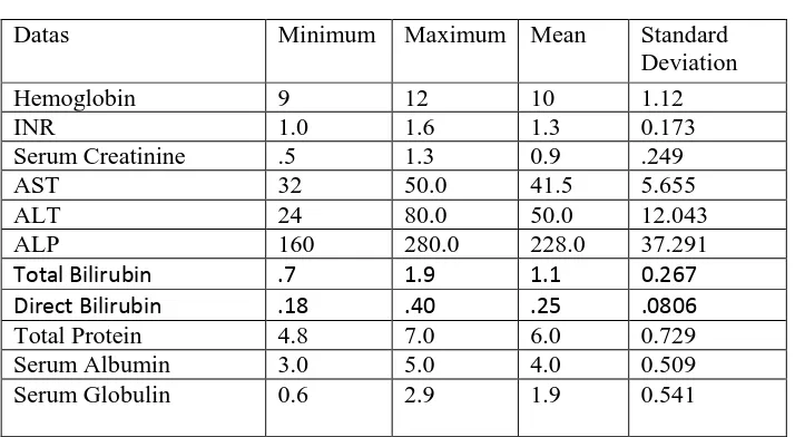
MATERIALS AND METHODS:
STUDY PLACE:
1) Department of Microbiology, Government Stanley Medical College,
Chennai.
2) Department of Surgical Gastroenterology, Government Stanley Medical
College, Chennai.
STUDY DESIGN:
Prospective study.
SAMPLE SIZE:
15 in numbers.
STUDY PERIOD:
October 2016 – July 2017.
ETHICAL CONSIDERATION:
Ethical and research clearance was obtained from the Ethical committee,
Stanley medical college. Permission to conduct the study was sought from the
respective hospital department authorities. An informed consent was obtained
STATISTICAL ANALYSIS:
The collected data were analysed with IBM.SPSS statistics software 23.0
Version. To describe about the data, descriptive statistics, frequency analysis,
percentage analysis were used for categorical variables and the mean &
Standard deviation (S.D) were used for continuous variables.
INCLUSION CRITERIA:
All patients undergoing liver transplant surgery at Institute of Surgical
Gastroenterology in Govt. Stanley Medical College, during the study period.
SAMPLE COLLECTION AND LABORATORY TESTING:
DONOR:
Complete blood investigations, Throat swab c/s, Nasal swab c/s, Blood c/s
& Urine c/s were done to rule out infections.
RECIPIENT:
Throat swab c/s, Nasal swab c/s, Blood c/s & Urine c/s before surgery to
rule out infections. Symptomatic investigations are done daily and biochemical
tests include, Complete blood investigations, Liver function test, Renal function
test, Coagulation profile, Serum pro-calcitonin, Tacrolimus or Cyclosporine
SAMPLE COLLECTION:5
THROAT SWAB:
It was made sure that the patient was not treated with antibiotics, 8 hours
before swabbing. In a good light from over the shoulder, the patient is instructed
to tilt the head back and breathe deeply. The tongue is depressed using a tongue
depressor to visualize the tonsillar fossae and posterior pharynx. The tonsillar
area and the posterior pharyngeal wall are rubbed using a sterile cotton wool
swab. Two swabs were taken, one for gram’s stain and other for culture.
NASAL SWAB:
Nasal swabs are obtained under direct vision using over-the-shoulder
illumination. With the thumb of one hand gently lift the tip of nose. Moisten the
tip of sterile cotton swab with sterile water or saline and gently insert into one
of the nares. Guide the swab backward and upward along the nasal septum until
a distinct feel of resistance indicates that a posterior pharynx has been reached.
URINE CULTURE:
After cleaning the external genitalia with soap and water, then rinse with
water and a clean voided mid-stream urine is collected in a sterile wide mouthed
container. In indwelling catheter (Foley’s catheter), disinfect the catheter
collection port and aspirate 5 – 10 ml of urine with sterile needle and syringe
BLOOD CULTURE:
The blood should be collected before antimicrobial treatment has started.To
increase the chances of isolating the pathogen, it is usually recommended that at
least two specimens are collected at different times and cultured. Under strict
aseptic precautions, about 5 – 10 ml of blood is collected using a sterile syringe
and needle in a sterile screw capped liquid culture bottle containing 50ml 0f
brain heart infusion broth. It is then incubated at 37ᵒ C for 1 week. When
growth is suspected, sub-culturing is done on blood agar and macconkey agar
and the organism was isolated.
CENTRALVENOUS CATHETER TIP CULTURE:
Disinfect the skin before removal and the catheter tip is collected in a
sterile screw cap container. It is cultured qualitatively by rolling the tip back and
forth across the blood agar and macconkey agar with sterile forceps four times.
BODY FLUIDS CULTURE: (Abdominal, peritoneal, bile, pleural and
drainage fluid)
Disinfect the skin before aspiration and aspirate about 2 – 3ml using a
sterile needle and syringe into a sterile test tube.
PUS CULTURE:
Pus from an abscess is best collected at the time, it is incised and drained,
from a drainage tube up to 5 ml of pus and transfer to a leak-proof sterile
container.
WOUND SWAB:
Using a sterile cotton-wool swab, sample is collected from the infected
site. Immerse the swab in a container of Amies transport medium. Usually, 2
swabs are collected, one for culture and the other for gram staining.
SPUTUM CULTURE:
A sterile, dry, wide-necked, leak-proof container was given to a patient
and requested to cough deeply to produce a sputum specimen. Sputum is best
collected in the morning soon after the patient wakes and before any
mouth-wash is used. When pulmonary tuberculosis is suspected, up to three specimens
may need to be examined to detect acid fast bacilli.
BRONCHO ALVEOLAR LAVAGE:
Bronchial and alveolar washings (broncho-alveolar lavage), are collected
using a bronchoscope. The specimen must contain alveolar exudate if cysts are