Advances in crustacean cell culture
176
0
0
Full text
(2) Advances in Crustacean Cell Culture. Thesis submitted by Kerry CLAYDON BA-BSc (Hons) Qld August, 2009. For the degree of Doctor of Philosophy in the School of Veterinary and Biomedical Sciences James Cook University.
(3) STATEMENT OF ACCESS DECLARATION I, the undersigned author of this work, understand that James Cook University will make this thesis available for use within the University Library and for use elsewhere via the Australian Digital Theses network. I understand that, as an unpublished work, a thesis has significant protection under the Copyright Act. I do not wish to place any further restriction on access to this work.. Kerry Claydon August 2009. ii.
(4) STATEMENT OF SOURCES DECLARATION I declare that this thesis is my own work and has not been submitted in any form for another degree or diploma at any university or other institution of tertiary education. Information derived from the published or unpublished work of others has been acknowledged in the text, and a list of references is included.. Kerry Claydon August 2009. STATEMENT OF SOURCES – ELECTRONIC COPY DECLARATION I, the undersigned the author of this work, declare that the electronic copy of this thesis provided to the James Cook University Library is an accurate copy, within limits of available technology, of the submitted printed thesis.. Kerry Claydon August 2009. iii.
(5) STATEMENT ON THE CONTRIBUTION OF OTHERS DECLARATION In undertaking the research for this PhD thesis, I received financial support through an APA Scholarship. In addition, a student stipend of $2,000 was supplied by the Graduate Research School of James Cook University for associated research expenses. The Veterinary and Biomedical Sciences Research Committee of James Cook University also contributed $1,000 to support presentation of this research at an international conference.. Editorial and critical feedback has been provided by my supervisor, Assoc. Prof. Leigh Owens.. All research procedures reported in this thesis received the approval of the relevant Ethics/Safety committees (Ethics permit A908).. Kerry Claydon August 2009. iv.
(6) ACKNOWLEDGEMENTS The biggest acknowledgement of all goes to my wonderful supervisor, Dr. Leigh Owens, for this most enjoyable (and frustrating) project. Thank you so much for having the faith and belief in me as a scientist to tackle this challenging project. I see now that three years is not enough time to change “our world,” and I look forward to an exciting working relationship in the future, with continued efforts and more crazy ideas towards the eventual establishment of a crustacean cell line.. Thank you to my co-supervisor Dr. Graham Burgess for your calm support in my times of confusion. Thank you, also, to Laurie Reilly for the wonderful expertise and great assistance in all aspects of histology, and enlightening life and science philosophy. There are not many people I enjoy consuming cool yeast beverages with more than you. Thank you to Dr. Ketheesan for your constant enlightening interruptions – especially on those few occasions that involved liquid from your filing cabinet. Thank you also to Dr. Val Gerard for your expert proofreading of this somewhat small document.. Big thanks to the Discipline technicians for their patience and understanding for all my unusual requests and demands. A special thanks to Karen Juntunen for sharing my peaks of excitement in cell culture. I thank my office buddy, „The Munro,‟ for keeping me sane throughout most of my project. I enjoyed sharing your life and our procrastinating talks, and I will never forget our crazy conference experience in Penang and Taipei. Thanks to all the postgrad students and other staff members of the Vet and Biomed Science team, who assisted me throughout this project, and for those that shared friendship. Worth special mention are Andrew Greenhill, Jo Penny, Jenny Scott, Kylie Bannister, Kirsty van Hennek, Gemma Berry, and Ryutaro Ueda.. v.
(7) ABSTRACT In 2006, for the first time in history, more seafood was produced from aquaculture raised animals than wild caught fisheries. However, crustacean aquaculture continues to be plagued by disease, due to the lack of sensitive investigative methods that assist diagnostics and pathogen control. Prevention and control of diseases are an absolute priority for the durability of the crustacean aquaculture industry.. The development of a permanent in vitro model for crustacean species is imperative. The research presented herein attempted to utilise novel modern technologies to develop the first continuous cell line from crustacea. The first investigation involved assessing methodologies that explored the existence of crustacean components in hybridised cell lines, which were developed by combining crustacean cells with immortal fish cells (Chapter 4). The methodologies involved cellular assessment from genomic and protein approaches, along with exploration of viral susceptibility of the cells. Using PCR, no crustacean 18S rRNA genes or haemocyanin genes were found in any of the seven hybrid cell lines. No crustacean proteins were detected, nor did any viral amplification occur when hybrid cells were inoculated with two crustacean viruses, indicating that these hybrid cell populations were not suitable for crustacean virological studies (Chapter 5).. The second investigation involved optimisation of in vitro methods using the Australian freshwater crayfish, Cherax quadricarinatus (Chapters 6 and 7). The approach included scientifically optimising the culture medium by comparing cell proliferation with three different cell culture media and 25 different media supplements. Leibovitz-15 medium, along with supplemental iron, copper, foetal bovine serum, and non-essential amino acids, was found to significantly increase cell proliferation (F = 6.231, df = 1,19, p< 0.05) and also augment the longevity of cells in vitro.. While spontaneous transformation of somatic cells can occur, transgenesis often expedites immortality. Therefore, the third investigation explored the transfection of. vi.
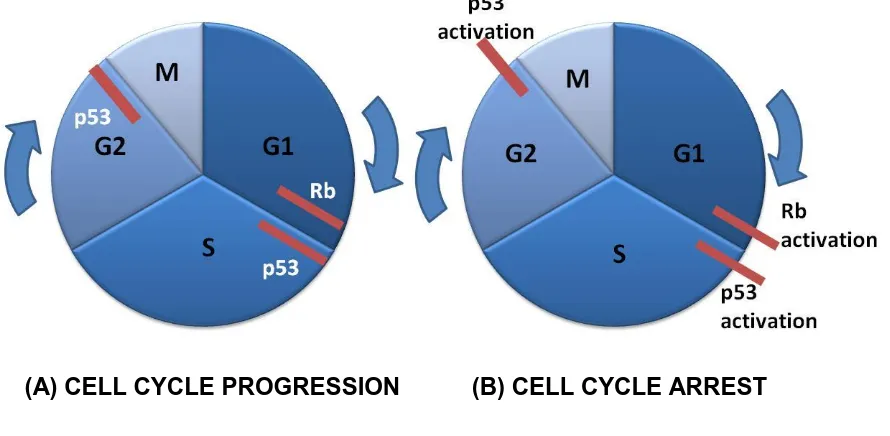
(8) C. quadricarinatus primary cells (Chapter 8). A lipofection reagent was used to introduce an observable green fluorescent protein vector into the cytoplasm of the cells. Transfection of the cells was then attempted using human oncogenes.. Papillomaviruses are non-enveloped, double-stranded DNA tumour viruses that play a critical role in the formation of human anogenital cancer. Early studies have demonstrated that the human papillomavirus-expressed E6 and E7 proteins function concomitantly to disrupt the p53 and Rb tumour suppressor genes, regulators of the cell-cycle checkpoints at the first gap (G1) phase of the cell cycle. To help C. quadricarinatus cells pass through the G1 phase and enter the DNA synthesis stage of the cell cycle, HPV E6 and E7 genes were transfected into the C. quadricarinatus cells. Successful transfection was demonstrated by the presence of oncogene mRNA by RT-PCR. At day 150, transfected cells remained viable, although cell proliferation was stagnant. It may be that, although transfection of the oncogenes was successful, no proliferation of the C. quadricarinatus cells was evident, due to a lack of telomere maintenance.. Overall, attempts to create a crustacean cell line remain elusive. Although the end product of a permanent cell line has not been forthcoming, this research made successful advancements in crustacean cell culture methodologies and explored new techniques and technologies that may assist eventual immortalisation.. vii.
(9) TABLE OF CONTENTS Page. Statement of Access. ii. Statement of Sources. iii. Statement on the Contribution of Others. iv. Acknowledgments. v. Abstract. vi. List of Tables. xiii. List of Figures. xiv. List of Abbreviations. xviii. Chapter 1. Introduction. 1. Chapter 2. Review of Literature. 5. 2.1. INTRODUCTION. 5. 2.2. CELL CULTURE MEDIA. 8. 2.3. CULTURE MEDIA COMPOSITION. 11. 2.3.1 Foetal bovine serum. 12. 2.3.2 Proteins. 13. 2.3.3 Lipids. 14. 2.3.4 Carbohydrates. 15. 2.3.5 Vitamins. 16. 2.3.6 Amino acids. 17. 2.3.6.1 Proline. 18. 2.3.6.2 Glutamine. 18. 2.3.7 Betaine. 19. 2.3.8 Cytokines, growth factors, and hormones. 20. 2.4. CRUSTACEA USED IN CELL CULTURE TRIALS. 23. 2.5. TISSUES USED IN CRUSTACEAN CELL CULTURE. 24. 2.6. CHARACTERISATION OF CULTURED CELLS. 27. 2.6.1 Cell identity. 27. viii.
(10) Page. 2.6.2 Cell viability. 29. 2.6.3 Cell proliferation. 29. 2.7. VIRAL INFECTIVITY STUDIES. 31. 2.8. DEVELOPMENT OF IMMORTAL CELL CULTURES. 34. 2.8.1 Transformation. 34. 2.8.2 Transfection and transgenesis. 36. 2.8.3 Cell hybridisation. 37. 2.8.4 Suppression of apoptosis. 38. 2.8.5 Cloning. 41. CRITICAL ANALYSIS OF CRUSTACEAN CELL LINE. 41. 2.9. DEVELOPMENT 2.9.1 Cell physiology and culture media. 42. 2.9.2 Vertebrate vs. invertebrate cell lines. 42. 2.9.3 Cell sensitivity. 43. 2.9.4 Cellular senescence and apoptosis. 44. CONCLUSION. 46. General Materials and Methods. 48. 3.1. DNA EXTRACTION. 48. 3.2. DETERMINATION OF DNA CONCENTRATION. 48. 3.3. PCR PRODUCT ANALYSIS AND GEL PURIFICATION. 48. 3.4. DNA AMPLIFICATION AND SEQUENCING. 49. 3.5. PRIMARY CELL CULTURE PROCEDURE AND MEDIA. 49. 3.6. OBSERVATION OF CULTURED CELLS. 50. 3.7. CELL VIABILITY COUNTS. 50. Assessment of Hybrid Crustacean Cell Populations. 51. 4.1. INTRODUCTION. 51. 4.2. MATERIALS AND METHODS. 52. 4.2.1 Cell culture. 52. 4.2.2 18S rRNA PCR. 54. 4.2.3 Haemocyanin PCR. 54. 2.10. Chapter 3. Chapter 4. ix.
(11) Page. 4.2.4 Immunohistochemistry using monoclonal. 55. and polyclonal antibodies. 4.3. 4.2.5 ELISA. 56. RESULTS. 57. 4.3.1 Cell culture. 57. 4.3.2 18S rRNA PCR. 58. 4.3.3 Haemocyanin PCR. 58. 4.3.4 Immunohistochemistry using monoclonal. 60. and polyclonal antibodies 4.3.5 ELISA. 61. DISCUSSION. 62. 4.4.1 Genomic approach. 62. 4.4.2 Protein approach. 63. Viral Reactivity in Hybrid Crustacean Cell Populations. 65. 5.1. INTRODUCTION. 65. 5.2. MATERIALS AND METHODS. 66. 5.2.1 Cell culture. 66. 5.2.2 Crustacean viruses. 66. 5.2.3 Viral purification. 67. 5.2.4 Verification of virus. 67. 5.2.5 Viral inoculation. 68. 5.2.6 Real-time PCR. 68. RESULTS. 69. 5.3.1 Cell culture. 69. 5.3.2 Verification of virus. 70. 5.3.3 Viral quantification. 70. DISCUSSION. 74. Optimisation of Primary Cell Culture Method for. 77. 4.4. Chapter 5. 5.3. 5.4. Chapter 6. Cherax quadricarinatus 6.1. INTRODUCTION. 77 x.
(12) Page. 6.2. MATERIALS AND METHODS. 78. 6.2.1 Putative virus-free C. quadricarinatus. 78. 6.2.2 Tissue comparison. 79. 6.2.3 Cell culture procedure. 79. 6.2.4 Cell culture media. 80. 6.2.5 Cell culture equipment. 81. RESULTS. 82. 6.3.1 Tissue comparison. 82. 6.3.2 Cell culture procedure. 83. 6.3.3 Cell culture media. 84. 6.3.4 Cell culture equipment. 86. DISCUSSION. 86. 6.4.1 Tissue source. 86. 6.4.2 Culture procedure and media. 87. 6.4.3 Culture equipment. 89. 6.4.4 Conclusions. 90. Culture Media Optimisation. 91. 7.1. INTRODUCTION. 91. 7.2. MATERIALS AND METHODS. 92. 7.2.1 Elemental composition of haemolymph. 92. 6.3. 6.4. Chapter 7. 7.2.2 Effects of media supplementation on cell proliferation 93 7.3. RESULTS. 95. 7.3.1 Elemental composition of haemolymph. 95. 7.3.2 Effects of media supplementation on cell proliferation 96 7.4. DISCUSSION. 100. 7.4.1 Elemental composition of haemolymph. 100. 7.4.2 Effects of media supplementation on cell proliferation 102 7.4.3 Conclusions. 105. xi.
(13) Page. Chapter 8. Transfection of Cherax quadricarinatus Primary. 107. Cell Cultures 8.1. INTRODUCTION. 107. 8.2. MATERIALS AND METHODS. 109. 8.2.1 Primary cell culture. 109. 8.2.2 Transfection vectors. 109. 8.3. 8.4. Chapter 9. 8.2.2.1. Green fluorescence protein vector. 109. 8.2.2.2. Human papilloma virus-16 vectors. 110. 8.2.3 Cell transfection. 112. 8.2.4 RT-PCR for mRNA. 113. RESULTS. 114. 8.3.1 pEGFP-N1 transfection. 114. 8.3.2 HPV-16 oncogenes. 114. 8.3.3 RT-PCR for mRNA. 115. 8.3.4 Cell culture morphology. 116. DISCUSSION. 118. General Discussion. 121. References. 126. Appendix 1 Animal Ethics Approval. 142. Appendix 2 Buffers and Solution Recipes. 143. Appendix 3 Key Crustacean Cell Culture Studies. 146. Appendix 4 Statistical Results for Optimised Culure Media. 154. Appendix 5 Presentations and Publications. 156. xii.
(14) LIST OF TABLES Page Table 2.1.. The non-essential amino acids that crustaceans are. 18. able to synthesise and essential amino acids that must be supplied in the diet or medium. Table 2.2.. Crustacean tissues used in cell culture attempts.. 26. Table 4.1.. Previously developed hybrid cell lines and media. 53. used for culture. Table 5.1.. Results of real time PCR using hybrid cell lines. 71. inoculated with HPLV. Table 5.2.. Results of real time PCR using hybrid cell lines. 72. inoculated with IHHNV. Table 6.1.. Characteristics of C. quadricarinatus cultured cells. 82. from different tissues. Table 6.2.. Osmolality of culture media, crustacean. 85. haemolymph, and L-15 medium supplemented with sodium chloride. Table 7.1.. Mean concentrations (n = 10) and standard deviations. 95. (SD) for iron, calcium, copper, and phosphate in C. quadricarinatus and P. monodon haemolymph, compared to concentrations in common culture media. Table 7.2.. Effect of culture medium supplements on proliferation of C. quadricarinatus cells. Shaded concentrations indicate peak absorbance readings, i.e. highest proliferation activity.. xiii. 96.
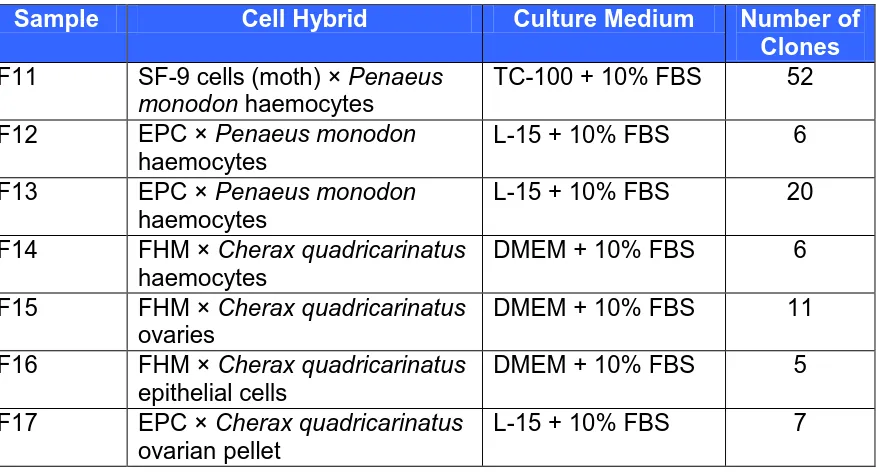
(15) LIST OF FIGURES Page. Figure 2.1. The variety of culture media used for crustacean cell. 9. culture studies. L-15 medium is the most common culture medium and is used at single and double strength.. Figure 2.2. Crustacean species used in cell culture attempts.. 24. Figure 2.3. Cytopathic effects of yellow head virus in cell culture.. 33. Figure 2.4. Cell cycle progression and inhibition.. 39. Figure 2.5. Schematic representation of the cell cycle.. 44. Figure 2.6. Progression of a typical cell culture.. 45. Figure 4.1. Schematic representation of cell hybridisation.. 52. Figure 4.2. (A) Cell growth of hybrid F12-clone C5 after 24 h.. 57. (B) Monolayer formed from F13-clone G10 after 72 h.. Figure 4.3. PCR results for the arthropod 18S gene in cell hybrids. 58. F11-F17, control cell lines, and haemocytes from C. quadricarinatus and P. monodon.. Figure 4.4. PCR for the presence of the crustacean haemocyanin. 59. gene in hybridised cells. No amplification was observed.. Figure 4.5. PCR for the presence of the crustacean haemocyanin gene in eight crustacean species.. xiv. 59.
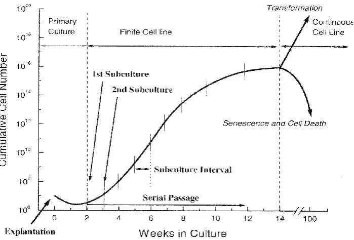
(16) Page. Figure 4.6. Immunostaining with the previously developed. 60. monoclonal antibody 1.9B2-5, raised against P. monodon haemocytes.. Figure 4.7. Immunostaining with a polyclonal antibody from. 61. P. monodon.. Figure 4.8. ELISA mean absorbance readings for EPC cells and. 62. EPC hybrid cell lines.. Figure 5.1. Hybrid cell line F14 inoculated with HPLV at day 3. 69. and day 14.. Figure 5.2. Results of viral purification PCR, showing bands at the. 70. expected amplicon size of 140bp for HPLV and 392bp for IHHNV.. Figure 5.3. Viral load in two hybrid cell lines over the 14 d. 73. experimental period.. Figure 6.1. Putative virus-free C. quadricarinatus were contained in. 79. six interconnecting tanks with recirculating water and biological filtration.. Figure 6.2. Photomicrographs of cultured cells from different tissues.. 83. Figure 6.3. Cultures of enzymatically disassociated cells exhibited. 85. more single cells than mechanically disassociated cells.. Figure 6.4. Comparison of cell proliferation in three culture media, based on the XTT assay. xv. 86.
(17) Page. Figure 6.5. Primary culture of C. quadricarinatus cells initiated. 86. with the Cytodex-1 microcarrier.. Figure 7.1. Plate layout used for WST-1 assay. Five. 93. concentrations of a culture media supplement were assessed simultaneously at four different time points.. Figure 7.2. Cell proliferation in optimised L-15 culture medium. 99. compared to standard L-15 medium with 10% FBS. Figure 7.3. Cultured C. quadricarinatus cells at 25 d in standard. 99. L-15 medium with 10% FBS and optimised L-15 medium. Figure 7.4. Cell proliferation in a primary cell culture for. 100. C. quadricarinatus and in an established cell line culture from bluegill fry (BF-2).. Figure 7.5. Controlled delivery of L-glutamine and its derivative,. 104. glutaMAX™, from medium to cells in culture. Figure 8.1. pEGFP-N1 vector map with restriction enzyme sites.. 110. Figure 8.2. Structures of the recombinant HPV-16 DNA vectors.. 111. Figure 8.3. The gene sequence of HPV-16 E7.. 112. Figure 8.4. Primary cells from hematopoietic tissue of. 114. C. quadricarinatus visualised under light microscopy and the same view exposed to UV light displaying fluorescence.. xvi.
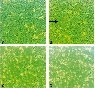
(18) Page. Figure 8.5. PCR of different clones of HPV-16 E6+E7 vector and. 115. HPV-16 E7 vector, using primers developed from the E7 oncogene.. Figure 8.6. HPV-16 E7 gene detection via PCR, following cDNA. 116. synthesis of total RNA extracted from transfected cells on days 1, 2, 10 and 14 post-exposure.. Figure 8.7. (A) Cultures of haematopoietic cells from C. quadricarinatus before transfection with HPV-16 vectors. (B) Cell aggregates began to form 4 d post-transfection with the HPV-16 E7 oncogene. (C) Viable cells remained in small aggregates (arrows) 150 d post-transfection with the HPV-16 E7 oncogene.. xvii. 117.
(19) LIST OF ABBREVIATIONS 2-ME. 2-mercaptoethanol. A260. Absorbance at 260nm. A280. Absorbance at 280nm. ABARE. Australian Bureau of Agricultural and Resource Economics. ADS. Appendage deformity syndrome. ANOVA. Analysis of variance. BF-2. Bluegill fry. bFGF. Basic fibroblast growth factor. BLAST. Basic local alignment search tool. BrdU. 5-bromo-2‟-deoxyuridine. CMRL. Connaught Medical Research Laboratories. CPE. Cytopathic effect. DEAE. N,N-diethylaminoethyl. DMEM. Dubecco‟s minimum essential medium. DNA. Deoxyribonucleic acid. dNTP. Deoxynucleotide triphosphate. E6. Human papillomavirus early gene 6. E7. Human papillomavirus early gene 7. EGF. Epidermal growth factor. ELISA. Enzyme-linked immunosorbant assay. EPC. Epithelioma papulosum cyprinid. FAO. Food and Agriculture Organisation. FBS. Foetal bovine serum. FCS. Foetal calf serum. FHM. Fathead minnow. GAMA. Gamma -amino butyric acid. GAV. Gill associated virus. GFP. Green fluorescence protein. H&E. Haematoxylin and eosin. HEPES. 4-(2-hydroxyethyl)-1-piperazine ethanesulfonic acid xviii.
(20) HGPRT. Hypoxanthine guanine phosphoribosyl transferase. HPLV. Hepatopancreatic parvo-like virus. HPV. Human papillomavirus. IHHNV. Infectious hypodermal and haematopoietic necrosis virus. IGF. Insulin growth factor. L-15. Leibovitz 15 culture medium. LO. Lymphoid organ. LTR. Long terminal repeat. M199. Medium 199. MAb. Monoclonal antibody. MEM. Minimum essential medium. mRNA. Messenger ribonucleic acid. NCTC. National Centre for Tissue Culture. p53. Protein 53. PAb. Polyclonal antibody. PCR. Polymerase chain reaction. PDGF. Platelet-derived growth factor. PEG. Polyethylene glycol. PRBV. Penaeid rod-shaped baculovirus. Rb. Retinoblastoma. RNA. Ribonucleic acid. RPMI. Roswell Park Memorial Institute. SF-9. Spodoptera frugiperda pupal ovarian tissue. SV40. Simian virus 40. TC-100. Tissue culture medium 100. TGF-β. Transforming growth factor-beta. TNF-α. Tumor necrosis factor-alpha. TRAP. Telomeric repeat amplification protocol. WSSV. Whitespot syndrome virus. WST-1. Water soluble tetrazolium salt. YHV. Yellowhead virus. xix.
(21) CHAPTER 1 INTRODUCTION. The global demand for aquatic food products is increasing. Production from capture fisheries has levelled off and most of the main fishing areas have reached their maximum potential (FAO, 2006). Therefore, seafood product from capture fisheries will not be able to meet the growing global demand for aquatic food. Aquaculture continues to make a significant contribution to this increasing demand. Currently, one billion people are dependent on aquatic animals as the principal source of animal protein (FAO, 2006). However, disease outbreaks have been increasingly recognised as a significant constraint on aquaculture production and trade, affecting the economic development of the industry in many countries.. Crustaceans represent one of the most economically important global aquaculture sectors, producing in excess of $10 billion USD annually (Johnson et al., 2008). Like other aquatic animals, crustaceans are susceptible to a wide variety of pathogens, including viruses, bacteria, fungi, and protozoa. During the last decade, disease was considered to be the most limiting factor for expansion of crustacean culture (Lin, 1995). In the mid 1990s, it was estimated that around 40% of worldwide prawn production, representing a value of over $3 billion USD, was lost due to diseases (Lundin, 1996). The main contributors to these losses were viruses (Lotz, 1997).. The stresses and close proximity of individuals under intensive aquaculture favour the spread of disease. However, modern practices for finfish aquaculture include highly effective routine vaccination against multiple pathogens, which has dramatically reduced the impact of disease (Johnson et al., 2008). These practices are effective, because teleost fish have adaptive immune responses and immune memory involving B cells and T cells, antibody and phagocytic cells. Understanding these immune mechanisms and how the pathogens interact at the cellular level has allowed aquatic animal health scientists to develop successful vaccines (Sommerset et al., 2005). On the other hand, invertebrates, such as crustaceans, rely solely on an innate immune 1.
(22) system characterised by generalised immune responses. The responses involve an array of pattern recognition receptors that interact with serine proteases to initiate encapsulation, phagocytosis, and an antimicrobial cascade based on the phenoloxidase system (Söderhall and Cerenius, 1998). No recognition system for viral infections has been documented. These animals are therefore highly susceptible to viral infections, which often result in mortality.. However, there is some recent evidence of specific immune memory in crustaceans (Johnson et al., 2008). To assist this understanding, and ultimately control viral disease outbreaks in crustacea, the study of these diseases must go beyond their basic description and identification, and the tools to investigate outbreaks must be developed. Given the breakthroughs obtained in human and veterinary health, the achievement of such measures greatly depends on the development of crustacean cell cultures that permit in vitro cultivation of the intracellular pathogenic agents. Such cell cultures could help us to determine and understand the host-pathogen relationship at the cellular and molecular level, as well as assisting us to understand immune functions.. Cell culture can be defined as the maintenance or growth of cells in a nutrient fluid following removal from an organism‟s body. The concept of cell culture is not new, but began over a hundred and twenty years ago, when in 1885 Wilhelm Roux discovered that chicken embryonic cells could survive outside the animal body (Freshney, 2005). Nearly sixty years later, the study of malignancy in vitro (Earle, 1943) led to the development of the first continuous cell line. Today, human cell cultures are powerful and important tools that are used to address many fundamental questions in biology and medicine. A large variety of cell lines exist for many vertebrate species. In contrast, in vitro models for invertebrate species, which make up 95% of the animal kingdom (Ruppert and Barnes, 1994), are far less advanced. Since the development of the first invertebrate cell line in 1961 (Grace, 1962), continuous cell lines have been established from more than 100 species of insects (Lynn, 1999). However, no permanent cell culture system exists for any aquatic invertebrate.. 2.
(23) Development of a cell line provides many advantages. Unlike virology of aquatic vertebrates, crustacean virology is still in its infancy, and the major goal of cell line development has been to improve knowledge of disease-causing agents. In vivo models have limitations for studying disease, and isolation of pathogens in vivo is difficult. The production of standardised in vitro systems provides a means to study the effects and mechanisms of pathogen invasion and provides a medium for isolation of infectious pathogens. In vitro systems also allow the study of cellular biochemistry and can facilitate studies of ecotoxicology (Freshney, 2005). Once cell lines have been established, they reduce the need for animal experimentation, which precludes legal, moral and ethical problems, as cells can be continually cultivated without the need to sacrifice whole organisms. Cell cultures also allow the culture of a single virus, instead of the mixed populations of viruses that are worked with in current bioassay systems (Owens, 1997).. Most of the crustacean species utilised in aquaculture come from the order Decapoda, of which Penaeidea (prawns), Astacidea (lobsters, crayfish), and Brachyura (true crabs) are representative orders. Penaeid species compose the largest group of crustaceans in aquaculture, with global production of approximately 3.16 million mt (FAO, 2006). Therefore, the penaeid species have been the primary focus of cell culture efforts during the past two decades. Chen et al., (1986) was first to report a short term tissue culture of Penaeus monodon, with a further nine other penaeid spp. undergoing recent cell culturing efforts. Primary cultures obtained from various penaeid organs are reported with increasing frequency (Wang et al., 2000; Mulford et al., 2001; LyonsAlcantara et al., 2002; Assavalapsakul et al., 2003; Chen-Lei et al., 2003; Gao et al., 2003; Maeda et al., 2003; Lang et al., 2004; Maeda et al., 2004; Jiang et al., 2005). These studies represent the first steps towards the establishment of cell lines and they provide useful information concerning the most suitable cell culture conditions involved in the survival and proliferative capacity of the various tissues (Toullec, 1999). Examination of the literature on establishment of crustacean and other invertebrate cell lines, as well as on recent advances in technology, identified a number of novel approaches for further investigation, including (1) cell hybridisation, (2) comparison of culture media supplementation 3.
(24) using cell proliferation assays, and (3) cell transformation by transfecting cells with oncogenes. The research presented in this thesis investigated these approaches with the goal of eventually establishing a permanent crustacean cell line.. 4.
(25) CHAPTER 2 REVIEW OF LITERATURE 2.1. INTRODUCTION. Cell culture, the maintenance or growth of cells in a nutrient fluid following removal from an organism‟s body, started over 120 years ago. Harrison (1907) maintained embryonic frog neural crest fragments in a culture fluid for several weeks and is considered the “father” of cell culture (Mothersill and Austin, 2000). Harrison‟s techniques initiated a wave of interest in the cultivation of animal tissue in vitro. Burrows (1910) further advanced the proliferation of chicken embryonic cells in culture and was the first to describe the process of mitosis in detail. Another significant milestone was the development of the first viral vaccine (Vaccinia) from cell culture (Carrel and Rivers, 1927). Following on from this, the role of malignancy was studied in vitro by Earle et al., (1943), which led to the development of the first continuous cell line. Earle‟s demonstration that human tumours could give rise to continuous cell lines increased interest in the development of human cell culture. Today, human cell cultures are a powerful and important tool that has been utilised to address many fundamental questions in biology and medicine. Recent advances have made it possible to use cultured human cells for therapeutic use. Cultured cells have been used to reconstruct skin, bone, and other tissues, including the endothelium (Freshney, 2005).. Today, a variety of cell lines exist for many vertebrate species. In contrast, in vitro models for invertebrate species, which make up 95% of the animal kingdom (Ruppert and Barnes, 1994), are far less advanced. Significant advances in invertebrate cell culture were made in the 1950s and 1960s, largely due to the availability of antibiotics and increasing knowledge of biochemistry (Mothersill and Austin, 2000). In 1961, Wyatt analysed insect haemolymph composition and designed a cell culture medium that showed promising results in enabling the survival of insect cells. Grace (1962) made some additions to Wyatt‟s culture medium and, through a technique described as “organised 5.
(26) neglect,” developed the first continuous invertebrate cell line from the gum moth, Antheraea eucalypti. Since then, over 400 continuous cell lines have been established from more than 100 species of insects (Lynn, 1999). Following the successful development of insect cell lines, some attempts were made to develop cell culture methodologies for aquatic invertebrates, including sponges (Pomponi et al., 1997), molluscs (Wen et al., 1993; Domart-Coulon et al., 1994), crustaceans (Chen et al., 1986), and ascidians (Raftos et al., 1990). Most of these species received attention, because their cells or tissues produce metabolites of possible pharmacological significance (Pomponi et al., 1997), or because the host serves as a vector for pathogens (Gong et al., 1997).. Fischer-Piette (1931) made the first attempt to establish tissue cultures for crustaceans, using simple saline solutions. Other studies were successful in establishing short-term crustacean cell cultures, but none were successful in establishing long-term cultures, and many were unable to subculture cells. As aquaculture began to develop and grow in the early 1980‟s, industry expansion was plagued by disease. The lack of crustacean cell lines for isolation and growth of crustacean viruses was a major setback for diagnosis and treatment. Most of the crustacean species in aquaculture come from the order Decapoda, of which Penaeidea (prawns), Astacidea (lobsters, crayfish), and Brachyura (true crabs) are representative orders. Penaeid species compose the largest group of crustaceans in aquaculture, with global production of approximately 3.16 million mt (FAO, 2006). Therefore, the penaeid species have been the primary focus of cell culture efforts during the past two decades. Since short-term tissue culture of Penaeus monodon was reported by Chen et al., (1986), culturing efforts have been made on another eleven penaeid species (Hu, 1990; Rosenthal and Diamant, 1990; Ellender et al., 1992; Luedeman and Lightner, 1992; Purushothaman et al., 1998; Chen and Wang, 1999; Owens and Smith, 1999; Gao et al., 2003; Maeda et al., 2004). Primary cultures obtained from various penaeid organs have been reported with increasing frequency (Wang et al., 2000; Mulford et al., 6.
(27) 2001; Uma et al., 2002; Assavalapsakul et al., 2003; Chen-Lei et al., 2003; Gao et al., 2003; Maeda et al., 2003; Lang et al., 2004; Maeda et al., 2004; Jiang et al., 2005). These studies represent the first steps towards the establishment of cell lines, and they provide useful information concerning suitable cell culture conditions for the survival and proliferation of various tissues (Toullec, 1999). To date, however, no valid and verified crustacean cell line has been established.. Development of a cell line provides many advantages. Unlike virology of aquatic vertebrates, crustacean virology is still in its infancy, and the major goal of cell line development has been to improve knowledge of disease-causing agents. In vivo models have limitations for studying disease, and isolation of pathogens in vivo is difficult. The production of standardised in vitro systems provides a means to study the effects and mechanisms of pathogen invasion and provides a medium for isolation of infectious pathogens. In vitro systems also allow the study of cellular biochemistry and can facilitate studies of ecotoxicology (Freshney, 2005). Once cell lines have been established, they reduce the need for animal experimentation, which precludes legal, moral and ethical problems, as cells can be continually cultivated without the need to sacrifice whole organisms. Cell cultures also allow the culture of a single virus, instead of the mixed populations of viruses that are worked with in current bioassay systems (Owens, 1997).. The purpose of this review is to summarise previous attempts at crustacean cell culture, and to compare this research with cell culturing efforts for other invertebrates and some vertebrates. Recent technological advances and novel approaches to cell culture are also discussed. Specific topics covered include crustacean cell culture media and their components, species and tissues used in cell culture studies, methods used to characterise cells in culture, viral infectivity, approaches to development of immortal crustacean cell lines, and problems related to crustacean cell physiology, cell sensitivity, and cellular senescence.. 7.
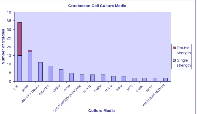
(28) 2.2. CELL CULTURE MEDIA. Most attempts at crustacean cell culture have focused on optimisation of the culture media. The culture medium is the most important element impacting on the survival of animal cells (Mothersill and Austin, 2000). The selection of culture medium is generally based on the requirements of the animal. With aquatic animals, this involves consideration of the external environment (marine or freshwater), the internal environment (specific requirements of the species), and whether carbon dioxide or any other buffering system will be used with the medium. There is virtually nothing known about the precise nutritional requirements of crustacean cells and, therefore, much of the medium selection process is guesswork (Mothersill and Austin, 2000).. Basic culture medium typically supplies essential nutrients for cellular growth, including salts, amino acids, and vitamins. Numerous studies have tested a variety of culture formulations (combinations of basal media and supplements) for their ability to support crustacean cell replication or maintenance. Since the development of more complex and defined media, many initial trials of crustacean cell culture utilised Grace‟s insect medium, due to its success in culturing other arthropod species (Luedeman and Lightner, 1992; Nadala et al., 1993; Frerichs, 1996; Toullec et al., 1996; Fraser and Hall, 1999). Twenty-five other media have also been tested with crustacean cells (Appendix 3); with Leibovitz‟s (L-15) and medium 199 (M199) the most commonly used (Figure 2.1).. 8.
(29) Crustacean Cell Culture Media 40. 30 25 20. Double strength. 15. Single strength. 10 5. M ED. IU. CT C. M. N. RL M C. M PS. M EM. M & M. EM D. M. 00 -1 TC. W N. KN O. IB IA N. N. R /U. AM PH. IS ED C. US TO M. EO N. PM I. EM EM. TR IA LS G R AC E' S. O FF. M. 19 9. 0 L1 5. 9. Number of Studies. 35. Culture Media. M & M = Maramorosch & Mitsuhashi media. Figure 2.1. The variety of culture media used for crustacean cell culture studies. L-15 medium is the most common culture medium, and is used at single and double strength. 9.
(30) The media M199 and L-15 have consistently supported successful establishment of primary cultures from various tissues. These media are very different in their composition and the proportion of their components (Lang et al., 2002a). Due to observed cellular growth in both media, it has been assumed that crustacean cells have a broad adaptability to media conditions (Lang et al., 2002a). Because these commercial culture media were primarily developed for vertebrate cell cultures, it is likely that they do not contain all the necessary de novo factors for invertebrate cell replication. Therefore the basal medium may not be as important as specific additives or supplements, especially for prolonged survival.. Although many researchers have examined the benefits of different media and/or modifications, few studies have attempted to identify specific physiological needs of crustacean cells (Mothersill and Austin, 2000). The method often employed by researchers to determine the physiological requirements of crustacean cells involves qualitative observations by light microscopy: either cells replicate or do not. This has been the basis for designing and modifying crustacean cell culture media. No previous studies have approached crustacean cell culture by analysing cell metabolism, i.e. by examining metabolic pathways and analysing the role of each nutrient. To date, only two studies focused on biochemical analysis of crustacean haemolymph, and both found that cultured cells performed better (established monolayers or delayed onset of cellular senescence) when the culture medium was optimised (Najafabadi et al., 1992; Shimizu et al., 2001). However, even with the optimised media, there was no evidence of sustained DNA or protein synthesis.. Regardless of the choice of basal medium, the culture medium must be adjusted to the physico-chemical requirements of the crustacean cells, depending on whether the species is freshwater or marine. The main factors that must be considered are osmolality, pH, and ionic strength. The osmotic pressure should be adjusted to 400-500 mOsm/kg (of water) for freshwater animals, and to 800-1000 mOsm/kg for marine species (Mothersill and Austin, 2000). In adjusting osmolality, ionic strength must also be taken into account, because the use of sodium chloride alone may result in an excessive 10.
(31) concentration of sodium (Mothersill and Austin, 2000). Some salts appear to be more important than others, particularly calcium, which has been described as the most important ion for the effectiveness of saline solutions in sustaining viability of crustacean cells (Wheatly, 1999).. The culture media should be relatively alkaline (pH 7.2-8.0), and non-CO2 dependent biological buffers, such as HEPES, appear to be preferable to bicarbonate buffer (Mothersill and Austin, 2000). However, in mammalian cell cultures, HEPES has induced vacuolisation of cultured cells in the absence or at low concentrations of serum (Bowers and Dahm, 1993).. It has recently been recognised that the medium-change regime may be more important than the selection of medium (Mothersill and Austin, 2000). Once the cells have been added to the medium, the culture develops a microenvironment that is optimal for growth and function of the cells. Too frequent medium changes can cause the loss of this micro-environment and destroy the developing cells. Conversely, medium changes are essential to ensure cells receive their specific nutritional requirements. Therefore, half medium changes may better sustain the microenvironment and, at the same time, replace exhausted nutrients (Mothersill and Austin, 2000).. Any medium chosen for cell culture should be refined to incorporate known requirements of the animal. Overall, cell culture media should be made up of a basal medium that includes essential amino acids and vitamins, have ionic strength and pH adjusted to the animal‟s physiology, and contain speciesspecific nutritional supplements.. 2.3. CULTURE MEDIA COMPOSITION. To stimulate cultured crustacean cells to carry on cell division, culture media have been supplemented with various nutrients, tissue extracts, and growth factors. Selection of these supplements has been based more on trial and error than on determination of essential cellular growth factors.. 11.
(32) The salt, amino acid, and lipid composition of haemolymph has been reported for two species of penaeids (Najafabadi et al., 1992; Shimizu et al., 2001), and the results demonstrate the variation in haemolymph composition within the genus Penaeus. Seasonal influences and varying water salinity had profound effects on haemolymph composition (Najafabadi et al., 1992). Other haemolymph metabolites varied from sample to sample, suggesting differences may have been caused by variations in diet and stress (Najafabadi et al., 1992). Therefore, it is not accurate to compare species from different locations with different environmental conditions and different feeding regimes, and attempt to formulate a generic medium to enable cell replication. Nevertheless, many culture media supplements, including proteins, lipids, carbohydrates, vitamins, amino acids and growth factors, have been tested by trial and error, and have assumed that a growing and, in some cases, dividing cell, has had its nutritional requirements satisfied.. 2.3.1 Foetal Bovine Serum Foetal bovine serum (FBS) is a common supplement to culture media. FBS is a complex supplement that acts as a source of minerals, lipids, and hormones, and contains growth and adhesion factors to promote cell attachment and proliferation (Freshney, 2005). The results of several studies indicated that FBS is essential for cell replication, although it is likely that does not provide all of the specific hormones and growth factors needed by crustacean cells (Mothersill and Austin, 2000; Mulford et al., 2001; Crane and Williams, 2002). Concentrations of FBS in culture media typically range between 5 and 20%, adjusted according to the tissue being cultured. The lipids provided by FBS have been found to be poor in quality for the requirements of crustacean cells (Mothersill and Austin, 2000). There is a low concentration of cholesterol and an imbalance between fatty acids and phospholipids. Extra supplementation of the culture medium with these lipids may be necessary for culturing crustacean cells (Fox et al., 1994; Mothersill and Austin, 2000). Mammalian serum may also lack hormones and growth factors needed by crustacean cells, so additional supplementation of these factors may be necessary to stimulate cell proliferation.. 12.
(33) There is a current trend to replace FBS with defined serum-free supplements, due to the inconsistent quality and increasing cost of FBS. Yeastolate, lactalbumin hydrolysate, peptones, and tryptose phosphate broth have been tested as replacements for bovine serum (Mothersill and Austin, 2000; Freshney, 2005). Yeastolate, an enzyme-hydrolysed yeast extract composed of a mixture of amino acids, peptides, water soluble vitamins, and carbohydrates, has been used as additive for crustacean cell culture (Maeda et al., 2003) and insect cell culture (Mendonca et al., 1999; Leite et al., 2003). It is often used in combination with lactalbumin hydrolysate, which reduces the cell requirements for serum by replacing required polypeptides. Yeastolate is also a source of peptide hormones and has been found to enhance cell replication and viability in crustacean cell cultures (Itami et al., 1999; Lang et al., 2002a; Lang et al., 2002b; Assavalapsakul et al., 2003; Maeda et al., 2003). It would be of interest to determine whether serum replacements developed for mammalian cell culture, containing defined nutrients and growth factors, can enhance or replace FBS in crustacean cultures.. Another alternative to supplementation with FBS is the use of isogenic or allogenic muscle extracts or plasma from haemolymph. Many studies of crustacean cell culture have combined these additives with sera and achieved varying results (Chen et al., 1986; Chen et al., 1988; Rosenthal and Diamant, 1990; Nadala et al., 1993; Lu et al., 1995b; Tapay et al., 1995; Tong and Miao, 1996; Chen and Wang, 1999; Fraser and Hall, 1999; Itami et al., 1999; Kasornchandra et al., 1999; Owens and Smith, 1999; Mulford et al., 2001).. 2.3.2 Proteins One of the main reasons for adding serum to culture media is to provide proteins necessary for cell survival and growth. Although proteins are a major component of serum, the functions of many proteins in vitro remain unclear (Freshney, 2005). It may be that relatively few proteins are required, other than as carriers for minerals, fatty acids, and hormones (Mothersill and Austin, 2000).. 13.
(34) The total protein concentration in crustacean haemolymph is highly variable depending on species, nutritional status, and moulting stage (Najafabadi et al., 1992; Mothersill and Austin, 2000). Najafabadi et al., (1992) found that double strength L-15, supplemented with lobster haemolymph and 10% serum, had a protein concentration one third of that measured in P. aztecus.. A number of different circulating proteins and proteases have been isolated and characterised in crustaceans. The release of some of these proteins from damaged or dying cells can lead to degradation of tissues and cells. Proteases, for example, play roles in homeostasis, such as regulation of endogenous proteases, haemolymph coagulation, and antimicrobial defence (MerriamWebster, 2003). Inhibition of proteases may be important to tissues rich in these enzymes, such as the hepatopancreas (Mothersill and Austin, 2000). The protease inhibitor phenyl methyl sulfonyl fluoride has been used to prevent autolysis of hepatopancreas cells by digestive enzymes released from damaged cells (Toullec et al., 1992). Other protease-inhibiting mixtures have been developed for cell culture to prevent proteolytic degradation by secreted proteins, but their effectiveness has not been tested with crustacean cells.. 2.3.3 Lipids Lipid requirements vary among different cell types and animal species. Lipid metabolism in crustacea, are controlled by the adipokinetic/hyperglycaemia hormones (Mothersill and Austin, 2000). Ingested lipids are enzymatically cleaved into diglycerides and monoglycerides in the gut lumen, absorbed, converted to phospholipids, and stored in the hepatopancreas (Lee and Puppione, 1978). Unlike vertebrates, which use fatty acids as the principle transport lipids, the principal circulating lipids in crustacea are phospholipids (Mothersill and Austin, 2000). Phospholipids synthesised by the hepatopancreas are released to the haemolymph, where they are available to other cells and tissues for structural and energetic processes. Chang and O‟Connor (1983) found that lipogenesis from carbohydrate catabolism is probably a deficient metabolic route in crustaceans. Therefore, lipids from vertebrate sera used as supplements in culture media may not be adequate to support crustacean cell replication. For example, levels of phospholipids in 14.
(35) prawn haemolymph (350-540 mg/L) are much higher than culture medium supplemented with 10% FBS (Najafabadi et al., 1992).. Crustaceans are incapable of de novo synthesis of sterols and require a dietary source of cholesterol for growth, development, and survival (Fox et al., 1994). The recommended level of cholesterol in crustacean diets varies from 0.4 to 1.4%, depending on species, age, and physiological condition (Teshima, 1997). Each species has an optimal level; with low cholesterol levels inhibiting growth and development, and high levels retarding growth (Teshima, 1997). Hernandez et al., (2004) found that Cherax quadricarinatus fed a 0.5% cholesterol diet had significantly higher weight gain than crayfish fed lower or higher levels of cholesterol. Kasornchandra et al., (1999) found supplementation with 0.01% cholesterol markedly enhanced cell replication of penaeid cells. Overall, it is important that animals are supported with diets containing necessary fatty acids and cholesterol prior to tissue extraction for the establishment of cell cultures, and that the cell cultures are supplemented with these factors to enable cell survival.. 2.3.4 Carbohydrates Like most nutrients, glucose levels in crustacean haemolymph vary widely depending on the species, diet, season, and moulting stage. Glucose supplements are important for insect cell cultures (Hink, 1976), and is therefore included in most culture media as an energy source. It is metabolised principally by glycolysis to form pyruvate, which may be converted to lactate or to acetoactetate and enter the citric acid cycle to form CO2 (Freshney, 2005). The accumulation of lactic acid in the medium suggests that the citric acid cycle many not function as in vivo. Carbon may be derived from glutamine, rather than glucose, and this may explain the high requirement of some cultured cells for glutamine (Freshney, 2005).. Many studies have found supplementation with glucose during the first 24 h of culture leads to rapid cell attachment and migration from explants (Hsu et al., 1995; Kasornchandra et al., 1999; Mulford et al., 2001; Crane and Williams, 2002; Assavalapsakul et al., 2003; Maeda et al., 2003). Without glucose, the 15.
(36) number of cells that attach to culture vessels is considerably reduced. Enhanced cell migration has also been observed by continual addition of one g/L glucose (Hsu et al., 1995; Mulford et al., 2001; Maeda et al., 2003). The need for supplemented glucose depends on the culture medium. L-15 is one of few culture media that contains galactose rather than glucose. Therefore, most studies that found the addition of glucose beneficial used L-15 as the basic culture medium (Hsu et al., 1995; Kasornchandra et al., 1999; Crane and Williams, 2002; Assavalapsakul et al., 2003; Maeda et al., 2003). Other carbohydrate supplements, such as sucrose and trehalose, have not been as successful as additives, resulting in a cell attachment rate of only 50%, compared to 80% when cultures are supplemented with glucose (Hsu et al., 1995).. 2.3.5 Vitamins Vitamin requirements of crustaceans have yet to be completely defined (Harlioglu and Barim, 2004). Results reported in the literature suggest that invertebrates have vitamin requirements similar to those of vertebrates, and both are unable to synthesise carotenes. Thiamine, riboflavin, nicotinamide, pyridoxine, pantothenic acid, biotin, and folic acid are regarded as essential vitamins for animals. Biotin and riboflavin, considered essential vitamins that aid in carboxylation reactions important for cell energy production, are lacking in L-15 medium (Luedeman and Lightner, 1992). Selenium is also lacking in most defined cell culture media; however, the effect of supplementation in P. monodon lymphoid organ culture was not commented on (Hsu, 1995). Keating and Dagbusan (1984) found that cell populations from water fleas could not be maintained without selenium supplementation at 0.1 ppb. In mammalian cell cultures, selenium deficiencies are managed by adding vitamin E, which lessens the absolute requirement for selenium, but does not wholly substitute for it (Freshney, 2005).. Few publications have reported on vitamin supplementation in vitro. However, vitamin deficiencies in dietary feeds have been reported. Kumar et al., (2004) found that carotenoid deficiency in the freshwater prawn Macrobrachium rosenbergii caused an increase in appendage deformity syndrome (ADS), 16.
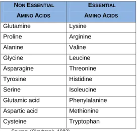
(37) resulting in 100% mortality after 18 days. However, supplementation with carotenoids allowed full recovery from ADS, and permitted regeneration of appendages. Carotenoids, which include beta-carotene, alpha-carotene and beta-cryptoxanthin (Merriam-Webster, 2003), come primarily from fruits and vegetables, and provide the sole source of vitamin A in the diet. Crustaceans used for cell culture may require diets containing carotenoids or vegetable matter to ensure that vitamin A requirements are met prior to initiation of cells into culture. Vitamin A may also be a required constituent of the culture medium.. The dietary requirement for vitamin E has been reported to be 20-100 mg/kg for fish and crustaceans (Harlioglu and Barim, 2004). Higher levels of vitamin E can cause hypervitaminosis, which is evident as retardation in growth. A major function of vitamin E is to prevent peroxidation of polyunsaturated fatty acids in the phospholipid and cholesterol of cellular and subcellular membranes (Harlioglu and Barim, 2004). Vitamin E added at 100 mg/kg increased pleopodal egg number and survival of juveniles of the crayfish, Astacus leptodactylus (Harlioglu and Barim, 2004). Addition of vitamin E, along with vitamins A and D, significantly improved growth and survival of P. vannamei (He et al., 1993). As most vertebrate-defined culture media probably do not provide all vitamins and minerals at optimal levels for cultured crustacean cells, experimentation with vitamin supplementation in vitro might be necessary to identify specific cellular requirements.. 2.3.6 Amino Acids The essential amino acids, i.e. those not synthesised within an organism, are required by cultured cells and should be included in any basic basal media. The individual requirements for amino acids vary from one cell to another, depending on cellular function (Freshney, 2005). Other, non-essential amino acids are often added to media, in case cells have lost their capacity to make them. The concentration of amino acids usually limits the maximum cell concentration attainable in cultures, and the balance may influence cell survival and growth rate (Mothersill and Austin, 2000). A list of the non-essential and essential amino acids of crustacean is listed in Table 2.1. 17.
(38) Table 2.1. The non-essential amino acids that crustaceans are able to synthesise and essential amino acids that must be supplied in the diet or culture medium.. NON ESSENTIAL. ESSENTIAL. AMINO ACIDS. AMINO ACIDS. Glutamine. Lysine. Proline. Arginine. Alanine. Valine. Glycine. Leucine. Asparagine. Threonine. Tyrosine. Histidine. Serine. Isoleucine. Glutamic acid. Phenylalanine. Aspartic acid. Methionine. Cysteine. Tryptophan. Source: (Claybrook, 1983). 2.3.6.1 Proline Proline acts as an osmolyte in a wide variety of marine invertebrates (Yancey et al., 1982), and is an energy source for insect flight muscles (Candy et al., 1997). Proline is one of the major free amino acids in the haemolymph of some prawn species (Fang et al., 1992; Shimizu et al., 2001). Shimizu et al., (2001) found that proline was 20-100 times lower in L-15 and M199, the two most frequently used culture media, than in the haemolymph of P. stylirostris. When proline was added to the medium, monolayers of cells were achieved, although there was no evidence of sustained DNA or protein synthesis. Mulford et al., (2001) also found the addition of proline to be beneficial to haematopoeitic tissue culture in the Dublin Bay prawn, Nephrops norvegicus.. 2.3.6.2 Glutamine Glutamine is required by most cells, and its addition to the culture medium has been found to greatly enhance cell survival (Freshney, 2005). Glutamine is 18.
(39) used by cultured cells as a source of energy and carbon (Butler and Christie, 1994). Energy metabolism is a requirement of the cell culture system for cellular growth. Therefore, the citric acid cycle must remain active, and it has become apparent that amino acids, particularly glutamine, can be utilised as a carbon source by oxidation to glutamate (Freshney, 2005). The deamination of the glutamine tends to produce ammonia, which is toxic to aquatic cells and can affect protein glycosylation, thereby limiting cell growth. However, dipeptides appear to minimise the production of ammonia and have the additional advantage of being more stable in the medium (Freshney, 2005). Thus, if glutamine is to be utilised as a supplement in crustacean cell cultures, it may be more advantageous to use glutamylalanine or glutamyl-glycine.. 2.3.7 Betaine Betaine was first identified in sugar beets and was later shown to be present in many plants and in animals. As animals are unable to synthesise betaine, it must be present in the environment or food source. Betaine is a highly water soluble compound that functions primarily as an osmotic agent (Petronini et al., 1993). Metabolised betaine is also a transient source of methyl (CH3) groups, which are required for methylation reactions. For example, methyl groups from betaine may be used to convert homocysteine to methionine, an essential amino acid (Zeisel, 2008).. Ronsch et al., (2003) investigated the impact of osmotic stress on lysine production in a bacterium and found that, under severe osmotic stress, proline was the prominent lysine-compatible solute in growing cells. Uptake of betaine, if available in the cell culture medium, reduced the concentration of proline from 750 to 300 mM, indicating that uptake of compatible solutes was preferred to synthesis. Furthermore, betaine was shown to have a higher efficiency to counteract osmotic stress than proline.. Few studies have examined effects of betaine on crustaceans. Penaeus monodon fed betaine exhibited reduced mortality when exposed to viral pathogens in the face of varying temperatures (Owens and Liessmann, 2004). Only two studies of crustacean cell culture have included betaine in the cell 19.
(40) culture medium (Ellender et al., 1992; Najafabadi et al., 1992). Neither study indicated the reason for its addition, or whether or not betaine was beneficial. Nevertheless, an osmotic agent capable of osmoprotection in vitro may be worth investigating.. 2.3.8 Cytokines, Growth Factors, and Hormones Cytokines are members of a protein family that are released by cells and have specific effects on cell behaviour, cellular interaction and communication (Merriam-Webster, 2003). Cytokines control cell growth and differentiation, and regulate immune and inflammatory responses. Although they are similar to hormones, cytokines can be released from a variety of cell types, rather than by specific cells, as is true for hormones. Cytokines that function as growth factors regulate several processes. Growth factors are essential to the normal cell cycle and thus, are vital elements in all animals. Growth factors may act synergistically or additively with each other or with other hormones and activity of some growth factors is dependent on the activity of a second growth factor (Phillips and Cristofalo, 1988). The results of four types of growth factors that have been experimented with in crustacean cell culture have been reported. These include insulin growth factors (IGF-I and IGF-II), epidermal growth factor (EGF), basic fibroblast growth factor (b-FGF), and transforming growth factor beta (TGF-β). Two other cytokines, interlukin-2 and a mouse nerve growth factor, have also been researched, however their ability to enhance cell growth in vitro was not commented on (Nadala et al., 1993).. EGF, like all growth factors, binds to specific high-affinity, low-capacity receptors on the surface of responsive cells. Tyrosine kinase is intrinsic to the EGF receptor, and tyrosine kinase activity is stimulated by EGF binding. In cell cultures, EGF has proliferative effects on cells of both epidermal and epithelial origin, particularly keratinocytes and fibroblasts (Freshney, 2005). In crustacean cell culture experiments, EGF stimulated or inhibited cell proliferation, depending on the cell type in culture. Lymphoid organ cultures of P. monodon treated with EGF at 3 ng/ml had higher attachment rates, but died earlier than control cells (Hsu et al., 1995). EGF at 10 ng/ml also increased the attachment of P. monodon ovary cells, but was detrimental to overall cell 20.
(41) survival (Fraser and Hall, 1999). At 20 ng/ml, EGF markedly enhanced P. stylirostris and P. vannamei lymphoid organ cell monolayers, but long term cell viable was not reported (Nadala et al., 1993). In contrast, Cancre (1995) successfully cultured hepatopancreatic cells of P. serratus using EGF at 60 ng/ml, and concluded that EGF stimulated protein synthesis. However, Mulford et al., (2001) found EGF had no appreciable effect on haematopoietic cells in N. norvegicus. Other studies that have utilised this growth factor have not commented on the effects of its addition to culture media (Lu et al., 1995b; Tapay et al., 1995). Further investigation is necessary to establish the effect of EGF on crustacean cells under various culture conditions.. Previous studies using mammal cells showed that basic fibroblast growth factor, bFGF, can bind to heparin-like molecules in the extracellular matrices of endothelial cells (Presta et al., 1989), thereby stimulating cell replication and differentiation (Ingber and Folkman, 1989). Moscatelli and Quarto (1989) demonstrated that high bFGF concentrations reduced the number of bFGF receptors and induced cell transformation in mouse cells. Four studies examined effects of bFGF on crustacean cell cultures. Hsu et al., (1995) found that lymphoid cells of P. monodon treated with bFGF at 20 ng/ml grew quickly and developed an extracellular matrix for cell attachment and growth. These cells also grew in suspension and could be subcultured more than 80 times. Fan and Wang (2002) showed that bFGF at 20ng/ml in combination with IGF-II at 80ng/ml elicited cell proliferation in embryonic cells of P. chinensis. In contrast, Mulford (2001) found that bFGF at 10-50 ng/ml did not produce any appreciable changes in cultured haematopoietic cells. Similarly, Fraser et al., (1999) concluded that bFGF at 20ng/ml had no effect on ovary cells of P. monodon in culture, and Crane and Williams (2002) found that addition of bFGF (20ng/ml) did not promote cell growth and division in a number of tissues.. IGF-I and IGF-II are similar to insulin in structure and function. Insulin is a peptide hormone that enhances the transportation and utilisation of sugar, as well as the absorption of amino acids by cells (Freshney, 2005). Insulin also stimulates DNA synthesis in some cells and acts as a growth factor (Hsu et al., 1995). IGF-I can stimulate cell proliferation after binding to cell receptors; 21.
(42) however, P. monodon lymphoid cells treated with IGF-I at 25 ng/ml became contaminated with yeast and had to be discarded (Hsu et al., 1995). IGF-I at 520 ng/ml had no observable stimulatory effect on haematopoietic cells in the Dublin prawn (Mulford et al., 2001).. IGF-II is almost exclusively expressed in embryonic tissue, and is required for early development (Pimentel, 1994). IGF-II has only been used in one study of crustacean cell culture. In combination with bFGF, IGF-II enabled embryonic cells of P. chinensis to grow rapidly and form confluent monolayers in three days (Fan and Wang, 2002). The cells could also be subcultured, providing they were continuously exposed to both growth factors. In other invertebrate cell cultures, Hatt et al., (2001) investigated the effects of IGF-I and IGF-II on a lepidopteran cell line, and found these supplements enabled the progression of the cell cycle, as evidenced by 5-bromo-2‟-deoxyuridine (BrdU) incorporation during DNA synthesis, but did not stimulate mitotsis. The transforming growth factor beta (TGF-β) has only been utilised in crustacean cell culture on one occasion. Hsu et al., (1995) found that lymphoid cells from P. monodon treated with TGF-β died within three days. Baker and Reddy (1998) found that TGF-β along with another cytokine, tumor necrosis factor-alpha (TNF-α), were potent and rapid inhibitors of proliferation, and induced growth arrest and apoptosis in vertebrate cells. A study of cultured cells from the insect, Lymantria dispar, showed that TGF-β1, along with plateletderived growth factor, PDGF-AB, partially inhibited the programmed cell death induced by a classical apoptosis inducer of mammalian cells (Ottavianai et al., 2004). The same study demonstrated that PDGF-AB and TGF-β1 are involved in the release of biogenic amines, which can be toxic to cells. PDGF-AB inhibited, and TGF-β1 stimulated amine release. Thus, PDGF-AB may be suitable as a growth stimulant for cultured crustacean cells.. Crustacean hormones regulate many different aspects of physiology, including larval development, growth, moulting behaviour, reproduction, and internal homeostasis (Mothersill and Austin, 2000). Supplementation with vertebrate serum, usually FBS, provides many of the hormones and growth factors 22.
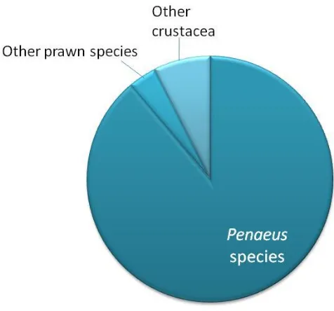
(43) necessary for cultured cells. However, there is no evidence that FBS provides all the hormones and growth factors required by crustacean cells (Mothersill and Austin, 2000; Mulford et al., 2001; Crane and Williams, 2002). Haemolymph and tissue extracts have been used as another source of hormones and growth factors (Chen et al., 1986; Chen et al., 1988; Rosenthal and Diamant, 1990; Nadala et al., 1993; Lu et al., 1995b; Tapay et al., 1995; Tong and Miao, 1996; Chen and Wang, 1999; Fraser and Hall, 1999; Itami et al., 1999; Kasornchandra et al., 1999; Owens and Smith, 1999; Mulford et al., 2001). None of these studies analysed the components of the tissue extract and or haemolymph additive, so the specific hormones or growth factors present were not known. Formulation of crustacean-defined media requires a hormonal profile, which would probably vary depending on the species and tissue used in culture.. 2.4. CRUSTACEA USED IN CELL CULTURE TRIALS. Efforts to develop crustacean cell lines have recently intensified, with emphasis on the commercially important penaeid species (Figure 2.2). Penaeids succumb to many viral infections in aquaculture and serve as excellent models for pathological studies. The most popular species used in cell culture experiments is P. monodon largely due to its worldwide availability and its popularity in aquaculture. Penaeus japonicus has been used in eight reported cell culture trials, P. stylirostris in six, and P. vannamei in five. Other penaeid species have been used in only one or two cell culture trials. Only four crustacean species other than prawns have been investigated for cell culture: the sand crab, Emerita asiatica; the swimming crab, Liocarcinus depurator; North American lobster, Homarus americanus; and the crayfish, Orconectes limosus. Out of the 17 species that have undergone cell culturing efforts, only two species are of freshwater origin, and the rest are marine (Appendix 3).. 23.
(44) Figure 2.2. Crustacean species used in cell culture attempts.. 2.5. TISSUES USED IN CRUSTACEAN CELL CULTURE. There is much disagreement regarding the best tissue for crustacean cell culture, which might be attributed to the different media and species being tested (Tong and Miao, 1996). Nevertheless, a few conclusions can be drawn on the basis of published results.. First, the age of the animal used for cell culture is likely to have a significant impact on cell proliferation; the younger the animal used as a tissue source, the more successful cell culture initiation is (Tong and Miao, 1996). Embryonic tissues provide undifferentiated and rapidly multiplying cells, which tend to adapt to in vitro growth more readily than the specialised cells of mature shrimp (Fan and Wang, 2002). Larval and embryonic cells in culture, furthermore, are less vulnerable than cells from adult animals to contamination from bacteria, yeast, and mould when attached to a substrate (Frank et al., 1994; Rinkevich, 1999) and have sometimes been found to survive culture conditions longer (Rinkevich, 1999; Fan and Wang, 2002).. 24.
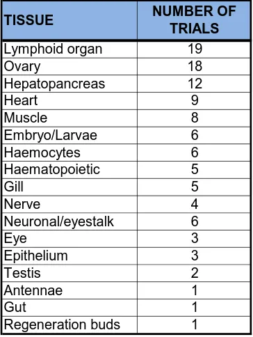
(45) The second conclusion from previous studies of crustacean cell culture is that tissues selected for culture should contain cells that are mitotically active. Embryonic tissues satisfy this criterion, as these tissues contain many undifferentiated and rapidly multiplying cells (Frerichs, 1996). Ovary tissue has also been a popular choice, as it is easy to locate, and promising for cell line establishment because of the numerous mitotically active germ stem cells (Luedeman and Lightner, 1992). Owens and Smith (1999) suggested that germinal tissues, such as haematopoietic, gonadal, and gut proliferative zones (e.g. distal hepatopancreatic cells and midgut caecae), subcutical areas at premoult, and embryonic tissues may provide the best source of proliferating cells. Neumann et al., (2000) may have created immortal cell cultures using neuronal and haematopoietic stem cells of the crayfish, Orconectes limosus, yet this lacks verification. No manipulation was carried out to transform cells, other than the selection and cloning of potential stem cells. These cells showed features of transformation, such as aneuploidy, anchorage independence, loss of contact inhibition, and low serum requirement. However, this cell line has not been verified or investigated for its ability to support viral growth. One of the main goals for the development of crustacean cell culture is disease diagnosis. Therefore, the potential cell line must be able to support viral growth in vitro. This is the reason why the lymphoid organ has been frequently selected as a tissue source. The lymphoid organ is the target for many infectious agents, so cells of the lymphoid organ are suitable for studying the mechanism of pathogen infection to host. To date, the longest survival time for primary culture of lymphoid organ cells is 43 days (Kasornchandra et al., 1999). Cells of the hepatopancreas have also been used to investigate the effect of disease and were maintained for 12 weeks (Uma et al., 2002). Many other tissues have been extracted from crustaceans in attempts to develop proliferating cell populations (Table 2.2).. 25.
(46) Table 2.2. Crustacean tissues used in cell culture attempts. TISSUE Lymphoid organ Ovary Hepatopancreas Heart Muscle Embryo/Larvae Haemocytes Haematopoietic Gill Nerve Neuronal/eyestalk Eye Epithelium Testis Antennae Gut Regeneration buds. NUMBER OF TRIALS 19 18 12 9 8 6 6 5 5 4 6 3 3 2 1 1 1. The method used to obtain cells from tissues influences the survival of the cells in culture. Two different methods are used: explants, in which cells migrate from the tissues, and dissociation (Toullec, 1999). In explants, tissues are removed using sterile techniques and placed in vessels containing the selected culture medium, based on the idea that cell aggregates have better ability than single cells to adhere to the substrate (Yavin and Yavin, 1974). This method has the potential to provide a large number of cell types and reduce stress to the cells; however, it is best adapted to loose tissues (Toullec, 1999).. Cell dissociation involves seeding culture flasks with tissues disrupted by either mechanical or enzymatic means. Mechanical dissociation can be achieved with magnetic stirring or by repeated aspiration using a pipette or syringe (Toullec, 1999). This technique is also suitable for loose tissues and can provide a large number of cells, but seems to reduce the ability of cells to attach to the substrate (Yavin and Yavin, 1974). Fragile cell types are often broken and ruptured during mechanical treatment, releasing proteases into the medium which leads to cell digestion (Freshney, 2005).. Enzymatic cell dissociation uses enzymes such as trypsin, pronase, collagenase, or enzyme mixes, such as collagenase/dispase. All enzymes are 26.
(47) not suitable for every tissue, and care must be taken to choose an appropriate enzyme. Trypsin, pronase, and collagenase seem to be too potent for crustacean tissues (Rosenthal and Diamant, 1990; Mothersill and Austin, 2000). Enzymatic dissociation is necessary for compact tissues, but can also be useful for loose tissues. This process is less drastic than mechanical dissociation and causes less cellular rupture. However, enzymatic treatment can weaken the cell membranes and decrease their ability to attach to the substrate. Cell cultures developed using enzymatic cell dissociation have been found to be capable of survival similar to cultures initiated by mechanical cell disruption. However, cell multiplication in enzyme dissociated tissues is low, and cell proliferation is limited upon sub-culture (Mothersill and Austin, 2000).. 2.6. CHARACTERISATION OF CULTURED CELLS. 2.6.1 Cell Identity The characterisation of cells in a cell culture involves confirming that the cells are authentic to the species and tissue of origin. Transformed cell lines can lose characteristics of the primary cultures from which they are derived (Crane, 1999). Therefore, identification of the cell types in culture is necessary, and selection of cell types that produce desired effects (e.g. virus susceptibility) is also important. To date, characterisation of crustacean cultures has been based mainly on determination of cellular morphology by light microscopy (Tong and Miao, 1996; Mulford et al., 2001). However, a variety of other methods have been developed, including immunohistochemistry, DNA fingerprinting, and identification of genes through the polymerase chain reaction (PCR).. Immunohistochemistry is one of the best methods for characterising specific phenotypic antigen expression (Freshney, 2005). Polyclonal or monoclonal antibodies have been developed for many crustacean molecules, including hormones, such as hyperglycaemic hormone, moulting inhibiting hormones, vitellogenesis-inhibiting hormone, and red pigment-concentrating hormone; neuropeptides, such as GABA, acetylcholine, serotonin, and histamine; and haemocytes. Antibodies or antiserum to phylogenetically conserved markers can also be used to identify crustacean cells in culture, including components of 27.
(48) the cytoskeleton, such as cytokeratins, actin, and myosin; specific enzymes, such as cytochromes and ATPases; and other proteins, such as heat shock proteins (Mothersill and Austin, 2000; Mulford et al., 2001). Results of previous studies demonstrates that some antibodies to mammalian epitopes cross-react with crustacean epitopes. DNA stained with the fluorescent marker, propidium, was used to differentiate hepatopancreas cells from chytrid parasites in cell cultures of the midgut of P. japonicus (Machii et al., 1988). Antigenic characterisation was achieved with hepatopancreas cells of N. norvegicus (Lyons-Alcantara et al., 1999) and a variety of tissues from the crustacean Pandalus borealis (Lyons-Alcantara et al., 2002). Thus, some cellular proteins are highly conserved in evolution and can be used to characterise crustacean cells in vitro.. DNA fingerprinting at the population level has permitted differentiation among species and has been successfully used with crustaceans. DNA fingerprinting can also identify a cell by visualising the structure of the repetitive component of genomic DNA, which is compared to a library of multilocus DNA fingerprints. The unique fingerprints enable the identification of cross-contamination, cell line variation, instability of cell lines, and inconsistent quality in cell stocks. This technique has been demonstrated to be both reliable and valuable for the identification of insect cell lines (Trisyono et al., 2000); however, it has not yet been applied to crustacean cell cultures.. Polymerase chain reaction (PCR) technologies can also be utilised to identify species-specific genes or DNA. DNA can easily be extracted from cells, and primers of highly conserved gene regions, either from crustaceans or speciesspecific markers can be used. These methods have been used to authenticate many vertebrate cell lines, including human, monkey, mouse, rats, and fish (Capasso et al., 2003). PCR amplification of the mitochondrial 18S rRNA gene fragment has also been used to characterise insect cell lines (Kshirsagar et al., 1997).. 28.
Figure
+7


Outline
Cytokines, Growth Factors, and Hormones
CHARACTERISATION OF CULTURED CELLS 1 Cell Identity
DEVELOPMENT OF IMMORTAL CELL CULTURES
CRITICAL ANALYSIS OF CRUSTACEAN CELL LINE DEVELOPMENT
Cell Culture Equipment
DISCUSSION 1 Tissue Source
CULTURE MEDIA OPTIMISATION
Elemental composition of crayfish haemolymph
Cell Culture Morphology
CHAPTER NINE GENERAL DISCUSSION
Related documents