Introduction
Transforming growth factor (TGF)-β1 is a cytokine that is involved in immune suppression, angiogenesis, apoptosis, cell growth, and epithelial to mesenchymal transitions (EMTs) during carcinogenesis [1–7]. TGF-β1 signals through the TGF-βtype I (TβRI) and TGF-βtype II (TβRII) transmembrane serine/threonine protein kinase receptors.
When TGF-β1 binds TβRII, TβRI is recruited to TβRII, and TβRII phosphorylates and activates the kinase activity of TβRI. Activated TβRI interacts with and phosphorylates a number of proteins, thereby activating many downstream signaling pathways, including the Smad (Sma/Mothers Against Decapentaplegic) pathway. The Smad signaling cascade is initiated by the phosphorylation of Smad2 or
DMEM = Dulbecco’s modified Eagle’s medium; EGF = epidermal growth factor; EMT = epithelial to mesenchymal transition; FBS = fetal bovine serum; HEK = human epidermal keratinocytes; HMEC = human mammary epithelial cell; KC = mouse keratinocytes transformed with K-ras; MEM = minimal essential medium; MK = mouse keratinocyte; PBS = phosphate buffered saline; Smad = Sma/Mothers Against Decapentaplegic; TβRI = TGF-βtype I receptor; TβRII = TGF-βtype II receptor; TGF = transforming growth factor; TTBS = Tris buffered saline with Tween-20; ZO = zonula occludens.
Research article
Induction by transforming growth factor-
ββ
1 of epithelial to
mesenchymal transition is a rare event in vitro
Kimberly A Brown
1,2, Mary E Aakre
1,2, Agnieska E Gorska
1,2, James O Price
1,3,4, Sakina E Eltom
5,
Jennifer A Pietenpol
1,6and Harold L Moses
1,21Vanderbilt-Ingram Cancer Center, Vanderbilt University Medical Center, Nashville, Tennessee, USA 2Department of Cancer Biology, Vanderbilt University Medical Center, Nashville, Tennessee, USA 3Department of Pathology, Vanderbilt University Medical Center, Nashville, Tennessee, USA 4Veterans Affairs Medical Center, Vanderbilt University Medical Center, Nashville, Tennessee, USA 5Department of Pharmacology, Meharry Medical College, Nashville, Tennessee, USA
6Department of Biochemistry, Vanderbilt University Medical Center, Nashville, Tennessee, USA
Corresponding author: Harold L Moses (e-mail: [email protected])
Received: 21 May 2003 Revisions requested: 20 Jun 2003 Revisions received: 10 Feb 2004 Accepted: 20 Feb 2004 Published: 17 Mar 2004
Breast Cancer Res 2004, 6:R215-R231 (DOI 10.1186/bcr778)
© 2004 Brown et al., licensee BioMed Central Ltd. This is an Open Access article: verbatim copying and redistribution of this article are permitted in all media for any purpose, provided this notice is preserved along with the article's original URL.
Abstract
Introduction: Transforming growth factor (TGF)-β1 is proposed to inhibit the growth of epithelial cells in early tumorigenesis, and to promote tumor cell motility and invasion in the later stages of carcinogenesis through the induction of an epithelial to mesenchymal transition (EMT). EMT is a multistep process that is characterized by changes in cell morphology and dissociation of cell–cell contacts. Although there is growing interest in TGF-β1-mediated EMT, the phenotype is limited to only a few murine cell lines and mouse models.
Methods:To identify alternative cell systems in which to study TGF-β1-induced EMT, 18 human and mouse established cell lines and cultures of two human primary epithelial cell types were screened for TGF-β1-induced EMT by analysis of cell morphology, and localization of zonula occludens-1, E-cadherin, and F-actin. Sensitivity to TGF-β1 was also determined by
[3H]thymidine incorporation, flow cytometry, phosphorylation of Smad2, and total levels of Smad2 and Smad3 in these cell lines and in six additional cancer cell lines.
Results:TGF-β1 inhibited the growth of most nontransformed cells screened, but many of the cancer cell lines were insensitive to the growth inhibitory effects of TGF-β1. In contrast, TGF-β1 induced Smad2 phosphorylation in the majority of cell lines, including cell lines resistant to TGF-β 1-mediated cell cycle arrest. Of the cell lines screened only two underwent TGF-β1-induced EMT.
Conclusion:The results presented herein show that, although many cancer cell lines have lost sensitivity to the growth inhibitory effect of TGF-β1, most show evidence of TGF-β1 signal transduction, but only a few cell lines undergo TGF-β 1-mediated EMT.
Keywords:E-cadherin, epithelial to mesenchymal transition, Smad, TGF-β1, ZO-1
Open Access
R216
Smad3 by TβRI. Phosphorylated Smad2 and Smad3 associate with Smad4 and are translocated to the nucleus, where they modulate transcription of many genes [8–13].
In early tumorigenesis TGF-β1 inhibits the growth of epithelial cells, and insensitivity to this growth inhibitory effect is associated with tumor progression [5,6]. Transgenic mice expressing a dominant negative TβRII in the epidermis and mammary glands exhibit accelerated tumor formation and malignant conversion [14,15]. Resistance to the antiproliferative effects of TGF-β1 is observed in head and neck squamous cell carcinomas [16–18], lung cancer [19], gastric cancer [20–22], colon cancer [22–25], pancreatic cancer [26], ovarian cancer [27,28], and in some recurrent breast cancers [29,30]. This loss of sensitivity to TGF-β1 is due to mutations in or transcriptional repression of the genes that encode TβRI and/or TβRII. Mutations in the genes encoding Smad2 (MADH2) and Smad4 (MADH4) also contribute to the insensitivity to TGF-β1 in many lung [31], pancreatic [32], and colorectal [33,34] carcinomas.
In contrast to the growth inhibitory effects of TGF-β1 in the early stages of carcinogenesis, TGF-β1 can also act as a promoter of tumor cell invasion and metastasis in the later stages of tumorigenesis [5,6]. Increased production of TGF-β1 is observed in epidermal [35], gastric [36], renal [37], breast [38–41], and prostate carcinomas [42] when compared with normal tissues. In mice with polyomavirus middle T antigen expression targeted to the mammary gland, blockade of TGF-β1 by administration of Fc:TβRII results in increased apoptosis in primary tumors and reduced tumor cell motility, intravasation, and metastasis [43]. Chronic exposure of mouse epidermal cells to TGF-β1 results in loss of TGF-β1-mediated growth inhibition and marked changes in cell morphology, downregulation of E-cadherin and cytokeratins, upregulation of vimentin, and formation of spindle cell carcinomas in mice [44,45]. Further studies show that carcinomas with excess TGF-β1 production are more motile and invasive, and exhibit increased tumor cell metastasis in athymic mice [36,40,45–51].
One mechanism by which TGF-β1 can promote tumor cell motility and invasion is through the induction of EMT [52]. EMT is a complex process that involves changes in cell morphology and dissociation of cell–cell contacts [53,54]. Cells undergoing EMT change from a cobblestone-like appearance to an elongated, mesenchymal phenotype. Accompanying this morphologic change is the delocaliza-tion of adherens and tight juncdelocaliza-tional proteins from the cell–cell junctions, and remodeling of the actin cyto-skeleton [53,54]. Characteristics associated with EMT, such as the dissociation of cell–cell and cell–extracellular matrix contacts, acquisition of an elongated cell
morpho-logy, and rearrangement of the cytoskeleton, can facilitate cell migration and invasion [55–57].
Although TGF-β1 is thought to play a key role in EMT in vivo, the frequency of TGF-β1-induced EMT in vitrois not known. To identify alternative cell systems in which to study TGF-β1-mediated EMT, we screened primary cultures of two human epithelial cell types and 18 established mouse and human cell lines for TGF-β1 responsiveness. We also included six additional cancer cell lines as a comparison for TGF-β1 responsiveness. We found that many of the cell strains displayed morpho-logical changes and exhibited actin stress fiber responses to TGF-β1. However, only in the NMuMG and MCT cells were those changes accompanied by a loss of E-cadherin and zonula occludens (ZO)-1 at cell–cell junctions after 48 hours of TGF-β1 treatment. Whereas all of the nontransformed cells were growth inhibited by TGF-β1, many of the cancer cell lines were insensitive to the growth inhibitory effects of TGF-β1. The TGF-β1-mediated growth inhibition was accompanied by an increase in phosphorylated Smad2 protein levels, but this was not unique to growth inhibited cells because changes in Smad2 phosphorylation occurred in a majority of cells after TGF-β1 treatment. In addition, prolonged TGF-β1 treatment induced a decrease in total Smad2 and/or total Smad3 in some cell lines. Our findings show that, although many cancer cells lost sensitivity to the growth inhibitory effect of TGF-β1, only two murine cell lines underwent TGF-β1-mediated EMT and these cells retain growth inhibitory response to TGF-β1.
Methods
Cell lines and culture conditions
R217 SCC012 and SCC028 cells were provided by D Sidransky
(Johns Hopkins University, Baltimore, MD, USA). Finally, UNC10 cells were provided by W Yarbrough (Vanderbilt University).
A549, Colo357, DU145, EpH4, H1299, HaCaT, HBL100, MCT, Panc-1, and 4T1 cells were maintained in Dulbecco’s modified Eagle’s medium (DMEM)/high-glucose medium (HyClone, Logan, UT, USA) supplemented with 10% fetal bovine serum (FBS). BT549 cells were maintained in RPMI-1640 medium (HyClone) supplemented with 10% FBS and 10µg/ml insulin. NMuMG cells were maintained in DMEM/high-glucose medium supplemented with 10% FBS and 10µg/ml insulin. HMEC-1016 and HMEC-1012 were grown in DMEM:F12 medium (1:1) (GibCoBRL, Grand Island, NY, USA) supplemented with 1% FBS, 10µg/ml ascorbic acid, 2 nmol/l β-estradiol, 35µg/ml bovine pituitary extract, 1 ng/ml cholera toxin, 12.5 ng/ml epidermal growth factor (EGF), 0.1 mmol/l ethanolamine, 0.1 mmol/l phospho-ethanolamine, 1µg/ml hydrocortisone, 1µg/ml insulin, 0.2 mmol/l L-glutamine, 10 nmol/l T3, 10µg/ml transferrin, and 15 nmol/l sodium selenite.
HEKs were maintained in EpiLife medium supplemented with human keratinocyte growth supplement (S-001-5) and 0.06 mol/l CaCl2 (Cascade Biologics, Portland, OR, USA). MCF-7 cells were maintained in minimal essential medium (MEM; GibCoBRL) supplemented with 10% FBS, 10µg/ml insulin, 1% nonessential amino acids, and 22µg/ml sodium pyruvate. MCF10A cells were cultured in (1:1) DMEM:F12 medium (Hyclone) supplemented with 5% horse serum, 100 ng/ml cholera toxin, 20 ng/ml EGF, 500 ng/ml hydrocortisone, and 10µg/ml insulin. MDA-MB-231, -361, -435S, and -436 cells were maintained in McCoy’s 5A medium (HyClone) supplemented with 10% FBS. MDA-MB-468 cells were grown in DMEM/high-glucose:McCoy’s 5A medium (1:1) supplemented with 10% FBS. MK and KC cells were maintained in DMEM/high-glucose medium supplemented with 8% dialyzed FBS, 5µg/ml calcium, and 4 ng/ml EGF. SCC012 and SCC028 cells were maintained in RPMI-1640 medium supplemented with 10% FBS, 1% insulin– transferrin–selenium A, 1% L-glutamine, and 1%
non-essential amino acids. UNC10 cells were grown in DMEM/ high-glucose medium supplemented with 10% FBS, 1% insulin–transferrin–selenium A, 1% L-glutamine, and 1%
nonessential amino acids.
All cell lines were maintained at 37°C in 5% CO2, except for the NMuMG, MK, and KC cells, which were grown in 7% CO2.
Antibodies and other reagents
TGF-β1 was from R&D Systems (Minneapolis, MN, USA). Antibodies to Smad2/3 (mouse) and E-cadherin (mouse) were from BD Transduction Laboratories (San Diego, CA,
USA); antibodies to phosphorylated (Ser 465/467) Smad2 (rabbit lot #21034 and #24432) were from Upstate Biotechnology (Lake Placid, NY, USA); and antibodies to ZO-1 (rat) were from Chemicon International (Temecula, CA, USA). Smad3 (rabbit) and ZO-1 (rabbit ZR-1) antibodies were obtained from Zymed Laboratories Inc. (San Francisco, CA, USA). Anti-actin (rabbit) antibodies and streptavidin–Cy3 conjugate were obtained from Sigma (St. Louis, MO, USA). Rabbit anti-goat, goat anti-mouse, and goat anti-rat biotinylated antibodies were obtained from Vector Laboratories Inc. (Burlingame, CA, USA).
[3H]Thymidine incorporation assays
Subconfluent cells were treated with TGF-β1 (5 ng/ml) for 46 hours in 12-well plates and pulsed for 2 hours with 4µCi/well [3H]thymidine (Perkin Elmer Life Sciences,
Boston, MA, USA). Cells were fixed with 1 ml 10% trichloroacetic acid for 30 min at 25°C, followed by two washes with 10% trichloroacetic acid. DNA was solubilized by incubation in 600µl 0.2 N NaOH for 30 min, and radioactivity was counted using 200µl solubilized DNA in 4 ml scintillation fluid.
Flow cytometry analyses
Subconfluent cells were treated with TGF-β1 (5 ng/ml) for 48 hours and approximately 106 cells were incubated in
propidium iodide solution (50µg/ml propidium iodide (Sigma), 5µg/ml RNase A, 0.1% Triton X-100, and 1µg/ml sodium citrate) for 5–10 min. Stained cells were analyzed using a FACS Caliber (Becton-Dickinson, San Jose, CA, USA) and the data stored as listmode files. DNA cell cycle histograms were analyzed and modeled using ModFit and WinList software (Verity Software House, Topsham, ME, USA). Fifteen thousand events were analyzed for each sample.
Cell lysis and immunoblot analyses
for phosphorylated Smad2 lot #24432, washed for 20 min in TTBS buffer, and incubated in secondary antibodies diluted in TTBS buffer plus 1% milk (1:10000) for 1 hour at 25°C. Membranes were washed two times in TTBS buffer (45 min/wash) and immunoreactive bands were visualized by enhanced chemiluminescence.
Immunofluorescence microscopy
Subconfluent cells, grown on 22 mm2 glass cover slips
(VWR Scientific, Atlanta, GA, USA), were treated for 48 hours or 6 days with 5 ng/ml TGF-β1. After treatment, cells were washed once with phosphate buffered saline (PBS) and fixed with 100% cold methanol (for rat ZO-1 and E-cadherin antibody use), 4% paraformaldehyde/PBS (for F-actin staining), or 1% paraformaldehyde/PBS (for rabbit ZO-1 antibody use) for 20 min at 25°C. Cells were washed four times with PBS, permeabilized by incubation with 0.2% Triton X-100 (in PBS) for 10 min at 25°C, and then washed two more times with PBS. For ZO-1 and E-cadherin staining, nonspecific binding sites were blocked by cellular incubation for 2 hours with 5% serum (goat serum for rabbit ZO-1 and E-cadherin antibody use and rabbit serum for rat ZO-1 antibody use) in PBS, incubated in primary antibodies diluted in 5% serum/PBS (rat ZO-1 1:1000, rabbit ZO-1 1:100, and E-cadherin 1:2500) for 1 hour at 25°C, followed by four washes with PBS. Cells were incubated in appropriate biotinylated secondary antibodies diluted in 5% serum/PBS (1:250) for 1 hour at 25°C, washed four times with PBS, and incubated in streptavidin-conjugated Cy3 diluted in 5% serum/PBS (1:1000) for 1 hour at 25°C. Cells were washed four times with PBS and nuclei were counter-stained by incubation with 0.1µg/ml Hoechst (33258) for 15 min at 25°C. For F-actin staining, cells were incubated in Texas Red-X phalloidin (Molecular Probes Inc., Eugene, OR, USA) diluted in PBS (1:200) for 30 min, washed with PBS three times, and were counterstained with Hoechst as described above.
After Hoechst staining, cells were washed three times with PBS and cover slips were mounted onto 25 × 75 mm microslides (Fisher Scientific, Pittsburgh, PA, USA) using AquaPolyMount (Polysciences, Warrington, PA, USA). Phase contrast images were captured using the Zeiss Epifluorescence inverted microscope and Zeiss AxioCam digital camera (Carl Zeiss, Jena, Germany). Fluorescent images were captured using the Zeiss Axiophot upright microscope (Carl Zeiss) and the Princeton Instruments cooled CCD digital camera (Princeton Scientific Instruments, Inc., Monmouth Junction, NJ, USA).
Results
Transforming growth factor-ββ1 induced epithelial to mesenchymal transition in NMuMG and MCT cells
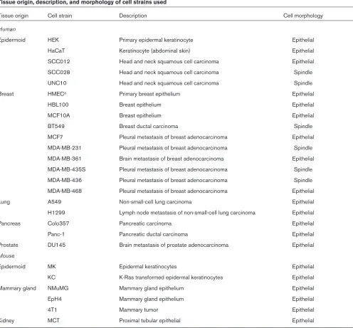
To determine the frequency of TGF-β1-mediated EMT in vitro, we performed a screen of 18 established cell lines
from human and mouse epidermis, mammary gland, lung, pancreas, kidney, and prostate (Table 1). In addition to the established cell lines, primary cultures of human epidermal keratinocytes (HEKs) and human mammary gland epithelial cells (HMEC-1012 and HMEC-1016) were also screened (Table 1). In the present study, EMT was defined as acquisition of a spindle-shaped morphology, loss of ZO-1 and E-cadherin from cell–cell junctions, and formation of actin stress fibers after TGF-β1 treatment. Using phase contrast and immunofluorescence microscopy, we determined that only two cell lines underwent EMT. After 48 hours of TGF-β1 treatment, the NMuMG and MCT cells became elongated (Fig. 1a,e,i,m), lost E-cadherin (Fig. 1b,f,j,n) and ZO-1 (Fig. 1c,g,k,o) staining at cell–cell junctions, and developed actin stress fibers (Fig. 1d,h,l,p).
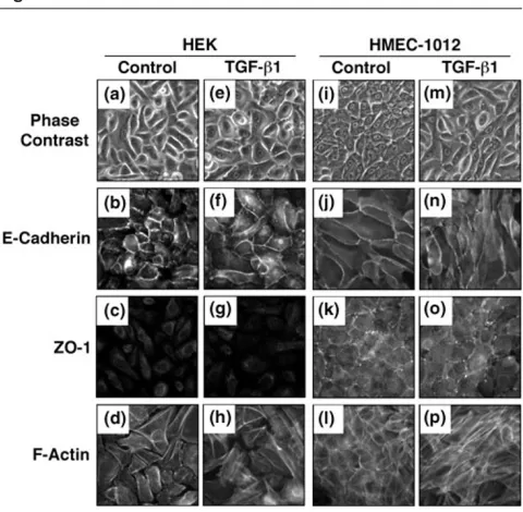
The primary cultures of breast epithelial cells (HMEC-1012 and HMEC-1016) acquired a spindle-shaped morphology, whereas the primary cultures of HEKs did not change cell morphology after 48 hours of TGF-β1 treatment (Fig. 2a,e,i,m; and data not shown). In addition, the HMECs and HEKs developed actin stress fibers after treatment (Fig. 2d,h,l,p; and data not shown). However, the HMECs and HEKs did not lose junctional E-cadherin staining (Fig. 2b,f,j,n; and data not shown) after TGF-β1 treatment for 48 hours. These cells also had little or no ZO-1 localized to their cell–cell junctions (Fig. 2c,g,k,o; and data not shown).
The human breast epithelial cell line MCF10A did not become elongated (Fig. 3a,e) or lose junctional E-cadherin or ZO-1 staining (Fig. 3b,f,c,g) in response to 48 hours of TGF-β1 treatment. After treatment, these cells exhibited some actin stress fiber formation (Fig. 3d,h). However, the MCF10A cells acquired a spindle-shaped morphology (Fig. 3i,m) and actin stress fibers (Fig. 3l,p), and lost much of the junctional E-cadherin and ZO-1 (Fig. 3j,n,k,o) after TGF-β1 treatment for 6 days. The HaCaT, UNC10, A549, Colo357, DU145, MK, KC, and 4T1 cells also exhibited an actin stress fiber response to TGF-β1, without delocali-zation of E-cadherin or ZO-1 from the cell–cell junctions, similar to the MCF10A cells after 48 hours of TGF-β1 treatment (Fig. 4b,f,r,v,c,g,s,w,d,h,t,x; and data not shown).
Other cell lines screened did not exhibit any changes in cell morphology or relocalization of E-cadherin, ZO-1, or actin after treatment with TGF-β1. These cell lines included SCC012, SCC028, HBL100, BT549, MCF7, MB-231, MB-361, MB-435S, MB-436, MDA-MB-468, H1299, Panc-1, and EpH4 cells (Fig. 4i–p; and data not shown). A summary of the TGF-β1-induced cellular response of the cell strains that exhibited acquisition of a spindle-shaped morphology, loss of junctional E-cadherin or ZO-1, or formation of actin stress fibers is presented in Table 2.
Transforming growth factor-ββ1-induced inhibition of S-phase
Previous studies analyzing TGF-β1 induction of EMT have emphasized the stages in which TGF-β1 acts as a promoter of carcinogenesis. It is proposed that cells first lose sensitivity to the growth inhibitory effects of TGF-β1, before TGF-β1 can promote EMT and cancer progression [59]. Therefore, we examined whether there was a correlation between TGF-β1-mediated EMT and loss of sensitivity to the growth inhibitory effects of TGF-β1 by examining [3H]thymidine incorporation and flow cytometry.
In [3H]thymidine incorporation experiments, it was observed
that 41% of the cell strains screened had a 0–25% reduction in [3H]thymidine incorporation (or 75% of the
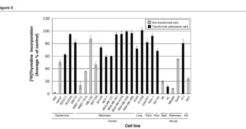
control) after 48 hours of TGF-β1 treatment. These cells included SCC028, UNC10, HBL100, MDA-MB-361, MDA-MB-435S, MDA-MB-436, MDA-MB-468, H1299, Colo357, Panc-1, and 4T1 (Fig. 5 and Table 3). Similarly, 37% of the cell strains screened had a 25–75% reduction in [3H]thymidine incorporation (or 75–25% of the control)
[image:5.612.60.556.108.569.2]after 48 hours of TGF-β1 treatment. These cells included HaCaT, SCC012, HMEC (1016), MCF10A, BT549, MCF7, R219
Table 1
Tissue origin, description, and morphology of cell strains used
Tissue origin Cell strain Description Cell morphology
Human
Epidermoid HEK Primary epidermal keratinocyte Epithelial
HaCaT Keratinocyte (abdominal skin) Epithelial
SCC012 Head and neck squamous cell carcinoma Epithelial
SCC028 Head and neck squamous cell carcinoma Spindle
UNC10 Head and neck squamous cell carcinoma Spindle
Breast HMECa Primary breast epithelium Epithelial
HBL100 Breast epithelium Epithelial
MCF10A Breast epithelium Epithelial
BT549 Breast ductal carcinoma Spindle
MCF7 Pleural metastasis of breast adenocarcinoma Epithelial
MDA-MB-231 Pleural metastasis of breast adenocarcinoma Spindle
MDA-MB-361 Brain metastasis of breast adenocarcinoma Epithelial
MDA-MB-435S Pleural metastasis of breast adenocarcinoma Spindle
MDA-MB-436 Pleural metastasis of breast adenocarcinoma Spindle
MDA-MB-468 Pleural metastasis of breast adenocarcinoma Epithelial
Lung A549 Non-small-cell lung carcinoma Epithelial
H1299 Lymph node metastasis of non-small-cell lung carcinoma Epithelial
Pancreas Colo357 Pancreatic carcinoma Epithelial
Panc-1 Pancreatic ductal carcinoma Epithelial
Prostate DU145 Brain metastasis of prostate adenocarcinoma Epithelial
Mouse
Epidermoid MK Epidermal keratinocytes Epithelial
KC K-Ras transformed epidermal keratinocytes Epithelial
Mammary gland NMuMG Mammary gland epithelium Epithelial
EpH4 Mammary gland epithelium Epithelial
4T1 Mammary tumor Epithelial
Kidney MCT Proximal tubular epithelial Epithelial
aHuman mammary epithelial cells (HMECs) from patients #1012 and #1016. HEK, primary human epidermal keratinocytes; MK, mouse
R220
Figure 1
NMuMG and MCT cells underwent transforming growth factor (TGF)-β1-mediated epithelial to mesenchymal transition. Phase contrast images of (a,e)NMuMG and (i,m)MCT cells with (TGF-β1) and without (control) TGF-β1 treatment (5 ng/ml) for 48 hours.
Immunofluorescence detection of (b,f,j,n)E-cadherin, (c,g,k,o)zonula occludens (ZO)-1, and (d,h,l,p)F-actin in NMuMG and MCT cells treated with 5 ng/ml TGF-β1 (TGF-β1) or without TGF-β1 (control) for 48 hours. TGF-β1 induced cell elongation, loss of cortical actin and junctional E-cadherin and ZO-1 staining, and formation of actin stress fibers in NMuMG and MCT cells. Micrographs are representative of at least three independent experiments.
Figure 2
Transforming growth factor (TGF)-β1 induced spindle-shaped morphology in nontransformed human mammary epithelial cells (HMECs) and human epidermal keratinocytes (HEKs). Phase contrast images of (a,e)HEK and (i,m)HMEC cells with (TGF-β1) and without (control) TGF-β1 treatment (5 ng/ml) for 48 hours.
[image:6.612.54.300.95.329.2]Immuno-fluorescence detection of (b,f,j,n)E-cadherin, (c,g,k,o)zonula occludens (ZO)-1, and (d,h,l,p)F-actin in HEK and HMEC cells treated with 5 ng/ml TGF-β1 (TGF-β1) or without TGF-β1 (control) for 48 hours. TGF-β1 induced cell elongation and formation of actin stress fibers in the HEK and HMEC cells. However, junctional E-cadherin staining remained intact after TGF-β1 treatment. ZO-1 was not detected in the HEK cells and little was observed in the HMEC cells with or without TGF-β1 treatment. Micrographs are representative of at least three independent experiments.
Table 2
Summary of transforming growth factor-ββ1-induced cellular responses in vitro
TGF-β1-induced responsea
Acquisition of Loss of junctional Actin stress fiber
Cell strain spindle shapeb E-cadherinc Loss of junctional ZO-1c formationc
HEK ± – N/Ad ±
HMECe ± – N/Ad +
HaCaT – – – ±
Colo357 – – – ±
Panc-1 – – – –
NMuMG + + + +
MCT + + + +
aTreatment with 5 ng/ml transforming growth factor (TGF)-β1 for 48 hours. bDetermined by phase contrast microscopy. cDetermined by E-cadherin,
zonula occludens (ZO)-1, or F-actin immunofluorescence. dHuman epidermal keratinocytes (HEKs) and human mammary epithelial cells (HMECs)
exhibited little to no junctional ZO-1 staining in the absence of TGF-β1 treatment. eHMECs from patients #1012 and #1016. Cellular responses to
MDA-MB-231, A549, DU145, and EpH4 (Fig. 5 and Table 3). Only 22% of the cell strains screened showed greater than a 75% reduction in [3H]thymidine incorporation (or
25% of the control) after 48 hours of TGF-β1 treatment (Fig. 5 and Table 3). These cells included HEK, HMEC (1012), MK, KC, NMuMG, and MCT (Fig. 5 and Table 3). Of note, established cell lines constituted 95% of the cells that did not have greater than 75% reduction in [3H]thymidine after TGF-β1 treatment (Fig. 5 and Table 3).
Most of the cell lines that did not have a greater than 75% reduction in [3H]thymidine incorporation after TGF-β1
treatment, did not exhibit changes in morphology or changes in actin, E-cadherin, or ZO-1 localization in this screen (Figs 1–5; and data not shown). Only the UNC10, A549, and DU145 cell lines did not have reduced [3H]thymidine
incorporation of greater than 75% and had increased actin stress fiber formation after TGF-β1 treatment (Figs 4 and 5; and data not shown). However, there was no change in ZO-1 localization in these cells (data not shown). In comparison, all of the cell lines that had decreased [3H]thymidine incorporation had increased actin stress
fiber formation after TGF-β1 treatment (Fig. 1d,h,l,p; Fig. 2d,h,l,p; Fig. 3d,h,I,p; Fig. 4d,h,t,x; Fig. 5; and data not shown). These cells included the HaCaT, MCF10A, Colo357, MK, KC, NMuMG, 4T1, and MCT cells, and the primary cultures of HMEC-1012 and HEK cells.
Flow cytometry was also performed on cells treated with TGF-β1 for 48 hours to confirm the data generated by [3H]thymidine incorporation. The data generated by flow
cytometry experiments were consistent with those of [3H]
thymidine incorporation (Figs 5 and 6, and Table 3). Fig. 6 includes histograms that were representative of cell strains with 0–25% (MDA-MB-436), 25–75% (EpH4), and 75–100% (NMuMG) reduction in S-phase after 48 hours of TGF-β1 treatment, when compared with the control cells.
Transforming growth factor-ββ1-mediated Smad2
phosphorylation did not correlate with S-phase reduction
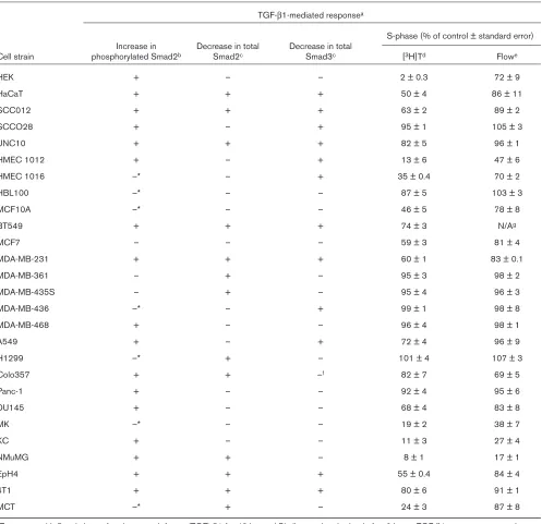
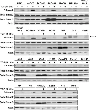
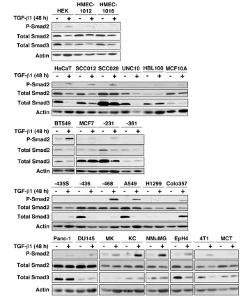
To determine the relationship of Smad2 phosphorylation with reduction in S-phase and EMT, after TGF-β1 treatment, we analyzed phosphorylated Smad2 protein levels. Western analyses of protein extracts showed that, after 2 hours of treatment with TGF-β1, phosphorylated Smad2 protein levels increased in all of the cell strains screened except for the MCF7, MDA-MB-361, and MDA-MB-435S cells when normalized to actin (Fig. 7 and Table 3). After treatment with TGF-β1 for 48 hours, levels of phosphory-lated Smad2 remained elevated in the cells with increased levels of phosphorylated Smad2 after 2 hours of treatment, except HMEC (1016), HBL100, MCF10A, MDA-MB-436, H1299, MK, and MCT when normalized to actin (Figs 7 and 8, and Table 3). Immunoblot analysis of the MCF7, MDA-MB-361, and MDA-MB-435S cells with the phosphorylated Smad2 antibody (lot #21034) resulted in a band (indicated by an asterisk) that migrated higher than the phosphorylated Smad2 protein (Fig. 7).
Cell strains that exhibited TGF-β1-induced phosphorylation of Smad2 did not necessarily exhibit a reduction in [3H]thymidine uptake. These cell strains included
SCC028, MDA-MB-436, MDA-MB-468, and H1299 (Figs 5 and 7, and Table 3). Although the MCF7 cells had [3H]thymidine uptake of 59% of the control after 48 hours
of TGF-β1 treatment, an increase in phosphorylation of Smad2 by Western analysis was not observed at 2 hours or at 48 hours (Figs 5, 7 and 8, and Table 3).
Prolonged transforming growth factor-ββ1 treatment led to decreased total Smad2 and Smad3 protein levels in many cell strains
[image:7.612.57.300.94.327.2]Total Smad2 and Smad3 protein levels decreased with TGF-β1 treatment in 51% and 48% of the cell lines, respectively (Fig. 8). Total Smad2 levels decreased in HEK, HaCaT, SCC012, UNC10, BT549, MDA-MB-231, MDA-MB-361, MDA-MB-435S, H1299, Colo357, NMuMG, EpH4, 4T1, and MCT cells when normalized to R221
Figure 3
R222
Figure 4
Transforming growth factor (TGF)-β1 did not induce epithelial to mesenchymal transition in Colo357, Panc-1, or HaCaT Cells. Phase contrast images of (a,e)Colo357, (i,m)Panc-1, and (q,u)HaCaT cells treated with (TGF-β1) or without (control) 5 ng/ml TGF-β1 for 48 hours. Immunofluorescence detection of (b,f,j,n,r,v)E-cadherin, (c,g,k,o,s,w)zonula occludens (ZO)-1, and (d,h,l,p,t,x)F-actin in Colo357, Panc-1, and HaCaT cells treated with (TGF-β1) or without (control) 5 ng/ml TGF-β1 for 48 hours. TGF-β1 treatment induced cell enlargement and formation of actin stress fibers in the Colo357 cells. However, junctional E-cadherin and ZO-1 remained intact after TGF-β1 treatment. Changes in cell morphology, E-cadherin, ZO-1, or actin localization were not observed in the Panc-1 and HaCaT cells. Micrographs are representative of two experiments.
Figure 5
Many cancer cell lines were not growth inhibited by transforming growth factor (TGF)-β1. Human primary cells and human and mouse established cell lines were treated with TGF-β1 (5 ng/ml) for 48 hours. The cells were then pulsed for 2 hours with [3H]thymidine, and incorporated radioactivity
was quantified. Results are presented as the average percentage of control (untreated cells) incorporation (± standard error) from three independent experiments with 6–12 replicates per experiment. The average percentage of control [3H]thymidine incorporation was calculated by dividing the
average [3H]thymidine incorporation of TGF-β1-treated cells (from three independent experiments) by the average [3H]thymidine incorporation of
[image:8.612.62.554.410.668.2]actin (Fig. 8). Total Smad3 levels decreased in HEK, HMEC (1012 and 1016), HaCaT, SCC012, SCC028, UNC10, BT549, MDA-MB-231, MDA-MB-436, A549, EpH4, and 4T1 cells when normalized to actin (Fig. 8).
[image:9.612.60.557.116.597.2]Decreased total Smad2 and total Smad3 protein levels did not correlate with an increased reduction in S-phase after TGF-β1 treatment for 48 hours. For instance, the HMEC (1012), MK, and KC cells had a 87%, 81%, and R223
Table 3
Summary of transforming growth factor-ββ1-mediated Smad signaling and effect on S-phase in vitro
TGF-β1-mediated responsea
S-phase (% of control ± standard error) Increase in Decrease in total Decrease in total
Cell strain phosphorylated Smad2b Smad2c Smad3c [3H]Td Flowe
HEK + – – 2 ± 0.3 72 ± 9
HaCaT + + + 50 ± 4 86 ± 11
SCC012 + + + 63 ± 2 89 ± 2
SCCO28 + – + 95 ± 1 105 ± 3
UNC10 + + + 82 ± 5 96 ± 1
HMEC 1012 + – + 13 ± 6 47 ± 6
HMEC 1016 –* – + 35 ± 0.4 70 ± 2
HBL100 –* – – 87 ± 5 103 ± 3
MCF10A –* – – 46 ± 5 78 ± 8
BT549 + + + 74 ± 3 N/Ag
MCF7 – – – 59 ± 3 81 ± 4
MDA-MB-231 + + + 60 ± 1 83 ± 0.1
MDA-MB-361 – + – 95 ± 3 98 ± 2
MDA-MB-435S – + – 95 ± 4 96 ± 3
MDA-MB-436 –* – + 99 ± 1 98 ± 8
MDA-MB-468 + – – 96 ± 4 98 ± 1
A549 + – + 72 ± 4 96 ± 9
H1299 –* + – 101 ± 4 107 ± 3
Colo357 + + –f 82 ± 7 69 ± 5
Panc-1 + – – 92 ± 4 95 ± 6
DU145 + – – 68 ± 4 83 ± 8
MK –* – – 19 ± 2 38 ± 7
KC + – – 11 ± 3 27 ± 4
NMuMG + + – 8 ± 1 17 ± 1
EpH4 + + + 55 ± 0.4 84 ± 4
4T1 + + + 80 ± 6 91 ± 1
MCT –* + – 24 ± 3 87 ± 8
aTreatment with 5 ng/ml transforming growth factor (TGF)-β1 for 48 hours. bSimilar results obtained after 2 hours TGF-β1 treatment except those
designated by an asterisk (detected increased phosphorylated Smad2 after 2 hours of TGF-β1 treatment). cNo decrease in total Smad2 or total
Smad3 was detected after 2 hours of TGF-β1 treatment. d[3H]Thymidine ([3H]T) experiments performed at least three times with 6–12 replicates.
Results are presented as the average percent of control of three independent experiments. eFlow cytometry experiments performed at least three
times in triplicate. Results are presented as the average percent of control of three independent experiments. fAn increase in total Smad3 was
observed in Colo357 cells 48 hours after TGF-β1 treatment. gAccurate flow cytometry analysis could not be performed on BT549 cells because
89% reduction in [3H]thymidine incorporation, respectively,
whereas there was no decrease in total Smad2 protein levels after 48 hours of TGF-β1 treatment (Figs 5 and 8, and Table 3). Similarly, the MK, KC, MCT, and NMuMG cells had 81%, 89%, 76%, and 92% reductions in [3H]thymidine incorporation, respectively, whereas there
was no decrease in total Smad3 protein levels after 48 hours of TGF-β1 treatment (Figs 5 and 8, and Table 3). In contrast, the HEK cells had a 98% reduction in [3H]thymidine incorporation and had no reduction of total
Smad2 or Smad3 after 48 hours of TGF-β1 treatment (Figs 5 and 8, and Table 3).
[image:10.612.90.438.100.484.2]Also, decreased total Smad2 and Smad3 protein levels did not correlate with an increase in the levels of phosphory-lated Smad2 protein after TGF-β1 treatment (Figs 7 and 8, and Table 3). An increase in phosphorylated Smad2 protein levels occurred in all cell strains screened after 2 hours of TGF-β1 treatment except for the MCF7, MDA-MB-361, and MDA-MB-435S cells (Fig. 7), whereas only a subset of those had decreased total Smad2 and/or Smad3 protein levels after TGF-β1 treatment for 48 hours (Fig. 8 and Table 3). Additionally, the MDA-MB-361 and the MDA-MB-435S cells did not have increased phosphorylated Smad2 levels or decreased total Smad3 R224
Figure 6
Transforming growth factor (TGF)-β1 induced a robust decrease in S-phase in non-cancer cell strains. Human primary cells and human and mouse established cell lines were treated with (TGF-β1) or without (control) 5 ng/ml TGF-β1 for 48 hours. The cells were then harvested and incubated with propidium iodide, and DNA content was determined using a FACS Caliber (Becton-Dickinson). Data were modeled and analyzed using ModFit and WinList software (Verity Software House). Fifteen thousand events were analyzed for each sample. Representative histograms are presented illustrating cells that had a robust decrease in S-phase (NMuMG), modest decrease in S-phase (EpH4), and no decrease in S-phase (MDA-MB-436) as a result of TGF-β1 treatment. G0/G1 and G2 phases are indicated by black shading and S phase is indicated by gray shading on the histograms. The percentage of cells in G0/G1 and in S phases of the cell cycle are indicated. Three independent experiments were
levels, but they did exhibit decreased total Smad2 levels after 48 hours of TGF-β1 treatment (Figs 7 and 8, and Table 3).
The decrease in total Smad2 and Smad3 protein levels, observed after TGF-β1 treatment, did not associate with the ability of the cell line to undergo TGF-β1-mediated EMT. NMuMG and MCT cells underwent EMT (Fig. 1) and they both exhibited decreased total Smad2 and no decrease in total Smad3 protein levels after TGF-β1 treatment for 48 hours (Fig. 8). The other cell lines that had decreased total Smad2 and no decrease in total Smad3 levels after TGF-β1 treatment did not undergo TGF-β1-induced EMT (Figs 2–4 and 8, and Table 3; and data not shown).
Discussion
TGF-β1 is an inhibitor of epithelial cell growth in the early stages of carcinogenesis. However, it also promotes tumor cell invasion and metastasis through the induction of EMT [52]. Many studies on the mechanism of TGF-β 1-induced EMT are limited to a few murine cell lines and mouse models. Therefore, to identify alternative cell systems in which to study TGF-β1-induced EMT, we performed a TGF-β1 sensitivity screen using a panel of human primary cells and established human and mouse cell lines.
[image:11.612.132.469.113.484.2]The TGF-β1-induced morphologic changes observed in the NMuMG, MCT, and HMEC cells 48 hours after TGF-β1 treatment are consistent with changes seen during EMT (Fig. 1a,e,i,m; Fig. 2i,m; and data not shown) [53,54]. R225
Figure 7
Changes in cell morphology, from a cobblestone-like appearance to more elongated shape, and actin stress fiber formation, were observed in the NMuMG and MCT cells, and in primary cultures of HMECs (Fig. 1a,d,e,h,i,l,m,p; Fig. 2i,l,m,p; and data not shown). However, only the NMuMG and MCT cells lost E-cadherin and ZO-1 staining at the cell–cell junctions after 48 hours of TGF-β1 treat-ment (Fig. 1b,f,j,n,c,g,k,o). E-cadherin is a protein found at adherens junctions that allows for cell–cell adherence, and ZO-1 is a tight junctional protein that forms a selective barrier between cells. Loss of these proteins at cell–cell junctions is used as a marker of EMT [54,60–63]. Cortical
[image:12.612.97.453.109.534.2]actin is a measure of cell integrity and loss thereof, or formation of actin stress fibers, is also used to define EMT [63,64]. Therefore, we concluded that only the NMuMG and MCT cells underwent TGF-β1-mediated EMT, after 48 hours of treatment, in this screen. Results from previous studies also show that NMuMG and MCT cells undergo TGF-β1-mediated EMT [60,61,63–65]. Other studies reported that TGF-β1-mediated EMT occurs in HaCaT, Colo357, and Panc-1 cells 48 hours after TGF-β1 treatment [66,67]; however, they did not undergo TGF-β 1-induced EMT according to the conditions used in the present study (Fig. 4). Other cell lines screened in the R226
Figure 8
study formed actin stress fibers with TGF-β1 treatment, but they did not lose ZO-1 from cell–cell junctions (Fig. 3c,d,g,h; and data not shown). We did not examine E-cadherin localization in all of the cell strains because not all of the cell strains exhibited morphologic changes, with TGF-β1 treatment, that were indicative of EMT.
Unlike the mouse NMuMG and MCT cells, none of the 14 human cell lines with an epithelial morphology screened underwent β1-mediated EMT within 48 hours of TGF-β1 treatment. The MCF10A human breast epithelial cell line took 6 days to undergo a morphologic change, lose junctional E-cadherin and ZO-1, and form actin stress fibers with TGF-β1 treatment (Fig. 3i–p). However, this extended treatment with TGF-β1 to induce EMT is not consistent with previous reports that used times up to and including 48 hours [60,61,66,67]. It may be that the extended treatment with TGF-β1 is necessary to activate secondary and tertiary signaling pathways that are not activated within 48 hours. The Panc-1, Colo357, and HaCaT cells were treated with TGF-β1 for longer than 48 hours because they were shown to undergo TGF-β 1-induced EMT in previous reports [66,67]. However, these cell lines did not undergo EMT when treated with TGF-β1 for 72 hours (data not shown). These data could suggest that human cells are more resistant to TGF-β1-mediated EMT. Alternatively, the TGF-β1 treatment times were not long enough to induce EMT, the small sample size used may not be representative, or the cells may not have the genetic alterations necessary for TGF-β1 to induce EMT.
Activation of non-Smad signaling pathways is also implica-ted in TGF-β1-mediated EMT. Many studies have shown a requirement for activated Ras/Raf/mitogen-activited protein kinase for TGF-β1-mediated EMT in human, rat, or mouse epidermal, pancreas, intestine, liver, prostate, and mammary epithelial cells [67–76]. In the mouse mammary gland epithelial cell line EpH4, TGF-β1 induces EMT and tumor formation in mice only when cells express oncogenic H-Ras [73]. Furthermore, it has been shown that TGF-β1 can activate Ras, and increased H-Ras levels are required for nuclear accumulation of Smad2 [70]. Additionally, signaling through integrin β1 [68] and activation of EGF receptor [62], RhoA [61], p38 mitogen-activited protein kinase [77], and phosphatidylinositol 3-kinase [60,70,71] pathways are involved in or required for TGF-β1-induced EMT.
The lack of TGF-β1-mediated EMT in the human cells does not necessarily refute a role for TGF-β1 in promoting EMT in vivoin human tumors. Human epithelial tumors can undergo mesenchymal conversions, providing a potential role for EMT in human metastasis. It has been shown that excess TGF-β1 can promote EMT in human tumors in vivo. Increased levels of TGF-β1 are observed in many
human tumors [35–40,42,51,78–80], are often localized
to the advancing edges of primary tumors and metastases, and are associated with poor clinical outcome [39,41].
It is somewhat unexpected that TGF-β1-induced EMT occurred in nontransformed cell lines, such as the NMuMG and MCT cells, because in vivo mouse studies show that TGF-β1 promotes tumor cell invasion and metastasis in the later stages of carcinogenesis [5,6]. In one of these studies, targeted expression of TGF-β1 to mouse suprabasal keratinocytes results in resistance to the formation of benign skin tumors, after long-term chemical carcinogenesis treatment. However, benign papillomas that develop become malignant at an accelerated rate and metastasis occur more rapidly than spontaneous tumors in control mice. Additionally, there are a high incidence of spindle cell carcinoma development in these mice, pointing to TGF-β1-induction of EMT in vivo [38]. NMuMG and MCT cells may have the genetic alterations that allow for TGF-β1-mediated EMT to occur.
Many studies on TGF-β1-mediated EMT have emphasized that TGF-β1 promotes carcinogenesis in stages. It is proposed that cells first lose sensitivity to the growth inhibitory effects of TGF-β1, and subsequently TGF-β1 can promote tumor progression and EMT [59]. MMH-D3 murine hepatocytes and EpH4 murine mammary gland epithelial cells undergo TGF-β1-mediated EMT only when rendered insensitive to TGF-β1-induced cell cycle arrest or apoptosis, by infection with active H-Ras [59,70,73,81]. However, the present study does not support this model because it shows that resistance to the growth inhibitory effects of TGF-β1 is not a prerequisite for TGF-β1-mediated EMT. The NMuMG and MCT cells exhibited a decrease in S-phase and underwent EMT after 48 hours of TGF-β1 treatment (Figs 1, 5, and 6). Consistent with the present study were experiments performed by Nicolas and coworkers [72] that showed that increased Smad3 in MDCK cells restored growth inhibitory responses to TGF-β1 but did not revert cells from a mesenchymal to an epithelial phenotype. Additionally, Chang and coworkers [49] observed that increased TGF-β1 expression in sarcoma cells increased cell tumori-genicity while inhibiting cell proliferation.
The trends observed with the analysis of the flow cytometry data were consistent with the [3H]thymidine
incorporation data (Figs 5 and 6 and Table 3). However, the values for the percentage of the control cells in S-phase were not identical between [3H]thymidine
incorporation and flow cytometry for each cell strain. This discrepancy can be accounted for by the fact that the data were generated using two different methodologies. [3H]thymidine incorporation examines DNA synthesis
[3H]thymidine incorporation and flow cytometry
experiments were not the same, the trends of TGF-β1-induced decreases in DNA synthesis were consistent.
All but three of the cell strains screened had an increase in phosphorylated Smad2 protein levels after 2 hours of TGF-β1 treatment, but not all of the cell lines that had increased phosphorylation of Smad2 had reduced S-phase after 48 hours of TGF-β1 treatment. The SCC028, MDA-MB-436, MDA-MB-468, and H1299 cells had an increase in phosphorylated Smad2 levels after 2 hours of TGF-β1 treatment, but they exhibited decreases in S-phase of only 5%, 1%, 4%, and 4%, respectively (Figs 5–7 and Table 3). The lack of S-phase decrease upon 48 hours of TGF-β1 treatment may point to inactivation of or mutations in downstream proteins that could abolish TGF-β1-mediated growth inhibition in these cells. The MCF7 cells did not exhibit an increase in phosphorylated Smad2 protein levels after 2 hours of TGF-β1 treatment, but there was a 41% decrease in S-phase on 48 hours of TGF-β1 treatment (Figs 5–7 and Table 3). This decrease in S-phase is not as great a decrease as was exhibited by some of the other cell strains when treated with TGF-β1 for 48 hours. The decrease could be explained by very low levels of phosphorylated Smad2 increase on TGF-β1 treatment that was not detected by immunoblot analysis. Alternatively, it is possible that the TGF-β1 signal was propagated by phosphorylation of Smad3.
Smad2 is not solely responsible for the propagation of TGF-β1 signals. Similar to Smad2, on TGF-β1 binding to the TβRII, Smad3 is phosphorylated by the TβRI, forms a complex with Smad4, translocates to nucleus, and regulates the activation of TGF-β1 target genes. However, mutations or deletions in Smad3 are rare in human cancers. Reports by Graham and coworkers [82] and Xu and coworkers [83] suggest that loss of Smad3 may largely be responsible for the nonresponsiveness of some cells to TGF-β1. The proliferation, migration, and invasion of normal extravillous trophoblast cells are under the control of TGF-β1. However, premalignant and malignant trophoblast cells, that have lost the Smad3 protein but retain functional Smad2, are resistant to the antiproliferative and anti-invasive effect of TGF-β1. It is therefore possible that the attenuation of inhibition of S-phase or the induction of EMT by TGF-β1 in the present study may be a result of mutation or loss of Smad3, even though the cells have increased phosphorylation of Smad2 on TGF-β1 treatment.
Interestingly, decreased total Smad2 and Smad3 protein levels were observed after TGF-β1 treatment regardless of changes in the levels of phosphorylated Smad2 protein or of whether cell proliferation was inhibited by TGF-β1 (Figs 5–8). Similar to our findings, decreased total Smad2 protein levels were reported in COS-1 monkey kidney
cells after TGF-β1 treatment [84]. In that report, the proteasome inhibitors MG-132 or lactacystin blocked Smad2 from TGF-β1-induced degradation. In addition, Smad3 decreases have been reported during TGF-β1-induced EMT in MDCK cells [72,85] and after TGF-β1 treatment of human lung epithelial cells [86]. In these studies, MDCK cells become refractory to the growth inhibitory effects of TGF-β1 [72,85]. TGF-β1 treatment of primary human fibroblasts and HaCaT cells also leads to decreased total Smad3 protein levels [87,88].
A negative feedback loop could explain the decreases in total Smad2 and Smad3 protein levels after 48 hours of TGF-β1 treatment. The E3 ubiquitin ligase Smurf has been shown to interact with Smads and promote their ubiquiti-nation [89,90]. In this model, phosphorylated, nuclear Smad2 is ubiquitinated by Smurf2 and degraded by proteasomes [89,90]. Smad7 also interacts with Smurf2 and induces TGF-β receptor degradation [91,92]. Phosphorylated Smad3 is also ubiquitinated by the ROC1-SCFFbw1aE3 ligase complex, and subsequently degraded
in the proteasome [88]. In order to prevent continuous Smad signaling in the absence of TGF-β1 stimulation, Smad2 and Smad3 are negatively regulated by a number of proteins. Smad6 and Smad7 inhibit Smad2 and Smad3 activation by competing with Smad2 and Smad3 for binding to the TGF-β receptors [93]. Smad6 and Smad7 are induced by activation of TGF-β1 signaling and form a negative feedback loop [94–96]. However, the mechanism of decrease in total Smad2 and/or Smad3 in different cell strains on prolonged TGF-β1 treatment remains unclear.
Conclusion
TGF-β1 induction of EMT is a rare occurrence in vitroand is not limited to transformed cell lines. In fact, TGF-β 1-mediated EMT was only observed in nontumorigenic cells in the present study. Furthermore, there does not appear to be a correlation between loss of TGF-β-mediated growth inhibition and EMT. The inability of TGF-β1 to induce EMT was not due to a total loss of TGF-β1 signaling, as was evidenced by induction of Smad2 phosphorylation. In conclusion, our data indicate that the
in vitro models used to study TGF-β1 induction of EMT may not be as relevant to in vivo cancer progression as was previously assumed in the field.
Competing interests
None declared.Acknowledgements
We thank the Vanderbilt University Toxicology Center tissue culture core facility (supported by NIH grant ES00267) and the Vanderbilt Uni-versity Medical Center Cell Imaging Core Resource (supported by NIH grants CA68485 and DK20593). We would also like to thank Dr Brian Law, Dr Neil Bhowmick, Dr Matthew Westfall, Jamie Hearnes, and Kristy Schavolt for critical reading of the manuscript. The present study was also supported by NIH training grant T32CA09592 to KAB and R01 CA85492 and R01 CA102162 to HLM.
References
1. Dennler S, Goumans MJ, ten Dijke P: Transforming growth factor beta signal transduction.J Leukoc Biol2002, 71:731-740.
2. Pepper MS: Transforming growth factor-beta: vasculogenesis, angiogenesis, and vessel wall integrity. Cytokine Growth Factor Rev1997, 8:21-43.
3. Schuster N, Krieglstein K: Mechanisms of TGF-beta-mediated apoptosis.Cell Tissue Res2002, 307:1-14.
4. Moustakas A, Pardali K, Gaal A, Heldin CH: Mechanisms of TGF-beta signaling in regulation of cell growth and differenti-ation.Immunol Lett2002, 82:85-91.
5. Derynck R, Akhurst RJ, Balmain A: TGF-beta signaling in tumor suppression and cancer progression.Nat Genet2001, 29:117-129.
6. Akhurst RJ, Derynck R: TGF-beta signaling in cancer: a double-edged sword.Trends Cell Biol2001, 11:S44-S51.
7. Wakefield LM, Roberts AB: TGF-beta signaling: positive and negative effects on tumorigenesis.Curr Opin Genet Dev2002, 12:22-29.
8. Kloos DU, Choi C, Wingender E: The TGF-beta–Smad network: introducing bioinformatic tools. Trends Genet 2002, 18:96-103.
9. Massague J: How cells read TGF-beta signals. Nat Rev Mol Cell Biol2000, 1:169-78.
10. de Caestecker MP, Piek E, Roberts AB: Role of transforming growth factor-beta signaling in cancer. J Natl Cancer Inst 2000, 92:1388-1402.
11. Ten Dijke P, Goumans MJ, Itoh F, Itoh S: Regulation of cell pro-liferation by Smad proteins.J Cell Physiol2002, 191:1-16. 12. Heldin CH, Miyazono K, ten Dijke P: TGF-beta signalling from
cell membrane to nucleus through SMAD proteins. Nature 1997, 390:465-471.
13. Massague J, Wotton D: Transcriptional control by the TGF-beta/Smad signaling system.Embo J2000, 19:1745-1754. 14. Gorska AE, Jensen RA, Shyr Y, Aakre ME, Bhowmick NA, Moses
HL: Transgenic mice expressing a dominant-negative mutant type II transforming growth factor-beta receptor exhibit impaired mammary development and enhanced mammary tumor formation.Am J Pathol2003, 163:1539-1549.
15. Amendt C, Schirmacher P, Weber H, Blessing M: Expression of a dominant negative type II TGF-beta receptor in mouse skin results in an increase in carcinoma incidence and an accelera-tion of carcinoma development.Oncogene1998, 17:25-34. 16. Garrigue-Antar L, Munoz-Antonia T, Antonia SJ, Gesmonde J,
Vel-lucci VF, Reiss M: Missense mutations of the transforming growth factor beta type II receptor in human head and neck squamous carcinoma cells.Cancer Res1995, 55:3982-3987. 17. Chen T, Yan W, Wells RG, Rimm DL, McNiff J, Leffell D, Reiss M:
Novel inactivating mutations of transforming growth factor-beta type I receptor gene in head-and-neck cancer metas-tases.Int J Cancer2001, 93:653-661.
18. Munoz-Antonia T, Li X, Reiss M, Jackson R, Antonia S: A muta-tion in the transforming growth factor beta type II receptor gene promoter associated with loss of gene expression. Cancer Res1996, 56:4831-4835.
19. Hougaard S, Norgaard P, Abrahamsen N, Moses HL, Spang-Thomsen M, Skovgaard Poulsen H: Inactivation of the trans-forming growth factor beta type II receptor in human small cell lung cancer cell lines.Br J Cancer1999, 79:1005-1011. 20. Park K, Kim SJ, Bang YJ, Park JG, Kim NK, Roberts AB, Sporn
MB: Genetic changes in the transforming growth factor beta (TGF-beta) type II receptor gene in human gastric cancer cells: correlation with sensitivity to growth inhibition by TGF-beta.Proc Natl Acad Sci USA1994, 91:8772-8776.
21. Kang SH, Bang YJ, Im YH, Yang HK, Lee DA, Lee HY, Lee HS, Kim NK, Kim SJ: Transcriptional repression of the transforming growth factor-beta type I receptor gene by DNA methylation results in the development of TGF-beta resistance in human gastric cancer.Oncogene1999, 18:7280-7286.
22. Myeroff LL, Parsons R, Kim SJ, Hedrick L, Cho KR, Orth K, Mathis M, Kinzler KW, Lutterbaugh J, Park K: A transforming growth factor beta receptor type II gene mutation common in colon and gastric but rare in endometrial cancers with microsatellite instability.Cancer Res1995, 55:5545-5547.
23. Markowitz S, Wang J, Myeroff L, Parsons R, Sun L, Lutterbaugh J, Fan RS, Zborowska E, Kinzler KW, Vogelstein B, Brattain M,
Wilson JKV: Inactivation of the type II TGF-beta receptor in colon cancer cells with microsatellite instability.Science1995, 268:1336-1338.
24. Grady WM, Myeroff LL, Swinler SE, Rajput A, Thiagalingam S, Lut-terbaugh JD, Neumann A, Brattain MG, Chang J, Kim SJ, Kinzler KW, Vogelstein B, Willson JK, Markowitz S: Mutational inactivation of transforming growth factor beta receptor type II in microsatellite stable colon cancers.Cancer Res1999, 59:320-324.
25. Markowitz S: TGF-beta receptors and DNA repair genes, coupled targets in a pathway of human colon carcinogenesis. Biochim Biophys Acta2000, 1470:M13-M20.
26. Goggins M, Shekher M, Turnacioglu K, Yeo CJ, Hruban RH, Kern SE: Genetic alterations of the transforming growth factor beta receptor genes in pancreatic and biliary adenocarcinomas. Cancer Res1998, 58:5329-5332.
27. Wang D, Kanuma T, Mizunuma H, Takama F, Ibuki Y, Wake N, Mogi A, Shitara Y, Takenoshita S: Analysis of specific gene mutations in the transforming growth factor-beta signal trans-duction pathway in human ovarian cancer.Cancer Res2000, 60:4507-4512.
28. Chen T, Triplett J, Dehner B, Hurst B, Colligan B, Pemberton J, Graff JR, Carter JH: Transforming growth factor-beta receptor type I gene is frequently mutated in ovarian carcinomas. Cancer Res2001, 61:4679-4682.
29. Lucke CD, Philpott A, Metcalfe JC, Thompson AM, Hughes-Davies L, Kemp PR, Hesketh R: Inhibiting mutations in the transforming growth factor beta type 2 receptor in recurrent human breast cancer.Cancer Res2001, 61:482-485.
30. Chen T, Carter D, Garrigue-Antar L, Reiss M: Transforming growth factor beta type I receptor kinase mutant associated with metastatic breast cancer. Cancer Res 1998, 58:4805-4810.
31. Yanagisawa K, Uchida K, Nagatake M, Masuda A, Sugiyama M, Saito T, Yamaki K, Takahashi T, Osada H: Heterogeneities in the biological and biochemical functions of Smad2 and Smad4 mutants naturally occurring in human lung cancers.Oncogene 2000, 19:2305-2311.
32. Hahn SA, Schutte M, Hoque AT, Moskaluk CA, da Costa LT, Rozenblum E, Weinstein CL, Fischer A, Yeo CJ, Hruban RH, Kern SE: DPC4, a candidate tumor suppressor gene at human chromosome 18q21.1.Science1996, 271:350-353.
33. Salovaara R, Roth S, Loukola A, Launonen V, Sistonen P, Avizienyte E, Kristo P, Jarvinen H, Souchelnytskyi S, Sarlomo-Rikala M, Aaltonen LA: Frequent loss of SMAD4/DPC4 protein in colorectal cancers.Mol Pathol2002, 55:385-388.
34. Eppert K, Scherer SW, Ozcelik H, Pirone R, Hoodless P, Kim H, Tsui LC, Bapat B, Gallinger S, Andrulis IL, Thomsen GH, Wrana JL, Attisano L: MADR2 maps to 18q21 and encodes a TGFbeta-regulated MAD-related protein that is functionally mutated in colorectal carcinoma.Cell1996, 86:543-552.
35. Krieg P, Schnapke R, Furstenberger G, Vogt I, Marks F: TGF-beta 1 and skin carcinogenesis: antiproliferative effect in vitro and TGF-beta 1 mRNA expression during epidermal hyperprolifer-ation and multistage tumorigenesis. Mol Carcinog 1991, 4: 129-137.
36. Maehara Y, Kakeji Y, Kabashima A, Emi Y, Watanabe A, Akazawa K, Baba H, Kohnoe S, Sugimachi K: Role of transforming growth factor-beta 1 in invasion and metastasis in gastric car-cinoma.J Clin Oncol1999, 17:607-614.
37. Gomella LG, Sargent ER, Wade TP, Anglard P, Linehan WM, Kasid A: Expression of transforming growth factor alpha in normal human adult kidney and enhanced expression of transforming growth factors alpha and beta 1 in renal cell car-cinoma.Cancer Res1989, 49:6972-6975.
38. Travers MT, Barrett-Lee PJ, Berger U, Luqmani YA, Gazet JC, Powles TJ, Coombes RC: Growth factor expression in normal, benign, and malignant breast tissue.Br Med J (Clin Res Ed) 1988, 296:1621-1624.
39. Gorsch SM, Memoli VA, Stukel TA, Gold LI, Arrick BA: Immuno-histochemical staining for transforming growth factor beta 1 associates with disease progression in human breast cancer. Cancer Res1992, 52:6949-6952.
40. Walker RA, Dearing SJ: Transforming growth factor beta 1 in ductal carcinoma in situ and invasive carcinomas of the breast.Eur J Cancer1992, 28:641-644.
advancing edges of primary tumors and to lymph node metastases of human mammary carcinoma. Am J Pathol 1993, 143:381-389.
42. Steiner MS, Zhou ZZ, Tonb DC, Barrack ER: Expression of transforming growth factor-beta 1 in prostate cancer. Endocrinology1994, 135:2240-2247.
43. Muraoka RS, Dumont N, Ritter CA, Dugger TC, Brantley DM, Chen J, Easterly E, Roebuck LR, Ryan S, Gotwals PJ, Koteliansky V, Arteaga CL: Blockade of TGF-beta inhibits mammary tumor cell viability, migration, and metastases.J Clin Invest 2002, 109:1551-1559.
44. Caulin C, Scholl FG, Frontelo P, Gamallo C, Quintanilla M: Chronic exposure of cultured transformed mouse epidermal cells to transforming growth factor-beta 1 induces an epithe-lial-mesenchymal transdifferentiation and a spindle tumoral phenotype.Cell Growth Differ1995, 6:1027-1035.
45. Portella G, Cumming SA, Liddell J, Cui W, Ireland H, Akhurst RJ, Balmain A: Transforming growth factor beta is essential for spindle cell conversion of mouse skin carcinoma in vivo: implications for tumor invasion.Cell Growth Differ 1998, 9: 393-404.
46. Arrick BA, Lopez AR, Elfman F, Ebner R, Damsky CH, Derynck R: Altered metabolic and adhesive properties and increased tumorigenesis associated with increased expression of trans-forming growth factor beta 1.J Cell Biol1992, 118:715-726. 47. Tobin SW, Douville K, Benbow U, Brinckerhoff CE, Memoli VA,
Arrick BA: Consequences of altered TGF-beta expression and responsiveness in breast cancer: evidence for autocrine and paracrine effects.Oncogene2002, 21:108-118.
48. Steiner MS, Barrack ER: Transforming growth factor-beta 1 overproduction in prostate cancer: effects on growth in vivo and in vitro.Mol Endocrinol1992, 6:15-25.
49. Chang HL, Gillett N, Figari I, Lopez AR, Palladino MA, Derynck R: Increased transforming growth factor beta expression inhibits cell proliferation in vitro, yet increases tumorigenicity and tumor growth of Meth A sarcoma cells.Cancer Res1993, 53: 4391-4398.
50. Walker RA, Dearing SJ, Gallacher B: Relationship of transform-ing growth factor beta 1 to extracellular matrix and stromal infiltrates in invasive breast carcinoma.Br J Cancer1994, 69: 1160-1165.
51. Welch DR, Fabra A, Nakajima M: Transforming growth factor beta stimulates mammary adenocarcinoma cell invasion and metastatic potential.Proc Natl Acad Sci USA1990, 87:7678-7682.
52. Akhurst RJ, Balmain A: Genetic events and the role of TGF beta in epithelial tumour progression.J Pathol1999, 187:82-90. 53. Thiery JP: Epithelial-mesenchymal transitions in tumour
pro-gression.Nat Rev Cancer2002, 2:442-454.
54. Savagner P: Leaving the neighborhood: molecular mecha-nisms involved during epithelial-mesenchymal transition. Bioessays2001, 23:912-923.
55. Birchmeier W, Birchmeier C: Epithelial-mesenchymal transi-tions in development and tumor progression.EXS1995, 74:1-15.
56. Birchmeier C, Meyer D, Riethmacher D: Factors controlling growth, motility, and morphogenesis of normal and malignant epithelial cells.Int Rev Cytol1995, 160:221-266.
57. Hay ED: Role of cell-matrix contacts in cell migration and epithelial-mesenchymal transformation.Cell Differ Dev1990, 32:367-375.
58. Sipes NJ, Lyons RM, Moses HL: Isolation and characterization of Kirsten murine sarcoma virus-transformed mouse ker-atinocytes resistant to transforming growth factor beta.Mol Carcinog1990, 3:12-19.
59. Lehmann K, Janda E, Pierreux CE, Rytomaa M, Schulze A, McMahon M, Hill CS, Beug H, Downward J: Raf induces TGFbeta production while blocking its apoptotic but not inva-sive responses: a mechanism leading to increased malig-nancy in epithelial cells.Genes Dev2000, 14:2610-2622. 60. Bakin AV, Tomlinson AK, Bhowmick NA, Moses HL, Arteaga CL:
Phosphatidylinositol 3-kinase function is required for trans-forming growth factor beta-mediated epithelial to mesenchy-mal transition and cell migration. J Biol Chem 2000, 275: 36803-36810.
61. Bhowmick NA, Ghiassi M, Bakin A, Aakre M, Lundquist CA, Engel ME, Arteaga CL, Moses HL: Transforming growth factor-beta1
mediates epithelial to mesenchymal transdifferentiation through a RhoA-dependent mechanism. Mol Biol Cell2001, 12:27-36.
62. Grande M, Franzen A, Karlsson JO, Ericson LE, Heldin NE, Nilsson M: Transforming growth factor-beta and epidermal growth factor synergistically stimulate epithelial to mesenchy-mal transition (EMT) through a MEK-dependent mechanism in primary cultured pig thyrocytes. J Cell Sci2002, 115:4227-4236.
63. Miettinen PJ, Ebner R, Lopez AR, Derynck R: TGF-beta induced transdifferentiation of mammary epithelial cells to mesenchy-mal cells: involvement of type I receptors. J Cell Biol1994, 127:2021-2036.
64. Piek E, Moustakas A, Kurisaki A, Heldin CH, ten Dijke P: TGF-(beta) type I receptor/ALK-5 and Smad proteins mediate epithelial to mesenchymal transdifferentiation in NMuMG breast epithelial cells.J Cell Sci1999, 112(Pt 24):4557-4568. 65. Okada H, Danoff TM, Kalluri R, Neilson EG: Early role of Fsp1 in
epithelial-mesenchymal transformation. Am J Physiol 1997, 273:F563-F574.
66. Zavadil J, Bitzer M, Liang D, Yang YC, Massimi A, Kneitz S, Piek E, Bottinger EP: Genetic programs of epithelial cell plasticity directed by transforming growth factor-beta. Proc Natl Acad Sci USA2001, 98:6686-6691.
67. Ellenrieder V, Hendler SF, Boeck W, Seufferlein T, Menke A, Ruhland C, Adler G, Gress TM: Transforming growth factor beta1 treatment leads to an epithelial-mesenchymal transdif-ferentiation of pancreatic cancer cells requiring extracellular signal-regulated kinase 2 activation. Cancer Res 2001, 61: 4222-4228.
68. Bhowmick N, Zent R, Ghaissi M, McDonnell M, Moses HL: Inte-grin b1 signaling is necessary for transforming growth factor-beta activation of p38MAPK and epithelial plasticity. J Biol Chem2001, 276:46707-46713.
69. Fujimoto K, Sheng H, Shao J, Beauchamp RD: Transforming growth factor-beta1 promotes invasiveness after cellular transformation with activated Ras in intestinal epithelial cells. Exp Cell Res2001, 266:239-249.
70. Gotzmann J, Huber H, Thallinger C, Wolschek M, Jansen B, Schulte-Hermann R, Beug H, Mikulits W: Hepatocytes convert to a fibroblastoid phenotype through the cooperation of TGF-beta1 and Ha-Ras: steps towards invasiveness. J Cell Sci 2002, 115:1189-1202.
71. Janda E, Lehmann K, Killisch I, Jechlinger M, Herzig M, Downward J, Beug H, Grunert S: Ras and TGF[beta] cooperatively regu-late epithelial cell plasticity and metastasis: dissection of Ras signaling pathways.J Cell Biol2002, 156:299-313.
72. Nicolas FJ, Lehmann K, Warne PH, Hill CS, Downward J: Epithe-lial mesenchymal transition in MDCK cells is accompanied by downregulation of Smad3 expression leading to resistance to TGFbeta-induced growth arrest.J Biol Chem2002, 278:3251-3256.
73. Oft M, Peli J, Rudaz C, Schwarz H, Beug H, Reichmann E: TGF-beta1 and Ha-Ras collaborate in modulating the phenotypic plasticity and invasiveness of epithelial tumor cells. Genes Dev1996, 10:2462-2477.
74. Oft M, Heider KH, Beug H: TGFbeta signaling is necessary for carcinoma cell invasiveness and metastasis.Curr Biol1998, 8:1243-1252.
75. Oft M, Akhurst RJ, Balmain A: Metastasis is driven by sequen-tial elevation of H-ras and Smad2 levels.Nat Cell Biol2002, 4: 487-494.
76. Park BJ, Park JI, Byun DS, Park JH, Chi SG: Mitogenic conver-sion of transforming growth factor-beta1 effect by oncogenic Ha-Ras-induced activation of the mitogen-activated protein kinase signaling pathway in human prostate cancer. Cancer Res2000, 60:3031-3038.
77. Bakin AV, Rinehart C, Tomlinson AK, Arteaga CL: p38 mitogen-activated protein kinase is required for TGFbeta-mediated fibroblastic transdifferentiation and cell migration. J Cell Sci 2002, 115:3193-3206.
78. Donnelly MJ, Patel V, Yeudall WA, Game SM, Scully C, Prime SS: Autocrine production of TGF-alpha and TGF-beta during tumour progression of rat oral keratinocytes.Carcinogenesis 1993, 14:981-985.
benign and malignant prostatic tumors. Prostate 1994, 25: 210-218.
80. Derynck R, Goeddel DV, Ullrich A, Gutterman JU, Williams RD, Bringman TS, Berger WH: Synthesis of messenger RNAs for transforming growth factors alpha and beta and the epider-mal growth factor receptor by human tumors. Cancer Res 1987, 47:707-712.
81. Filmus J, Zhao J, Buick RN: Overexpression of H-ras oncogene induces resistance to the growth-inhibitory action of trans-forming growth factor beta-1 (TGF-beta 1) and alters the number and type of TGF-beta 1 receptors in rat intestinal epithelial cell clones.Oncogene1992, 7:521-526.
82. Graham CH, Connelly I, MacDougall JR, Kerbel RS, Stetler-Stevenson WG, Lala PK: Resistance of malignant trophoblast cells to both the anti-proliferative and anti-invasive effects of transforming growth factor-beta.Exp Cell Res1994, 214:93-99.
83. Xu G, Chakraborty C, Lala PK: Expression of TGF-beta signal-ing genes in the normal, premalignant, and malignant human trophoblast: loss of smad3 in choriocarcinoma cells.Biochem Biophys Res Commun2001, 287:47-55.
84. Lo RS, Massague J: Ubiquitin-dependent degradation of TGF-beta-activated smad2.Nat Cell Biol1999, 1:472-478.
85. Nicolas FJ, Lehmann K, Warne PH, Hill CS, Downward J: Epithe-lial to mesenchymal transition in Madin-Darby canine kidney cells is accompanied by down-regulation of Smad3 expres-sion, leading to resistance to transforming growth factor-beta-induced growth arrest. J Biol Chem 2003, 278: 3251-3256.
86. Yanagisawa K, Osada H, Masuda A, Kondo M, Saito T, Yatabe Y, Takagi K, Takahashi T: Induction of apoptosis by Smad3 and down-regulation of Smad3 expression in response to TGF-beta in human normal lung epithelial cells.Oncogene1998, 17:1743-1747.
87. Mori N, Morishita M, Tsukazaki T, Giam CZ, Kumatori A, Tanaka Y, Yamamoto N: Human T-cell leukemia virus type I oncoprotein Tax represses Smad-dependent transforming growth factor beta signaling through interaction with CREB-binding protein/p300.Blood2001, 97:2137-2144.
88. Fukuchi M, Imamura T, Chiba T, Ebisawa T, Kawabata M, Tanaka K, Miyazono K: Ligand-dependent degradation of Smad3 by a ubiquitin ligase complex of ROC1 and associated proteins. Mol Biol Cell2001, 12:1431-1443.
89. Lin X, Liang M, Feng XH: Smurf2 is a ubiquitin E3 ligase medi-ating proteasome-dependent degradation of Smad2 in trans-forming growth factor-beta signaling.J Biol Chem2000, 275: 36818-36822.
90. Zhang Y, Chang C, Gehling DJ, Hemmati-Brivanlou A, Derynck R: Regulation of Smad degradation and activity by Smurf2, an E3 ubiquitin ligase.Proc Natl Acad Sci USA2001, 98:974-979. 91. Kavsak P, Rasmussen RK, Causing CG, Bonni S, Zhu H,
Thomsen GH, Wrana JL: Smad7 binds to Smurf2 to form an E3 ubiquitin ligase that targets the TGF beta receptor for degra-dation.Mol Cell2000, 6:1365-1375.
92. Ebisawa T, Fukuchi M, Murakami G, Chiba T, Tanaka K, Imamura T, Miyazono K: Smurf1 interacts with transforming growth factor-beta type I receptor through Smad7 and induces receptor degradation.J Biol Chem2001, 276:12477-12480. 93. Hayashi H, Abdollah S, Qiu Y, Cai J, Xu YY, Grinnell BW,
Richard-son MA, Topper JN, Gimbrone MA, Jr., Wrana JL, Falb D: The MAD-related protein Smad7 associates with the TGFbeta receptor and functions as an antagonist of TGFbeta signaling. Cell1997, 89:1165-1173.
94. Nakao A, Afrakhte M, Moren A, Nakayama T, Christian JL, Heuchel R, Itoh S, Kawabata M, Heldin NE, Heldin CH, ten Dijke P: Identi-fication of Smad7, a TGFbeta-inducible antagonist of TGF-beta signalling. Nature1997, 389:631-635.
95. Afrakhte M, Moren A, Jossan S, Itoh S, Sampath K, Westermark B, Heldin CH, Heldin NE, ten Dijke P: Induction of inhibitory Smad6 and Smad7 mRNA by TGF-beta family members. Biochem Biophys Res Commun1998, 249:505-511.
96. Ishisaki A, Yamato K, Nakao A, Nonaka K, Ohguchi M, ten Dijke P, Nishihara T: Smad7 is an inducible inhibitor of activin-induced growth arrest and apoptosis in mouse B cells.J Biol Chem1998, 273:24293-24296.
Correspondence
Harold L Moses, Vanderbilt University, 691 Preston Research Building, 2220 Pierce Avenue South, Nashville, TN 37232-6838, USA. Tel: +1 615 936 1507; fax: +1 615 936 1790; e-mail: [email protected]