S H O R T R E P O R T
Open Access
Predictability of thermo-lesions using electrodes
for deep brain stimulation - an in vitro study
Thomas L Hauska
1*, Hermann Lanmüller
2†, Wolfgang Kainz
3†, François Alesch
1†Abstract
Background:Typically, electrodes for Deep Brain Stimulation (DBS) are used for chronic stimulation. However, there are conditions where this therapy has to be discontinued. In such cases using the DBS electrodes as a tool for thermo-lesioning (coagulation) could be used for an alternative treatment. The aim of this study was to determine if it is possible to generate coagula with a predictable geometry and to define their dimensions as a function of power and time in an in vitro model (egg white at room temperature). Furthermore, we tested if repetitive (cumulative) coagulation has an impact on the overall form and size of the clot.
Findings:Coagulation-growth was achieved as a function of power and duration of coagulation; reproducible well-formed thermocoagulations could be achieved. When using two adjacent electrodes a power range between 1.25 Watt and 2.00 Watt resulted in homogenous ovoid coagula. After two minutes of coagulation the clots reached a maximum in size and further growth could not be achieved. It was also possible to increase the size of a preformed clot by repetitive coagulation either by increasing the power level or the duration of the coagulation process.
Conclusions:We could show that it is possible to obtain predictable coagula in-vitro when using DBS electrodes for thermocoagulation even though they have not been developed for that specific purpose. However, until in-vivo safety and efficacy of DBS electrodes for ablation purposes is properly assessed, only approved electrodes should be used for brain ablation.
Background
Today Deep Brain Stimulation (DBS) is state-of-the-art for treating various cases of movement disorders and it has widely replaced conventional therapeutic lesioning (thalamotomy, pallidotomy) [1]. The adaptability and reversibility of DBS makes it superior to ablative proce-dures. Tremor, rigidity, and dystonia can be efficiently treated. However, there are conditions where the ther-apy has to be discontinued. Complications such as infec-tion or erosion, loss of effect, tolerance, pain or discomfort and device failure can result in terminating the DBS treatment [2]. Also financial limitations can be a reason for terminating the DBS treatment [3]. To us it seems feasible and practical to perform lesions using DBS electrodes which are already at the right location. However, it remained unclear whether these lesions are
predictable in size and geometry, a key factor for a safe use of ablative procedures.
The aim of this study was to determine if it is possible to generate coagula with a predictable geometry and to define their dimensions as a function of power and time in an in vitro model (egg white at room temperature). Furthermore, we tested if repetitive coagulations could lead to a predictable change of size and shape of a pre-existent coagulum.
Methods
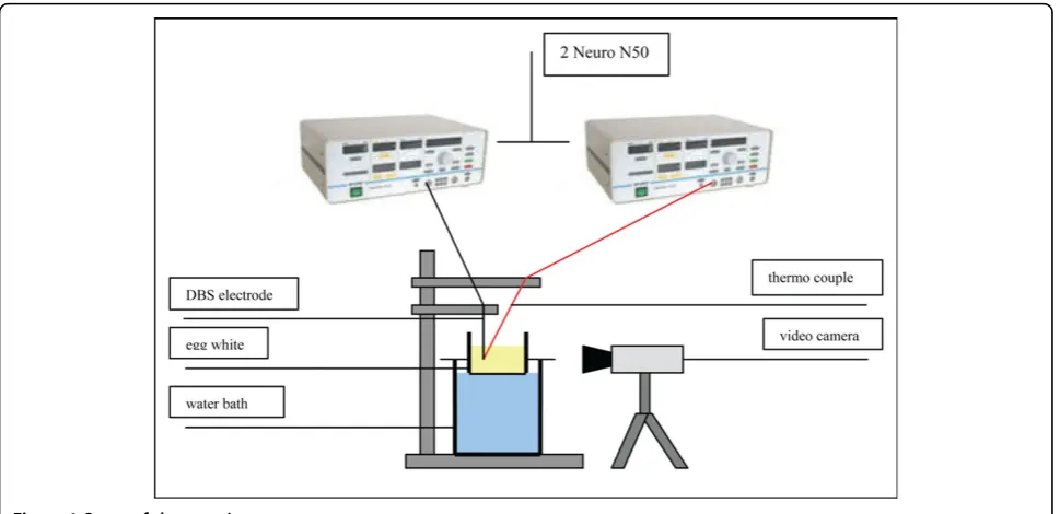
Our laboratory setup was similar to the ones described in [4-6]. In a container filled with egg white we slowly inserted the DBS probe. The slow insertion of the DBS probes into the egg white, which we kept at room tem-perature, prevented the formation of air bubbles leading to optimised visibility. A thin copper plate on the inner side of one of the flat walls of the container acted as reference electrode for monopolar coagulations. Ther-mocouples placed at a maximum distance of 1 mm next * Correspondence: [email protected]
†Contributed equally
1Department of Neurosurgery, Medical University Vienna, Währinger Gürtel
18-20, 1090 Vienna, Austria
Hauskaet al.BMC Research Notes2010,3:84 http://www.biomedcentral.com/1756-0500/3/84
to the electrode were used to monitor temperature development (figure 1, table 1).
A 500 kHz radio frequency (RF) generator (Neuro N50, Stockert GmbH) delivering the high frequency cur-rent to the DBS electrodes was connected to a PC through an analogue/digital converter.
The software EPWIN-N50 (Stockert GmbH) con-trolled the ablation current and graphically displayed the electrode impedance, current and voltage over time. The ablation process was monitored using a digital video camera. Subsequently taken screenshots allowed to mea-sure the coagulum size at different stages of its growth.
To compare the mean and standard deviation values of the coagulum growth we systematically increased the coagulation power and duration. For repeatability each combination of power and time was repeated five times in separate samples. To investigate coagulum growth of cumulative ablation, we performed a primary coagula-tion and after a short cooling period different para-meters were used for another coagulation process. Pictures of the coagulum after standardized time points allowed measuring the coagulum size (the diameters of the probe and the lesion were measured in pixels in
each picture and as the diameter of the probe is known, the size of the coagulum could be calculated).
Findings
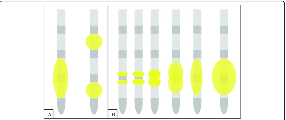
For the quadripolar electrodes we examined all six possible locations for bipolar ablation. However, only when choosing two adjacent poles as active ablation electrodes homogenous coagulation ovoids formed (figure 2a). The origin of the resulting coagulations was situated near the two adjacent active poles. Abla-tion over distant poles resulted in two smaller single coagula with the same shape and size as monopolar coagulations (figure 2b).
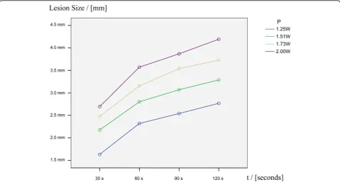
Lesion size is as a function of power and duration. Increasing power and/or lesioning time showed the trend of resulting in larger coagula (figure 3 and 4). Pre-dictable and reproducible results were obtained in the power range between 1.25 Watt and 2.00 Watt over the duration of a maximum of 120 seconds. With less power than 1.25 Watt no well-formed thermocoagula-tions could be obtained. Applying more than 2.00 Watt resulted in vaporisation and it was not possible to achieve homogenous coagula. The incremental increase in coagulation depth changed over time (figure 4). After two minutes of coagulation the clots reached a maxi-mum size and further growth could not be achieved (plateau). The resulting clots were homogeneous and ovoid in shape.
[image:2.595.57.542.88.323.2]Cumulative coagulation, in terms of increasing the size of a preformed coagulum, could be achieved. However, further coagulum growth could only be managed either by coagulating longer or with more power. For example, Figure 1Setup of the experiment.
Table 1 Means and standard deviations of temperatures reached in function of lesioning duration and power.
1.25 W 1.51 W 1.73 W 2.00 W
[image:2.595.58.290.666.732.2]a coagulum generated with 1.25 Watt over 60 seconds could either be increased with 1.51 Watt over 60 sec-onds or with 1.25 Watt over the duration of more than 60 seconds. Applying the same parameters as the pri-mary clot has been formed with - in this example 1.25 Watt over 60 seconds - did not lead to an increase in size. Table 2 summarizes the coagula’s sizes for different power levels and coagulation durations.
Discussion
There are conditions where patients treated with chronic stimulation discover long-term complications such as infection or erosion, loss of effect, tolerance, pain or discomfort. In these cases the therapy has to be discontinued and alternatives have to be found.
We tested if it was possible to perform reproducible lesions with the already implanted and well-located DBS electrodes. We could show in our in-vitro egg white experiments that DBS electrodes can be used to form predictable, reproducible and homogeneous ovoid coa-gula. Due to a limited number of coagulations, we could only show the trend of their increase of size in function of power and/or lesioning duration.
[image:3.595.57.541.88.292.2]The incremental increase in coagulation size reached a maximum after two minutes. We assume that various non-linear parameters are causing this effect. Change of electric properties due to increasing impedance caused by continuous tissue damage could be one explanation. Certain tissue properties, such as relative density and electrical conductivity, also vary in different anatomical Figure 2Schematic illustration of lesion development. a) Only when choosing two adjacent poles as active ablation electrodes homogenous coagulation ovoids formed. Ablation over distant poles resulted in two smaller single coagula with the same shape and size as monopolar coagulations. b) The origin of the resulting coagulations was situated near the two adjacent active poles of the quadripolar electrodes.
Figure 3Example of the development of a lesion obtained with 1.73 Watt over 120 seconds. Homogenous ovoid lesions increased in size in function of lesioning duration.
Hauskaet al.BMC Research Notes2010,3:84 http://www.biomedcentral.com/1756-0500/3/84
[image:3.595.62.538.327.509.2]structures (e.g. grey and white matter of the brain). Their impact on lesion size and shape should be observed by modern finite element methods. Although it has been stated that lesions’ sizes obtained in egg white models are correlating with those obtained in vivo [6], a generalization of this method to neural tissue could be limited. Thus, to fully understand the efficacy and safety of using DBS electrodes for ablation pur-poses, the comparison of in vitro, computational (FEM) and in vivo experiments appear to be mandatory. At this point in time it is important to mention that these electrodes are not approved for ablation procedures and only approved electrodes should be used for brain ablation.
Next steps in our research are to use computational prediction of thermo ablation procedures in homoge-neous media and then in anatomically correct models of the human anatomy. These predictions will include non-linear tissues parameters and account for blood flow. Using these novel techniques we hope to develop ablation electrodes for various coagula shapes using
multiple electrodes and computer controlled ablation parameters.
Conclusion
Using DBS electrodes it is possible to produce reprodu-cible and homogeneous thermo-coagulations with pre-dictable size and shape in egg white.
Acknowledgements
Thanks to Inomed Medizintechnik GmbH, especially to Rudi Mattmüller and Dieter Mussler who provided professional surroundings for carrying out the experiments.
We also thank Stockert GmbH who assisted in using the HF generator.
Author details
1Department of Neurosurgery, Medical University Vienna, Währinger Gürtel
18-20, 1090 Vienna, Austria.2Center for Biomedical Technics and Physics,
Medical University Vienna, Währinger Gürtel 18-20, 1090 Vienna, Austria.
3Center for Devices and Radiological Health, FDA, 10903 New Hampshire
Avenue, Silver Spring, MD 20993, USA.
Authors’contributions
[image:4.595.58.538.89.346.2]FA conceived of the study, and participated in its design and coordination and helped to draft the manuscript. TLH carried out the experiments and Figure 4Result of the variance analysis. Lesion growth shows the trend to be as a function of time and/or duration.
Table 2 Means and standard deviations of lesions’maximum diameter depending on power and duration.
1.25 W 1.51 W 1.73 W 2.00 W
30 s 1.63 +/- 0.67 mm 2.18 +/- 0.39 mm 2.48 +/- 0.34 mm 2.69 +/- 0.29 mm
60 s 2.32 +/- 0.42 mm 2.80 +/- 0.46 mm 3.15 +/- 0.51 mm 3.57 +/- 0.20 mm
90 s 2.54 +/- 0.32 mm 3.07 +/- 0.52 mm 3.54 +/- 0.58 mm 3.87 +/- 0.34 mm
[image:4.595.55.540.399.463.2]performed the statistical analysis. HL participated in the study’s design and helped with technical and physical questions. WK participated in the study’s design and revised the results critically.
All authors read and approved the final manuscript.
Competing interests
The authors declare that they have no competing interests.
Received: 13 October 2009 Accepted: 26 March 2010 Published: 26 March 2010
References
1. Hariz MI, Krack P,et al:Multicentre European study of thalamic stimulation for parkinsonian tremor: a 6 year follow-up.J Neurol Neurosurg Psychiatry2008,79(6):694-699.
2. Marras C, Zorzi G, Lenardi C, Rizzi M, Messina G, Alimehmeti R, Nardocci N, Franzini A:Deep Brain Stimulation Electrode Used for Radiofrequency Lesion of the Globus Pallidus Internus in Dystonia.Stereotact Funct
Neurosurg2009,87:348-352.
3. Benabid AL, Chabardes S, Mitrofanis J, Pollak P:Deep brain stimulation of the subthalamic nucleus for the treatment of Parkinson’s disease.Lancet Neurol2009,8(1):67-81.
4. Oh MY,et al:Deep brain stimulator electrodes used for lesioning: proof of principle.Neurosurgery2001,49(2):363-369.
5. Moringlane JR,et al:Experimental radiofrequency (RF) coagulation with computer-based on line monitoring of temperature and power.Acta
Neurochir1989,96:126-131.
6. Eriksson O,et al:Experimental radiofrequency brain lesions: a volumetric study.Neurosurgery2002,51(3):781-788.
doi:10.1186/1756-0500-3-84
Cite this article as:Hauskaet al.:Predictability of thermo-lesions using electrodes for deep brain stimulation - an in vitro study.BMC Research Notes20103:84.
Submit your next manuscript to BioMed Central and take full advantage of:
• Convenient online submission
• Thorough peer review
• No space constraints or color figure charges
• Immediate publication on acceptance
• Inclusion in PubMed, CAS, Scopus and Google Scholar
• Research which is freely available for redistribution
Submit your manuscript at www.biomedcentral.com/submit Hauskaet al.BMC Research Notes2010,3:84
http://www.biomedcentral.com/1756-0500/3/84