C A S E R E P O R T
Open Access
An electronic brachytherapy technique for
treating squamous cell carcinoma in situ of the
digit: a case report
V Elayne Arterbery
1*and Alice C Watson
2Abstract
Background:Squamous cell carcinoma in situ of the digit presents a complex management problem, which is
usually treated with surgery or radiation or topical agents. The outcome of the surgical treatment can be an undesirable cosmetic result and loss of function. We report a unique Electronic Brachytherapy technique to treat the digit, which uses a 50 Kv miniaturized X-ray source with specialized applicators.
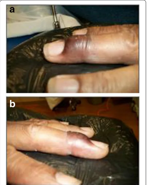
Case presentation:A 62-year-old African-American male was presented with a 12-month history of gradual darkening of the dorsal-distal middle left finger. Examination revealed a hyper pigmented scaly patch on the proximal to lateral nail fold of the L 3rd finger, nail dystrophy, and vertical split in the lateral section of the nail. The patient underwent evaluation of the lesion by Plastic Surgery with the removal of the lateral nail and a nail bed biopsy. Pathology revealed squamous cell carcinoma in situ with a possible focal positive, deep margin. The patient deliberated over surgical opinions, and eventually decided on radiation. A high dose rate Electronic Brachytherapy system using the XOFT Accent controller delivered dose of 4000 cGy in eight fractions, twice weekly, with at least 48 hours between fractions and treatment prescribed to a depth of 0 to 2 mm. The Xoft unit has specialized skin applicators that permit superficial treatment. Parameters assessed included the efficacy, cosmetic results feasibility, and acute safety of the Electronic Brachytherapy technique.
Conclusions:The patient exhibited moderate redness, hyperpigmentation erythema, desquamation, and Grade 1
to 2 edema acutely (following radiation), which resolved within 1 month of the treatment. Electronic brachytherapy treatment delivery took about 6 minutes, and the total procedure time was about 15 minutes. At the median follow-up of one year, the area revealed excellent cosmesis, and there was no infection or fat necrosis,
desquamation, no cancer recurrence, and no evidence of fibrosis at the last follow-up. This suggests that Electronic Brachytherapy was a viable treatment option for this particular patient.
Keywords:Skin cancer, Radiation therapy, Electronic brachytherapy
Background
Skin cancers are a worldwide health problem. In the United States, over two million patients are consistently detected with basal cell or squamous cell carcinoma per year [1]. Squamous cell carcinoma (SCCA-in-Situ) is a cu-taneous malignancy, first described by Bowen in the 1900’s. The normal manifestation is an appearance of a slowly growing, sharply defined erythematous, scaly plaque. In darker skin types, it can appear as a hyper
pigmented, scaly patch or plaque, with the darker skin type masking the pink color of erythema. It, typically, oc-curs in the sun exposed areas and in patients over the age of 60, but any age and location can be affected. Eight per-cent of untreated cases may, eventually, progress to inva-sive carcinoma. While surgical removal is, typically, the standard treatment, other well-defined treatment options include cryotherapy [2], curettage and electrocautery, sur-gical excision, laser ablation [3], photodynamic therapy (PDT), topical 5-FU and topical imiquimod [1,4-7]. Con-ventional radiation is another treatment option. The treat-ment options include electron beam or photons given daily over a series of 3- 6 weeks. The challenge with * Correspondence:arterbery@gmail.com
1
Crittenton Radiation Oncology Center, 1901 Star Batt Drive, Rochester Hills, MI 48309, USA
Full list of author information is available at the end of the article
week for a total dose of 4000 cgy), is identical to that used with High dose rate after-loaded techniques. Electronic Brachytherapy technique (EBT) can be used as a treat-ment for skin malignancy without the use of a radio-active isotope, but uses a similar hypo fractionated regimen. It is a form of high-dose-rate Electronic Brachytherapy but using a miniaturized x-ray tube, in-stead of a live isotope. Either surgery or radiation would be a standard treatment approach for squamous cell carcinoma in Situ of the digit [2-4,8].
In general, the dose ranging from 35 to 65 gray with standard fractionation is acceptable. Other fractionation regimens are also acceptable that are not necessarily daily. Optimal external beam radiation treatment regi-men, typically, includes fractions that range from 2 to 3.5 centigray. This method ensures greater benefit in terms of better normal tissues preservation and superior aesthetic functional outcome [10].
The risk factors that can predispose individuals to these conditions include prolonged sun exposure, chronic immunosuppressive, and arsenic exposure [11]. The traditional management of Bowen’s disease is surgi-cal excision or Mohs surgery [12]. However, when Bowen’s disease or squamous cell carcinoma occurs in the digits, surgical removal can lead to scar contracture, loss of the fingernail, or amputation in extensive cases. Alternative treatments, such as cryotherapy, photo-dynamic therapy, and topical creams have other draw-backs, such as variable response rate, dependence on patient compliance for topical creams, and variability in the technique (cryotherapy and PDT8). Others have tried a combination of photodynamic therapy and radi-ation [13]. There are some additional limited data that suggest carbon dioxide laser vaporization for Bowne’s disease may be effective. All of these nonsurgical and non-radiation approaches are considered in order to spare function but have limitations since they have been only moderately successful [14].
Case presentation
In this case study, the patient was, primarily, treated with a superficial Electronic Brachytherapy technique
priate surface areas for the custom applicator. Accept-able clinical target volume around the tumor was a 3 mm margin due to the sharp fall off of the source.
[image:2.595.305.540.396.692.2]Then, a customized lead shield (used as a cut out) and a 3.5 cm cone applicator were selected. The half-value layer of the lead was calculated in order to block the unin-tended areas and decrease the amount of dose to the un-involved tissues. After simulation, the depth calculation was based on the thickness of the lesion, and then, the treatment was delivered to the patient. The Xoft skin ap-plicators are calibrated with fixed depth dose curves. For example if you prescribe to .5 cm from the applicator
surface, your surface dose is 1000 cGy. The comprehen-sive calibration processes ensured correct dose output of these applicators prior to each treatment. Primary obser-vations revealed the average normal dose-rate output of the skin surface for the 35 mm applicator was 1.35 gray
per minute with a positive/negative 5% variation. The out-put variation is within 2%. Calculations of the effective source-surface distance were carried out on the air-gap measurements for all applicator sizes, and the field flatness and symmetry were well within 5%. Calibration in this case study was identical to cases reported elsewhere on quality assurance for this technique [15].
Clinically, the patient had a fair amount of keratotic epi-thelialization, and thus, the initial treatment prescription was to 2 mm in depth. After the first three fractions, the patient developed severe irritation and the lesion appeared to flatten, and thus, the doses for the remaining fractions delivered at the skin-surface. The patient received a total dose of 4000 centigray delivered in 500 centigray fractions over eight fractions twice a week with 48 hours between fractions. The total time for radiation delivery was about 5 minutes, and the total mean procedure time was about 15 minutes from the start to finish (including the clinical set up). The patient tolerated the treatment well and had no recurrence of the cancer in the one-year follow-up and overall cosmesis was excellent in the patient. There was no infection, fat necrosis, desquamation, or cancer recur-rences. There was no evidence of fibrosis, at the last follow-up session. There has been no recurrence of tumor. At the site of the radiation application, mild skin des-quamation developed in the weeks following the treat-ment. This reaction healed with minimal fibrosis and epithelialized quickly. There was no adverse reaction to the radiation, except for the appearance of a small 2 mm area of moist desquamation, appropriately one week after the last treatment, which resolved with topical agents. The patient also experienced hypopigmentation that is slowly resolving.
Discussion
[image:3.595.57.289.88.387.2]Ever since its introduction in 2009, the Xoft Accent Electronic Brachytherapy unit with skin applicators has Figure 2Clinical set up of treatment with Xoft machine.
Selected treatment cone is placed directly on the surface of the finger. The xoft machine and the electronic source are inserted directly into the hollow cone to allow the dose delivery directly to the area.
[image:3.595.303.538.89.239.2]Figure 3Customized lead cut out used on the digit to protect uninvolved areas.The half-value of lead is calculated to shield 100% of non-involved tissues. The cut out is designed based on the clinical shape and size of the lesion with a small margin.
[image:3.595.57.290.535.685.2]tively sophisticated, much localized, non-invasive, and allows superficial delivery of radiation in a variety of set-tings. Because of the twice-a-week treatment and only a total of 8 fractions, this technique has a higher degree of patient acceptance and compliance. It may be suitable for lesions in an unusual location, the elderly, and in cases when patients are unable to tolerate five weeks of daily treatment. The significant advantage is patient con-venience and the lack of regulatory issue associated with an after-loaded source. This technique requires less physicist supervision and the machine can be operated by the therapists, increasing treatment efficiency. There is limited data about the long term effectiveness of this technique and the largest experience has short follow up and excellent tumor control similar to what is seen with external beam therapy (16) Although, we did see acute forms of skin reaction, such as erythema, dry desquam-ation, and mild moist desquamdesquam-ation, these conditions healed quickly with minimal toxicity, and are due to the high surface dose in this technique. Hyperpigmentation or hypopigmentation of radiated skin may develop as chronic reaction within a couple of months after com-pletion of the treatment, but this reaction will fade. It may also progress to a vitiligo appearance in some pa-tients [16].
Conclusion
Superficial skin tumors of the digit are a challenging man-agement problem, and can be effectively treated by Elec-tronic Brachytherapy (EBT) using the superficial skin applicators. EBT to the skin, for squamous cell carcinoma in situ, is a feasible alternative to surgery for squamous cell carcinoma of the digit. Further exploration and more extensive follow-up on this method will help to gain better insights and shed light in the direction of clinical useful-ness of the procedure. This case study positively indicates that Electronic Brachytherapy using superficial skin appli-cators is a local therapeutic solution for the treatment of this case. It is a convenient option and a non-invasive therapy with satisfactory cosmetic results and outcome for this patient. The convenience and portability of this tech-nique can allow more patients access to care.
the manuscript deciding treatment guidelines, and clinical delivery. She also has written, proofread and edited this submission. AW provided assistance on the proofreading and formatting of the paper as well as content review. She has read and edited this submission. Both authors have read and approved the final manuscript.
Authors’information
VEA is a radiation oncologist with an interest in Electronic Brachytherapy and emerging technologies with radiation techniques. She has a Master’s degree in Health Services Administration from the University of Michigan as well as MD from that institution. VEA is interested in novel treatments that produce excellent outcome with improved cost benefit and quality of life for patients.
Acknowledgements
The authors would like to thank Kim Grammes and Renu Sharma for their efforts with this study and for their administrative assistance with the manuscript.
Author details
1Crittenton Radiation Oncology Center, 1901 Star Batt Drive, Rochester Hills,
MI 48309, USA.2Department of Dermatology, University Physician Group-Georgetown, Professional Building, Suite 208, Sterling Heights, MI 48310, USA.
Received: 14 August 2012 Accepted: 27 February 2013 Published: 15 April 2013
References
1. Rogers HW, Weinstock MA, Harris AR, Hinckley MR, Feldman SR, Fleischer AB, Coldiron BM:Incidence Estimate of Non-melanoma Skin Cancer in the United States.Arch Derm2006,142:729–735.
2. Ahmed I, Jones B, Holmes C, O' Callaghan C, Ilchyshyn A:Comparison of cryotherapy with curettage in the treatment of Bowen's disease: a prospective study.Br J Dermatol2000,143(4):759–766.
3. Tantikun N:Treatment of Bowen's disease of the digit with carbon dioxide laser.J Am Acad Dermatol2000,43(6):1080–1083.
4. Palungwachira P, Ogawa H:Treatment of multiple lesions of Bowen's disease and squamous cell carcinoma with topical imiquimod.
J Dermatol2005,32(12):1005–1009.
5. Patel GK, Goodwin R, Chawla M,et al:Imiquimod 5% cream monotherapy for cutaneous squamous cell carcinoma in situ (Bowen’s disease): a randomized, double-blind, placebo-controlled trial.J Am Acad Dermatol
2006,54:1025–1032.
6. Hansen JP, Drake AL, Walling HW:Bowen's Disease: a four-year retrospective review of epidemiology and treatment at a university center.Derm Surg2008,34(7):878–883.
7. Leibovitch I, Huilgol SC, Selva D, Franzco AAA, Richards S, Paver R:
Cutaneous squamous carcinoma in situ (Bowen’s disease): Treatment with Mohs micrographic surgery.J Am Acad Dermatol2005,52:997. 8. Herman JM, Pierce L, Sandler HM, Griffith K, Jabbari S, Hiniker S, Johnson T:
Radiotherapy using the water bath in the treatment of Bowen's disease of the digit.Radiother Oncol2008,88(3):398–402. Epub.
10. Zygogianni A, Kouvaris J, Tolia M, Kyrgias G, Beli I, Kantzou I, Soulimioti G, Kouloulias V:The Potential Role of Radiation Therapy in Bowen's Disease: A Review of the Current Literature.Rev Recent Clin Trials2011: ISSN: 1876-1038. 11. Foo CC, Lee JS, Guilanno V, Yan X, Tan SH, Giam YC:Squamous cell
carcinoma and Bowen's disease of the skin in Singapore.Annual Acad Med Singapore2007,36(3):189–193. 17450264.
12. Leibovitch I, Huilgol SC, Selva D, Richards S, Paver R:Cutaneous squamous carcinoma in situ (Bowen's disease): treatment with Mohs micrographic surgery.J Am Acad Dermatol2005,52(6):997–1002.
13. Nakano A, Watanabe D, Akita Y, Kawamura T, Tamada Y, Matsumoto Y:
Treatment efficiency of combining photodynamic therapy and ionizing radiation for Bowen's disease.J Eur Acad Dermatol Venereol2011,25(4):475–478. 14. Gordon KB, Garden JM, Robinson JK:Bowen's disease of the distal digit
outcome of treatment with carbon dioxide laser vaporization.Dermatoll Surg1996,22(8):723–728.
15. Rong Y:Surface applicator calibration and commissioning for skin applicators for electronic brachytherapy.Med Phys2010,37(10):5509–5517. 16. Shimm DS, Cassady JR:The skin. InMoss' radiation oncology.7th edition.
Edited by Cox JD. St. Louis: Mosby–Year Book Inc; 1994:99–118.
doi:10.1186/1756-0500-6-147
Cite this article as:Arterbery and Watson:An electronic brachytherapy technique for treating squamous cell carcinoma in situ of the digit: a case report.BMC Research Notes20136:147.
Submit your next manuscript to BioMed Central and take full advantage of:
• Convenient online submission
• Thorough peer review
• No space constraints or color figure charges
• Immediate publication on acceptance
• Inclusion in PubMed, CAS, Scopus and Google Scholar
• Research which is freely available for redistribution