Abstract
Gene expression profiling is a relatively new technology for the study of breast cancers, but within the past few years there has been a rapid rise in interest in its potential to improve the clinical management of breast cancer. This technology has contributed to our knowledge of the molecular pathology of breast tumours and shows promise as a tool to predict response to therapy and outcome, such as risk of metastasis. Microarray technology is continually developing and it is becoming apparent that, despite the various platforms available, robust conclusions can still be drawn that apply across the different array types. Gene expression profiling is beginning to appear in the breast cancer clinic but it is not yet fully evaluated. This review explores the questions that must be addressed before this technology can become an everyday clinical tool.
Introduction
Ongoing development of a wide spectrum of therapies for breast cancer has increased the complexity of breast cancer management in recent years. Clinicians must make decisions regarding the best treatment for an individual woman, but the disease parameters currently used to decide upon treatment are relatively unsophisticated. Furthermore, although these parameters may predict proportions of the population in which an outcome may occur fairly accurately, they cannot identify in which women the outcome will occur. This leads to over-treatment of many patients and inadequate treatment of others. High-throughput genomic technologies have been used to delineate tumour subtypes and response to therapy, and to identify patient characteristics that may influence tumour behaviour. One of the keys to improving breast cancer management is the targeting of treatment to those who will truly benefit, thereby avoiding iatrogenic morbidity in those who will not. The use of gene expression profiling is
unfortunately not straightforward, because differences between the platforms used in studies have led to inconsis-tencies in results and considerable scepticism. However, as is discussed below, these problems are not insurmountable and gene expression profiling shows considerable promise, but large clinical studies are now needed.
Historical perspective
Clinically measurable characteristics (for instance, tumour size, spread to lymph nodes, distant metastases and histo-logical appearance [tumour grade]) and patient characteristics (such as age and menopausal status) give an approximate guide to tumour behaviour and formed the basis of the earliest prognostic indices. Advances in immuno-histochemistry added oestrogen receptor (ER)-α and progesterone receptor status, and most recently growth factor receptor status (epidermal growth factor receptor and ERBB2 over-expression), Ki-67 and others. Some or all of these factors are included in indices such as The International Consensus Guidelines of St Gallen [1] and the Nottingham Prognostic Index [2], which are widely used to guide treatment. It has been obvious for years that breast cancer is a heterogeneous disease, and more recently it has become apparent that dissection of the molecular basis of malignancies classifies tumours into increasingly large numbers of subtypes that require individualized treatment. The advent of genomics and proteomics could be regarded as a natural progression from the use of only a few markers to the combination of large numbers of markers – ‘tumour signatures’ – that can define tumour subtypes and predict outcome.
Review
High-throughput genomic technology in research and clinical management of
breast cancer
Exploiting the potential of gene expression profiling:
is it ready for the clinic?
Andrew H Sims
1,2, Kai Ren Ong
1,2, Robert B Clarke
2and Anthony Howell
21AHS and KRO contributed equally to the review
2Breast Biology Group, University of Manchester, Paterson Institute for Cancer Research, Wilmslow Road, Manchester, M20 4BX, UK
Corresponding author: Anthony Howell, [email protected]
Published: 11 October 2006 Breast Cancer Research2006, 8:214 (doi:10.1186/bcr1605) This article is online at http://breast-cancer-research.com/content/8/5/214
© 2006 BioMed Central Ltd
Gene expression approaches to classification,
prognostication and determining response to
treatments
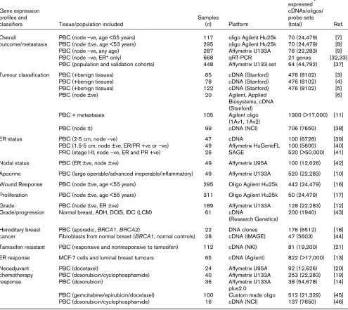
The primary focus of work using gene expression microarrays in the breast cancer field has been the molecular sub-classification of breast cancer put forward by Perou and Sorlie and their coworkers [3-6] and the prognosis profiles proposed by the Amsterdam [7,8] and Rotterdam [9] groups (Table 1). The luminal and basal-like subtypes have been
repeatedly identified and validated by gene expression analysis as the most distinctive of three or more molecular subtypes of breast cancer. Tumours that are positive for ERBB2 are predominantly identified as a distinct subtype of breast cancer by the ‘intrinsic’ subtype classification [3-6] and among the ‘molecular apocrine’ tumours [10]. However, ERBB2-positive tumours are also found within other classes, which presumably reflects heterogeneous expression of ER and their distinctive therapeutic and clinical attributes. The
Table 1
Gene expression studies of breast carcinomas aiming to improve clinical management
Number of differentially expressed
Gene expression cDNAs/oligos/
profiles and Samples probe sets
classifiers Tissue/population included (n) Platform (total) Ref.
Overall PBC (node –ve, age <55 years) 117 oligo Agilent Hu25k 70 (24,479) [7]
outcome/metastasis PBC (node ±ve, age <53 years) 295 oligo Agilent Hu25k 70 (24,479) [8]
PBC (node –ve, any age) 287 Affymetrix U133A 76 (22,283) [9]
PBC (node –ve, ER+only) 668 qRT-PCR 21 genes [32,33]
PBC (population and validation cohorts) 448 Affymetrix U133 set 64 (44,792) [37]
Tumour classification PBC (+benign tissues) 65 cDNA (Stanford) 476 (8102) [3]
PBC (+benign tissues) 78 cDNA (Stanford) 476 (8102) [4]
PBC (+benign tissues) 122 cDNA (Stanford) 476 (8102) [5]
PBC (node ±ve) 20 Agilent, Applied [6]
Biosystems, cDNA (Stanford)
PBC + metastases 105 Agilent oligo 1300 (>17,000) [11]
(1Av1, 1Av2)
PBC (node ±) 99 cDNA (NCI) 706 (7650) [38]
ER status PBC (2-5 cm, node –ve) 47 cDNA 100 (6728) [39]
PBC (1.5-5 cm, node ±ve, ER/PR +ve or –ve) 49 Affymetrix HuGeneFL 100 (5600) [40] PRC (stage I-II, node –ve, ER and PR +ve) 26 SAGE 520 (>50,000) [41]
Nodal status PBC (ER ±ve, node ±ve) 49 Affymetrix U95A 100 (12,626) [42]
Apocrine PBC (large operable/advanced inoperable/inflammatory) 49 Affymetrix U133A 520 (22,283) [10]
Wound Response PBC (node ±ve, age <55 years) 295 Oligo Agilent Hu25k 442 (24,479) [16]
Proliferation PBC (node ±ve, age <55 years) 311 Oligo Agilent Hu25k 50 (24,479) [17]
Grade PBC (node ±ve, ER ±ve) 189 Affymetrix U133A 128 (22,283) [12]
Grade/progression Normal breast, ADH, DCIS, IDC (LCM) 61 cDNA 200 (1940) [43]
(Research Genetics)
Hereditary breast PBC (sporadic, BRCA1, BRCA2) 22 DNA clones 176 (6512) [18]
cancer Fibroblasts from normal breast (BRCA1, normal controls) 28 cDNA (IMAGE) 47 (5603) [44] Tamoxifen resistant PBC (responsive and nonresponsive to tamoxifen) 112 cDNA (NKI) 81 (19,200) [21]
ER response MCF-7 cells and luminal breast tumours 65 cDNA (Agilent) 822 (>17,000) [13]
Neoadjuvant PBC (docetaxel) 24 Affymetrix U95A 92 (12,626) [20]
chemotherapy PBC (doxorubicin/cyclophosphamide) 40 Affymetrix U133A 253 (22,283) [19]
response PBC (doxorubicin) 36 Affymetrix U133A 38 (54,678) [14]
plus2.0
PBC (gemcitabine/epirubicin/docetaxel) 100 Custom made oligo 512 (21,329) [45]
PBC (doxorubicin/cyclophosphamide) 16 cDNA (NCI) 137 (7650) [46]
[image:2.612.58.553.143.584.2]molecular subtypes identified are associated with significantly different clinical outcomes [4,11] and are likely to respond best to different treatment approaches.
Traditional classifications of tumours may provide clear-cut treatment options in high and low risk cases, but often tumours fall into an ‘intermediate’ group; it is in these borderline cases that improvements are most urgently required. In these cases the ‘safe’ option is to over-treat, benefiting a relatively small minority of cases and exposing the rest to side effects unnecessarily. Conversely, a more conservative approach may avoid unwarranted treatment and additionally reduce costs, but some women that would benefit may go untreated. Studies that examine links between gene expression and known prognostic factors such as grade [12] and ER status [13] may be beneficial for this intermediate group. Tumour grade is an excellent example; grade 3 tumours are at much higher risk of recurrence than are grade 1 tumours, but 30-60% of tumours are classified as histological grade 2. Sotiriou and coworkers [12] recently used gene expression profiling to reclassify grade 2 tumours into two groups resembling grade 1 and grade 3 tumours with low and high risk of recurrence, respectively, thereby better characterizing these tumours conventionally considered to be ‘intermediate risk’.
Expression profiling has uncovered signatures associated with distinct phenotypes (Table 1), such as profiles exhibited by hypoxic tumours or inflammatory breast cancers [14]. A recent study [15] showed that medullary breast cancer is a subgroup of basal breast cancers. Novel aspects of tumour behaviour have been identified by the expression of specific functional sets of genes such as ‘wound response’ genes [16]. A ‘proliferation signature’ has been shown to identify particular patient groups who have an extremely poor outcome in a subpopulation of breast cancer patients [17]. Gene expression studies have been used to distinguish between sporadic tumours and tumours from women with BRCA1and BRCA2germline mutations [18]. Tumours from BRCA1mutation carriers have been found to be frequently of the basal subtype, whereas BRCA2tumours fall mainly within the luminal A category [5]. Observations such as these may further our understanding of the pathogenesis of tumours in mutation carriers.
Response to treatment has also been studied using expression microarrays. A gene expression profile from pretreatment tumour biopsies has been found to predict response to combination doxorubicin/cyclophosphamide treatment [19]. The genes comprising this profile differed from those in the same group’s previously reported profile for predicting response to docetaxel therapy [20]. Similarly, response to tamoxifen can be predicted by expression profiling [21]. These exciting results suggest that predictive profiles could potentially be found for any given chemotherapy or endocrine regimen.
Influences of genetic background on outcome
The finding that tumours from BRCA1 mutation carriers are predominantly of the basal subtype clearly demonstrates that genetic background can predispose to a particular tumour subtype and outcome. Furthermore, a study showed that oncogene-induced mouse mammary tumors from 31 inbred strains exhibit differing rates of metastasis [22]. The study also reported distinct gene expression profiles according to genetic background. This suggests that the tendency for metastatic disease to develop may have a germline component. The existence of a common 11-gene ‘death from cancer’ signature from both epithelial and nonepithelial malignancies [23] may lend support to this hypothesis. The implication of a germline polymorphism as opposed to a somatic mutation for assessment of metastasis risk is that risk assessment can be done using any tissue at the time of diagnosis or even before diagnosis of a primary tumor [24]. The existence of inherited metastasis risk factors (or prospective metastatic biomarkers) has potentially highly significant implications for our models of metastasis, clinical prognosis and the development of tailored treatment. However, the specific genes and mechanisms responsible for this heritable effect on metastasis remain to be fully identified, and more evidence is required to establish their influence on human disease.Cost/benefits
Although the actual cost of performing gene expression analysis is likely to be higher than those of determining conventional clinical and histopathological markers, the potential savings in terms of avoiding over-treatment, based on existing guidelines, could be substantial.
Reliability of microarray data
The genes that make up a gene expression signature are by their nature dependent on the patient and tumour characteristics, array platform, normalization method and statistical thresholds for gene selection or the classification algorithm employed. Using a particular dataset to generate a predictive profile has its own inherent bias based on its attributes. By simply changing the members of ‘training’ and ‘test’ sets, Ein-Dor and coworkers [26] were able to identify more than 1000 genes that are related to survival, even when using the same dataset and methods as van’t Veer and colleagues [7,8], and found that many different but equally predictive lists of 70 genes could have been produced from the same analysis. Rather than indicating that the data are meaningless, this suggests that the predictive value is real but highly context dependent.
The comparability of different microarray platforms is a major issue. The lack of overlap (three genes) between the 70-gene signature of the Amsterdam group (cDNA arrays) [7,8] and the 76-gene signature of the Rotterdam group (Affymetrix oligonucleotide arrays) [9] has been claimed as evidence that genomic approaches are unreliable. A multiple random validation strategy has been suggested to combat this problem [27]. However, although validation is essential, a predictive signature determined from a highly selected group of samples cannot possibly be expected to replicate the exact findings when applied to a different group of samples using a different gene expression platform. The differences between signatures in different studies are due to varying inclusion criteria (age, lymph node status, diameter of tumour, adjuvant treatment, among other factors), the platform (cDNA or oligonucleotide arrays, or reverse transcription polymerase chain reaction) and data analysis methods used in each study.
Nevertheless, Sorlie and coworkers [6] recently showed that even with different array platforms the breast cancer subtypes are distinguishable at the unsupervised level. Despite some variation in the most differentially expressed genes identified by each array platform, there was a highly significant overlap at the pathway level. This confirms that distinct molecular mechanisms underlie the clinically relevant subtypes of breast cancer, and that perturbations in these mechanisms can be detected reliably by different platforms. Similarly, Hu and colleagues [11] recently described and validated a refined ‘intrinsic’ classification signature that is conserved across microarray platforms and also uncovered a possible ‘new’ subtype characterized by the high expression of interferon-regulated genes.
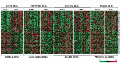
Validation of new findings with independent data is essential for us to believe that the results are indeed ‘real’, and this has been encouraged and facilitated by making it a prerequisite for publication that gene expression datasets are publicly accessible according to the MIAME (Minimum Information
About a Microarray Experiment) guidelines [28], allowing complete datasets to be downloaded. Public access has permitted studies in which gene expression data from several studies are re-analyzed. For example, meta-analyses of multiple experiments using different platforms have resulted in new predictive signatures that perform as well or better than the platform specific signature [29,30] (Figure 1). These approaches remove the inherent bias of a single microarray platform and are able to concentrate on genes that are consistently differentially expressed, regardless of the technology used. However, they may be limited by the number of common genes represented.
Improvements in technology and analysis
Advances in gene expression technology are continually being made, with increasing numbers of more clearly defined transcripts represented on each new generation of array. Improvements in RNA extraction, quantification and quality assessment (such as the RNA integrity number [31]) will increase reliability of expression profiling. Improved labelling and amplification protocols enable profiling to be performed on smaller amounts of tissue.Laser capture microdissection has been used to ensure that RNA is isolated from tumour rather than normal tissue. Alternatively, small amounts of RNA have been isolated from formalin fixed, paraffin embedded blocks rather than fresh frozen tissue, and can be used in assays such as the multigene reverse transcription polymerase chain reaction classifier commercially known as OncotypeDX [32,33]. These are important considerations because taking fresh tissue for gene expression analysis from a tiny tumour can compromise histopathology reporting. These improved methods will be required to circumvent the logistical difficulties, costs and time associated with collecting fresh tissue straight from a tumour at the time of surgery. Multicentre studies will be facilitated by techniques for extraction of intact RNA from preserved tissues, because these will obviate the need to collect and transport frozen material.
Although much of the additional information from expression profiling of primary tumours is consistent with that for existing markers, there have been some conflicting results; for example, some women found to be ERBB2 positive by immunohistochemistry were classified in the luminal cluster and not the ErbB2 group [4]. It may be difficult to choose the most appropriate treatment when different assays conflict, and of course existing technologies should not be discounted. Fluorescent in situ hybridization is still considered the most reliable and consistent method to determine ERBB2 status [34].
Trials: what is required
MINDACT (Microarray for Node-negative Disease Avoids Chemotherapy) trial, which will be run under the auspices of the TRANSBIG consortium and coordinated by the EORTC (European Organisation for Research and Treatment of Cancer). It will study women with node-negative disease who are aged 18-75 years. One of its primary aims is to establish whether women with a good prognosis gene expression signature (assessed using Mammaprint, a commercially available form of the 70-gene profile reported by van’t Veer and coworkers [7,8]) but poor prognosis clinicopathological status (as assessed using the Adjuvant! Online software [35]) can be spared chemotherapy without reducing metastasis-free survival. One of the secondary objectives is to identify and validate expression profiles that predict response to chemotherapy and endocrine therapy. Another large trial currently underway is being run by the US Intergroup PACCT (Program for the Assessment of Clinical Cancer Tests) to assess the OncotypeDX signature in 10,046 women aged 18-75 years with operable, node negative, ER positive and/or progesterone receptor positive breast cancer. In this study, patients with an intermediate risk OncotypeDX score will be randomized to combination chemotherapy and endocrine therapy (the usual treatment) or endocrine therapy alone. These two studies are seen as the best way to establish whether the genomic signatures will lead to the desired goal of better
targeting treatment to those who will benefit and so yielding improvements in breast cancer mortality and morbidity. With individualization of therapy, it is inevitable that women in a study will be undergoing many different treatment regimens. Consequently, it may become more difficult to obtain sufficiently high numbers of women with similar regimens to compare with each other to evaluate new tests with adequate statistical power. Many of the profiles have been developed in well defined groups of women (Table 1), and clearly it is important that validation takes place in equivalent groups of women undergoing the same treatment, because prognostic differences evident in one group may not be apparent in a differently treated group. It is likely that new profiles will be developed in the years ahead, and the need for large numbers of patients will necessitate large multinational trials.
New drugs are rapidly being developed. In order for gene expression profiling to be useful, the initiation of trials evidently needs to keep pace with the introduction of new treatments that are shown to be best practice.
Conclusion
[image:5.612.63.551.99.350.2]Fundamentally, there is a major leap from the observation that gene expression profiles can predict outcome to the use of
Figure 1
these profiles in treatment decisions. In recent years great advances in our knowledge of the molecular biology of breast tumours have been achieved using genomic approaches. It is perhaps not surprising that genomic signatures tend to outperform the pre-existing clinical models when one considers the large numbers of additional factors that are being taken into account, and that precision of measurement of these factors is continuous (gene expression levels) rather than categorical (for example, tumour grade and lymph node status). Despite their promise, some caution is required with the new approaches. A study comparing the power of gene expression measurements with that of conventional prognostic markers found that transcriptional profiling approaches did not perform noticeably better than indices constructed from the clinical variables [36].
The challenge that lies ahead is to convert the increased information potentially available from gene expression profiling of breast tumours into useful tools that can optimize clinical decisions and tailor treatment regimens to an individual patient, ultimately improving outcome and reducing overall costs. This will take place through advances in sample processing, microarray technology and statistical analysis techniques to obtain consistency. Finally, investment in trials of this genomic technology is needed now if savings in costs of treatment, particularly with the rise in availability of expensive drugs, are to be made in the long term.
Competing interests
The authors declare that they have no competing interests
References
1. Goldhirsch A, Glick JH, Gelber RD, Coates AS, Thurlimann B, Senn HJ: Meeting highlights: international expert consensus on the primary therapy of early breast cancer 2005.Ann Oncol
2005, 16:1569-1583.
2. Haybittle JL, Blamey RW, Elston CW, Johnson J, Doyle PJ, Camp-bell FC, Nicholson RI, Griffiths K: A prognostic index in primary breast cancer.Br J Cancer 1982, 45:361-366.
3. Perou CM, Sorlie T, Eisen MB, van de Rijn M, Jeffrey SS, Rees CA, Pollack JR, Ross DT, Johnsen H, Akslen LA, et al.: Molecular portraits of human breast tumours.Nature 2000, 406:747-752. 4. Sorlie T, Perou CM, Tibshirani R, Aas T, Geisler S, Johnsen H, Hastie T, Eisen MB, van de Rijn M, Jeffrey SS, et al.: Gene expression patterns of breast carcinomas distinguish tumor subclasses with clinical implications. Proc Natl Acad Sci USA
2001, 98:10869-10874.
5. Sorlie T, Tibshirani R, Parker J, Hastie T, Marron JS, Nobel A, Deng S, Johnsen H, Pesich R, Geisler S,et al.: Repeated obser-vation of breast tumor subtypes in independent gene expres-sion data sets.Proc Natl Acad Sci USA 2003, 100:8418-8423.
6. Sorlie T, Wang Y, Xiao C, Johnsen H, Naume B, Samaha RR, Bor-resen-Dale AL: Distinct molecular mechanisms underlying clin-ically relevant subtypes of breast cancer: gene expression analyses across three different platforms. BMC Genomics
2006, 7:127.
7. van ‘t Veer LJ, Dai H, van de Vijver MJ, He YD, Hart AA, Mao M, Peterse HL, van der Kooy K, Marton MJ, Witteveen AT, et al.: Gene expression profiling predicts clinical outcome of breast cancer.Nature 2002, 415:530-536.
8. van de Vijver MJ, He YD, van’t Veer LJ, Dai H, Hart AA, Voskuil DW, Schreiber GJ, Peterse JL, Roberts C, Marton MJ, et al.: A gene-expression signature as a predictor of survival in breast cancer.N Engl J Med 2002, 347:1999-2009.
9. Wang Y, Klijn JG, Zhang Y, Sieuwerts AM, Look MP, Yang F, Talantov D, Timmermans M, Meijer-van Gelder ME, Yu J, et al.: Gene-expression profiles to predict distant metastasis of lymph-node-negative primary breast cancer. Lancet 2005, 365:671-679.
10. Farmer P, Bonnefoi H, Becette V, Tubiana-Hulin M, Fumoleau P, Larsimont D, Macgrogan G, Bergh J, Cameron D, Goldstein D,et al.: Identification of molecular apocrine breast tumours by microarray analysis.Oncogene 2005, 24:4660-4671.
11. Hu Z, Fan C, Oh DS, Marron JS, He X, Qaqish BF, Livasy C, Carey LA, Reynolds E, Dressler L,et al.: The molecular portraits of breast tumors are conserved across microarray platforms.
BMC Genomics 2006, 7:96.
12. Sotiriou C, Wirapati P, Loi S, Harris A, Fox S, Smeds J, Nordgren H, Farmer P, Praz V, Haibe-Kains B,et al.: Gene expression pro-filing in breast cancer: understanding the molecular basis of histologic grade to improve prognosis. J Natl Cancer Inst
2006, 98:262-272.
13. Oh DS, Troester MA, Usary J, Hu Z, He X, Fan C, Wu J, Carey LA, Perou CM: Estrogen-regulated genes predict survival in hormone receptor-positive breast cancers.J Clin Oncol 2006, 24:1656-1664.
14. Dressman HK, Hans C, Bild A, Olson JA, Rosen E, Marcom PK, Liotcheva VB, Jones EL, Vujaskovic Z, Marks J, et al.: Gene expression profiles of multiple breast cancer phenotypes and response to neoadjuvant chemotherapy. Clin Cancer Res
2006, 12:819-826.
15. Bertucci F, Finetti P, Cervera N, Charafe-Jauffret E, Mamessier E, Adelaide J, Debono S, Houvenaeghel G, Maraninchi D, Viens P,et al.: Gene expression profiling shows medullary breast cancer is a subgroup of basal breast cancers.Cancer Res 2006, 66: 4636-4644.
16. Chang HY, Nuyten DS, Sneddon JB, Hastie T, Tibshirani R, Sorlie T, Dai H, He YD, van’t Veer LJ, Bartelink H,et al.: Robustness, scalability, and integration of a wound-response gene expres-sion signature in predicting breast cancer survival. Proc Natl Acad Sci USA 2005, 102:3738-3743.
17. Dai H, van’t Veer L, Lamb J, He YD, Mao M, Fine BM, Bernards R, van de Vijver M, Deutsch P, Sachs A,et al.: A cell proliferation sig-nature is a marker of extremely poor outcome in a subpopula-tion of breast cancer patients.Cancer Res 2005, 65:4059-4066. 18. Hedenfalk I, Ringner M, Ben-Dor A, Yakhini Z, Chen Y, Chebil G, Ach R, Loman N, Olsson H, Meltzer P,et al.: Molecular classifi-cation of familial non-BRCA1/BRCA2 breast cancer.Proc Natl Acad Sci USA 2003, 100:2532-2537.
19. Cleator S, Tsimelzon A, Ashworth A, Dowsett M, Dexter T, Powles T, Hilsenbeck S, Wong H, Osborne CK, O’Connell P,et al.: Gene expression patterns for doxorubicin (Adriamycin) and cyclophosphamide (Cytoxan) (AC) response and resistance.
Breast Cancer Res Treat 2006, 95:229-233.
20. Chang JC, Wooten EC, Tsimelzon A, Hilsenbeck SG, Gutierrez MC, Elledge R, Mohsin S, Osborne CK, Chamness GC, Allred DC,et al.: Gene expression profiling for the prediction of ther-apeutic response to docetaxel in patients with breast cancer.
Lancet 2003, 362:362-369.
21. Jansen MP, Foekens JA, van Staveren IL, Dirkzwager-Kiel MM, Rit-stier K, Look MP, Meijer-van Gelder ME, Sieuwerts AM, Portengen H, Dorssers LC, et al.: Molecular classification of tamoxifen-resistant breast carcinomas by gene expression profiling. J Clin Oncol 2005, 23:732-740.
22. Qiu TH, Chandramouli GV, Hunter KW, Alkharouf NW, Green JE, Liu ET: Global expression profiling identifies signatures of tumor virulence in MMTV-PyMT-transgenic mice: correlation to human disease.Cancer Res 2004, 64:5973-5981.
This article is part of a review series on High-throughput genomic technology in research and
clinical management of breast cancer, edited by Yudi Pawitan and Per Hall. Other articles in the series can be found online at
23. Glinsky GV, Berezovska O, Glinskii AB: Microarray analysis identifies a death-from-cancer signature predicting therapy failure in patients with multiple types of cancer.J Clin Invest
2005, 115:1503-1521.
24. Hunter KW, Crawford NP: Germ line polymorphism in metasta-tic progression.Cancer Res 2006, 66:1251-1254.
25. Oestreicher N, Ramsey SD, Linden HM, McCune JS, van’t Veer LJ, Burke W, Veenstra DL: Gene expression profiling and breast cancer care: what are the potential benefits and policy implications?Genet Med 2005, 7:380-389.
26. Ein-Dor L, Kela I, Getz G, Givol D, Domany E: Outcome signa-ture genes in breast cancer: is there a unique set? Bioinfor-matics 2005, 21:171-178.
27. Michiels S, Koscielny S, Hill C: Prediction of cancer outcome with microarrays: a multiple random validation strategy.
Lancet 2005, 365:488-492.
28. Microarray Gene Expression Data Society: MGED Society [www.mged.org]
29. Warnat P, Eils R, Brors B: Cross-platform analysis of cancer microarray data improves gene expression based classifica-tion of phenotypes.BMC Bioinformatics 2005, 6:265.
30. Shen R, Ghosh D, Chinnaiyan AM: Prognostic meta-signature of breast cancer developed by two-stage mixture modeling of microarray data.BMC Genomics 2004, 5:94.
31. Schroeder A, Mueller O, Stocker S, Salowsky R, Leiber M, Gassmann M, Lightfoot S, Menzel W, Granzow M, Ragg T: The RIN: an RNA integrity number for assigning integrity values to RNA measurements.BMC Mol Biol 2006, 7:3.
32. Paik S, Shak S, Tang G, Kim C, Baker J, Cronin M, Baehner FL, Walker MG, Watson D, Park T, et al.: A multigene assay to predict recurrence of tamoxifen-treated, node-negative breast cancer.N Engl J Med 2004, 351:2817-2826.
33. Paik S, Tang G, Shak S, Kim C, Baker J, Kim W, Cronin M, Baehner FL, Watson D, Bryant J,et al.: Gene expression and benefit of chemotherapy in women with node-negative, estro-gen receptor-positive breast cancer. J Clin Oncol 2006, 24: 3726-3734.
34. Perez EA, Suman VJ, Davidson NE, Martino S, Kaufman PA, Lingle WL, Flynn PJ, Ingle JN, Visscher D, Jenkins RB: HER2 testing by local, central, and reference laboratories in specimens from the North Central Cancer Treatment Group N9831 intergroup adjuvant trial.J Clin Oncol 2006, 24:3032-3038.
35. Adjuvant! Online. Decision making tools for health care pro-fessionals[www.adjuvantonline.com]
36. Eden P, Ritz C, Rose C, Ferno M, Peterson C: "Good Old" clini-cal markers have similar power in breast cancer prognosis as microarray gene expression profilers. Eur J Cancer 2004, 40:1837-1841.
37. Pawitan Y, Bjohle J, Amler L, Borg AL, Egyhazi S, Hall P, Han X, Holmberg L, Huang F, Klaar S, et al.: Gene expression profiling spares early breast cancer patients from adjuvant therapy: derived and validated in two population-based cohorts.Breast Cancer Res2005, 7:R953-964.
38. Sotiriou C, Neo SY, McShane LM, Korn EL, Long PM, Jazaeri A, Martiat P, Fox SB, Harris AL, Liu ET: Breast cancer classification and prognosis based on gene expression profiles from a pop-ulation-based study. Proc Natl Acad Sci USA 2003, 100: 10393-10398.
39. Gruvberger S, Ringner M, Chen Y, Panavally S, Saal LH, Borg A, Ferno M, Peterson C, Meltzer PS: Estrogen receptor status in breast cancer is associated with remarkably distinct gene expression patterns.Cancer Res 2001, 61:5979-5984. 40. West M, Blanchette C, Dressman H, Huang E, Ishida S, Spang R,
Zuzan H, Olson JA, Jr., Marks JR, Nevins JR: Predicting the clini-cal status of human breast cancer by using gene expression profiles.Proc Natl Acad Sci USA 2001, 98:11462-11467. 41. Abba MC, Hu Y, Sun H, Drake JA, Gaddis S, Baggerly K, Sahin A,
Aldaz CM: Gene expression signature of estrogen receptor alpha status in breast cancer.BMC Genomics 2005, 6:37. 42. Huang E, Cheng SH, Dressman H, Pittman J, Tsou MH, Horng
CF, Bild A, Iversen ES, Liao M, Chen CM,et al.: Gene expres-sion predictors of breast cancer outcomes.Lancet 2003, 361: 1590-1596.
43. Ma XJ, Salunga R, Tuggle JT, Gaudet J, Enright E, McQuary P, Payette T, Pistone M, Stecker K, Zhang BM,et al.: Gene expres-sion profiles of human breast cancer progresexpres-sion.Proc Natl Acad Sci USA 2003, 100:5974-5979.
44. Kote-Jarai Z, Williams RD, Cattini N, Copeland M, Giddings I, Wooster R, tePoele RH, Workman P, Gusterson B, Peacock J,et al.: Gene expression profiling after radiation-induced DNA damage is strongly predictive of BRCA1 mutation carrier status.Clin Cancer Res 2004, 10:958-963.
45. Thuerigen O, Schneeweiss A, Toedt G, Warnat P, Hahn M, Kramer H, Brors B, Rudlowski C, Benner A, Schuetz F, et al.: Gene expression signature predicting pathologic complete response with gemcitabine, epirubicin, and docetaxel in primary breast cancer.J Clin Oncol 2006, 24:1839-1845. 46. Sotiriou C, Powles TJ, Dowsett M, Jazaeri AA, Feldman AL,
Asser-sohn L, Gadisetti C, Libutti SK, Liu ET: Gene expression profiles derived from fine needle aspiration correlate with response to systemic chemotherapy in breast cancer.Breast Cancer Res