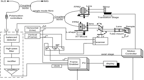
Optical-coherence tomography, extension to imaging of low coherence interferometry
Full text
Figure


Related documents
We show that if a particular (but most natural) aver- age case reduction from SBB to GSBB exists, then the Strong Di¢ e-Hellman (SDH) and the Computational Di¢ e-Hellman (CDH) have
It can be concluded that the long-term sustainable growth in the labour market is secured in both countries, based on an analysis of the development of indicators affecting the
Flavoring and medicinal values of the yellow pigment produced by Monascus ruber 4066 strain cultivated on static malt agar medium fungal endophytes from Garcinia
Figures 9- 11 show heat transfer rate on the hot and cold heat sources in the form of the average Nusselt number, average temperature of the chamber and maximum
This study aims to find the best mixture formula based on caloric value, best particle size (mesh), and calculate the physical properties of the best biomass pellets
(8) A viral infection is caused by the virus itself and NSP4 protein responsible, directly or through a messenger, for activation of the enteric nervous system
For example, the most recent version of the rubric accounts for the scenario of a student who submits an original post by Tuesday, but who does not submit a response
