Structure-function analysis of an
autoactive derivative of the tomato Cf-9
disease resistance protein
Choon Yang (Kevin) Tee
June 2016
A thesis submitted for the degree of Doctor of Philosophy
i
DECLARATION
I declare that this thesis is my own work and has not been submitted in any form for another degree or diploma at any university or other institution of higher education. Information derived from the published or unpublished work of others has been acknowledged in the text and a list of references is given. Materials obtained for use in this study that were generated by others have been acknowledged accordingly in the text.
ii
ACKNOWLEDGEMENTS
This work would not have been possible without the support from the following people to whom I express my thanks:
To my principal supervisor Assoc. Prof. Dr David Jones, for his continuous guidance, patience and input in this thesis, although there have been great challenges especially in the writing phase. My PhD advisors Assoc. Prof. Adrienne Hardham and Assoc. Prof. Peter Solomon for their previous advice, Dr Claire Anderson for her initiation of the M205 project and helpful discussions, and Dr Ming-Bo Wang for allowing access to the multiwell fluorescence platereader facility in CSIRO for MUG assay experiments.
To the past and present members of Jones lab for the stimulating research environment, including Dr Ginny Lim, Dr Neil Horner, Dr Maryam Rafiqi, Cahya Prihatna, Thilaga Vellusamy, Ursula Hurley, Dr Ann-Maree Cantanzariti, Dr Yvonne Gonzalez-Cendales, Huong Do, Laura Rolston, Nadya Farah and Jaime Simbaqueba. Special thanks to the plant culture service team Christine Larsen, Jenny Rath and Steve Dempsy for their excellent horticultural assistance.
iii
ABSTRACT
An autoactive chimera of the tomato extracellular leucine-rich repeat receptor-like protein Cf-9, designated Hcr9-M205 has been characterized previously as exhibiting characteristics of constitutive defence activation (Barker et al., 2006b). The initial work of this thesis (Chapter 3) involved generation and assessment of transgenic tobacco containing an E22 (PR-5) promoter: gusA reporter construct as a quantitative reporter for Hcr9-M205 autoactivity in Agrobacterium-mediated transient expression (agroinfiltration) assays. Time course analysis showed that the induction of E22 promoter preceded the necrotic response induced by Hcr9-M205, providing an early indication of defence activation. Further characterization of the E22 promoter (Chapter 4) by incubating the E22: gusA tobacco leaf disks in different defence-inducing compounds using a multi-well plate set-up indicated the defence-inducible nature of E22 promoter including antagonistic regulation by salicylic acid and jasmonic acid, activation by ethylene and synergistic activation by salicylic acid and cytokinin; demonstrating the applicability of the leaf disks assays in screening potential plant defence activators.
iv
18 had the greatest effects in signalling repression: Y389 of LRR 13 and E411 of LRR 14 did not significantly affect autoactivity, A433 of LRR 15 marginally repressed autoactivity whereas L457 of LRR 16 completely abolished autoactivity, similar to L481 of LRR 17 shown by Anderson et al. (in preparation). These findings were consistent with the notion that Cf-9 is autoinhibited by interactions between LRRs 15-17 and LRR 18. Unexpectedly, introduction of C387 of LRR 13 into Hcr9-M205 enhanced autoactivity. Sequence analysis comparing the Hcr9-M205(L389C) mutant containing C387 in Hcr9-M205, the CLB103V(14) domain swap that exhibited enhanced autoactivity and domain swaps that did not indicated that this phenomenon only occurred with an additional Hcr9-9A substitution spanning LRRs 14-17, suggesting that C387 may enhance signal activation upon Avr9-induced derepression and a possible role of E411 of LRR 14 in signalling repression. The data revealing some of the specificity-determining residues in signalling repression suggest that Avr9 recognition may directly compete with the autoinhibitory interactions mediated by these residues for Cf-9 activation.
v
LIST OF FIGURES
CHAPTER 1
Figure 1.1 Overview of the plant immune system.
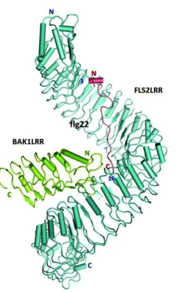
Figure 1.2 Crystal structure of the FLS2-BAK1-flg22 complex.
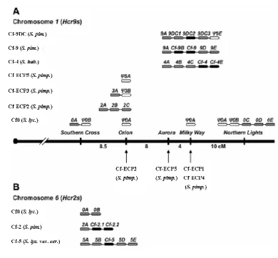
Figure 1.3 Structural organization of Hcr9 and Hcr2 gene clusters in the tomato genome. Figure 1.4 Structural domains of the tomato Cf-9 resistance protein.
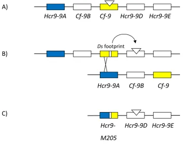
Figure 1.5 Hcr9-M205 gene was generated by a complex transposon-induced recombination event.
CHAPTER 3
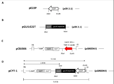
Figure 3.1 Diagrammatic representation of the intermediate plasmids involved in the generation of the pCYT-1 binary vector containing an E22 promoter (E22P): gusA reporter: E22 terminator (E22T) cassette.
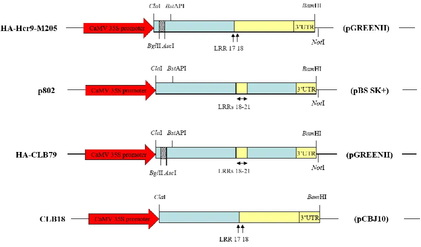
Figure 3.2 Constructs used for induction of the E22: gusA reporter.
Figure 3.3 DNA gel blots of nine F2 kanamycin resistant transgenic pCYT-1 tobacco plants for line 3B.
Figure 3.4 DNA gel blot showing segregation of 46 F2 kanamycin resistant transgenic pCYT-1 tobacco plants for line 14.
Figure 3.5 DNA gel blots of pCYT-1 primary transformants for lines 16B, 20A, 20B, 24, 30A and 30B.
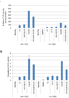
Figure 3.6 GUS activity induced by agroinfiltration of defence-activating constructs in transgenic pCYT-1 tobacco lines 14(8) and 14(2) at 5 dpi (days post infiltration).
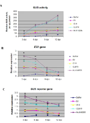
Figure 3.7 Time-course analysis comparing A) GUS activity B) expression of the endogenous E22 gene and C) expression of the gusA reporter gene induced by agroinfiltration of selected defence-activating constructs at 3, 6, 9 and 12 dpi (days post infiltration).
vi CHAPTER 4
Figure 4.1 Tissue-specific GUS activity in the flower parts of healthy transgenic E22 promoter: gusA reporter (pCYT-1) tobacco detected by GUS histochemical staining. Figure 4.2 GUS activity in leaves of mature and senescing transgenic E22 promoter: gusA reporter (pCYT-1) tobacco plants.
Figure 4.3 Effects of SA and JA on GUS activity in E22 promoter: gusA leaf disks after 48 hours of incubation.
Figure 4.4 Time-course analysis of the induction of GUS activity in E22 promoter: gusA leaf disks by salicylic acid (SA), cytokinin (CK) and ethylene (ET) after 12, 24, 36, 48, 60 and 72 hours of incubation.
Figure 4.5 GUS activity in the transgenic E22 promoter: gusA reporter (pCYT-1) tobacco leaf disks incubated with 2 mM salicylic acid (SA) after 24, 48 and 72 hours of incubation revealed by GUS histochemical staining.
Figure 4.6 Effects of combined cytokinin (CK) and salicylic acid (SA) application on GUS activity in E22 promoter: gusA leaf disks after incubation for 60 hours.
Figure 4.7 Induction of GUS activity in E22 promoter: gusA leaf disks by 50 mM NaCl after 48 hours of incubation.
CHAPTER 5
Figure 5.1 Autoactivity of Hcr9-M205 protein, its progenitors Cf-9 and Hcr9-9A, and the reciprocal domain swap CLB101.
Figure 5.2 Domain swaps that define the position of the junction between Hcr9-9A and Cf-9 required for autoactivity.
Figure 5.3 Domain swaps dissecting the C-terminal region required for signal transduction of Hcr9-M205.
Figure 5.4 Domain swaps dissecting the N-terminal Hcr9-9A sequence required for Hcr9-M205 autoactivity.
Figure 5.5 Comparison between Cf-9 and Hcr9-9A proteins.
Figure 5.6 Existing plasmids used in this study for construction of domain swaps and generation of site-directed mutants.
vii
Figure 5.8 Generation of HA-tagged CLB103 and CLB104 in a pGREENII binary vector.
Figure 5.9 Generation of HA-tagged CLB93 in a pGREENII binary vector. Figure 5.10 Generation of CLB103 domain swap derivatives.
Figure 5.11 Dissection of the N-terminal Hcr9-9A sequence required for Hcr9-M205 autoactivity by domain swapping analysis.
Figure 5.12 Protein expression of domain swaps defining the signalling repression domain in LRRs 10-17.
Figure 5.13 Role of the Cf-9 specificity-determining residues in autoactivity.
Figure 5.14 GUS reporter activity induced in E22 promoter: gusA leaf disks by site-directed mutants of Hcr9-M205 and Cf-9(SR).
Figure 5.15 Protein expression of Hcr9-M205 site-directed mutants defining the role of the Cf-9 specificity-determining residues in LRRs 13-16 in Hcr9-M205 autoactivity. Figure 5.16 L481 in LRR17 is required for Avr9-dependent necrosis.
Figure 5.17 Protein expression of Cf-9 and Cf-9(L481S) mutant. Figure 5.18 Molecular determinants of enhanced autoactivity.
Figure 5.19 Amino acid residues that may contribute to autoinhibition.
CHAPTER 6
viii
LIST OF TABLES
CHAPTER 1
Table 1.1 eLRR RLPs with demonstrated function in plant immunity. Table 1.2 Dramatis personӕ in tomato-Cladosporium fulvum interaction. Table 1.3 Specificity-determining region of Cf resistance proteins.
CHAPTER 3
Table 3.1 Primers used for generation of an E22 promoter: gusA reporter: E22 terminator (pCYT-1) cassette.
Table 3.2 Generation of tobacco primary transformants containing the E22 promoter: gusA reporter: E22 3’UTR (pCYT-1) T-DNA transgene.
Table 3.3 Segregation analysis of the self-progenies (T2) of independent transgenic E22
promoter: gusA reporter: E22 3’UTR (pCYT-1) tobacco lines under selection for kanamycin resistance.
Table 3.4 Segregation analysis of the test cross progenies (TC1) of independent
transgenic E22 promoter: gusA reporter: E22 3’UTR (pCYT-1) tobacco lines under
selection for kanamycin resistance.
CHAPTER 4
Table 4.1 Preparation of phytohormone or salt solutions.
Table 4.2 List of putative cis-acting elements identified in the E22 promoter.
CHAPTER 5
ix
LIST OF ABBREVIATIONS
ABA abscisic acid
ANOVA Analysis of Variance
BAP 6-Benzylaminopurine
bp base pairs
BS Bluescript
BSA Bovine Serum Albumin
Rubisco ribulose-1,5-bis-phosphate carboxylase/oxygenase CaMV cauliflower mosaic virus
CK cytokinin
CTAB cetyl trimethyl ammonium bromide DNA deoxyribonucleic acid
d day(s)
dNTP deoxynucleoside triphosphate
DTT dithiothreitol
EDTA ethyelenediaminetetraacetic acid
ETH ethylene
ETI effector-triggered immunity
GUS β-glucuronidase
x g gravitational force
h hour(s)
Hcr Homologues of Cladosporium fulvum resistance gene
HR hypersensitive response
JA jasmonic acid
kb kilobase
kDa kilodalton
LB Luria-Bertani
LRR leucine-rich repeat
M molar
MAMP/PAMP microbe/pathogen-associated molecular pattern MCS multiple cloning site
MES 2-(N-morpholino)ethanesulfonic acid
x
mM millimolar
MS Murashige and Skoog
MTI/PTI MAMP/PAMP-triggered immunity MUG 4-Methylumbelliferyl-β-D-glucuronide NAA 1-Naphthaleneacetic acid
OD optical density
PAGE polyacrylamide gel electrophoresis PCR polymerase chain reaction
PRR pattern recognition receptor
PR pathogenesis-related
RNA ribonucleic acid
rpm rotation per minute
RT reverse transcription
s second(s)
SA salicylic acid
SAR systemic acquired resistance
SDS sodium dodecyl sulphate
TAE Tris-Acetate-EDTA buffer
T-DNA transfer DNA
TMV tobacco mosaic virus UTR untranslated region
xi
TABLE OF CONTENTS
DECLARATION ……… i
ACKNOWLEDGEMENTS ……….. ii
ABSTRACT ………..… iii
LIST OF FIGURES ……….….……. v
LIST OF TABLES ……….… viii
LIST OF ABBREVATIONS ………...… ix
TABLE OF CONTENTS ………. xi
CHAPTER 1: Literature Review ………. 1
1.1The plant immune system ………...… 3
1.1.1 Non-host resistance ……….. 3
1.1.2 Host-specific resistance ………...… 4
1.2Plant resistance proteins ……….… 8
1.2.1 NB-LRR receptors ………... 8
1.2.2 eLRR RLKs and RLPs ……… 9
1.2.2.1Tomato Ve1 receptor ………. 13
1.2.2.2Tomato Eix1 and Eix2 receptors ………...… 13
1.2.2.3The requirement for signalling partners in eLRR RLP function ………... 14
1.2.2.4Effector-triggered defence (ETD) – a new concept in plant defence ………… 14
1.3Pathogen recognition – direct and indirect recognition ………...………. 16
1.3.1 The LRR domain ……….………..… 16
1.3.2 Direct recognition by R proteins ….……….. 18
1.3.3 Indirect recognition by R proteins..……… 19
1.4The tomato-Cladosporium fulvum pathosystem ……….……….… 20
1.4.1 The tomato-C. fulvum compatible and incompatible interactions ……….…… 23
1.4.2 The C. fulvum effectors ……….…… 23
xii
1.4.4 Molecular and genetic basis of Cf-/Avr interactions …….……… 28
1.4.4.1How does Cf-9 recognize Avr9? ………..……..…………...… 28
1.4.4.2The Cf-2-Rcr3-Avr2 interaction ……….…...……… 28
1.4.4.3The interaction between Cf-4 and Avr4 ……… 29
1.4.5 Structure and function of Cf proteins ………....………....… 30
1.4.6 Cf-/Avr-mediated downstream signalling ………...…….………. 34
1.5Autoactive R proteins ………...…...……….………. 35
1.5.1 Mutations in the R protein ……….…...………...….. 36
1.5.1.1Autoactive NB-LRR proteins ……….………...… 36
1.5.1.2Autoactive eLRR proteins ……….…...…..………37
1.5.2 Heterologous expression of R proteins in foreign plant species ….…...……..… 38
1.6A tomato mutant that contains a recombinant Hcr9 gene encoding an autoactive protein ………...……….….. 40
1.7 Thesis aims ………...……...………...……….……….. 41
CHAPTER 2: General Materials & Methods …………..………..………... 43
2.1 DNA isolation ………...……….…...………..… 44
2.1.1 Isolation of plasmid DNA ……….………...………. 44
2.1.2 Genomic DNA extraction ……….…...………. 44
2.2 Molecular cloning procedures ……….…….…...………. 45
2.2.1 PCR amplification ……….... 45
2.2.2 Site-directed mutagenesis ………...….………. 46
2.2.3 Restriction digestion ………...…….………. 46
2.2.4 Gel electrophoresis and DNA gel purification ………..…..………. 47
2.2.5 TA cloning and ligation ……….…...……… 47
2.2.6 Preparation of electrocompetent E. coli Mach1 cells …....………... 47
2.2.7 Transformation of electrocompetent E. coli Mach1 cells ….………...…………. 48
2.2.8 DNA sequencing ………... 48
2.3 Agrobacterium tumefaciens-mediated transient gene expression in tobacco .... 49
2.3.1 Preparation of Agrobacterium tumefaciens GV3101 cells ….…….…..………... 49
2.3.2 Transformation of A. tumefaciens GV3101 by electroporation …………...…… 49
2.3.3 Transient gene expression in tobacco via agroinfiltration …...….……… 50
2.4 DNA gel blots ………...………..………..……….. 50
xiii
2.5.1 Total RNA extraction ………..…..………... 51
2.5.2 Reverse transcription and cDNA synthesis ……....………... 52
2.5.3 Quantitative real-time PCR (qRT-PCR) ………..…..………...… 53
2.6 Protein gel blots ………...………..… 53
2.6.1 SDS-Polyacrylamide Gel Electrophoresis (PAGE) ....……….. 53
2.6.2 Western Blot ………...…….………. 54
2.7 β-Glucuronidase (GUS) reporter assays …...……….……….. 55
2.7.1 Quantitative MUG assay ………..…………..………..…… 55
2.7.2 GUS histochemical assay …………..………..………. 55
2.8 Statistical analysis ………...…………..……….……….... 56
CHAPTER 3: Generation and Examination of a Quantitative Reporter for Hcr9-M205-mediated Defence Activation in Agroinfiltration Assays ……….. 57
3.1 Introduction ………..………....………. 58
3.1.1 Pathogenesis-related (PR) genes …………...……….……….. 59
3.1.2 PR-5 genes ………...……….……… 60
3.2 Materials and methods ………...……….……….………. 62
3.2.1 Generation of an E22 promoter: gusA reporter: E22 terminator (pCYT-1) cassette ……….... 62
3.2.2 Growth of tobacco seedlings in tissue culture …………..……..………..… 64
3.2.3 Agrobacterium-mediated transformation of tobacco ……..……..………... 64
3.2.4 Screening for homozygous transgenic pCYT-1 tobacco lines …………..……... 65
3.2.5 Constructs used for induction of the E22: gusA reporter ……….…...………….. 66
3.3 Results …...………….………. 68
3.3.1 Generation of pCYT-1 primary transformants …..……..……....………. 68
3.3.2 Segregation analysis of T2 progeny ………..…..……….. 70
3.3.3 Characterization of pCYT-1 tobacco transformants by DNA gel-blot analysis ... 72
3.3.4 Selection of candidate transgenic lines for quantitative measurement of Hcr9-M205-mediated defence activation ………...……….……… 76
3.3.5 Induction of the E22: gusA reporter by Hcr9-M205 and its domain swap derivative CLB79 in a time course analysis ………...……….... 78
3.3.6 Protein expression of Hcr9-M205 and domain swap CLB79 …...……….... 81
xiv
3.4.1 PR-5 may be a defence-activation marker specifically suited for infiltration
experiments ……… 85
3.4.2 Advantages and limitations of the E22: gusA reporter system ………. 87
CHAPTER 4: Transcriptional Regulation of a Tobacco Pathogenesis-Related (PR) 5 Gene in Plant Defence Signalling ……… 91
4.1 Introduction ………..………. 92
4.2 Materials and methods ….………...…….………. 94
4.2.1 Promoter sequence analysis and identification of cis-acting elements …………. 94
4.2.2 Tobacco leaf disks assays ………. 94
4.3 Results ………...……….………. 96
4.3.1 Structure and sequence analysis of the E22 promoter and E22 protein ……...… 96
4.3.2 Developmental regulation of the E22 promoter in healthy transgenic pCYT-1 tobacco plants ………...………... 100
4.3.3 Regulation of the E22 promoter by PR gene regulators ………..……...… 103
4.4 Discussion ….………...………...………...………... 109
4.4.1 Developmental regulation of the E22 promoter in healthy transgenic pCYT-1 tobacco plants ………...………... 109
4.4.2 Regulation of the E22 promoter by PR gene regulators ……..………...… 110
4.4.3 Conclusion ………...……….……….. 114
CHAPTER 5: Structure-Function Analysis of an Autoactive Recombinant Cf-9 Protein, Hcr9-M205 ………...………… 117
5.1 Introduction ……….……….... 118
5.2 Materials and methods ………...………. 124
5.2.1 Plant materials ………...………...…….. 124
5.2.2 Starting plasmids ………...………...….. 130
5.2.3 Construction of CLB103 domain swap derivatives, 9(L481S) mutant and Cf-9(SR) mutant ……….... 130
5.2.4 Construction of Hcr9-M205 site-directed mutants ………...………….. 132
5.2.5 Transfer of binary vectors into Agrobacterium tumefaciens and A. tumefaciens -mediated transient gene expression in tobacco ………...………. 132
xv
5.3.1 A minimum Hcr9-9A substitution in LRRs 15-17 of Cf-9 is sufficient for
autoactivity ………...………....133
5.3.2 Role of the Cf-9 specificity-determining residues in signalling repression .….. 137
5.3.3 L481 in LRR 17 is required for Avr9-dependent necrosis ……….…….143
5.4 Discussion ……….………...……….… 145
5.4.1 Role of the specificity-determining residues in Cf-9 activation ………. 147
5.4.2 The contribution of other polymorphic residues in LRRs 13-17 in autoinhibition ………148
CHAPTER 6: General Discussion ……… 151
6.1 Possible mechanisms of Cf-9 autoinhibition and activation ……..………... 152
6.2 Future Directions ………...……… 158
REFERENCES ………..……… 161
1
CHAPTER 1:
3
In the natural environment, plants are constantly exposed to various biotic and abiotic stresses. Although plants are sessile organisms, they are able to make appropriate adjustments and respond at the cellular level to counter these stresses. The responses of plants to biotic stress have been of major interest to many researchers because plant diseases cause significant losses to agriculture worldwide. In developing countries, an estimated 30-40% of crop production is lost to pests and diseases (Flood, 2010). Furthermore, the increasing human population imposes a higher demand on global food supply. Therefore, it is important to tackle plant diseases affecting crop plants. Understanding the mechanisms underlying plant defence has given rise to applications that enable intervention against plant diseases such as the genetic engineering of crop plants for improved resistance traits (Lusser et al., 2012). The ultimate goal of studying plant defence systems is to develop durable disease resistance.
1.1 The plant immune system
Plant diseases are caused by a diverse array of pathogens ranging from bacteria, fungi, oomycetes, viruses, nematodes to piercing-sucking insects. Like animals and insects, plants possess an innate immune system that effectively precludes infection by most potential pathogens. However, plants lack an adaptive immune system consisting of a blood circulatory system that delivers specialized immune cells to the sites of infection as found in animals but rather they rely on an evolutionarily ancient innate immune system that operates at a single-cell level (Ausubel, 2005). To circumvent pathogen infections, plant defence occurs at the non-host and host-specific levels (Hammond-Kosack and Parker, 2003; Jones and Takemoto, 2004).
1.1.1 Non-host resistance
4
cinerea in Arabidopsis (Reina-Pinto and Yephremov, 2009). In addition to these physical barriers, plants also possess constitutive chemical barriers present on the leaf surface. These include phytoanticipins, which represent a diverse group of antimicrobial compounds constitutively present on host surfaces prior to pathogen infection (van Etten et al., 1994; González-Lamothe et al., 2009). At the attempted infection sites, plant cells undergo rapid cytoskeletal reorganization, local callose deposition and accumulation of antimicrobial compounds to prevent pathogen entry (Hardham et al., 2007). However, if pathogens breach these primary barriers e.g. through stomata or wounding, plants rely on the inducible innate immune system to counter these attacks. Pattern recognition receptors (PRRs) constitute the front line of the plant innate immune system by recognizing conserved microbial structures essential for the function and survival of pathogens called microbe/pathogen-associated molecular patterns (MAMPs/PAMPs) (Segonzac and Zipfel, 2011). Typically, PRRs are cell surface localized extracellular leucine-rich repeat (eLRR) receptor-like kinases (RLKs) or receptor-like proteins (RLPs) (Monaghan and Zipfel, 2012; Zhang and Thomma, 2013). Examples of MAMPs include bacterial flagellin, lipopolysaccharides of Gram-negative bacteria and fungal chitin (Newman et al., 2013). The result of this recognition is the activation of a series of immune responses inside the cells including changes in cellular ion fluxes, induction of the mitogen-activated protein kinases (MAPK) cascades, production of reactive oxygen species (ROS) and accumulation of pathogenesis-related (PR) proteins, leading to antimicrobial effects (Asai et al., 2002; Boller and Felix, 2009). Such non-specific defence responses, which constitute non-host resistance are referred to as MAMP-triggered immunity (MTI).
1.1.2 Host-specific resistance
5
Figure 1.1 Overview of the plant immune system. The plant immune system consists of two tiers of receptors, one located at the cell surface predominantly consisting of extracellular leucine-rich repeat (eLRR) receptors and the other comprising nucleotide-binding leucine-rich repeat (NB-LRR) receptors found inside the cells. The eLRR receptor-like kinases (RLKs) and receptor-like proteins (RLPs) recognize extracellular pathogen molecules such as the conserved microbe-associated molecular patterns (MAMPs) and/or apoplastic effector proteins. The cytoplasmic NB-LRR receptors, such as MLA10 (a CC-NB-LRR) and L6 (a TIR-NB-LRR) proteins recognize intracellular pathogen effectors, which are delivered by the type III secretion system (T3SS) of Gram-negative bacteria or via the haustorium formed by fungal or oomycete pathogens. The cell surface eLRR receptors recruit additional signalling partner(s) for their function, for instance, the eLRR RLKs BRI1-Associated Receptor Kinase 1 (BAK1) and SUPPRESSOR OF BIR1-1 (SOBIR1) are required for the function of FLS2 (an RLK) and Cf-9 (an RLP), respectively. These surveillance receptors act as molecular switches that govern plant defence activation upon detection of pathogen molecules, leading to MAMP-triggered immunity (MTI) or effector-triggered immunity (ETI) via activation of mitogen-activated protein kinases (MAPKs). Figure adapted from Wirthmueller et al. (2013).
8
1.2 Plant resistance proteins
1.2.1 The NB-LRR receptors
The NB-LRR receptors constitute the majority of R genes with approximately 150 and 600 encoding genes identified in Arabidopsis and rice, respectively (Marone et al., 2013). These NB-LRR receptors consist of a C-terminal LRR domain, a central nucleotide binding (NB) domain and an N-terminal domain composed of a coiled-coil (CC) or a Toll/interleukin-1 receptor (TIR) cytoplasmic-domain-like structure that define the two broad groups of these receptors. This class of resistance proteins is very similar to the mammalian intracellular nucleotide-binding oligomerization domain (NOD)–like receptors (NLRs) (Maekawa et al., 2011b). In general, the C-terminal LRR domain of the NB-LRR receptors is more commonly involved in pathogen recognition (Takken and Goverse, 2012) (Section 1.3). However, some exceptions exist in which other domains have also been implicated in pathogen recognition. For instance, some alleles of the flax L resistance protein (a TIR-NB-LRR receptor) that confer different pathogen specificity contain identical LRR domains, suggesting that regions outside the LRR domain can also determine ligand specificity (Luck et al., 2000). Furthermore, Ravensdale et al. (2012) showed that co-operative interactions between the TIR, NB and LRR domains of the flax L5 and L6 receptors influence the binding of their corresponding AvrL567 ligands and the resulting HR. By contrast, the N-terminal CC or TIR domain is more commonly involved in signal transduction. This can be observed when the expression of the CC and TIR domains alone of the barley mildew A (MLA) and flax L6 proteins, respectively, was sufficient to trigger an effector-independentHR via domain homodimerization (Bernoux et al., 2011; Maekawa et al., 2011a).
9
are activated following recognition of the corresponding effectors from these pathogens remains to be elucidated. As this dissertation focuses on an autoactive plant disease resistance protein, Section 1.5 will further discuss NB-LRR receptors involved in autoactivity.
1.2.2 The eLRR RLKs and RLPs
10
The eLRR RLP class of resistance proteins is represented by the tomato Cf proteins, which confer resistance to different races of the leaf mould fungus Cladosporium fulvum and includes the Cf-9 protein encoded by one of the first isolated R genes (Jones et al., 1994; Rivas and Thomas, 2005); the tomato Ve1 protein, which confers resistance to Verticillium wilt disease caused by race 1 of Verticillium dahliae and strains of Verticillium albo-atrum (Kawchuk et al., 2001; Fradin et al., 2009); and the apple HcrVf proteins, which confers resistance to the scab fungus Venturia inaequalis (Belfanti et al., 2004). In addition, the tomato eLRR RLPs Eix1 and Eix2, which detect fungal xylanase, are considered PRRs. Xylanase is a potent elicitor of plant defence responses typical of MAMP-induced responses in specific cultivars of tomato and tobacco, including induction of ethylene biosynthesis (Ron and Avni, 2004). Among all the classes of plant resistance proteins, little is known about the molecular activation of the eLRR RLPs because the cytosolic domain lacks an obvious signalling function. While this class of resistance proteins has received less attention to date, there is an increasing number of RLPs that have been shown to play a role in plant immunity and mediate disease resistance to various pathogens (Table 1.1). These include the rapeseed Brassica napus LepR3 (Leptosphaeria maculans Resistance 3) protein, which confers resistance to races of the fungal pathogen Leptosphaeria maculans that secrete the AvrLM1 effector (Larkan et al., 2013); the wheat RLP1.1 protein involved in resistance against stripe rust Puccinia striiformis f. sp. tritici (Jiang et al., 2013b) and the Arabidopsis RFO2 (Resistance to Fusarium oxysporum 2) protein that mediates quantitative resistance to vascular wilt disease caused by F. oxysporum f. sp. matthioli (Shen and Diener, 2013).
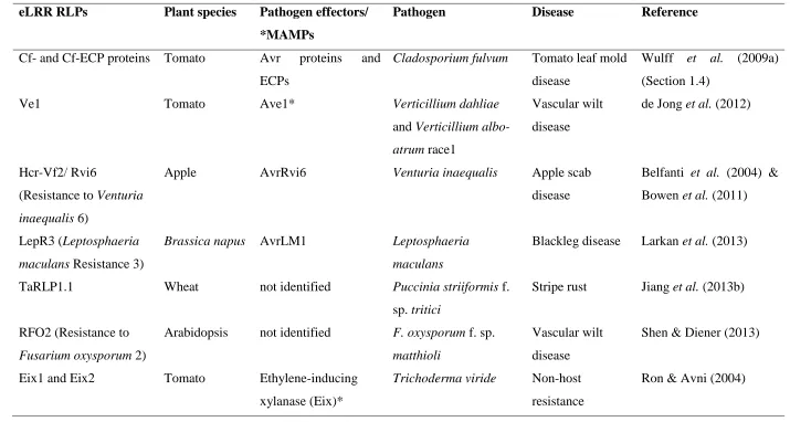
Table 1.1 eLRR RLPs with demonstrated function in plant immunity. Pathogen-derived molecules marked with asterisks are MAMPs.
eLRR RLPs Plant species Pathogen effectors/ *MAMPs
Pathogen Disease Reference
Cf- and Cf-ECP proteins Tomato Avr proteins and ECPs
Cladosporium fulvum Tomato leaf mold disease
Wulff et al. (2009a) (Section 1.4)
Ve1 Tomato Ave1* Verticillium dahliae
and Verticillium albo-atrum race1
Vascular wilt disease
de Jong et al. (2012)
Hcr-Vf2/ Rvi6
(Resistance to Venturia inaequalis 6)
Apple AvrRvi6 Venturia inaequalis Apple scab
disease
Belfanti et al. (2004) & Bowen et al. (2011)
LepR3 (Leptosphaeria maculans Resistance 3)
Brassica napus AvrLM1 Leptosphaeria maculans
Blackleg disease Larkan et al. (2013)
TaRLP1.1 Wheat not identified Puccinia striiformis f.
sp. tritici
Stripe rust Jiang et al. (2013b)
RFO2 (Resistance to Fusarium oxysporum 2)
Arabidopsis not identified F. oxysporum f. sp. matthioli
Vascular wilt disease
Shen & Diener (2013)
Eix1 and Eix2 Tomato Ethylene-inducing xylanase (Eix)*
Trichoderma viride Non-host resistance
Ron & Avni (2004)
EILP (Elicitor inducible LRR protein)
Tobacco not identified Pseudomonas syringae pv. glycinea and
P. syringae pv. tabaci
Non-host resistance
Takemoto et al. (2000)
ReMax (Receptor for eMax
Arabidopsis eMax* Xanthomonas Non-host
resistance
Jehle et al. (2013)
RPBG1 (Responsiveness to Botrytis
Polygalacturonases 1)
Arabidopsis Endopolygalacturo-nases (PGs)*
Botrytis cinerea
(a necrotroph)
Non-host resistance
Zhang et al. (2014a)
RLP30 Arabidopsis Sclerotinia Culture
Filtrate Elicitor 1 (SCFE1)*
Sclerotinia
sclerotiorum
(a necrotroph)
Non-host resistance
Zhang et al. (2013)
RLP52 Arabidopsis Fungal chitin* Erysiphe
cichoracearum
(a powdery mildew pathogen)
Non-host resistance
Ramonell et al. (2005)
13
1.2.2.1 Tomato Ve1 receptor
The tomato Ve locus mediates resistance against race 1 strains of Verticillium dahliae and Verticillium albo-atrum. The two genes Ve1 and Ve2 at this locus encode eLRR RLPs containing 37 LRRs with 84 % amino acid identity but only Ve1 mediates resistance against race 1 Verticillium strains in tomato (Fradin et al., 2009; Fradin et al., 2014). It has been shown that Ve1 remains functional upon interfamily transfer to Arabidopsis and that Ve1-mediated resistance involves similar downstream signalling components to that in tomato (Fradin et al., 2011), indicating conservation of defence signalling mediated by this RLP between different plant species. Recently, the corresponding Ave1 effector protein from race 1 strains of V. dahliae and V. albo-atrum, which activates Ve1-mediated resistance, was identified via a comparative genomic approach using RNA sequencing technology (de Jonge et al., 2012). Domain swapping between Ve1 and the non-functional homologue Ve2 demonstrated that the first 30 LRRs are required for Ve1/Ave1-mediated HR induction and disease resistance (Fradin et al., 2014).
1.2.2.2 Tomato Eix dual receptor system
14
1.2.2.3 The requirement for signalling partners in eLRR RLP function
The recent identification of the tomato homologue of the Arabidopsis eLRR RLK SUPPRESSOR OF BIR1-1 (SOBIR1), also known as EVERSHED (EVR) (Gao et al., 2009; Leslie et al., 2010), which interacts with several eLRR RLPs involved in immunity to fungal pathogens including Cf-2, Cf-4, Cf-9, Ve1 and Eix2(Liebrand et al., 2013), has resolved the search for the long anticipated signalling partner required for the function of the eLRR RLPs (Jones et al., 1994; Rivas and Thomas, 2005). SOBIR1 and its homologue SOBIR1-like protein were shown to be required for Cf-2, Cf-4 and Ve1-mediated resistance by virus-induced gene silencing (VIGS). Likewise, VIGS of SOBIR1 and SOBIR1-like protein also compromised Cf-4/Avr4- and Ve1/Ave1-induced necrosis, demonstrating a role for SOBIR1 and SOBIR-like protein in Cf- and Ve1-mediated immune responses (Liebrand et al., 2013). Of note, the involvement of SOBIR1 appeared to be exclusive to the function of the eLRR RLPs, including that of CLV2 but not for the eLRR RLKs such as FLS2 and CLV1 (Liebrand et al., 2013; Liebrand et al., 2014). By contrast, the SOMATIC EMBRYOGENESIS RECEPTOR KINASES (SERKs), another class of ‘short eLRR RLKs’ similar to SOBIR1, are required for the function of both RLPs and RLKs (Liebrand et al., 2014). For instance, through VIGS analysis, SERK1 was demonstrated to be involved in both Cf-4- and Ve1-mediated disease resistance whereas SERK3/BRI1-associated kinase 1 (BAK1) was only essential for Ve1-mediated resistance (Fradin et al., 2009). Furthermore, BAK1 has been shown to act as a co-receptor for BRI1 and FLS2 function in the recognition of brassinosteroid and flagellin, respectively (Santiago et al., 2013; Sun et al., 2013).
1.2.2.4 Effector-trigerred defence (ETD) – a new concept in plant
defence
15
also been identified in other Dothideomycete species (Section 1.4). The presence of conserved effector homologues across fungal species has led to the proposition to reclassify the eLRR RLPs including Ve1 as a PRR involved in MTI instead of an R protein that mediates ETI (Thomma et al., 2011; de Jonge et al., 2012). One current view about eLRR RLPs is that this class of resistance proteins is involved in ‘effector-triggered defence’ (ETD), a newly introduced concept of plant defence involving apoplastic fungal pathogens (Stotz et al., 2014). The blurred definition of some eLRR RLPs based on their involvement in both race-specific resistance (and so these receptors should be classified as R proteins involved in ETI) and recognition of conserved effector molecules (and therefore should be classified as PRRs involved in MTI) has led the introduction of this new concept of plant defence as distinct from ‘effector-triggered immunity’ (ETI) involving the NB-LRR receptors that recognize intracellular pathogen effectors.
16
1.3 Pathogen recognition – direct and indirect recognition
1.3.1 The LRR domain
17
Studies on the eLRR RLKs i.e. FLS2 and BRI1 have been at the forefront in understanding the structural basis of plant cell surface receptors with recent X-ray crystallography data elucidating the eLRR domain of these receptors interacting with their corresponding ligands (Hothorn et al., 2011; She et al., 2011; Sun et al., 2013). The solved crystal structure of FLS2 revealed that its eLRR domain adopts a superhelical structure (Figure 1.2) (Sun et al., 2013), similar to that of BRI1 (Hothorn et al., 2011; She et al., 2011) and the antifungal bean polygalacturonase-inhibiting protein (PGIP) (Di Matteo et al., 2003). This differs from the horseshoe-like structure formed by the LRR domains of the porcine ribonuclease inhibitors (RI) and the mammalian Toll-like receptors (Bell et al., 2003), owing to the β1-β2-310 helix LRR structure encoded by a 23- to 25-amino acid consensus plant-specific eLRR motif LxxLxxLxxLxLxxNxLxGxIPxx typical of plant cell surface eLRR receptors (Jones and Jones, 1997).
[image:35.595.238.416.339.634.2]18
It is thought that the eLRR domain of the cell surface receptors provides a platform for receptor complex assembly (Jaillais et al., 2011; Li, 2011). Upon perception of the flg22 epitope from bacterial flagellin, FLS2 undergoes a rapid heterodimerization with BAK1 (BRI1-associated Kinase 1) to form a signalling active FLS2-BAK1 complex, followed by reciprocal phosphorylation to activate plant defence (Chinchilla et al., 2007; Heese et al., 2007; Schulze et al., 2010; Sun et al., 2013). As shown in Figure 1.2, FLS2 and BAK1 form a monomeric heterodimer, whereby the BAK1 LRR domain interacts directly with the C-terminal portion of FLS2 LRR domain in an orientation resulting in both transmembrane domains being located in close proximity to one another (Sun et al., 2013). While the crystal structure of FLS2 did not show that the receptor undergoes homodimerization, FLS2 homodimerization remains possible (Sun et al., 2012). Apart from its interaction with the LRR domain of FLS2, BAK1 also interacts with the C-terminus of flg22, demonstrating its role as a co-receptor in flg22 perception. The flg22 epitope binds to the concave surface of FLS2 from LRR 3 to LRR 16 with its C terminus sandwiched between the LRR domain of FLS2 and the N-terminal cap domain of BAK. The crystal structures of the free and flg22-bound LRR domain of FLS2 are nearly identical, suggesting that conformational changes may not be necessary for FLS2 activation. By contrast, a comparison of the crystal structures of BRI1 on its own or in the presence of its brassinolide ligand showed that the receptor undergoes local structural rearrangements at the loop-out region and the two flanking LRR domains upon binding of the hormone molecule in an ‘induced-fit’ manner (She et al., 2011). This subsequently forms a docking platform for the co-receptor BAK1 to bind and allow signal transduction (Santiago et al., 2013).
1.3.2 Direct recognition by R proteins
19
et al., 2010). Additionally, the flax L6 and M proteins have been shown by yeast two-hybrid assays to interact directly with their corresponding Melampsora lini fungal effectors AvrL567 and AvrM (Dodds et al., 2006; Ellis et al., 2007; Catanzariti et al., 2010).
1.3.3 Indirect recognition by R proteins
While some R/Avr interactions have been shown to be direct, most other cases are not. These R/Avr interactions appeared to be mediated by a host protein, which can be envisioned as an adaptor protein that mediates Avr protein recognition by the corresponding R protein. To account for indirect R/Avr interactions, van der Biezen and Jones, (1998) proposed the ‘guard model’ wherein R proteins act as guards of effector targets (guardees) by sensing modifications of a guardee by a pathogen effector, resulting in activation of plant defence. This model was originally formulated to explain the recognition of P. syringae effector AvrPto by the two tomato proteins Pto and Prf (an NB-LRR protein)(van der Biezen and Jones, 1998). In this case, Prf acts as the guard that detects structural changes in Pto (the guardee) caused by AvrPto and then activates defence.
20
targeted by multiple effectors and guarded by multiple R genes. An indirect R/Avr recognition enables a limited R gene repertoire to recognize diverse pathogens because the same host proteins are often targets for effectors produced by different pathogens (Dangl and Jones, 2001).
1.4 The tomato-Cladosporium fulvum pathosystem
Table 1.2 Dramatis personӕ in tomato-Cladosporium fulvum interaction. Proteins indicated in brackets are encoded by genes that have not yet been cloned. Proteins indicated with a question mark are either hypothetical in the case of the Cf-ECP6 or Cf-ECP7 proteins or their role as an effector or effector target is not determined in the case of PhiA, the high-affinity binding site (HABS) and the Nicotiana benthamiana necrosis-inducing protein (NbNIP). *Proteins shown to contribute to pathogenicity. Mycosphaerella fijiensis Avr4, MfAvr4. Table adapted from Wulff et al. (2009a).
Cf proteins C. fulvum effectors (or indicated
otherwise)
Structure/ function Effector target(s) Reference
Cf-2 Avr2* Secreted cysteine
protease inhibitor
Rcr3 and Pip1
(Secreted cysteine proteases)
Dixon et al. (1996) (Cf-2)
Luderer et al. (2002) and van’t Klooster et al. (2011) (Avr2)
Dixon et al. (2000) (Rcr3pimp)
Shabab et al., (2008) and van Esse et al. (2008) (Pip1)
Nematode VAP1 protein*
Secreted venom allergen-like protein
Rcr3 Lozano-Torres et al. (2012) (VAP1)
Cf-4 Avr4* Secreted
chitin-binding protein
Thomas et al. (1997) (Cf-4) Joosten et al. (1994) (Avr4)
MfAvr4* Secreted
chitin-binding protein
Stergiopoulos et al. (2010) (MfAvr4)
Cf-4E Avr4E Secreted
cysteine-rich protein
Takken et al. (1999) (Cf-4E) Westerink et al. (2004) (Avr4E)
Cf-5 Avr5* Secreted
cysteine-rich protein
Dixon et al. (1998) (Cf-5) Mesarich et al. (2014) (Avr5)
Cf-9 Avr9* Secreted cystine-knot
protein
HABS? Jones et al. (1994) (Cf-9)
van den Ackerveken et al. (1992) (Avr9) Kooman-Gersmann et al. (1996) (HABS)
Cf-9B (Avr9B) Unknown NbNIP orthologue? Panter et al. (2002) (Cf-9B) Chakrabarti et al. (2009) (NbNIP) (Cf-ECP1) AvrECP1* Secreted
cysteine-rich protein
Soumpourou et al. (2007) (Cf-ECP1) van den Ackerveken et al. (1993) (ECP1) (Cf-ECP2) AvrECP2* Secreted
disulphide-bonded (?) protein
Laugé et al. (1998) and Haanstra et al. (1999) (Cf-ECP2)
van den Ackerveken et al. (1993),
Stergiopoulos et al. (2010) and de Wit et al. (2012) (ECP2)
(Cf-ECP4) AvrECP4 Secreted cysteine-rich protein
Soumpourou et al. 2007 (Cf-ECP4) Laugé et al. (2000) (ECP4)
(Cf-ECP5) AvrECP5 Secreted cysteine-rich protein
Haanstra et al. (2000) (Cf-ECP5) Laugé et al. (2000) (ECP5)
(Cf-ECP6?) ECP6* Secreted LysM
domain protein, possible chitin-binding protein
None? Bolton et al. (2008) (ECP6)
(Cf-ECP7?) ECP7 Secreted
cysteine-rich protein
Bolton et al. (2008) (ECP7)
? PhiA? Phialide protein
homologue
None? Bolton et al. (2008) (PhiA)
23
1.4.1 The tomato-C. fulvum compatible and incompatible interactions
In a susceptible tomato genotype lacking Cf resistance genes, C. fulvum can successfully infect and establish a compatible interaction with the host. This fungus infects the abaxial surface of the tomato leaves, starting with conidia that germinate on the leaf surface. At approximately 3 days post-infection, the conidia produce runner hyphae that enter the host leaf through open stomata and colonize the apoplastic space between mesophyll cells. Ten to 14 days later, conidiophores re-emerge from the stomata to release massive amounts of conidia and spread the disease. This results in stomatal clogging and impaired gas exchange, leading to wilting of leaves, defoliation and in the case of severe infections, host death occurs (Bond, 1938; Thomma et al., 2005; de Wit et al., 2012). While C. fulvum does not form feeding structures such as the haustoria, the fungal hyphae form close contact with the host cells by appressing the walls of mesophyll cells, perhaps as a way to obtain nutrients from the host (Bond, 1938; Lazarovits and Higgins, 1976).
Infection in incompatible interactions between specific races of C. fulvum and tomato cultivars carrying the corresponding Cf genes is similar to compatible interactions with respect to the initial stages of infection i.e. conidial germination, runner hyphae formation and stomatal penetration, but the majority of the hyphae do not grow out of
the stomata as a consequence of reduced hyphal development in the apoplast and there
is little or no conidia formation. Host defence responses such as callose deposition and
rapid accumulation of pathogenesis-related proteins including PR-2 (β-1,3-glucanases)
and PR-3 (chitinases) occur in the apoplastic space, resulting in an arrest of fungal
growth 1 or 2 days after penetration (Lazarovits and Higgins, 1976; de Wit, 1977).
Eventually, the mesophyll cells adjacent to the intracellular hyphae and occasionally,
some guard cells and epidermal cells collapse, which also results in release of
antimicrobial compounds from the cells (Joosten and de Wit, 1999).
1.4.2 The C. fulvum effectors
24
et al., 2009a). To date, 13 C. fulvum effector genes that encode Avr (avirulence) proteins or ECPs (extracellular proteins) have been cloned (Table 1.2 and references therein). Ten of these effector genes i.e. Avr2, Avr4, Avr4E, Avr5, Avr9, Ecp1, Ecp2-1, Ecp4, Ecp5 and Ecp6, known to function as avirulence genes, are able to elicit a plant defence response in plants carrying the corresponding Cf genes Cf-2, Cf-4, Cf-4E, Cf-5, Cf-9, Cf-Ecp1, Cf-Ecp2, Cf-Ecp4, Cf-Ecp5 and Cf-Ecp6, respectively (Stergiopoulos and de Wit, 2009; Wulff et al., 2009a; Mesarich et al., 2014). Most of these effectors, including those encoded by avirulence genes i.e. Avr2, Avr4, Avr4E, Avr5 and Avr9, are small, secreted cysteine-rich proteins with no sequence similarity to one another.
25
The ECP genes are more conserved across the various races of C. fulvum in comparison to the Avr genes (Stergiopoulos et al., 2007; Bolton et al., 2008). This is probably due to commercial deployment of Cf genes which has imposed selection pressure against the corresponding Avr genes (Stergiopoulos et al., 2007). As a result, the Avr genes have evolved to escape recognition through a number of mechanisms that shape the polymorphism observed in these genes today. These include gene deletions (which occurs in the case of Avr4E and Avr9), transposon insertions (Avr2) and point mutations either involving single nucleotide polymorphisms (SNPs) that result in nonsynonymous amino acid substitutions (Avr4 and Avr4E) or indels (insertions or deletions) of nucleotides that result in a frame-shift mutation (Avr4)(Joosten et al., 1997; Luderer et al., 2002; van den Burg et al., 2003; Westerink et al., 2004; Stergiopoulos et al., 2007; Wulff et al., 2009a). For instance, natural variants of C. fulvum strains virulent on Cf-4 tomato contain point mutations in Avr4 that destabilize the effector, thereby avoiding recognition by Cf-4 (van den Ackerveken et al., 1992; Joosten et al., 1997; van den Burg et al., 2003) whereas, some C. fulvum strains circumvent Cf-2-mediated resistance via alleles of Avr2 truncated by transposon insertion (Luderer et al., 2002).
26
1.4.3 The tomato Cf genes
[image:44.595.55.446.124.486.2]27
28
thereby contributing to the diversity of recognitional specificity encoded by these genes (Parniske et al., 1997; Wulff et al., 2009a).
1.4.4 Molecular and genetic basis of Cf-/Avr interactions
1.4.4.1 How does Cf-9 recognize Avr9?
Despite the use of various biochemical approaches to investigate the interaction between Cf-9 and Avr9, no interaction has been detected between these proteins (Luderer et al., 2001). By contrast, Avr9 was found to interact with a high affinity binding site (HABS) present in plasma membranes of tomato and other Solanaceous plants independent of the presence of Cf-9 (Kooman-Gersmann et al., 1996). Interestingly, the binding affinity of Avr9 to the HABS positively correlates with its ability to induce a Cf-9-dependent HR, suggesting that Cf-9 recognizes Avr9 indirectly by sensing the interaction of Avr9 with the HABS to then activate defence (Kooman-Gersmann et al., 1998; de Jong et al., 2002). Consistently, co-expression of Cf-9 and Avr9 proteins in tobacco and potato was sufficient to induce an HR. This correlates with the presence of a HABS in these plant species (Hammond-Kosack et al., 1998).
1.4.4.2 The Cf-2-Rcr3-Avr2 interaction
29
death requires specific structural modifications of Rcr3. The Rcr3pimp homologue from S. lycopersicum, Rcr3lyc was found to trigger an Avr2-independent necrosis among F2 segregants when a tomato line carrying Rcr3lyc was crossed to a line carrying Cf-2 (Krüger et al., 2002). The Rcr3lyc protein differs from its S. pimpinellifolium homologue by six amino acid substitutions and one amino acid deletion, supporting the model of Cf-2 activation that involves conformational changes of Rcr3. Interestingly, Cf-2 was found to mediate resistance to the root parasitic nematode Globodera rostochiensis that secretes a venom allergen-like protein designated VAP1 that has no sequence similarity to Avr2 (Lozano-Torres et al., 2012), demonstrating the ability of Cf-2 to recognize two independently evolved pathogen effectors. Similar to Avr2, VAP1 was also found to bind and inhibit the active site of Rcr3 which in turn allowed its recognition by Cf-2. In addition, tomato plants lacking Cf-2 but carrying Rcr3 showed increased susceptibility to G. rostochiensis infection, indicating that VAP1 is involved in pathogenicity and Rcr3 is a virulence target of VAP1 (Lozano-Torres et al., 2012).
1.4.4.3 The interaction between Cf-4 and Avr4
30
1.4.5 Structure and function of Cf proteins
The tomato Cf resistance genes (i.e. the Hcr9 and Hcr2 genes) encode eLRR RLPs, consistent with their proposed role as receptors that recognize C. fulvum effectors in the apoplast (Kruijt et al., 2005; Rivas and Thomas, 2005). These resistance proteins adopt a typical eLRR RLP structure as illustrated by the schematic representation of the Cf-9 protein in Figure 1.4.
Figure 1.4 Structural domains of the tomato Cf-9 resistance protein. Domain A is a cleavable signal peptide, B and D are cysteine-containing LRR flanking domains, C is the LRR domain containing 27 leucine-rich repeat (LRR) motifs divided into two blocks (C1 and C3) by a non-LRR loop-out region (C2), E is an acidic domain, F is a transmembrane (TM) domain and G is a highly basic cytosolic tail. The N- and C- termini of the protein are denoted by N and C, respectively. Figure adapted from (Barker, 2002)
31
al., 2005). Furthermore, Cf-9 and Cf-4 were shown to interact in vivo with several endoplasmic reticulum-resident chaperone proteins, which were thought to be required for proper folding of these eLRR receptors before being transported to the plasma membrane as functional receptor proteins (Liebrand et al., 2012). The N-terminal and C-terminal cysteine-containing LRR-flanking domain (domains B and D) are proposed to function as ‘caps’ that stabilize the eLRR domain. However, only the conserved cysteine-rich motifs in domain B but not domain D were shown to be essential for Cf-9 function, possibly by maintaining the overall protein structure through intramolecular interactions via the formation of disulphide bridges (van der Hoorn et al., 2005).
Sequence comparison among 39 Hcr9 proteins revealed that domain C1, particularly in LRRs 4-18, is highly variable (Parniske et al., 1997; Wulff et al., 2009b). In this region, a higher rate of non-synonymous amino acid substitutions to synonymous amino acid substitutions was found notably at the interstitial solvent-exposed positions in the central LRRs (Parniske et al., 1997). By contrast, the C-terminal portion of Hcr9 proteins is more conserved and this sequence conservation can be also found in the more distantly related Hcr2 proteins (Dixon et al., 1996; Parniske et al., 1997). These observations suggest a role of the variable N-terminus in ligand recognition whereas the conserved C-terminus is more likely to be involved in the interaction with a common signalling partner(s) in signal transduction (Jones and Jones, 1997; Parniske et al., 1997). For instance, Cf-9 and Cf-4 proteins are completely identical in their C-termini (from LRR 17 to the C-terminus of Cf-9) and yet these proteins recognize the sequence unrelated Avr9 (28 amino acids) and Avr4 (86 amino acids) elicitors (Thomas et al., 1997; Joosten and de Wit, 1999). This indicates that the recognition of Avr9 and Avr4 effectors by Cf-9 and Cf-4, respectively, is likely to be mediated by the differing N-termini of these proteins.
32
may be an important determinant of ligand recognitional specificity as it determines the spacing between solvent-exposed residues in contact with ligand (Jones and Jones, 1997). Domain swapping between Cf-9 and its close homologue, Cf-9B led to greater resolution in the identification of Cf-9 specificity-determining residues located at the solvent-exposed positions in the β-strand/ β-turn motif in the central LRRs of the protein (Chakrabarti et al., 2009, Wulff et al., 2009b). An advantage of using Cf-9B as a domain-swapping template is the greater similarity between Cf-9 and Cf-9B compared to the similarity between Cf-9 and Cf-4 in their proposed recognition domains (84 % identity versus 72 % identity from the N-terminus to LRR 15 of these proteins). Furthermore, Cf-4 also lacks two LRRs that correspond to LRRs 11 and 12 of Cf-9 whereas both Cf-9 and Cf-9B contain 27 LRRs. Collectively, the aforementioned studies have pinpointed five key amino acid residues i.e. C387 and Y389 in LRR 13, E411 in LRR 14, A433 in LRR 15 and L457 in LRR 16 as the major specificity determinants of Cf-9. In addition, through Cf-9/Cf-9B domain swaps, Chakrabarti et al. (2009) showed via agroinfiltrations in Avr9-expressing tobacco that LRRs 10-12 located upstream of the major specificity-determining region (LRRs 13-16) contributed to the strength of HR induction in wild type Cf-9,
33
Table 1.3 Specificity-determining region of Cf resistance proteins.
The loop-out region of Cf-9 has no demonstrated function as yet. This region has been proposed to act as a molecular hinge between domain C1 and C3, which may allow the protein to take on a conformational change during ligand interaction (van der Hoorn et al., 2005). Not much is known about the cytosolic tail (domain G) of Cf proteins. The conserved KKRY motif in this domain appeared to be required for its retrieval from the Golgi apparatus to endoplasmic reticulum of Cf-9 and hence may be involved in the quality control of Cf-9 biogenesis (Jones et al., 1994; Benghezal et al., 2000), although it may not determine the final location of the protein ((Piedras et al., 2000; van der Hoorn et al., 2001b). Yeast two-hybrid assays using the cytosolic domain of Cf-9 as bait identified several interacting proteins including a Cf-9-interacting thioredoxin (CITRX) and a ‘VAP27’ protein (Laurent et al., 2000; Rivas et al., 2004). VIGS of CITRX resulted in enhanced Cf-9/Avr9-mediated HR and defence responses, indicating that it plays a negative regulatory role in Cf-9 signalling. However, the role of CITRX appeared to be specific to 9 and not 2 as silencing of CITRX did not alter Cf-2/Avr2-mediated defence responses (Rivas et al., 2004). By contrast, the function of VAP27, a vesicle-associated membrane protein (VAMP)-like protein with a predicted role in protein trafficking in Cf-9-mediated resistance is yet to be demonstrated (Laurent et al., 2000). Additionally, a Cf-2/Cf-9 domain swap containing the first 34 N-terminal LRRs of Cf-2 fused to the three C-terminal LRRs and remaining portion of Cf-9 including its cytosolic tail has been shown to mediate an Avr2- and Rcr3-dependent HR and resistance to C. fulvum infection (Krüger et al., 2002). While CITRX is not involved in the regulation of Cf-2 signalling, silencing of CITRX enhanced an Avr2-dependent HR mediated by the Cf-2/Cf-9 domain swap (Rivas et al., 2004), indicating that this chimeric protein induces plant defence through the Cf-9 signalling pathway.
Cf proteins Number of LRRs Specificity-determining region References
Cf-2 38 LRRs 3-27 Seear and Dixon (2003)
Cf-4 25 LRRs 13-16 van der Hoorn et al. (2001a)
Cf-5 32 LRRs 3-21 Seear and Dixon (2003)
Cf-9 27 LRRs 10-16 Chakrabarti et al. (2009) Wulff et al. (2009b)
Cf-9B 27 N-terminus
to LRR 15
34
Altogether, these data support a role of the variable N-terminus in ligand recognition and the conserved C-terminus in signal transduction of Cf proteins.
1.4.6 Cf-/Avr-mediated downstream signalling
Transgenic tobacco suspension-culture cells expressing Cf-9 or Cf-4 genes have been used successfully to identify an array of signalling events following activation of Cf-/Avr-mediated defence responses by the corresponding Avr9 and Avr4 elicitors. Early responses include changes in ion fluxes, reactive oxygen species (ROS) bursts, alkalization of the culture medium and activation of a calcium-dependent protein kinase (CDPK) and several mitogen-activated protein kinases (MAPKs), including a salicylic acid-induced protein kinase (SIPK) and a wound-induced protein kinase (WIPK) (Piedras et al., 1998; Blatt et al., 1999; de Jong et al., 2000; Romeis et al., 2000a; Romeis et al., 2000b). Interestingly, activation of a phospholipase C (PLC)-mediated signalling pathway was shown to be involved in Cf-4/Avr4-mediated defence response (de Jong et al., 2004). PLC signalling may to contribute to ROS production and downstream phosphorylation events involving protein kinases such as CDPK (de Jong et al., 2004). VIGS of the tomato PLC isoforms 4 and 6 (SlPLC4 and SlPLC6) showed that both were essential for Cf-4–mediated resistance to C. fulvum whereas Cf-4/Avr4-induced HR appeared to be specifically mediated by SlPLC4 but not SlPLC6 (Vossen et al., 2010).
35
1). Interestingly, NRC1 was found to be required for HR mediated by several resistance proteins from both the cell surface eLRR and cytoplasmic CC-NB-LRR classes as silencing of NRC1 abolished their HR induction (Gabriëls et al., 2007), indicating an integration point in the downstream signalling of the two major different classes of R proteins.
Late responses observed following Cf-/Avr-mediated defence activation include accumulation of salicylic acid and ethylene (Hammond-Kosack et al., 1996; Etalo et al., 2013). For instance, infiltration of Cf-2 and Cf-9 tomato cotyledons with intercellular washing fluids (IF) containing the Avr2 and Avr9 peptides, respectively, resulted in accumulation of salicylic acid within 8 to 12 hours (Hammond-Kosack et al., 1996). However, while salicylic acid was shown to be involved in the initiation of cell death response, it is not required for Cf-9- and Cf-2-mediated resistance to C. fulvum (Brading et al., 2000), suggesting other defence signalling pathways are involved.
1.5 Autoactive R proteins
36
activation of plant defence. This section will review a number of factors that can contribute to autoactivity in plants as a prelude to the subject of this thesis, which involves research about an autoactive mutant of the tomato Cf-9 gene (Section 1.6). There are several types of mutations that can contribute to autoactivation of plant defence. These include mutations affecting the R protein itself or trans-acting components regulating R protein activity. Alternatively, mutations involving components that are not directly related to R protein activity such as the cyclic nucleotide-gated ion channel proteins involved in the defence no death (dnd) mutants, can also contribute to autoactivity (Lorrain et al., 2003). These mutants are called ‘lesion mimic’ mutants but will not be further discussed here as they are not the subject of this study.
1.5.1 Mutations in the R protein
1.5.1.1 Autoactive NB-LRR proteins
37
Point mutations that lead to autoactivity in NB-LRR proteins include those located both inside the LRR domain (Bendahmane et al., 2002; Farnham and Baulcombe, 2006) and outside (Shirano et al., 2002; Hwang and Williamson, 2003; Zhang et al., 2003). In particular, mutations in the conserved motifs involved in nucleotide binding and hydrolysis can cause autoactivity or loss of resistance function in these proteins (Howles et al., 2005; Tameling et al., 2006; van Ooijen et al., 2008; Williams et al., 2011). As autoactive NB-LRR proteins are often associated with ATP binding, it has been hypothesised that the ‘on’ and ‘off’ states of NB-LRR receptors are determined by binding of ATP and hydrolysis to ADP, respectively (Takken et al., 2006). Collectively, it has been proposed that activation of NB-LRR receptors is regulated via interaction between the NB domain and adjacent N-terminal LRRs and involves ATP binding and hydrolysis (Collier and Moffett, 2009; Takken and Goverse, 2012). Although the NB-LRR proteins differ from the eNB-LRR class in both their structure and function, reports on autoactive NB-LRR proteins nevertheless indicate negative inhibitory regulation of R protein activity in the absence of their cognate pathogen avirulence proteins.
1.5.1.2 Autoactive eLRR proteins
38
A suppressor screen in Arabidopsis searching for signalling components functioning in NONEXPRESSOR OF PATHOGENESIS-RELATED GENES 1 (NPR1)-independent signalling identified two autoactive RLPs (Zhang et al., 2010). RLP51 (also known as SNC2 for SUPPRESSOR OF NPR1-1 CONSTITUTIVE 2) and a close homologue RLP55 (SNC3), both contain point mutations in the conserved GXXXG motif in the transmembrane domain (Zhang et al., 2010), which is also conserved among the Cf proteins, suggesting that this motif is important for the negative regulation of these RLPs. Both the snc2 and snc3 mutants showed dwarf morphology, accumulation of high levels of endogenous salicylic acid and enhanced resistance to the virulent oomycete Hyaloperonospora arabidopsidis Noco2 (Zhang et al., 2010). The GxxxG motif has been implicated in homo-/heterodimerization of cell surface eLRR receptors (Zhang and Thomma, 2013). By contrast, a similar mutation in Cf-9 did not result in autoactivity but loss of function of the protein (Wulff et al., 2004b), indicating different mechanisms may be involved in preventing autoactivity among these RLPs.