COMPARATIVE ANALYSIS OF CONVENTIONAL METHODS WITH CATRIDGE BASED PCR METHOD IN THE DIAGNOSIS OF PAEDIATRIC TUBERCULOSIS
Dissertation submitted to
THE TAMIL NADU DR. M.G.R MEDICAL UNIVERSITY
in partial fulfillment of the regulations for the award of the degree of
M.D. (MICROBIOLOGY)
BRANCH – IV
GOVT. STANLEY MEDICAL COLLEGE AND HOSPITAL
THE TAMIL NADU DR. M.G.R MEDICAL UNIVERSITY
CHENNAI, INDIA
CERTIFICATE
This is to certify that this dissertation entitled “COMPARATIVE
ANALYSIS OF CONVENTIONAL METHODS WITH CATRIDGE
BASED PCR METHOD IN THE DIAGNOSIS OF PAEDIATRIC
TUBERCULOSIS” is the bonafide original work done by Dr.S.ABINAYA,
MD Postgraduate in Microbiology (2015 – 2018), under the guidance and supervision in the Department of Microbiology, Govt Stanley Medical College, Chennai, in partial fulfillment of the regulations of The Tamil Nadu Dr. M.G.R Medical University for the award of M.D Degree in MICROBIOLOGY (BRANCH – IV).
GUIDE DR.R.SELVI M.D.,
Professor and HOD Department of Microbiology
Stanley Medical College
CHENNAI - 600001
Prof. Dr. PONNAMBALA NAMASIVAYAM, MD., DA., DNB.
Dean
Stanley Medical College & Hospital, Chennai – 600 001
DR.R.SELVI M.D.,
Professor and HOD
DECLARATION
I solemnly declare that this dissertation “COMPARATIVE
ANALYSIS OF CONVENTIONAL METHODS WITH CATRIDGE
BASED PCR METHOD IN THE DIAGNOSIS OF PAEDIATRIC
TUBERCULOSIS” is the bonafide work done by me during my post graduate course in MD Microbiology (2015 – 2018), at the Department of Microbiology, Govt Stanley Medical College, Chennai, under the guidance and supervision of
Prof. Dr.R.SELVI, M.D., Professor and Head, Department of Microbiology, Govt Stanley Medical College, Chennai - 600001. This dissertation is submitted to The Tamil Nadu Dr. M.G.R Medical University, Chennai in partial fulfillment of the University regulations for the award of M.D Degree in MICROBIOLOGY (BRANCH – IV) examinations to be held in May 2018.
Place: Chennai Dr.S.ABINAYA
ACKNOWLEDGEMENT
My sincere and heartfelt thanks to Dr.Ponnambalanamasivayam, M.D.,DA.,DNB, DEAN Government Stanley Medical College and Hospital
for giving me permission for the study and use of resources of the
institution.
I owe my sincere and profound gratitude to Prof. Dr. R.Selvi. M.D.,
Prof. and Head of Department of Microbiology for her guidance and encouragement during the study. I would like to acknowledge her
constant support which she provided throughout the study without which
this work would not have been possible. My sincere thanks to
Prof. D.Aravind, Professor and Head of Department of Institute of social paediatrics, Dr. Syed Hissar, Scientist-C, NIRT for his encouragement and valuable inputs for the study.
I extend my thanks and gratitude to Associate professor
Dr.N.Thilagavathy M.D., Assistant professors Dr.B.Shanthi, M.D., Dr.Sheeba.V,M.D., Dr.P.Ponnammal M.D., Dr.A.Madhhumathy, M.D., Dr.B.Gomathi Manju M.D., Dr. Sivagama Sundari, of Department of Microbiology for their valuable help and guidance. I also thank all my
seniors, juniors and co-postgraduates for their moral support and
I express my thanks to all the technical staff and other staff
members of the Department of Microbiology for their kind help during
CONTENTS
S.NO. TITLE PAGE
1. INTRODUCTION 1-3
2. AIMS AND OBJECTIVES 4
3. REVIEW OF LITERATURE 5-41
4. MATERIALS AND METHODS 42-66
5. RESULTS 67-84
6. DISCUSSION 85-94
7. SUMMARY 95-96
8. CONCLUSION 97
9. BIBLIOGRAPHY
10. ANNEXURES
1. PROFORMA
2. CONSENT FORM
INTRODUCTION
Tuberculosis is a great menace to mankind since ancient times.
The genus Mycobacterium was known to originate 150 million years ago.
The skeletal deformities of tuberculosis have been demonstrated in
Egyptian mummies1. But the causative agent of this dreadful disease was
not known for longer periods. The discovery of Mycobacterium
tuberculosis, the causative agent for tuberculosis was by Robert Koch in
1882 using light microscopy and a special staining technique2.
India, accounts for 20% of the new 10.4 million TB cases detected
globally in 2015. The incidence of TB in India in 2015 was estimated to
be 2,840,0003. According to the estimates, childhood TB constitutes
10–20% of all TB in high burden countries like India and the annual risk
of tuberculosis in children is around 2-5%4,5. Drug-resistant (DR) strains
of Mycobacterium tuberculosis are also highly prevalent among the
paediatric population. The death rate due to tuberculosis in children is
nearly 8-20%5,6,7.
Tuberculosis among the pediatric population is underestimated in
developing countries like India due to lack of ideal diagnostic methods. In
addition to this, the hurdles in diagnosing a case of paediatric tuberculosis
include very low bacillary count8, 9, atypical clinical presentations10,
difficulty in obtaining specimens and amount of sample collected is often
Till now, microscopy has an important role in the diagnosis of
tuberculosis as it is simple, economical and less time consuming. But, its
usage in detecting a case of paediatric tuberculosis is limited because they
are paucibacillary and the lower limit of detection using microscopy is
1000011. Fluorescent microscopes with mercury vapour lamps or LED
has a limited role as it is costlier and requires technical expertise.
Although culture of M. tuberculosis using solid or liquid medium is the
gold standard in the diagnosis of TB, it requires specialized laboratories
and highly skilled staff. Solid culture takes a longer duration
(6-8 weeks).12 Though, liquid cultures detects M.TB earlier (10-14 days),
it is easily prone for contamination. Drug susceptibility testing using
these phenotypic (culture) methods can be done only after obtaining
primary growth in the media. This further increases the time taken for
detecting resistance (3-4 months) among anti-tuberculous drugs. This
affects the patient treatment response and spread of resistant tuberculosis
among the population.
Newer automated systems like BACTEC 9000; Versa TREK,
mycobacteria growth indicator tube (MGIT) and microscopic observation
drug susceptibility (MODS) assay are faster and accurate but have high
operational cost13. The nucleic acid amplification techniques like
conventional and real-time polymerase chain reaction is rapid in
diagnosis but has not reached the people at core level in countries
Despite the discovery of many newer methods in the diagnosis of
tuberculosis, a robust point of care test is required for effective
management of Paediatric tuberculosis. Though Tuberculosis shows a
decreasing trend in India32, tuberculosis in children15 and adolescents,
MDR TB, XDR TB, TB-HIV co-infection are increasing at an alarming
rate and have hindered our progress towards an END-TB strategy32.
CBNAAT (Catridge based nucleic acid amplification tests) has
now become a boon in the early and prompt diagnosis of tuberculosis
especially in paediatric TB cases and extrapulmonary TB cases. The
CBNAAT can be done in paucibacillary samples as the limit of detection
(LOD) is 131CFU/ml. The CBNAAT besides providing faster results also
detects resistance to rifampicin simultaneously. It is now being used as a
first line of investigation in HIV –TB coinfection and in extrapulmonary
tuberculosis. Newer generations of CBNAAT are in the pipeline to create
a low cost near patient technology for the diagnosis of tuberculosis16.
The aim of our study is to compare the cartridge based PCR assay
(CBNAAT) with the conventional methods for a better and earlier
diagnosis of paediatric tuberculosis. The purpose is to fill the lacunae and
AIMS AND OBJECTIVES
AIM:
The primary aim of this study is to compare CBNAAT with the
conventional methods in the diagnosis of Paediatric Tuberculosis.
OBJECTIVES:
1. To determine the distribution of tuberculosis among symptomatic
patients in paediatric age group in a tertiary care centre by CBNAAT and
conventional methods.
2. To compare the sensitivity of CBNAAT with the conventional
methods in the diagnosis of Paediatric Tuberculosis.
3. To determine Rifampicin resistance prevalent among the paediatric
age group.
4. To determine the percentage of pulmonary and extra pulmonary
REVIEW OF LITERATURE
Historical Perspective:
Tuberculosis, one of the earliest disease known to mankind dates
back to 8000 BC and 5800 BC (Neolithic); from Egyptian mummies
(2400 BC) and Assyrian tablets (700 BC)17,18. Hippocrates, Greece in 460
BC gave the first clinical description for tuberculosis “phthisis/ wasting
away”19
. The history of tuberculosis was changed dramatically on March
24, 1882, when Hermann Heinrich Robert Koch announced the discovery
of M.TB20 from human and animal sources for which he received the
Nobel Prize in 1905. Koch mentioned the bacteria as “Tubercle bacillus”,
so the disease came to be known as tuberculosis. In 1890, he also
discovered tuberculin, a substance derived from tubercle bacilli that can
be used for diagnostic purposes2.
Demonstration of Mycobacterium tuberculosis
Robert Koch (1882) demonstrated tubercle bacilli from the grey
tubercles of animals lungs hardened with alcohol and stained with
methylene blue and caustic potash21. The acid fast nature of the bacillus
was demonstrated by Ehrlich in 188222. Other diagnostic discoveries
include X-rays by Wilhelm Konrad Roentgen in 1895, development of
the tuberculin skin test by Von Pirquet and Mantoux in 1907-1908 and
Tuberculosis epidemiology Global burden
The World Health Organization (WHO) estimated about 8 million
new cases and 3 million deaths due to TB worldwide and 1.3 million new
cases and 450,000 deaths among children less than 15 years of age in
1990. According to the estimations, in 1994, the global incidence of
tuberculosis (TB) in children aged 0–14 years would be more than 1
million cases by 2000 and most of them occurring in the sub-Saharan
Africa.24 World Health Organization (WHO) declared TB a global
emergency25 as this would cause a worldwide increase of 36% from the
1990 estimate26. Majority of the patients lived in the most populous
countries of Asia - Bangladesh, China, India, Indonesia and Pakistan.
These countries constituted about half (49 %) of all new TB cases arising
worldwide in 2005.25 The 22 highest-burden countries account for about
80% of all new cases each year.27
Regional data from the World Health Organization (WHO) in 2007
showed that smear-positive TB in children aged <14 years accounted for
0.6-3.6% of reported cases.
However, because ∼95% of cases in children <12 years of age are
smear negative, these data underestimate the true burden of TB. In 2010,
of the ∼1 million estimated cases of TB in children worldwide, 75%
In the SEA region, estimated incidence of all forms of TB in 2013
was highest in India (62.4%), followed by Indonesia (13.7%) and
Bangladesh (10.4 %). Similarly, the estimated prevalence of all forms of
TB in SEAR in 2013 was highest in India (58 %), followed by Indonesia
(15.2 %) and Bangladesh (14.1 %) 29.
In 2015, the world had an estimated 10.4 million new TB cases.
Over half of these were among men (5.9 million), and women constituted
over a third (3.5 million). Ten per cent of cases were among children.
Among the 30 high-TB-burden countries, six SEA regions have an
increased incidence rate in the order, the Democratic People’s Republic
Korea, Indonesia, Myanmar, Bangladesh, India and Thailand. In 2015, in
the SEA Region, the incidence was estimated to be 4.74 million including
HIV+TB co-infection. In the SEA region, estimated incidence of
MDR/RR-TB was 200 000, with India alone accounting for 130 0003.
Burden in India
India is the largest country in the SEA region and has an annual
incidence of TB cases that depicts one fourth of global TB rate30. In 2015,
India ranks first in incidence and sixth in TB incidence rate among the
SEA region. India ranks sixth in mortality rate and first in mortality
numbers due to TB among the SEA regions. The incidence of TB in India
in 2015 was estimated to be 2,840,000. The incidence of MDR-TB was 1,
aim of WHO END-TB strategy is to decrease the incidence rate due to
TB by 80% and to reduce TB deaths by 90% by 2030, compared with
20153.
India has a National TB Control Program (NTP) since 1962 which
was later formulated into the Revised National TB Control Program
(RNTCP). RNTCP adopted the internationally recommended Directly
Observed Treatment Short-course (DOTS) strategy in 1993. The control
measures undertaken by RNTCP created increased awareness among the
people and there was 58% reduction in TB mortality along with 55%
reduction in TB prevalence rate in 2014 as compared to the 199030.
HIV-TB coinfection
The human immunodeficiency virus (HIV) epidemic contributed to
the spread of TB by increasing the host susceptibility to both primary as
well as reactivation of old TB. HIV infected people have 30 times higher
risk of developing active TB than others. The immune response to TB
bacilli increases HIV replication leading to rapid progression of the
disease. HIV-TB co-infection accounted for 11% of all new TB cases
estimated globally in 2015, while in the SEAR this accounts for 4.7% or
an estimated 2,27,000 cases. According to the estimates, 74, 000 patients
with HIV-associated TB died in 2015 in the SEA Region. According to
the estimates, 1.1 lakh HIV associated TB occurred in 2015 and among
MDR-TB
Since the usage of anti-TB drugs from 1943, development of
resistance among M.TB has been a great problem in both
immune-compromised and immune-competent as only few drugs are available to
effectively treat M.TB infection.
According to the global estimates, there were 580 000 (range,
520 000– 640 000) incident cases of MDR-TB in 2015, which accounted
for 83% of the total cases. There were about 250 000 (range,
160000-340000) deaths from MDR-TB in 20153. The average proportion of
MDR-TB cases with XDR-TB was 9.5% (95% CI: 7.0-12.1%), similar to
estimates for previous years (9.7% in 2014 and 9.0% in 2013)32.
In India, the earliest report on drug resistant M.TB is based on the
ICMR survey during the 1960s which reported INH resistance of 8.2 %,
STR 5.8%: to both INH and STR 6.5 %33. Later, gradual increase in the
prevalence of resistance in new cases over the next decades was reported
by TRC, Chennai. There was 3 % - 17 % rise for INH and 3 % - 13 % for
STR. MDR-TB was reported to be prevalent in 0.5 % - 3 % in new cases
and 12 % among the retreatment cases33,34,35.
The estimated number of MDR-TB cases in India in the year 2015
was 1.3 lakh3. The actual prevalence of MDR-TB in children in India is
not known. According to a study in Mumbai, the prevalence of DR-TB
increase in prevalence of XDR-TB and XXDR-TB from 6.1 % to 10.6 %
and 0 % to 5.6 % has been reported36. The estimated number of
laboratory confirmed XDR-TB cases in India in the year 2015 is 3048 out
of which only 2130 cases undertook proper treatment3.
Paediatric tuberculosis
Tuberculosis in young children, a vulnerable population differs
from adults in many ways like atypical radiological features, non-specific
symptoms, difficulty in obtaining samples, lower bacillary load, absence
of a gold standard test for diagnosis and by being commonly extra
pulmonary or disseminated at its presentation37,38. Tuberculosis can
mimic common diseases like pneumonia, generalized bacterial or viral
infections, malnutrition and HIV39.
In the national programs for control of tuberculosis also, paediatric
population remains neglected6. This leads to increased morbidity and
mortality among children. Children in developing countries have latent
tuberculosis most commonly and its progression to active tuberculosis
depends on various factors. Age, nutrition, HIV status, contact with a
known case of tuberculosis, diseases like measles and pertussis determine
the progression of tuberculosis in children40. The recent advances in
diagnosis and newer drug formulations have not yet reached the
Pulmonary tuberculosis and intra-thoracic lymphadenopathy
constitutes 60%-80% of the paediatric cases. Among the extrapulmonary
tuberculosis, lymphadenopathy is the most common (67%), followed by
central nervous system involvement (13%, pleural (6%), disseminated
(5%) and skeletal (4%) TB. Disseminated (miliary) disease and TB
meningitis are commonly seen in very young children and/or
HIV-infected children6.
Mycobacterium tuberculosis
Morphology
Mycobacterium tuberculosis is a non-motile, non-spore forming,
acid fast, aerobic or microaerophilic, straight or slightly curved
rod-shaped bacteria measuring 0.2-0.6× 1.0-10µm. They are intracellular
pathogen11.
Structure and properties of cell wall
The Mycobacterium tuberculosis cell wall has two components, the
peptidoglycan layer and the lipid layer. The peptidoglycan layer is
attached to arabinogalactan which in turn is attached to mycolic acids.
This forms the cell wall core- the mycolyl arabinogalactan-peptidoglycan
(mAGP) complex. Mycolic acids are unique alpha-branched lipids which
make up 50% of the dry weight of the mycobacterial cell envelope. They
are hydrophobic and affect permeability at the cell surface. Because of
commonly used basic aniline dyes at room temperature and takes up these
stains when treated with increased staining time along with application of
heat or acidified organic compounds. But once stained, they resist
decolourisation with acid-alcohol. This characteristic feature is called as
“Acid Fastness” and the bacterium is called as “Acid Fast Bacilli (AFB)”.
Cell wall proteins such as phosphatidylinositol mannosides (PIMs),
lipomannan and lipoarabinomannan are present. Cord Factor/ Trehalose
6,6’-dimyolaate (TDM), a glycolipid with two long chain
β-hydroxyl-α-branched fatty acids of variable length is responsible for serpentine cord
formation42. TDM acts as a virulence factor by stimulating NADase
activity, leading to lower levels of host NAD, especially in lung, liver and
spleen. Cord factor has a direct effect on mitochondrial membranes,
resulting in disruption of electron flow along the mitochondrial
respiratory chain and oxidative phosphorylation. Sulfolipids were
implicated in phagosome- lysosome fusion. Cording is a strict in-vitro
phenomenon43,44.
Staining methods
Dr. Franz Ziehl (1857-1926), a German bacteriologist in Lubek,
introduced Carbol fuchsin for staining tubercle bacillus in 1882. Ziehl
along with Friedrich Neelsen (1854-1894), a pathologist developed the
Ziehl-Neelsen stain, which is commonly used for acid fast staining. In
hot which melts the mycolic acid layer and increases the permeability of
the cell wall45.
Acid fast’s cold method uses increased concentrations of basic
fuchsin and phenol for staining acid fast organisms46,47. In Gabbet’s cold
staining method, methylene acts as both decolorizer and counterstain48. A
combination of Acid fast and Gabbet’s staining techniques was used by
Tax Thiam Hok49. In Rao et al. method of acid fast staining, chloroform
was used as solvent instead of ethanol and there was no increase in
concentrations of basic fuchsin-phenol50.
In 1937, Hagemann was first to stain Mycobacterium tuberculosis
with berberine sulphate for demonstrating fluorescence. In 1938, he
devised a better technique using phenol-auramine. From then on, many
fluorescent compounds including phenol acridine orange were used for
staining51.
The fluorescent staining techniques have decreased the time for
screening each slide as it covers a larger surface area using lesser
magnification. It increases the chances of false positive results due to
Cultural characteristics Growth requirements
Mycobacterium tuberculosis is an obligate aerobe but, presence of
carbon-di-oxide will enhance its growth. The organism grows well at
37°C (optimal temperature). It has slow generation time, 15-20 hours and
the time for detection of visible growth in solid media takes 2-8 weeks
differentiating it from other non-pathogens in the genus which shows
growth in 2 to 3 days44.
Solid media
Robert Koch had used the cattle blood serum solid medium which
was devised by Professor John Tyndall. At the end of two weeks, very
tiny colonies were seen through the magnifying lens. There are two types
of solid media – egg based media, Lowenstein Jensen medium and
colestos or agar based middlebrook 7H10 and 7H11 agar53.
The egg based media are cheaper and have long shelf life. They
can provide nutrients for the slow growers and can be used for primary
culture and sub-culture of the organism. The growth can be confirmed
using biochemical tests and AFB staining. Drug susceptibility testing can
Liquid media
Proskauer and Beck liquid medium were used earlier for
sub-culture of H37Rv strain. The liquid media available for sub-culture of
Mycobacterium tuberculosis includes Kirchner medium, Dubos Tween
albumin broth and Middlebrook7H9 broth. The growth of Mycobacterium
tuberculosis in liquid medium is in the fashion of serpentine cords which
is defined as the rope like aggregates of AFB bacilli which joins end to
end and side to side along the long axes of the bacteria56. The turnaround
time for detection of Mycobacterium tuberculosis using liquid media is
10-14 days. This gives faster results when compared with solid culture55.
Once growth is suspected, it is confirmed with AFB staining and
biochemical tests.
Biochemical characteristics Niacin accumulation test
The Mycobacterium tuberculosis accumulates the niacin produced
which occurs as a result of NAD activity. The test detects the presence of
cyanogen chloride which breaks niacin ring and forms aldehyde gamma
carboxyglutamate. This in turn reacts with aromatic amine to form yellow
Nitrate reductase test
This test detects the presence of nitrate reductase, an enzyme
present in the cell membrane of mycobacteria, reducing nitrates to
nitrites. Fuchsia colour is formed when sulfalinamide and
dihydrochloride-N-naphthyl ethylamide are added to the growth
medium58.
Catalase test
In Mycobacterium tuberculosis, thermolabile catalase which is an
antioxidant enzyme responsible for elimination of hydrogen peroxide is
present58. On adding 30%H2O2 and 10% Tween 80 to the medium, brisk
effervescence occurs59.
Urease test
This test detects the presence of urease enzyme which hydrolyses
urea to ammonia and this is used in the process of biosynthesis by
mycobacteria. Fuchsia colour is formed when the reaction occurs in
medium containing red phenol58.
Pyrazinamidase test
This test detects the intracellular enzyme pyrazinamidase which
converts pyrazinamide to pyrazinoic acid. Mycobacterium tuberculosis
Acid phosphatase test
Some Mycobacteria contains acid phosphatase which separates
phenolphthalein from phenolphthalein diphosphate and produces red
colour. Mycobacterium tuberculosis gives negative test61.
Arylsulfatase test
This test detects the presence of aryl sulfatase enzyme which
hydrolyses phenolphthalein disulfate potassium to free phenolphthalein in
the presence of alkali producing red colour. Mycobacterium tuberculosis
gives negative test61.
Tuberculosis in children
Tuberculosis in children is different from others in the way that
they are more vulnerable to severe disease and death following infection.
The children with latent infection become reservoir and leads to
reactivation in adulthood. Paediatric tuberculosis has been neglected for
long periods and now it has emerged as a global public health emergency
particularly in developing countries. Tuberculosis in children represents
ongoing transmission in the community as the infection most commonly
Pathogenesis
Mode of infection and transmission
Children usually contract infection from an infectious case of adult
TB by inhalation of tubercle bacilli in aerosolized droplets. A household
contact is considered as an important factor in childhood tuberculosis.
Other factors includes social factors, community TB prevalence, age of
the child, nutritional status, vaccination, immune status of the child,
duration of exposure and proximity of exposure to an infectious case63.
The social factors involved are poverty, poor housing, urban
environment and overcrowding. The age at which the child gets exposed
is directly proportional to the severity of the disease64. The chance of
transformation of infection to disease is more in case of infants than in
older children65.
Pathogenesis of pulmonary tuberculosis
The alveolar macrophages and dendritic cells are the first to
encounter the tubercle bacilli which limit its growth and also recruit other
immune cells. In children, there is impairment of both macrophage and
monocyte activity and their recruitment allowing the mycobacteria to
predominate and spread the disease66. In infants, there is a delay in
antigen specific response due to relative inactiveness of dendritic cells in
presenting the antigen to T- cells. The dendritic cells are functionally
mediated immunity in infants is highly impaired. Collectins such as
Mannose binding protein and surfactant proteins A & D bind to
mycobacteria and affect opsonisation, complement activation and
Toll-like receptor signaling. The fate of tubercle bacilli once they enter the
macrophage are they can cause primary TB disease, latent tuberculosis
infection, get eliminated or reactivate at a later stage to active disease39.
Pathogenesis of extra pulmonary tuberculosis and sites affected
The primary complex in children usually heals spontaneously by
fibrosis and calcification either with or without treatment68. Sometimes, it
may spread through bronchial airways, lymphatic spread through the
hilum or blood-spread resulting in distant metastases. In case, pulmonary
artery is affected, military tuberculosis occurs and if pulmonary vein is
affected, CNS, bone or kidney metastases occur. When an infant gets
infected with the tubercle bacilli, extrapulmonary dissemination and
progression to death is more common. This depends upon factors like
host genetics, microbial virulence and immune status of the child.
Although dissemination can occur at any site, the most commonly
affected part is the CNS. TB meningitis commonly occurs three to six
Clinical manifestation Pulmonary tuberculosis
Among Paediatric tuberculosis, parenchymal disease and
intrathoracic adenopathy are the most common clinical manifestations.
Primary complex consists of pulmonary infiltrates (sub pleural focus) and
lymph node enlargement in hilar and paratracheal regions. This causes
narrowing of bronchi and trachea which leads to atelectasis and
obstructive emphysema. The symptoms most commonly encountered are
persistent respiratory disease, unresponsive to antibiotics, fever, cough
and weight loss70.
Extra pulmonary tuberculosis
Lymphadenopathy is the most common extrapulmonary
manifestation followed by central nervous system, pleural, military or
disseminated and skeletal TB. Disseminated disease and TB meningitis
are common in very young children and HIV-infected children.
Superficial lymphadenopathy is common in preschool children and
Laboratory diagnosis of tuberculosis Microscopy
Light Microscopy
Microscopy in the diagnosis of tuberculosis still holds its position due to its easy availability and quicker results. This method is more
economical but has the disadvantage of low sensitivity (>105 / ml) and
cannot differentiate between live and dead bacilli. The sensitivity mainly
depends on the nature of sample, ranging from 71%-78% and the
specificity is 97%-100%. The respiratory samples have greater sensitivity
than other samples. Centrifugation of samples also increases the
sensitivity. Ziehl-Neelsen and Acid fast methods of acid fast staining are
in routine use nowadays. The tubercle bacilli are seen as thin, long,
slender, slightly curved occasionally beaded pink bacilli against a blue
background by light microscopy using acid fast staining techniques. The
smears are graded according to RNTCP guidelines72.
Fluorescence Microscopy
In fluorescent microscope, the bacilli appear as slender bright
yellow fluorescent rods against a dark background. Fluorescent
microscopy increases the sensitivity by 10% when compared with
conventional light microscopy73. It takes lesser time for screening of
smears. The disadvantage being high cost of installation of microscope
rooms for examination. LED microscopes are relatively less expensive
and LED bulbs have longer life span of 10000 hours. WHO has suggested
LED microscopes in the place of conventional and fluorescent
microscopy. The sensitivity of mercury vapour lamp microscopy and
LED microscopy were 82.4% and 83.1% respectively73,74.
Concentration techniques
Since culture remains the gold standard in the diagnosis of
tuberculosis, decontamination is a critical step in isolation of the
organism. The most common method used for decontamination is
Petroff’s method. This is an effective digestion and decontamination
method. In, NALC- NaOH method, the solution is added in equal
amounts to the specimen with brief vortexing. The mixture is incubated
for 15 minutes at room temperature. Phosphate buffer is added and the
volume is brought upto 50ml. It is then centrifuged at 3000 rpm for 15
minutes. The supernatant is discarded and the deposit is resuspended
using phosphate buffer which can be used for inoculating cultures. The
use of phosphate buffer shifts the pH, dilutes the toxic substances and
decreases the specific gravity and centrifugation further increases the
effectiveness75.
Modified petroffs method uses 4% NaOH in equal volumes of the
specimen which is homogenized in a shaker for 15 minutes. After
and the deposit is neutralized with 20 ml of sterile distilled water. The
recovery of Mycobacterium tuberculosis using NALC-NaOH method was
superior when compared with modified Petroff’s method and Petroff’s
method. Other methods are Oxalic acid method, Sulphuric acid method
and Cetylpridinium chloride method76.
Culture and semi-automated Solid medium
The Lowenstein Jensen (LJ) medium is the commonly used solid
media for culture of Mycobacterium tuberculosis. The processed samples
are inoculated into the LJ slopes and observed for growth daily for the
first one week and then weekly for 8 weeks. A minimum of 10 to 100
viable organisms are required for the culture to be positive. The typical
colony morphology of Mycobacterium tuberculosis on LJ media is rough,
tough and buff coloured colonies with warty, granular surface. The
Mycobacterium tuberculosis is differentiated from non-tuberculous
mycobacteria by the growth rate, optimum temperature for growth,
colony morphology, pigmentation and certain biochemical tests77.
Liquid medium
Liquid medium have an increased sensitivity for the growth of M.
tuberculosis (up to 20% increase in positivity) and a reduced delay in the
more with liquid cultures in comparison with the solid culture. WHO now
recommends the use of traditional solid media along with liquid media in
primary isolation of mycobacteria78.
Semi-automated methods BACTEC 460
BACTEC 460 uses BACTEC 12B medium which consists of
Middlebrook 7H9 broth with supplements such as casein hydroxylate,
bovine serum albumin, catalase, 14C-labeled substrate, Polymyxin B,
Amphotericin B, nalidixic acid, trimethoprim, azlocillin, pyridoxal
hydrochloride and polyoxyethylene-40-stearate. The mycobacterium
utilizes the radioactive substrate by decarboxylation and releases 14CO2
which is measured by the instrument. The measure of radioactivity is
given as growth index from 0-999. When ATT is introduced into the
medium, there occurs a decrease in growth index which represents
sensitivity.
The BACTEC 460 have considerably decreased the time for
detection of Mycobacterium tuberculosis but high cost of installation of
the devices, generation of radioactive wastes and labor intensiveness
makes it unsuitable for routine diagnostics. The mean times for detection
BACT ALERT
The BacT Alert system is based upon continuous colorimetric CO2
detection device which is used to indicate mycobacterial growth in a
closed system. The colorimetric indicator present at the base of each vial
changes from green to yellow when CO2 is produced in the vial. The
compartment of the instrument where the vials are incubated contains a
reflectometer and a detection unit. The results are updated every
10 minutes by the system.79
MGIT
MGIT uses Middlebrook 7H9 broth in association with a
fluorescence quenching based oxygen sensor. The mechanism of
fluorescence sensor is to detect oxygen depletion which results from
microbial metabolism. The fluorescence increases with the growth of
microorganisms. The medium allows for good growth of Mycobacterium
tuberculosis. The oxygen sensor is made up of silicon rubber impregnated
with a ruthenium pentahydrate. This system provides easy and faster
means of Mycobacterium tuberculosis detection and susceptibility testing.
The advantages of MGIT include less cost for instrumentation, easy
handling and it is non-radiometric.
The rate of recovery of Mycobacterium tuberculosis and time for
detection makes it usage in a wider perspective. Contamination rates are
higher since it uses a liquid medium. So, using both a liquid and a solid
for detection of Mycobacterium tuberculosis using MGIT in case of
smear positive and smear negative are 9.9 and 20.3 days respectively80.
MODS
MODS is a liquid culture technique which serves as a rapid,
sensitive and specific test for early diagnosis of tuberculosis in children.
It detects live tubercle bacilli based upon two following two factors. The
growth of Mycobacterium tuberculosis in liquid medium is quite faster
than in solid medium and the bacilli is seen as cords, strings or tangles.
MODS media consists of Middlebrook 7H9 broth with OADC and
PANTA mixture was added. A 48 well microtitre plate is used for
inoculation81. This method is more sensitive than smear but less sensitive
than MGIT. But this has similar sensitivity with MGIT in case of smear
negative cases of paediatric tuberculosis. It is cheaper than other rapid
molecular methods82.
Thin layer agar (TLA)
The TLA method uses Middlebrook 7H11 agar with added OADC
and PANTA mixture in the detection of tubercle bacilli. The growth is
detected by the appearance of micro-colonies/ cording using optical
microscope. The mean time for detection is 12.5 days and with smear
positive cases it is 10.6 days. TLA is rapid and can be used as an
economical alternative to LJ culture and MGIT in low resource countries.
Contamination rates are also lower when compared with liquid media83.
Molecular methods
Microscopy has high specificity but low sensitivity84. The Gold
standard for detection of tubercle bacilli is by cultivation on solid and
liquid media. The main disadvantage with culture on solid media is the
delay in detection (6-8 weeks). Liquid culture medium such as
BacT/ALERT system, Mycobacterial Growth Indicator Tube (MGIT),
MODS, Bactec 460TB systems take about 7-10 days only, but are costlier
and require subculture onto solid media85.
In order to overcome these inadequacies in the conventional
methods and for early diagnosis of paediatric tuberculosis, there is an
increased focus on Nucleic acid amplification techniques (NAAT). A
positive NAAT test is helpful in ruling in a diagnosis of TB, however a
negative NAAT test does not rule out TB86.
Conventional PCR and target sites
Among the nucleic acid amplification techniques for diagnosis of
tuberculosis, in-house PCR or conventional PCR was the first to emerge.
The use of specific primers and targets play an important role in
determining the sensitivity and specificity of the assay. Primers such as
Repetitive Insertion sequence IS6110 are commonly used. They are
specific to the members of the Mycobacterium tuberculosis complex
(M.TBC) and are present in multiple copies in the genome of M.TBC.
They are: Insertion sequence IS6110, MPB64,Insertion sequence IS986,
Heat shock proteins (hsp) 65KDa, 38KDa protein, TRC4, etc 87,88,89.
The decreased sensitivity of the test is due to the presence of
inhibitors of PCR present in the samples. The decreased specificity is due
to cross contamination from heavy loaded samples, laboratory machinery
or work surfaces contaminated with the amplicons.Different studies from
India have reported varying sensitivity (61% -100%) and specificity (75
% to 100%) using in-house PCR methods targeting different genes89. The
reasons for such variations in the accuracy of PCR include usage of
different primers, variation in the diagnostic criteria and the use of
different protocols for processing of samples. Thus, the usefulness of
PCR methods as a standard method to diagnose TB remains
controversial.
The most promising gene target appears to be IS6110, MPB64 (an
immunogenic protein specific to M.TB) with 90-100 % sensitivity and
100 % specificity88,90. Therefore, IS6110 primer together with MPB64
can be used effectively for detection of M.TB91.
Real time PCR
Real Time PCR technology enables direct identification of
Mycobacteria in clinical samples and gives faster results than other
conventional methods. The advantage in using the method is the presence
required. In general, real time PCR is an alternative for the conventional
PCR for detection of Mycobacteria from samples, especially to
differentiate its species and to detect the drug resistance92.
Quantification of the bacterial load is also possible with this
real-time technology. This provides easy visualization of amplification curves.
The assay has highly specific probes which results in accurate and
sensitive detection of Mycobacterium tuberculosis. This cannot be used
for determining efficacy of treatment and disease progression as it cannot
differentiate between viable or non-viable organisms. Real time PCR has
increased sensitivity and specificity for both pulmonary and
extrapulmonary samples93.
Gene-Xpert
CBNAAT was developed by FIND (Foundation for Innovative
New Diagnostics). It was launched in 2004 and the process was
completed in 2008. CBNAAT uses a single disposable cartridge which
contains a sample processing system and an automated heminested real
time PCR. It can simultaneously detect the presence of M.tuberculosis
and its resistance to rifampicin (RIF). CBNAAT detects M. tuberculosis
and rifampicin resistance by amplification of the rifampin resistance
determining region (RRDR) of the rpoB gene and subsequent probing of
rifampicin resistant tuberculosis cases have mutations in 81-bp region.
The limit of detection (LOD) is 131 CFU per ml of clinical specimen95.
This is developed as a near patient technology in the diagnosis of
tuberculosis. WHO propagated the use of CBNAAT from the year 2010.
The sensitivity of the test is 98% to 100% and specificity is 100%. In case
of smear negative cases, the sensitivity is 72%. CBNAAT requires less
technical expertise, minimal laboratory facilities and gives result within
two hours. This serves as an efficient tool in early diagnosis and
treatment of drug resistant tuberculosis especially in case of
extrapulmonary tuberculosis, HIV-TB coinfection and paediatric
tuberculosis. Thus it is more effective in paucibacillary cases95.
It cannot be used for monitoring the efficacy of treatment as it
cannot differentiate between live and dead bacilli. The disadvantages of
the test includes the cost of each test, requirement of stable electrical
supply, annual recalibration of the instrument, and a short shelf life of 18
months.
To overcome the above difficulties, newer technologies such as
CBNAAT Ultra and Omni are emerging. CBNAAT Ultra, a second
generation test, which seems to be a good alternative to CBNAAT. This
uses the same machine as for CBNAAT but with a different cartridge
system. This system is designed to increase the sensitivity of the test by
specimen. On 24 March 2017, WHO recommended the use of CBNAAT
Ultra as it has increased sensitivity in case of smear negative cases,
paediatric tuberculosis, extrapulmonary tuberculosis and in HIV
co-infection96.
CBNAAT Omni is also under development which is considered as
a point of care test in the diagnosis of tuberculosis and rifampicin
resistance. It uses the same cartridge as for CBNAAT but the machine
will be smaller, lighter and portable. It has advanced microfluidics system
and the whole testing process occurs within the same cartridge in a fully
automated manner from sample processing to detection of tubercle
bacilli. It is enough to load the patient sample alone for getting results. It
is more economical as the cost of each test is much lower than CBNAAT.
This device comes with an inbuilt 4-hour battery and low power
consumption. Rechargeable supplemental battery is also available with
the system97. A study on Xpert XDR will be started by FIND in 20183.
Line probe assay
WHO recommended Line probe assay from the year 2008. Line
probe assays combines PCR with reverse hybridization technologies for
earlier detection of Mycobacterium tuberculosis and its resistance to both
rifampicin and isoniazid. The method consists of three steps-DNA
The advantages of this method are it can detect both rifampicin and
isoniazid resistance simultaneously and gives quicker results when
compared with the routine phenotypic DST methods. This method also
differentiates NTM from Mycobacterium tuberculosis99.
The disadvantages include requirement of a good infrastructure and
equipment for performing the test, good technical expertise, biosafety
measures to prevent spread of infection and cross contamination between
samples as these assays have an open tube format. LPA cannot be done
for smear negative samples.
The sensitivity and specificity for determining rifampicin
resistance is 98.1% and 98.7% and for isoniazid resistance, it is 95% and
99.5% respectively. Line probe assay for second line drugs is also under
trial98.
Loop mediated isothermal amplification (LAMP)
LAMP is a type of nucleic acid amplification occurring in
isothermal conditions. This assay uses four to six loop primers
complimentary to the target gene and 100-1000 times amplification can
be achieved in 30 minutes. DNA binding fluorescent dyes such as SYBR
green can be used for visualizing the turbid fluorescent product with the
naked eye. WHO has recommended the use of TB-LAMP as a
replacement for smear microscopy in case of adult pulmonary
tuberculosis. Its role in diagnosing paediatric tuberculosis remains
The advantages include non-requirement of a sophisticated
laboratory, minimal biosafety requirements, short time for performing the
test (30-35min), feasible and economical for use in developing countries.
Although the assay is more specific, the sensitivity is 89%, lesser than
CBNAAT but is more sensitive than conventional PCR. The assay does
not provide any information regarding resistance to anti-TB dugs101.
Other technologies in pipeline
The need for ideal point of care technology with low cost, lead to
the emergence of newer technologies. Timely results serve to provide
cure and prevent spread of infection to the mass population.
The” END TB” goal of WHO by 2030 is possible only with the
development of an ideal rapid diagnostic technology reaching the
developing countries. The newer technologies in pipeline includes
GenoType M.TBDRplus (Line probe assay for RIF + INH resistance),
GenoType M.TBDRsl (Line probe assay for FQ + SLID resistance),
Genedrive M.TB/RIF (Portable RT-PCR for M.TB + RIF resistance) and
Truenat M.TB (Chip-based NAAT with RT-PCR on handheld device for
M.TB)3.
The other recently developed methods includes single strand
conformation polymorphism, DNA microarrays, RNA/RNA mismatch,
Anti-tuberculosis drug susceptibility testing
Though the first line anti-TB drugs remain the backbone in
controlling the disease, resistance has appeared to both first and second
line drugs and newer drugs are under trial but have not reached the
masses. Thus it becomes a great challenge in developing new anti-TB
drugs. Early diagnosis and treatment of multi-drug resistant tuberculosis
cases can prevent spread of infection and control of tuberculosis.
Phenotypic (culture-based)
The conventional methods for drug susceptibility testing (DST)
performed using solid media are 1% proportion method, absolute
concentration method and resistance ratio method. The time required for
providing results include the time for primarily isolating the organism
using culture and DST results are obtained in 6-12 weeks. This not only
leads to clinical deterioration of the patient but also to the spread of
infection to the community.
The MGIT uses liquid culture media for DST and the time has
been reduced to 3-4 weeks. The limitations are requirement of a stable
power supply and good technical expertise. MODS can also be used for
DST due to its rapidity when compared with solid culture and low cost.
The thin layer agar method detecting microcolonies of Mycobacteria is
The colorimetric redox indicator method and nitrate reductase
method depends on the oxidizing and reducing properties of
mycobacteria. This does not require specific equipment and can be used
by low income countries. It is easier to interpret results using colour
changes.103
Genotypic (nucleic acid amplification testing–based)
This method is based upon the detection of various genetic
mutations related to resistance of anti-TB drugs. The genes targeted are
katG, inhA, ahpC, kasA, rpoB, pncA, rpsL, rrs, tlyA, gyrA, gyrB and
embCAB. Various molecular assays are available including GeneXpert,
Line probe assay, PCR-SSCP (single strand conformation
polymorphisms), multi allele specific assays (MAS), hybridization
assays, DNA microarrays, PCR- restriction fragment length
polymorphism analysis (PRA) and real time PCR techniques.103
Others Mantoux
Mantoux test is not used to measure immunity but the degree of
hypersensitivity to tuberculin antigen. This test uses a standard dose of 5
TU (0.1ml) which is injected intradermally and read after 48-72 hours. In
developing countries, there is a high prevalence of malnutrition, parasitic
and infective illnesses including HIV infection which limit the usage of
differentiate between latent tuberculosis infection and active disease.
BCG vaccination and atypical mycobacteria produces false positive
results. Various factors which influence its interpretation are age,
immunological status and co-existing illness.104
IGRA
The interferon gamma release assays (IGRAs) is used for
demonstration of past or current infection with tuberculosis. This method
measures the amount of interferon gamma released when patient blood is
incubated with specific M. tuberculosis antigens. The antigens used are
early secretory antigenic target-6 (ESAT-6), culture filtrate protein 10
(CFP-10), and the tuberculosis 7.7 antigens. IGRAs cannot differentiate
between latent M. tuberculosis infection and active tuberculosis disease.
The IGRAs have many advantages over TSTs as the numbsr of false
positives caused by nontuberculosis mycobacteria or bacille
Calmette-Gue´rin are decreased and the patients need not visit the lab again for
assessing the reaction.105, 106
Lactate deaminase, ADA, LDH
Adenosine deaminase (ADA) is considered to be an excellent
surrogate marker in the diagnosis of extrapulmonary TB. This is mainly
used in detecting tubercle bacilli in serosal fluids like pleural effusion
(TB pleural effusion), pericardial effusion (TB Pericarditis) and
serosal fluids range from 90-100%. ADA is a simple, inexpensive, rapid
test which can be employed in the diagnosis of extra- pulmonary
tuberculosis.107 The ADA value gets decreased on treatment and so it can
be used as a good prognostic marker. ADA can also be elevated in other
diseases like brucellosis, coccidomycosis, histoplasmosis etc., thus
decreasing the specificity of the test.108 Similar to ADA, LDH also shows
some good sensitivity with diagnosis of TB in pleural fluid and ascitic
fluid. It is also elevated in other conditions like Pneumocystis carinii
pneumonia, pulmonary embolism, hodgkins lymphoma etc., thus
decreasing its specificity.109Hence these tests cannot be relied upon for
effective diagnosis of paediatric tuberculosis as they lack specificity.
Serology
WHO has a negative policy towards the use of serological tests in
the diagnosis of tuberculosis. In 2011, WHO banned the use of
serological tests as they were inaccurate, inconsistent, and imprecise in
detecting the active form of tuberculosis. They cannot differentiate
between active and latent infection. India banned the manufacture, import
Treatment
In adults: The DOTS regimen used for treatment of tuberculosis under RNTCP30 is
Treatment groups
Type of patient Regimen Intensive phase (IP) Continuation phase (CP) New case (Cat I)
New sputum smear positive New sputum smear negative New extra‐pulmonary New others
2 H3R3Z3E3 4 H3R3
Previously treated (Cat II)
Smear positive relapse Smear positive failure
Smear positive treatment after default
Others
2 H3R3Z3E3 S3 +
1 H3R3Z3E3
5 H3R3E3
In children
Under RNTCP, the same weekly thrice regimen as for adults is
being followed for children with alteration in drug dosages. The daily
doses (mg per kg of body weight per day) given to children include
Rifampicin 10-12 mg/kg (max 600 mg/day), Isoniazid 10 mg/kg (max
300 mg/day), Ethambutol 20-25mg/kg (max 1500mg/day), PZA
30-35mg/kg (max 2000mg/day) and Streptomycin 15mg/kg
(max 1gm/day).110 The treatment schedule for MDR TB is also the same
Treatment of MDR TB
TYPE OF RESISTANCE DEFINITION
Mono Resistance Resistant to only one first line drug
Poly Resistance Resistant to more than one first line
drug other than rifampicin, isoniazid.
Multi Drug Resistance Resistant to both rifampicin and
isoniazid
Extensive Drug Resistance Resistant to fluoroquinolones and
second line injectable anti-TB drugs.
Monoresistance:
Monoresistance TB cases are treated using a combination of
following drugs – a second line injectable (SLD), a fluoroquinolone (FQ),
Rifampicin(R) and any two of these drugs( H, E, Z)111
Polyresistance:
Polyresistance is treated using SLD + FQ + R + any FLD which is
sensitive + any one of following drugs (Eto, Cs, PAS) 111
Multidrug resistant TB
The MDR TB cases are treated for a short course therapy
(6-12 months) with a combination of drugs like a fluroquinolone, a
second line injectable, pyrazinamide and any two of the following
Bedaquilline and Delamanid are the newly developed drugs under
trial for treatment of MDR TB.111
Treatment of XDR
TYPE OF
TB CASE
INTENSIVE PHASE CONTINUATION PHASE
XDR (6-12)Cm, PAS, Mfx, high dose H, Cfz, Lzd, Amx/Clv
(18)PAS, Mfx, high dose H, Cfz, Lzd, Amx/Clv
Prevention
BCG vaccine was developed in the year 1921 and till now it is the
only vaccine currently available for preventing severe forms of
tuberculosis like disseminated TB and TB meningitis. It is universally
given over the left arm for all new born children under the national
immunization schedule. BCG vaccine does not prevent primary infection
and reactivation of latent tuberculosis infection in children.112
RNTCP
NTCP was started in 1962 with an aim to detect cases at the
earliest and treat them. However the treatment success rate was very low
and the death and default rate was really high. In order to combat these
problems, revised national tuberculosis control programme came into
action in 1993 with the concept of DOTS. By 2006, the entire country
85% and detection at the rate of 70%. According to RNTCP, two sputum
samples are collected from the TB suspect patients and even if one shows
acid fast bacilli, he is declared as positive case and treatment is started.30
RNTCP works at three levels – National reference laboratories,
Intermediate reference laboratories and Designated microscopy centres.
The National Reference Laboratories are NTI Bangalore, TRC Chennai
and LRS New Delhi. The Intermediate Reference Laboratories works at
state level. A network of Designated Microscopy Centers (>11,000) are
present which includes microscopy centers in medical colleges. One
MATERIALS AND METHODS
PLACE OF STUDY
1. Department of Microbiology, Government Stanley Medical
College and Hospital, Chennai.
2. Institute of social Paediatrics, Government Stanley Medical
College and Hospital, Chennai.
STUDY DESIGN Prospective study
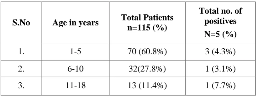
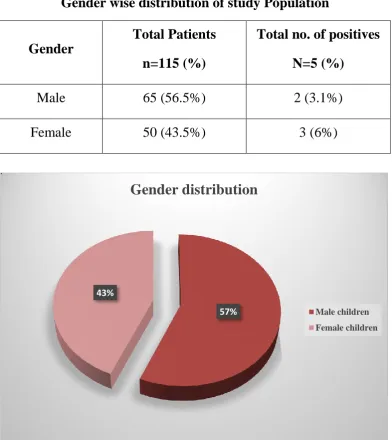
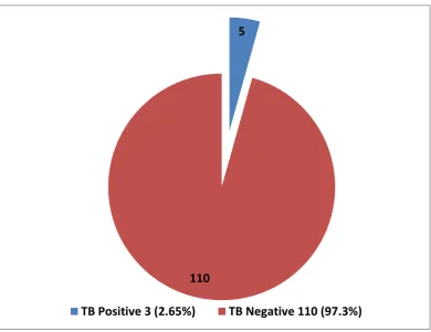
SAMPLE SIZE 115
STUDY PERIOD
May 2016 to September 2017
ETHICAL CONSIDERATION
Ethical and research clearance was obtained from the Ethical
committee, Stanley Medical College. Permission to conduct the study
was sought from the respective hospital department authorities. Personal
interview was conducted by the author to every child’s attender. A
standard proforma was filled up at the time of interview and informed
STATISTICAL ANALYSIS
The data was analyzed with SPSS 16.0 version. To find the
significance in categorical data, Chi square was used.
INCLUSION CRITERIA
1. Children <18 years of age with symptoms of persistent fever or
cough for more than two weeks.
2. Children <18 years of age with loss of appetite or unexplained
weight loss/ no weight gain in the past three months.
3. Children <18 years of age who has history of contact with an
infectious TB case.
4. Children <18 years of age with significant superficial
lymphadenopathy.
5. Children <18 years of age with severe respiratory distress and
abnormal shadows in X-Ray.
6. Children <18 years of age with failure to thrive symptoms.
EXCLUSION CRITERIA
8. Patients <18 years of age who is a known case of tuberculosis and
on anti-tubercular treatment.
9. Patients <18 years of age who are relapse, default and failure cases.
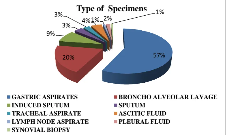
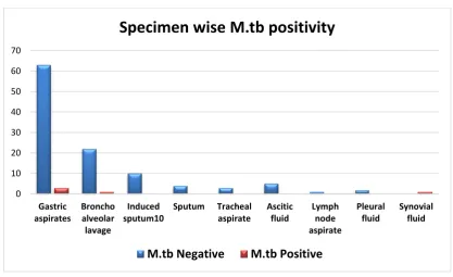
SPECIMEN COLLECTION GASTRIC ASPIRATE
The child was kept fasted for at least four hours. Early morning as
soon as the child wakes up, he/she was asked to rest on back. After
wearing sterile gloves, the distance between the nose and the stomach
was measured and slowly nasogastric tube was inserted through the
child’s nose into the stomach. Using a syringe, the gastric contents
(2-5ml) are aspirated. When no fluid was aspirated, 5-10 ml of normal
saline was injected through the tube after confirming its position in
stomach, and the same was aspirated and transferred into a sterile wide
mouth container. An equal volume of sodium bicarbonate was added to it.
The same procedure was followed the consecutive day.113
SPUTUM
The child was instructed to rinse his /her mouth with water and
asked to take two deep breaths, holding for few seconds and then
exhaling slowly. After this, the child was asked to breathe in and exhale
forcefully with a cough. The sputum produced was collected in a sterile
INDUCED SPUTUM
After giving a bronchodilator, nebulized hypertonic saline
(3% NaCl) was administered to the child for 15 minutes. Thereafter, the
same procedure as for sputum collection was followed.115
BRONCHO ALVEOLAR LAVAGE
After proper sedation, the flexible fibreoptic bronchoscope was
inserted into the right middle lobe and 5-20 ml of pre-warmed sterile
saline was injected in fractions using a syringe into the suction channel
along with sufficient air. The fluid was then aspirated using a syringe and
transferred onto a sterile container.116
TRACHEAL ASPIRATE
The tracheal aspiration probe was introduced through the ETT until
resistance and retracted 2cm proximally. Vacuum was then released and
the probe was removed from which the secretions were aspirated into a
sterile tube.117
ASCITIC FLUID
The child was asked to lie down in lateral decubitus position. A
point two finger breadth medial to the anterior superior iliac spine was
chosen. The skin was retracted caudally and the needle was inserted until
fluid comes out. The fluid was aspirated using syringe and transferred
PLEURAL FLUID
The child was positioned in sitting posture with arms and head in
resting position. The thoracocentesis site was selected in the interspace
below the point of dullness to percussion in the mid posterior line.
Anesthesia was given over the insertion site and the needle was inserted
over the top of the rib. The fluid was aspirated using the syringe and
transferred into a sterile container.
LYMPH NODE ASPIRATE
The child was asked to lie down and topical anaesthetic was
applied over the node to be aspirated. The node was positioned and the
needle was introduced into it. The fluid aspirated was transferred into a
sterile container.119
SYNOVIAL BIOPSY
The child was positioned supine with knee in an extended position.
A local anaesthetic was injected subcutaneously into the insertion site and
a small incision was made at that point. The forceps was forwarded
through the suprapatellar pouch and tissue sample was retrieved using the
PROCESSING OF COLLECTED SPECIMENS PREPARATION OF NALC-NAOH SOLUTION121
1. 4%NaOH was prepared by dissolving 20g of NaOH to 0.5L
distilled water.
2. Sodium citrate was prepared by adding 14.5g of Na citrate to 0.5L
distilled water.
3. The two solutions were mixed and autoclaved for 20min at 121oC,
15lbs.
4. 0.5g of NALC powder was added to 100 ml of the NaOH solution
just before using.
PREPARATION OF PHOSPHATE BUFFER121
Phosphate buffer was prepared by mixing stock solutions A and B.
1. Stock solution A (Disodium phosphate) was prepared by adding
9.47g of Na2HPO4 to 1L of distilled water.
2. Stock solution B (Mono Potassium phosphate) was prepared by
adding 9.07g of KH2PO4 to 1L of distilled water.
3. Both the solutions were mixed together and the pH was adjusted to
6.8. The solutions were autoclaved and kept in small bottles for
MATERIALS REQUIRED
Biosafety cabinet
Centrifuge
Vortex mixer
Lysol bin
Falcon tubes
Wire racks for falcon tubes
4%NaOH
NALC powder
Phosphate buffer solution
Timer
PROCEDURE FOR DIGESTION AND DECONTAMINATION OF SAMPLES121
The specimens such as gastric aspirates, broncho alveolar lavage,
induced sputum, sputum and tracheal aspirate were decontaminated
using the following procedure.
The specimens collected were divided into equal portions – one
half of the sample was used for CBNAAT and another half was
Equal volumes of 4%NaOH were added to the centrifuge tube.
The cap of the centrifuge tube was tightened and vortexed for few
seconds to digest the specimen.
The mixture was allowed to stand for 15 minutes at room
temperature.
Phosphate buffer was added up to the 50-ml mark on the centrifuge
tube and then vortexed for few seconds.
The tube was centrifuged at 3000g for 15 minutes.
The supernatant solution was carefully poured into a discard can
containing 5% phenol.
The deposit collected was re-suspended using 2ml of phosphate
buffer solution and used for further proceedings.
PROCESSING OF STERILE SAMPLES122
The decontamination process was not required for sterile
specimens such as pleural fluid, ascitic fluid and lymph node
aspirate.
These specimens were concentrated by means of centrifugation to