Multiple Cellular Signaling Pathways
Youxing Li,aQi Zhang,a,bYin Liu,aZhen Luo,aLei Kang,aJing Qu,aWeiyong Liu,aXueshan Xia,cYingle Liu,a,bKailang Wu,a,b and Jianguo Wua,b
State Key Laboratory of Virology, College of Life Sciences, and Chinese-French Liver Disease Research Institute at Zhongnan Hospital, Wuhan University, Wuhan, Chinaa ; Wuhan Institutes of Biotechnology, Wuhan, Chinab
; and College of Life Sciences, Kunming University of Science and Technology, Kunming, Chinac
Hepatitis C virus (HCV) infection is associated with numerous liver diseases and causes serious global health problems, but the
mechanisms underlying the pathogenesis of HCV infections remain largely unknown. In this study, we demonstrate that signal
transducer and activator of transcription 3 (STAT3), matrix metalloproteinase-2 (MMP-2), and B-cell lymphoma 2 (Bcl-2) are
significantly stimulated in HCV-infected patients. We further show that HCV activates STAT3, MMP-2, Bcl-2, extracellular
reg-ulated protein kinase (ERK), and c-Jun N-terminal kinase (JNK) in infected Huh7.5.1 cells. Functional screening of HCV
pro-teins revealed that nonstructural protein 4B (NS4B) is responsible for the activation of MMP-2 and Bcl-2 by stimulating STAT3
through repression of the suppressor of cytokine signaling 3 (SOCS3). Our results also demonstrate that multiple signaling
cas-cades, including several members of the protein kinase C (PKC) family, JNK, ERK, and STAT3, play critical roles in the
activa-tion of MMP-2 and Bcl-2 mediated by NS4B. Further studies revealed that the C-terminal domain (CTD) of NS4B is sufficient for
the activation of STAT3, JNK, ERK, MMP-2, and Bcl-2. We also show that amino acids 227 to 250 of NS4B are essential for
lation of STAT3, JNK, ERK, MMP-2, and Bcl-2, and among them, three residues (237L, 239S, and 245L) are crucial for this
regu-lation. Thus, we reveal a novel mechanism underlying HCV pathogenesis in which multiple intracellular signaling cascades are
cooperatively involved in the activation of two important cellular factors, MMP-2 and Bcl-2, in response to HCV infection.
H
epatitis C virus (HCV) persistent infection is a major cause of
chronic liver diseases, including hepatic steatosis, cirrhosis,
and hepatocellular carcinoma (HCC), which affect approximately
200 million people worldwide (
12
,
36
,
38
). However, the
mecha-nisms by which HCV infection causes chronic human liver
dis-eases remain largely unknown. HCV is a small and enveloped
RNA virus belonging to the
Hepacivirus
genus of the
Flaviviridae
family (
26
). The HCV genome consists of a single-stranded
posi-tive-sense RNA of approximately 9.6 kb that contains a single
open reading frame encoding a polyprotein precursor of
approx-imately 3,000 residues. The polyprotein precursor is then cleaved
into at least 10 distinct proteins, including 4 structural proteins
(core, E1, E2, and p7) and 6 nonstructural proteins (NS2, NS3,
NS4A, NS4B, NS5A, and NS5B) (
52
).
Signal transducers and activators of transcription (STATs) are
a family of cytoplasmic proteins with Src homology-2 (SH2)
do-mains that act as signal messengers and transcription factors and
participate in normal cellular responses to cytokines and growth
factors (GFs). After stimulation of cytokine-receptor complexes
and GF-receptor complexes following ligand binding, STATs are
activated via the tyrosine phosphorylation cascade (
40
,
59
,
66
).
Among the STAT proteins characterized to date, STAT3 has been
implicated in the transduction of cellular signals involved in the
development of cardiac hypertrophy and in the induction of gene
expression in response to cytokine receptor stimulation (
20
,
40
).
After tyrosine phosphorylation, STAT3 is dimerized and
translo-cated to the nucleus, where it activates downstream target genes
(
20
,
40
), including c-Fos, cyclin D1 (CCND1), cell division cycle
25A (CDC25A), c-Myc, proviral integration site 1 (Pim1), and
B-cell lymphoma 2 (Bcl-2) (
5
). Bcl-2 inhibits apoptosis and
con-tributes to cell survival and the resistance of cells against damaging
influences (
23
). The Bcl-2-related genes regulate cell death and are
considered to correlate with the pathogenesis and progression of
cancers (
15
,
28
,
63
). STAT3 also promotes metastasis and
angio-genesis by inducing expression of a metastatic gene, matrix
met-alloproteinase-2 (MMP-2), as well as a potent angiogenic gene,
vascular endothelial growth factor (VEGF) (
15
). STAT3 activation
is often associated with cell growth or transformation, and
disrup-tion of STAT3 causes embryonic lethality.
Mitogen-activated protein kinases (MAPKs) play important
roles in viral infection. In multicellular organisms, there are three
well-characterized subfamilies of MAPKs, including the
extracel-lular signal-regulated kinases (ERKs; ERK1 and ERK2), the c-Jun
N-terminal kinases (JNKs; JNK1, JNK2, and JNK3), and the p38
enzymes (p38
␣
, p38

, p38
␥
, and p38
␦
). The JNK and ERK
path-ways have been implicated in relaying extracellular signals to the
nucleus to mediate specific responses, such as proliferation,
dif-ferentiation, apoptosis, and stress, by regulating transcription
fac-tor activity (
25
,
33
,
53
). It has been reported that the cooperation
of tyrosine and serine phosphorylation is necessary for the full
activation of STAT3 (
4
,
9
,
61
). Members of the suppressors of
cytokine signaling (SOCS) family negatively regulate STAT3
ac-tivity.
Members of the protein kinase C (PKC) superfamily play
key regulatory roles in many cellular processes, ranging from
the control of fundamental cell autonomous activities (such as
proliferation) to more organismal functions (such as memory).
These kinases can be activated by phosphatidylserine (PS) and
Received9 May 2012Accepted27 August 2012
Published ahead of print5 September 2012
Address correspondence Jianguo Wu, [email protected].
Copyright © 2012, American Society for Microbiology. All Rights Reserved. doi:10.1128/JVI.01136-12
on November 7, 2019 by guest
http://jvi.asm.org/
diacylglycerol (DAG) in a Ca
2⫹-dependent manner and also by
tumor-promoting phorbol esters such as phorbol 12-myristate
13-acetate (PMA) (
46
). PKC
␣
-mediated ERK, JNK, and p38
regulate the myogenic program in human rhabdomyosarcoma
cells (
45
).
Our previous studies have shown that HCV infection activates
the Ras/Raf/MEK pathway, which in turn facilitates HCV
replica-tion via attenuareplica-tion of the interferon (IFN)-JAK-STAT pathway
(
67
). We also demonstrated that HCV infection activates the
ex-pression of the major vault protein (MVP), which is involved in
multidrug resistance, nucleocytoplasmic transport, and cell
sig-naling through the NF-
B and Sp1 pathways (
42
). More
interest-ingly, virus-activated MVP can suppress HCV replication by
in-ducing type I IFN expression (
42
). These findings suggested that
HCV infection activates multiple cellular signaling pathways.
Thus, in this study we investigated the signal transduction
net-works regulated by HCV infection and the molecular mechanisms
underlying such regulation.
Here, we found that STAT3, MMP-2, and Bcl-2 were
signifi-cantly stimulated in peripheral blood mononuclear cells (PBMCs)
isolated from patients with HCV infection and in cell cultures
infected with HCV. In addition, we demonstrated that HCV
reg-ulates MMP-2 and Bcl-2 by activating the STAT3 signaling
cas-cade. Further studies revealed that the ERK, JNK, and PKC
signal-ing pathways are involved in the upregulation of STAT3 activity in
response to HCV infection. We also discuss the mechanism
un-derlying the role of HCV NS4B in controlling multiple signaling
pathways and in the regulation of genes involved in cell
transfor-mation, apoptosis, and tumorigenesis in response to HCV
infec-tion.
MATERIALS AND METHODS
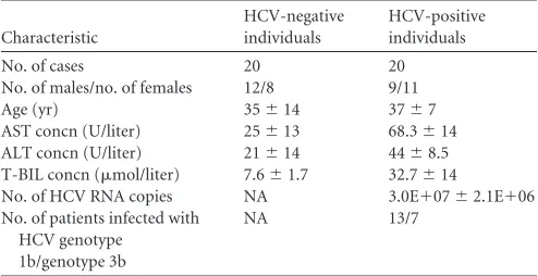
Blood specimens.Peripheral blood specimens were obtained from 20 patients with chronic HCV infection in Yunnan Province, China (Table 1). All patients were confirmed to be HCV positive and were negative for concomitant HBV, HDV, or HIV infection. They did not suffer any con-comitant disease at the moment of sampling, did not show any serological markers suggestive of autoimmune disease, and had not received any antiviral or immunomodulatory therapy prior to this study. Matched by
sex and age, 20 HCV-negative individuals with no history of liver disease were randomly selected from a local blood donation center as controls. The Institutional Review Board of the College of Life Sciences, Wuhan University, approved the collection of blood samples for this research, in accordance with guidelines for the protection of human subjects. Written informed consent was obtained from each participant.
Isolation of PBMCs.PBMCs were obtained by density centrifugation of peripheral blood samples diluted 1:1 in pyrogen-free saline over His-topaque (Haoyang Biotech). Cells were washed twice in saline and sus-pended in culture medium (RPMI 1640) supplemented with penicillin (100 U/ml) and streptomycin (100 mg/ml).
Plasmids.Plasmid pGL3-APRE-luc, in which the reporter gene is un-der the control of the STAT3 gene promoter, was obtained from Li Liu of Tsinghua University, China. Plasmids expressing small interfering RNA (siRNA) against STAT3 and members of the protein kinase C (PKC) family were constructed by ligating corresponding pairs of oligonucleo-tides based on target sequences described previously (41, 43) to pSilencer2.1-U6 neo (Ambion, Inc., Austin, TX). The siRNAs against SOCS3, JNK, and ERK (si-SOCS3, si-JNK, and si-ERK, respectively) and the siRNA control (si-Ctrl) used in our study were synthesized by the Ribobio Company and purchased directly from the Ribobio Company. ERK1 and ERK2 mutants (mERK1 and mERK2) were gifts from Melanie Cobb of the University of Texas Southwestern Medical Center, while the JNK mutant (mJNK) was from Michael Karin of the University of Cali-fornia at San Diego, San Diego, CA (41,43). V12 encoding activated hemagglutinin (HA)-Ras was cloned into pCMV-Tag2A vector (Strat-agene) as described previously (67). Plasmids expressing HCV genotype 2a proteins were generated in the State Key Laboratory of Virology, Wu-han University, as described previously (42). Truncated NS4B genes were generated by reverse transcription-PCR (RT-PCR) using HCV RNA as the template. The amplified DNA fragments were subcloned into pCMV-Tag2A to generate plasmids pCMV-NS4BCTD, pCMV-NS4B⌬C1, pCMV-NS4B⌬C2, and pCMV-NS4B⌬C3. Mutated NS4B genes (D228A, L237E, S239W, T241A, L245D, and H250E) were constructed by PCR-based site-directed mutagenesis using plasmid pCMV-NS4B as the tem-plate. In these mutated NS4B genes, amino acids 228D, 237L, 239S, 241T, 245L, and 250H were replaced by 228A, 237E, 239W, 241A, 245D, and 250E, respectively. All constructs were verified by automatic DNA se-quencing. The primers used in this study are listed inTable 2.
Antibodies and reagents.Antibodies against ERK (sc-153), phos-phorylated ERK (p-ERK) 7383), phosphos-phorylated JNK (p-JNK) (sc-7988), STAT3 (sc-476), MMP-2 (sc-13594), and Bcl-2 (sc-7382) were purchased from Santa Cruz Biotechnology (Santa Cruz, CA). Antibody against HCV NS3 (ab65407) was purchased from Abcam (Cambridge, United Kingdom). Antibodies against p-STAT3 (9145) and JNK (9252) were purchased from Cell Signaling Technology (Beverly, MA). Antibod-ies against -actin and glyceraldehyde-3-phosphate dehydrogenase (GAPDH) were purchased from CWBio (Beijing CoWin Biotech, China). The protein kinase inhibitors (U0126 and SP100625) were purchased from Tocris Bioscience (Bristol, United Kingdom) and dissolved in di-methyl sulfoxide (DMSO; Sigma Chemical Co, St. Louis, MO) upon use. Cells and viruses.Huh7.5.1 cells were kindly provided by Francis Chisari. Huh7.5.1 and Huh7 cells were cultured in Dulbecco modified Eagle medium (Gibco BRL, Grand Island, NY) supplemented with 10% fetal calf serum (Gibco BRL), 100 U/ml penicillin, and 100g/ml strep-tomycin sulfate. Cells were maintained at 37°C in a 5% CO2incubator.
HCV genotype 2a strain JFH-1 was kindly provided by Takaji Wakita. Huh7.5.1 cells were infected with JFH-1 at a multiplicity of infection (MOI) of between 0.1 and 5. HCV was propagated for 6 days before collection. Virus stock was obtained after filtering of the cell supernatant. Viral titers were quantified using a commercial kit (HCV RNA qPCR diagnostic kit; KHB Company, Shanghai, China). Aliquots were stored at ⫺80°C prior to use.
[image:2.585.40.287.87.214.2]Transient-transfection and luciferase assays.Cells were seeded in 24-well dishes and transfected with Lipofectamine 2000 (Invitrogen, TABLE 1Demographic and baseline characteristics of HCV-negative
individuals and HCV-positive patientsa
Characteristic
HCV-negative individuals
HCV-positive individuals
No. of cases 20 20
No. of males/no. of females 12/8 9/11
Age (yr) 35⫾14 37⫾7
AST concn (U/liter) 25⫾13 68.3⫾14 ALT concn (U/liter) 21⫾14 44⫾8.5 T-BIL concn (mol/liter) 7.6⫾1.7 32.7⫾14
No. of HCV RNA copies NA 3.0E⫹07⫾2.1E⫹06 No. of patients infected with
HCV genotype 1b/genotype 3b
NA 13/7
a
All HCV-positive individuals were confirmed to be negative for HBV, HDV, and HIV-1, were not suffering from any concomitant illness, did not show any serological markers suggestive of autoimmune disease, and had not received any antiviral or immunomodulatory therapy prior to this study. Matched by sex and age, 20 HCV-negative individuals with no history of liver disease were randomly selected as controls. AST, aspartate aminotransferase; ALT, alanine transaminase; T-BIL, total bilirubin; NA, not applicable.
on November 7, 2019 by guest
http://jvi.asm.org/
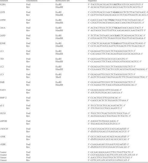
TABLE 2Primers used in these studiesa
Mutation Orientation Restriction site Sequence
D228A Fwd EcoRI 5=-TACGTGACGGAGTCGGCTGCGTCGCAGCGTGT-3=
Rev HindIII 5=-ACACGCTGCGACGCAGCCGACTCCGTCACGTA-3=
L237E Fwd EcoRI 5=-CGTGTGACCCAACTAGAGGGCTCTCTTACTATAACC-3=
Rev HindIII 5=-GGTTATAGTAAGAGAGCCCTCTAGTTGGGTCACACG-3=
S239W Fwd EcoRI 5=-GACCCAACTACTTGGCTGGCTTACTATAACCAG-3=
Rev HindIII 5=-CTGGTTATAGTAAGCCAGCCAAGTAGTTGGGTC-3=
T241A Fwd EcoRI 5=-ACTACTTGGCTCTCTTGCAATAACCAGCCTACT-3=
Rev HindIII 5=-AGTAGGCTGGTTATTGCAAGAGAGCCAAGTAGTT-3=
L245D Fwd EcoRI 5=-TCTTACTATAACCAGCGACCTCAGAAGACTCCAC-3=
Rev HindIII 5=-GTGGAGTCTTCTGAGGTCGCTGGTTATAGTAAGA-3=
H250E Fwd EcoRI 5=-CTACTCAGAAGACTCGAGAATTGGATAACTGAGG-3=
Rev HindIII 5=-CCTCAGTTATCCAATTCTCGAGTCTTCTGAGTAG-3=
oC1 Fwd EcoRI 5=-GGAGAATTCCGCCTCTAGGGCGGCTCT-3=
Rev HindIII 5=-CACAAGCTTCTACAGAATGGCCGCGCAGATGA-3=
CTD Fwd EcoRI 5=-GGCGAATTCGCGCCGCCACGTG-3=
Rev HindIII 5=-CCAAAGCTTCTAGCATGGGATGGGGCAGTCC-3=
oC2 Fwd EcoRI 5=-GGAGAATTCCGCCTCTAGGGCGGCTCT-3=
Rev HindIII 5=-CGAAAGCTTCTACTCCGTCACGTAGTGAGTAGGGG-3=
oC3 Fwd EcoRI 5=-GGAGAATTCCGCCTCTAGGGCGGCTCT-3=
Rev XholI 5=-AGTCTCGAGCTAGTGGAGTCTTCTGAGTAGGCTGG-3=
NS4B Fwd EcoRI 5=-GGAGAATTCCGCCTCTAGGGCGGCTCT-3=
Rev HindIII 5=-CCAAAGCTTCTAGCATGGGATGGGGCAG-3=
STAT3 Fwd 5=-GGAGGAGGCATTCGGAAG-3=
Rev 5=-ATCTGTGTGACACCAACGA-3=
MMP-2 Fwd 5=-CCACTGCCTTCGATACAC-3=
Rev 5=-GAGCCACTCTCTGGAATCTTAAA-3=
Bcl-2 Fwd 5=-TCCCTCGCTGCACAAATACTC-3=
Rev 5=-TTCTGCCCCTGCCAAATCT-3=
SOCS3 Fwd 5=-TGCCTCCTGACTATGTCTGGCTAA-3=
Rev 5=-AGTGGGGACCTGGTGGCTCTGCTC-3=
GAPDH Fwd 5=-AAGGCTGTGGGCAAGG-3=
Rev 5=-TGGAGGAGTGGGTGTCG-3=
si-SOCS3 Fwd 5=-CACCUGGACUCCUAUGAGAdTdT-3=
Rev 5=-dTdTGUGGACCUGAGGAUACUCU-3=
si-JNK Fwd 5=-GCCCAGUAAUAUAGUAGAUdTdT-3=
Rev 5=-dTdTCGGGUCAUUAUAUCAUCAU-3=
si-ERK Fwd 5=-CAAGAAGACCUGAAUUGUAdTdT-3=
Rev 5=-dTdTGUUCUUCUGGACUUAACAU-3=
F1outer Fwd 5=-GCAACAGGGAACCTTCCTGGTTGCTC-3=
R1outer Rev 5=-CGTAGGGGCCAGTTCATCATCATCATCAT-3=
F1inner Fwd 5=-AACCTTCCTGGTTGCTCTTTCTCTAT-3=
R1inner Rev 5=-GTTCATCATCATATCCCATGCCAT-3=
a
Mutated nucleotides are in boldface. D228A, L237E, S239W, T241A, H250, and H250E, point mutations of HCV NS4B;oC1,oC2, andoC3, truncations of HCV NS4B; CTD, carboxy-terminal domain of the NS4B protein; Fwd, forward primers; Rev, reverse primers; NS4B, wild-type NS4B protein of HCV; STAT3, quantitative RT-PCR analysis of the signal transducer and activators of transcription 3; MMP-2, quantitative RT-PCR analysis of the matrix metalloproteinase-2; Bcl-2, quantitative RT-PCR analysis of the B-cell lymphoma 2; SOCS3, semiquantitative RT-PCR analysis of the suppressor of cytokine signaling 3; GAPDH, quantitative RT-PCR analysis of the glyceraldehyde-3-phosphate dehydrogenase; si-SOCS3, si-JNK, and si-ERK, siRNAs against SOCS3, JNK, and ERK, respectively, were purchased directly from Ribobio Company; F1outer, outer sense primer from the 5=noncoding region of the HCV(493 to 518 nucleotides); R1outer, outer antisense primer from the 5=noncoding region of the HCV genome (987 to 964 nucleotides); F1inner, inner sense primer from the 5=noncoding region of the HCV genome (502 to 527 nucleotides); R1inner, inner antisense primer from the 5=noncoding region of the HCV genome (975 to 952 nucleotides).
on November 7, 2019 by guest
http://jvi.asm.org/
Carlsbad, CA) for 24 h and then serum starved for an additional 24 h prior to harvest.Renillaluciferase plasmid pRL-TK was used as an internal control. Luciferase assays were performed with a dual-luciferase kit (Pro-mega, Madison, WI) according to the manufacturer’s instructions.
Quantitative RT-PCR analysis and nested PCR.Total RNA was ex-tracted by the TRIzol reagent (Invitrogen) and reverse transcribed with random primers. Real-time PCR was performed with a LightCycler 480 apparatus (Roche, Basel, Switzerland) with GAPDH as the internal con-trol. To generate the HCV RNA references for determination of the sen-sitivity and specificity of the strand-specific RT-PCR, RNA was extracted from the patients’ PBMCs. Subsequently, RNA was retrotranscribed with primers specific for the 5=untranslated region of HCV (antisense primer R1outer for the plus RNA strand and sense primer F1outer for the minus RNA strand). A nested PCR was then performed using two different primer sets: F1outer and R1outer in the first step and F1inner and R1inner in the second step. The amplified product (474 bp) was visualized on an ethidium bromide-stained agarose gel. Primers used for the quantitative RT-PCR and nest-PCR analysis are listed inTable 2.
MMP zymography.The collagenolytic activity was determined on a gelatin-impregnated (1 mg/ml; Difco, Detroit, MI) 8% SDS-polyacryl-amide gel. Protein samples were separated under nonreducing condi-tions, followed by 30 min incubation in 2.5% Triton X-100 (BDH, Eng-land). The gels were then incubated for 16 h at 37°C in 50 mM Tris, 0.2 M NaCl, 5 mM CaCl2, 0.02% Brij 35 (wt/vol) at pH 7.6. At the end of the
incubation period, the gels were stained with 0.5% Coomassie G250 (Bio-Rad, Richmond, CA) in methanol-acetic acid-H2O (30:10:60). MMP-2
standards were loaded into each gel for band identification, and the pro-teolytic band intensities were quantified by scanning densitometry.
Nuclear extraction.After serum starvation for 24 h, cells were washed twice with cold phosphate-buffered saline (PBS). They were then har-vested and incubated in 2 volumes of buffer A (10 mM HEPES, pH 8.0, 0.5% Nonidet P-40, 1.5 mM MgCl2, 10 mM KCl, 0.5 mM dithiothreitol [DTT], and 200 mM sucrose) for 5 min at 4°C with tube flipping. The crude nuclei were collected by centrifugation for 30 s; pellets were rinsed with buffer A, resuspended in 1 volume of buffer B (20 mM HEPES, pH 7.9, 1.5 mM MgCl2, 420 mM NaCl, 0.2 mM EDTA, and 1.0 mM DTT),
and incubated on a shaking platform for 30 min at 4°C. Nuclei were centrifuged for 5 min, and supernatants were collected. Cocktail protease inhibitor tablets were added to each type of buffer. Nuclear extracts were stored at⫺70°C before use.
Western blot analysis.Cells were washed with ice-cold PBS and col-lected, and the pellets were resuspended in radioimmunoprecipitation assay buffer (50 mM Tris, pH 7.5, 150 mM NaCl, 1% Triton X-100, 1% sodium deoxycholate, 0.1% SDS, 1 mM sodium formate, 1 mM phenyl-methylsulfonyl fluoride, and 10% cocktail protease inhibitor). Lysates were centrifuged at 12,000 rpm for 10 min. The protein concentration in each sample was determined using a Bradford assay kit (Bio-Rad, Hercu-les, CA). Cultured cell lysates (100g) were electrophoresed on a 12% SDS-polyacrylamide gel and transferred to a nitrocellulose membrane (Amersham). Nonspecific sites were blocked with 5% nonfat dried milk before being incubated with a specific antibody. Blots were analyzed using a luminescent image analyzer (LAS-4000; Fujifilm, Tokyo, Japan).
Immunofluorescence.Huh7 cells grown on sterile coverslips were transfected with pCMV-NS4B or control vector at 40% confluence. After 48 h, cells were fixed with 4% paraformaldehyde for 15 min, washed three times with PBS, and permeabilized with PBS containing 0.5% Triton X-100 for 5 min. They were then washed three times with PBS and blocked with PBS containing 4% bovine serum albumin for 1 h at room temper-ature. Then, the cells were incubated with the primary antibody overnight at 4°C, followed by incubation with Alexa 492-labeled secondary antibod-ies (ProteinTech Group) for 1 h. Mounting was done with Vectashield mounting medium with DAPI (4=,6-diamidino-2-phenylindole; Vector Laboratories), and the cells were visualized by confocal laser microscopy (Fluoview FV1000; Olympus, Tokyo, Japan).
Statistics.All experiments were reproducible, and each set was re-peated at least three times with similar results. Parallel samples were ana-lyzed for a normal distribution by the Kolmogorov-Smirnov test. Abnor-mal values were eliminated according to a follow-up Grubbs test. Levene’s test for equality of variances was performed and provided information for Student’sttest to distinguish the equality of means. Means were illus-trated using a histogram with error bars representing⫾the standard deviation (SD), and aPvalue of⬍0.05 was considered statistically signif-icant.
RESULTS
HCV stimulates STAT3 activity through regulating the JNK and
ERK signaling cascades, resulting in the activation of MMP-2
and Bcl-2.
We first investigated the effect of HCV infection on the
regulation of STAT3, an important protein associated with cell
cycle progression, growth, transformation, metastasis, and
angio-genesis. PBMCs were isolated from HCV-positive individuals
(
n
⫽
20) and HCV-negative individuals (
n
⫽
20) (
Table 1
).
Real-time RT-PCR showed that the relative mRNA levels of STAT3
were approximately 3-fold higher in HCV-positive individuals
than in HCV-negative individuals (
Fig. 1A
), suggesting that
STAT3 is activated during HCV infection. Since STAT3 is the
upstream target of MMP-2 and Bcl-2 (
5
,
14
), we examined the
expression status of MMP-2 and Bcl-2 in the clinical samples.
Similar to STAT3, the relative mRNA levels of MMP-2 (
Fig. 1B
)
and Bcl-2 (
Fig. 1C
) were increased in HCV-positive individuals
compared to HCV-negative individuals. These results
demon-strate that HCV activates STAT3, resulting in the upregulation of
MMP-2 and Bcl-2. HCV replication in the PBMCs of HCV
pa-tients was determined by nested PCR analyses, which showed that
HCV plus-strand RNA was detected in the PBMCs of 12 of the 20
patients and minus-strand RNA was detected in the PBMCs of 7 of
the 20 patients (
Fig. 1D
), confirming that HCV replicated in the
PBMCs of HCV patients.
The mechanism involved in the activation of STAT3 by
HCV infection was further investigated
in vitro
by infecting
Huh7.5.1 cells with HCV JFH-1 for different times. Western
blot analyses revealed that the HCV NS3 protein was detected
at 3 days postinfection, indicating that HCV replicated well in
infected cells (
Fig. 1E
). The levels of the p-STAT3, MMP-2, and
Bcl-2 proteins in infected cells were increased in a
time-depen-dent manner, the levels of STAT3 proteins were slightly
in-creased by 1.17-fold at 6 days postinfection, and the level of

-actin proteins remained relatively constant during HCV
in-fection (
Fig. 1E
). It has been reported that phosphorylation of
STAT3 is regulated by the JNK and ERK signaling cascade (
4
,
9
,
61
). We also revealed that the levels of the p-ERK and p-JNK
proteins were increased over time after HCV infection, while
the levels of the ERK, JNK, and

-actin proteins were relatively
unchanged during HCV infection (
Fig. 1E
). In addition,
Huh7.5.1 cells were infected with JFH-1 at different
concentra-tions. The results showed that the levels of the NS3, p-STAT3,
MMP-2, Bcl-2, p-ERK, and p-JNK proteins were enhanced in
cells infected with HCV in a dose-dependent fashion and the
level of the STAT3 protein was slightly increased by 1.22-fold,
while the levels of the ERK, JNK, and

-actin proteins were
relatively unchanged during HCV infection (
Fig. 1F
). Thus, we
demonstrate that HCV stimulates STAT3 activity through the
JNK and ERK signaling cascades, resulting in the activation of
MMP-2 and Bcl-2 in hepatocytes. These
in vitro
results (in
Huh7.5.1 cells transiently infected with HCV) are consistent
on November 7, 2019 by guest
http://jvi.asm.org/
with our
in vivo
data (in PBMCs chronically infected with
HCV).
The NS4B protein of HCV activates MMP-2 and Bcl-2
ex-pression by repressing SOCS3 production and stimulating
STAT3 activity.
We then determined which of the 10 proteins of
HCV is responsible for the regulation of STAT3. Huh7 cells were
cotransfected with the reporter plasmid pGL3-APRE-Luc, along
with plasmids expressing each of the 10 HCV genes constructed
previously (
42
). A luciferase activity assay showed that STAT3
promoter activity was activated by NS4B or NS5A, while other
proteins had a slight or no effect on the STAT3 promoter (
Fig.
2A
). These results suggest that the HCV NS4B and NS5A proteins
are involved in the regulation of STAT3 expression.
HCV NS5A is known to activate STAT3, interact with Bax, and
inhibit apoptosis in hepatocellular carcinoma (
10
,
21
,
55
).
More-over, it has been reported that the nucleotide binding motif of
hepatitis C virus NS4B can mediate cellular transformation and
tumor formation without HA-Ras cotransfection (
14
). Thus, we
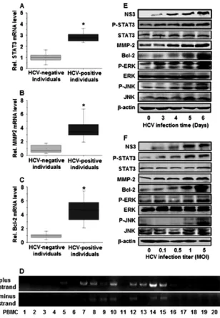
FIG 1Expression of STAT3, MMP-2, Bcl-2, ERK, and JNKin vivoandin vitro. (A to C) PBMCs were isolated from peripheral blood samples obtained from 20 patients with HCV infection and 20 HCV-negative individuals. The mRNA levels of MMP-2, Bcl-2, and STAT3 were measured using real-time PCR. GAPDH was used as an internal reference. MMP-2, Bcl-2, and STAT3 levels in each sample were normalized by dividing by the GAPDH quantity; *,P⬍0.05; Rel., relative. (D) HCV plus- and minus-strand RNAs from the PBMCs of HCV-infected patients were detected by nested PCR using two different primer sets for the 5=
noncoding region, which is the best conserved among different HCV isolates. (E) Huh7.5.1 cells were infected with JFH-1 at an MOI of 1 for different times, as indicated. Proteins were detected by Western blot analyses using appropriate antibodies. The HCV NS3 protein level was measured to confirm HCV replication in the cells. (F) Huh7.5.1 cells were infected with JFH-1 at different titers for 6 days, as indicated. Proteins were detected by Western blot analyses using appropriate antibodies. HCV NS3 protein levels were monitored to ensure that HCV was replicating in the cells. NS3, HCV NS3 protein; p-STAT3, phosphor-ylated STAT3 protein; STAT3, total STAT3 protein; p-ERK, phosphorphosphor-ylated ERK protein; ERK, total ERK protein; p-JNK, phosphorphosphor-ylated JNK protein; JNK, total JNK protein.
on November 7, 2019 by guest
http://jvi.asm.org/
[image:5.585.139.447.65.511.2]explored only the role of NS4B in STAT3 activation in this study.
To determine the effects of NS4B on MMP-2 and Bcl-2
expres-sion, Huh7 cells were transfected with NS4B or
pCMV-Tag2A. RT-PCR results showed that STAT3, MMP-2, and Bcl-2
mRNAs were stimulated by NS4B but SOCS3 (a suppressor of
STAT3) mRNA was repressed by NS4B (
Fig. 2B
). These results
indicate that HCV NS4B activates STAT3 expression by repressing
its suppressor, SOCS3.
STAT3 is activated by phosphorylation, which induces its
translocation from the cytosol to nucleus to regulate target genes.
We examined the effect of NS4B on the phosphorylation and
translocation of STAT3. Huh7 cells were transfected with
pCMV-NS4B or pCMV-Tag2A at different concentrations. Results
re-vealed that the SOCS3 protein was decreased but that in the
pres-ence of NS4B the STAT3, p-STAT3, MMP-2, and Bcl-2 proteins
were increased in a dose-dependent fashion, while the STAT3
pro-tein was slightly increased by 1.45-fold (
Fig. 2C
). In addition,
re-sults from gelatin zymography analyses showed that in the
pres-ence of NS4B the gelatinolytic activity of MMP-2 was increased in
a dose-dependent manner (
Fig. 2D
). Further investigation
re-vealed that the level of the STAT3 protein was slightly affected by
NS4B in whole-cell lysates and was reduced by NS4B in the cytosol
(
Fig. 2E
) but significantly enhanced by NS4B in the nucleus (
Fig.
2F
), suggesting that the HCV NS4B protein may play a role in
stimulating STAT3 translocation from the cytosol to the nucleus.
Moreover, immunofluorescence assays showed that the STAT3
protein was mainly located in the cytosol in the absence of NS4B
but shifted its distribution to the nucleus in the presence of NS4B
(
Fig. 2G
). These results demonstrate that NS4B activates the
phos-phorylation of STAT3 and promotes the translocation of STAT3
from the cytosol to the nucleus, resulting in the activation of the
target genes, MMP-2 and Bcl-2.
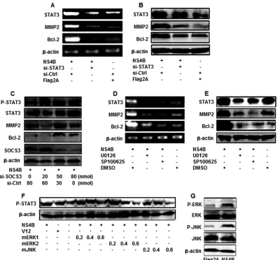
ERK, JNK, and SOCS3 play critical roles in the regulation of
STAT3 signaling mediated by NS4B.
To further explore the role
of STAT3 in MMP-2 and Bcl-2 expression mediated by NS4B,
Huh7 cells were cotransfected with pCMV-NS4B, pCMV-flag2A,
si-STAT3, or si-Ctrl, as indicated. We showed that the levels of
STAT3 mRNAs and protein were repressed in the presence of
si-STAT3 but the levels of

-actin mRNA and protein were not
affected by si-STAT3 (
Fig. 3A
and
B
), indicating that si-STAT3 is
specific and effective. Results also showed that the levels of
MMP-2 and Bcl-2 mRNAs and proteins were activated by NS4B
but significantly reduced by si-STAT3 (
Fig. 3A
and
B
). These
re-sults suggest that overexpression of STAT3 upregulates MMP-2
and Bcl-2, while knockdown of STAT3 downregulates MMP-2
and Bcl-2, supporting the suggestion that STAT3 activates
MMP-2 and Bcl-2 in the presence of HCV NS4B.
To verify the role of SOCS3 in this regulation, Huh7 cells were
transfected with pCMV-NS4B and treated with SOCS3 or
si-Ctrl at different concentrations. The results indicate that while
SOCS3 was reduced, the p-STAT3, STAT3, MMP-2, and Bcl-2
proteins were enhanced in the presence of si-SOCS3 in
dose-de-pendent fashion and STAT3 and

-actin were not affected by
si-SOCS3 (
Fig. 3C
). These results indicate that HCV NS4B activates
STAT3 expression by repressing its suppressor, SOCS3.
Given the critical roles of STAT3 in liver inflammation and
carcinogenesis, it is important to understand the key upstream
signaling pathways involved in the regulation of STAT3 in human
liver cells. It has been reported that the ERK and JNK signaling
cascade regulates several transcription factors that are important
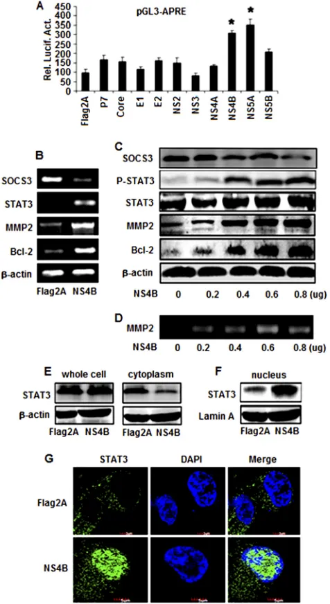
FIG 2Effects of HCV proteins in the regulation of STAT3, MMP-2, and Bcl-2 expression. (A) Huh7 cells were cotransfected with the reporter pGL3-APRE-Luc containing the luciferase gene under the control of the STAT3 promoter and Flag2A, core, E1, E2, P7, NS2, NS3, NS4A, NS4B, NS5A, or pCMV-NS5B expressing the corresponding HCV proteins. Luciferase activity was measured as described in Materials and Methods. Results are expressed as the mean⫾SD of independent experiments performed in triplicate and normal-ized using a-galactosidase assay. *,P⬍0.05 versus pCMV-Flag2A; Rel. Lucif. Act., relative luciferase activity. (B) Huh7 cells were transfected with pCMV-NS4B or pCMV-Flag2A. At 48 h posttransfection, total RNA was isolated and used as the template for RT-PCR using primers specific to Bcl-2, MMP-2, STAT3, SOCS3, and-actin. (C) Huh7 cells were transfected with pCMV-NS4B at different concentrations, as indicated. Proteins were detected by Western blot analysis using antibodies to p-STAT3, STAT3, MMP-2, Bcl-2, SOCS3, and-actin. (D) Huh7 cells were transfected with pCMV-NS4B or pCMV-Flag2A at the indicated concentrations. MMP-2 activity was measured by gelatin zymography at 48 h posttransfection. (E and F) Huh7 cells were transfected with pCMV-NS4B or pCMV-Flag2A. The STAT3 protein levels in whole-cell lysates, the cytoplasm, and the nucleus were determined by Western blot analysis using antibodies to STAT3. (G) Effect of NS4B on the transloca-tion of STAT3 from the cytosol to the nucleus. Huh7 cells were transfected with pCMV-NS4B or control vector for 48 h. After fixation, the cells were immunostained with antibody for STAT3. The nuclei were stained by DAPI.
on November 7, 2019 by guest
http://jvi.asm.org/
[image:6.585.45.283.68.504.2]in the proinflammatory response and that the regulation of
STAT3 expression depends on the kinase activators in various cell
types (
4
,
9
,
61
). Thus, we identified the components of cellular
signaling cascades involved in the regulation of STAT3 in response
to HCV infection. Huh7 cells were transfected with pCMV-NS4B
and treated with U0126 (an ERK-specific inhibitor) and SP600125
(a JNK-specific inhibitor). The results indicate that the levels of
STAT3, MMP-2, and Bcl-2 mRNA (
Fig. 3D
) and protein (
Fig. 3E
)
were enhanced by NS4B but repressed by SP600125 and U0126,
suggesting that ERK and JNK are involved in the regulation of
STAT3, MMP-2, and Bcl-2 mediated by NS4B.
The roles of ERK and JNK in the activation of STAT3 mediated
by NS4B were further evaluated by introducing three dominant
kinase-inactive mutants (mERK1, mERK2, and mJNK) which
block the corresponding kinase activities by competing with
en-dogenous kinases (
43
). Huh7 cells were cotransfected with
pCMV-NS4B V12 (a constitutively active form of Ras that
acti-vates ERK) and each of the three kinase mutants, mERK1,
mERK2, and mJNK. We found that the p-STAT3 protein was
activated by NS4B and V12 but repressed by mERK1, mERK2, and
mJNK in a dose-dependent manner (
Fig. 3F
), demonstrating that
ERK and JNK are involved in the activation of STAT3 regulated by
NS4B.
To evaluate the effect of NS4B on the activation of the JNK and
ERK signaling cascades, Huh7 cells were transfected with
pCMV-NS4B or pCMV-Tag2A. The results showed that the levels of the
p-ERK and p-JNK proteins were significantly enhanced by NS4B
but the levels of the ERK, JNK, and

-actin proteins were
unaf-fected by NS4B (
Fig. 3G
). These results suggest that NS4B plays a
role in the activation of the ERK and JNK signaling pathway
through phosphorylation of these two protein kinases, resulting in
the activation of STAT3, MMP-2, and Bcl-2.
FIG 3Roles of SOCS3, ERK, and JNK in the regulation of STAT3, MMP-2, and Bcl-2. (A and B) Huh7 cells were transfected with pCMV-NS4B or pCMV-Flag2A or treated with si-STAT3 or si-Ctrl. STAT3, MMP-2, and Bcl-2 mRNA levels were determined by RT-PCR, and TAT3, MMP-2, and Bcl-2 protein levels were determined by Western blot analysis. (C) Huh7 cells were transfected with pCMV-NS4B and treated with si-SOCS3 or si-Ctrl at different concentrations, as indicated. STAT3, p-STAT3, MMP-2, and Bcl-2 protein levels were determined by Western blot analysis. (D and E) Huh7 cells were transfected with pCMV-NS4B or pCMV-Flag2A and then treated with protein kinase inhibitors SP600125 and U0126. STAT3, MMP-2, and Bcl-2 mRNA levels were measured at 48 h posttransfection by RT-PCR, and STAT3, MMP-2, and Bcl-2 protein levels were determined by Western blot analysis. The blot is a representative of three experiments with similar results. (F) Huh7 cells were cotransfected with pCMV-NS4B, V12, or dominant-negative mutants (mERK1, mERK2, and mJNK) of ERK or JNK at different concentrations, as indicated. p-STAT3 and-actin proteins were measured by Western blot analysis. (G) Huh7 cells were transfected with pCMV-NS4B or pCMV-Flag2A. p-ERK, ERK, p-JNK, JNK, and-actin proteins were determined by Western blot analysis.
on November 7, 2019 by guest
http://jvi.asm.org/
[image:7.585.95.495.65.437.2]Members of the PKC family are involved in activation of
STAT3, MMP-2, and Bcl-2 regulated by NS4B.
It is known that
members of the PKC family comprise a class of intracellular
ser-ine/threonine-specific kinases (
62
). Depending on the isoform,
activation of PKCs is typically initiated by Ca
2⫹, lipid second
mes-sengers, or protein activators. PKC
ε
interacts with STAT3 and
regulates its constitutive activation in prostate cancer and also
sensitizes skin to UV radiation-induced cutaneous damage and
the development of squamous cell carcinomas (
62
). PKC
␣
acti-vates ERK, JNK, and p38 to regulate the myogenic program in
human rhabdomyosarcoma cells (
45
).
We extended our study to identify the specific PKC proteins
involved in STAT3 activation regulated by NS4B. siRNA
mole-cules specific to each of the PKC isozymes were designed and used
previously (
43
). Huh7 cells were cotransfected with pCMV-NS4B
and si-PKC
␦
, si-PKC
␣
, si-PKC

, si-PKC
ε
, si-PKC
, or si-Ctrl, as
indicated. Results showed that MMP-2 and Bcl-2 mRNA levels
were increased in the presence of NS4B, while they were repressed
by si-PKC
␣
, si-PKC

, and si-PKC
ε
and were not affected by
si-PKC
␦
, si-PKC
, or si-Ctrl (
Fig. 4A
and
B
). Similarly, JNK,
p-ERK, p-STAT3, MMP-2, and Bcl-2 proteins were stimulated by
NS4B, while they were repressed by si-PKC
␣
, si-PKC

, and
si-PKC
ε
and not affected by si-PKC
␦
, si-PKC
, or si-Ctrl (
Fig. 4C
and
D
). These results suggest that PKC
␣
, PKC

, and PKC
ε
are
involved in the NS4B-mediated activation of STAT3.
The results presented above demonstrate that PKC
␣
, PKC

,
FIG 4Effects of the members of the PKC family on the regulation of MMP-2, Bcl-2, and STAT3 mediated by HCV NS4B. (A to C) Huh7 cells were transfected with plasmids expressing si-PKC␦, si-PKC␣, si-PKC, si-PKCε, si-PKC, or si-Ctrl. A similar vector (pSilencer 2.0) containing an irrelevant sequence that does not have significant homology to any human gene was provided by Ambion, Inc., and used as a negative control. The relative levels of MMP-2 and Bcl-2 mRNA were measured by real-time PCR at 48 h posttransfection, and the levels of p-STAT3, MMP-2, Bcl-2, and-actin proteins were determined by Western blot analysis. GAPDH was used as an internal reference. MMP-2 and Bcl-2 levels in each sample were normalized by dividing by the GAPDH quantity. Data are expressed as the mean⫾SD of three independent experiments. (D) Huh7 cells were transfected with plasmids expressing si-PKC␣, si-PKC, si-PKCε, si-PKC␦, si-PKC, or si-Ctrl. The levels of SOCS3, p-ERK, ERK, p-JNK, JNK, and-actin proteins were determined by Western blot analysis at 48 h posttransfection. (E) Huh7 cells were transfected with pCMV-NS4B and treated with si-ERK or si-Ctrl at the indicated concentrations, and the levels of the ERK, SOCS3, p-JNK, JNK, and-actin proteins were determined by Western blot analysis. (F) Huh7 cells were transfected with pCMV-NS4B and treated with si-JNK or si-Ctrl at different concentrations, as indicated. The levels of the JNK, SOCS3, p-ERK, ERK, and-actin proteins were determined by Western blot analysis. (G) Huh7 cells were transfected with pCMV-NS4B and treated with si-ERK (80 nmol), si-JNK (80 nmol), or si-ERK (40 nmol) and si-JNK (40 nmol). The protein levels of p-STAT3, STAT3, MMP-2, Bcl-2, and-actin were determined by Western blot analysis.
on November 7, 2019 by guest
http://jvi.asm.org/
[image:8.585.128.468.64.441.2]PKC
ε
, ERK, JNK, and SOCS3 play critical roles in the regulation
of STAT3 activity medicated by the HCV NS4B protein. To
fur-ther evaluate the correlation between ERK, JNK, and SOCS3,
Huh7 cells were transfected with pCMV-NS4B and treated with
si-JNK or si-ERK at different concentrations. The results indicate
that the ERK and p-JNK proteins are reduced in the presence of
si-ERK, while the SOCS3, JNK, and

-actin proteins are not
af-fected by si-ERK (
Fig. 4E
). Equally interesting, JNK and p-ERK
proteins were repressed in the presence of si-JNK, while SOCS3,
ERK, and

-actin proteins were not affected by si-JNK (
Fig. 4F
).
Furthermore, Huh7 cells were transfected with pCMV-NS4B and
treated with si-ERK (80 nmol), si-JNK (80 nmol), or si-ERK (40
nmol) and si-JNK (40 nmol) (
Fig. 4G
). The activity of STAT3 was
significantly reduced when cells were transfected with both si-JNK
(40 nmol) and si-ERK (40 nmol). Results show that the two
com-ponents of these pathways (ERK and JNK) have synergistic effects.
Thus, these results suggest that there is a correlation between ERK
and JNK but SOCS3 is independent of ERK/JNK.
The NS4B CTD is sufficient for the activation of STAT3, JNK,
ERK, MMP-2, and Bcl-2, and amino acids 237L, 239S, and 245L
of NS4B are crucial for this regulation.
HCV NS4B has four
transmembrane domains (TMDs) containing 70 amino acids
(residues 192 to 261) within its carboxy terminus, which is called
the NS4B carboxy-terminal domain (CTD) (
44
). The NS4B CTD
is located on the cytosolic side of the endoplasmic reticulum (ER)
membrane, and most of the residues in this region are required for
HCV genome replication, perhaps through an interaction with
viral and host factors (
2
). Here, we evaluated the role of the NS4B
CTD in the regulation of the STAT3 signaling cascade. To
deter-mine the sequences of NS4B protein required for the activation of
STAT3, we generated 4 deletion mutants of the NS4B protein,
named NS4B
⌬
C1, NS4BCTD, NS4
⌬
C2, and NS4B
⌬
C3 (
Fig. 5A
).
The 4 mutated NS4B genes were then subcloned into the
expres-sion vector to yield four plasmids, pCMV-NS4B
⌬
C1,
pCMV-NS4BCTD, pCMV-NS4B
⌬
C2, and pCMV-NS4B
⌬
C3.
Huh7 cells were transfected with each of the plasmids, and the
effects of these mutant NS4B proteins on the regulation of
MMP-2, Bcl-2, STAT3, ERK, and JNK were evaluated. Real-time
PCR analyses indicated that the relative levels of MMP-2 mRNA
(
Fig. 5B
) and Bcl-2 mRNA (
Fig. 5C
) were activated by NS4B,
NS4B CTD, and NS4B
⌬
C3 but not by NS4B
⌬
C1 or NS4B
⌬
C2.
These results suggest that NS4B CTD is sufficient for the
activa-tion of MMP-2 and Bcl-2 and that the 24 residues (amino acids
227 to 250) of the NS4B CTD are essential for the regulation of
MMP-2 and Bcl-2. The results also showed that the p-STAT3,
MMP-2, Bcl-2, p-ERK, and p-JNK proteins were upregulated by
NS4B, NS4B CTD, and NS4B
⌬
C3 but not by NS4B
⌬
C1 or
NS4B
⌬
C2 (
Fig. 5D
). However, the levels of the STAT3, ERK, JNK,
and

-actin proteins were relatively unchanged in the presence of
NS4B and its mutants (
Fig. 5D
). These results demonstrate that
the NS4B CTD is sufficient for the activation of STAT3, ERK, and
JNK and suggest that amino acids 227 to 250 of NS4B play a
crit-ical role in the regulation of the signaling components.
Based on the results presented above, we attempted to
deter-mine which of the 24 residues of NS4B protein are required for the
activation of MMP-2, Bcl-2, STAT3, ERK, and JNK. A series of
NS4B point mutations was constructed by site-directed
mutagen-esis, in which amino acids 228D, 237L, 239S, 241T, 245L, and
250H were replaced by 228A, 237E, 239W, 241A, 245D, and 250E,
respectively (
Fig. 5E
). Huh7 cells were transfected with plasmids
expressing NS4B and each of the mutants. Real-time PCR showed
that MMP-2 mRNA (
Fig. 5F
) and Bcl-2 mRNA (
Fig. 5G
) were
activated by NS4B, D228A, T241A, and H250E but not by L237E,
S239W, and L245D. These results suggest that amino acids 237L,
239S, and 245L are essential for the activation of MMP-2 and Bcl-2
but that amino acids 228D, 241T, and 250H are not essential for
such regulation. Western blot analyses indicated that the
p-STAT3, MMP-2, Bcl-2, p-ERK, and p-JNK proteins were
en-hanced by NS4B, D228A, T241A, and H250E but not by L237E,
S239W, or L245D (
Fig. 5H
). These results demonstrate that amino
acids 237L, 239S, and 245L play critical roles in the regulation of
STAT3, ERK, and JNK, while amino acids 228D, 241T, and 250H
are not involved in this regulation.
DISCUSSION
HCV persistent infection is associated with a variety of human
liver diseases, including HCC. However, the mechanisms by
which HCV infection causes chronic liver diseases remain unclear.
Here, we demonstrated that STAT3, MMP-2, and Bcl-2 are
signif-icantly stimulated in the PBMCs of patients with HCV infection
and in cell cultures infected with HCV. These results are consistent
with those of previous studies showing that MMP-2 and Bcl-2
levels are elevated in the serum of patients with HCC, cirrhosis,
and chronic hepatitis (
15
,
23
,
28
,
63
).
It has been reported that HCV is able to infect not only
hepa-tocytes but also PBMCs (
37
,
54
). Immediately after discovery of
the virus in 1989, different groups showed that HCV replicates in
lymphoid cells by infecting macrophages and B and T
lympho-cytes (
8
,
24
,
58
). Moreover, several reports described the presence
of the replicative intermediate or negative strand in PBMCs (
47
,
48
,
70
). Our results show that both HCV plus-strand RNA and
minus-strand RNA can be detected in the PBMCs of patients with
HCV infection, and these findings are consistent with previous
reports (
6
,
11
,
27
). The presence of the replicative form of HCV in
PBMCs suggests that these cells can be a virus reservoir capable of
reinfecting the liver in transplant patients or in subjects treated
with IFN (
56
,
57
). This lymphotropism may explain the
associa-tion between this virus and certain lymphoproliferative disorders
(LPDs) (
51
). It is likely that HCV infection of T and B lymphocytes
is responsible, at least in part, for the many extrahepatic disorders
often observed in individuals chronically infected with the virus
(
17
,
18
,
19
).
Although the HCV core and NS5A proteins function as
transcriptional transactivators for many cellular genes, such as
NF-
B, AP-1, SRE, and STAT3 (
10
,
16
,
32
,
49
), the molecular
basis for hepatocellular MMP-2 and Bcl-2 expression in
pa-tients with chronic HCV infection remains unknown. We
re-vealed that HCV activates MMP-2 and Bcl-2 expression by
regulating the STAT3 signaling pathway. It has been reported
that STAT3 induces MMP-2 and Bcl-2 expression (
5
), and we
demonstrate for the first time that STAT3 plays a role in the
regulation of gene expression during HCV infection. Following
phosphorylation, STAT3 is dimerized and translocated to the
nucleus to activate downstream target genes (
20
,
40
). We
dem-onstrate that the HCV NS4B protein activates STAT3 by
en-hancing the phosphorylation and translocation of STAT3,
thereby activating MMP-2 and Bcl-2 expression.
STAT3 is involved in the transduction of cellular signals and
the induction of gene expression in response to cytokine receptor
stimulation (
20
,
40
). The primary function of MMP-2 is to
on November 7, 2019 by guest
http://jvi.asm.org/
grade proteins in the extracellular matrix. Physiologically, MMP-2
plays a role in normal tissue remodeling events such as embryonic
development, reproduction, and tissue remodeling, as well as in
disease processes, such as arthritis, angiogenesis, and metastasis.
Bcl-2 is a proto-oncogene that suppresses apoptosis in a variety of
cell systems, regulates cell death by controlling mitochondrial
membrane permeability, and inhibits caspase activity either by
preventing the release of cytochrome
c
from the mitochondria or
by binding to apoptosis-activating factor 1 (APAF-1).
The mechanism by which HCV regulates STAT3, MMP-2,
and Bcl-2 was investigated in this study. We found that HCV
NS4B activates the JNK and ERK signaling cascade, resulting in
FIG 5Function of HCV NS4B CTD in the regulation of STAT3, MMP-2, Bcl-2, ERK, and JNK. (A) Schematic diagram of the NS4B deletion mutants. The numbers indicate the amino acid residues located in the NS4B protein. (B to and D) Huh7 cells were transfected with plasmids expressing NS4B protein or its truncated mutant proteins. The relative levels of MMP-2 and Bcl-2 mRNA were measured at 48 h posttransfection using real-time PCR, and the levels of the p-STAT3, STAT3, MMP-2, Bcl-2, p-ERK, ERK, p-JNK, JNK, and-actin proteins were determined by Western blot analysis. GAPDH was used as an internal reference. The MMP-2 and Bcl-2 levels in each sample were normalized by dividing by the GAPDH quantity. (E) Sequences of amino acids 226 to 250 of wild-type NS4B and its mutants. Mutated amino acids are underlined. (F to and H) Huh7 cells were transfected with plasmids expressing NS4B protein or its mutants. The relative levels of MMP-2 and Bcl-2 mRNA were measured at 48 h posttransfection using real-time PCR, and the levels of the p-STAT3, STAT3, MMP-2, Bcl-2, p-ERK, ERK, p-JNK, JNK, and-actin proteins were determined by Western blot analysis.
on November 7, 2019 by guest
http://jvi.asm.org/
[image:10.585.82.492.67.557.2]the activation of STAT3 activity and MMP-2 and Bcl-2
expres-sion. ERK and JNK have been implicated in relaying
extracel-lular signals to the nucleus and in mediating proliferation,
dif-ferentiation, apoptosis, and stress by regulating transcription
factors (
25
,
33
,
53
). Thus, our results demonstrating that HCV
activates STAT3, MMP-2, and Bcl-2 may reveal a novel
mech-anism underlying the pathogenesis and tumorigenesis caused
by HCV infection.
STAT3 is thought to regulate the MMP-2 and Bcl-2 expression
mediated by a variety of stimulating agents (
5
,
7
,
29
,
35
). In this
study, we showed that the HCV NS4B protein regulates STAT3,
MMP-2, and Bcl-2 in the human liver cell line Huh7. STAT3 is
essential for NS4B-induced gene expression, because knockdown
of STAT3 prevents NS4B from activating MMP-2 and Bcl-2. In
addition, we demonstrated that NS4B activates MMP-2 and Bcl-2
expression by stimulating STAT3 signaling and repressing the
suppressor of cytokine signaling (SOCS3) expression and that
PKC
␣
, PKC

, and PKC
ε
are involved in NS4B-mediated STAT3
activation. Members of the SOCS family are known to negatively
regulate the activity of STAT3. Our results indicating that HCV
NS4B stimulates STAT3 activity by inhibiting the activity of its
repressor, SOCS3, suggest that the activation of STAT3 regulated
by NS4B is through repression of SOCS3. PKC comprises a family
of serine/threonine protein kinases that play important roles in
cellular signal transduction cascades. Thus, in this study we
dem-onstrated that multiple cellular signaling cascades are involved in
the regulation of STAT3, MMP-2, and Bcl-2 in response to HCV
infection.
NS4B is a poorly characterized hydrophobic protein that
may play a role in the induction of cell transformation and
tumorigenesis (
14
). Other activities associated with NS4B
in-clude modulation of NS5A hyperphosphorylation (
30
,
34
),
transactivation of interleukin 8 (IL-8) (
31
), and induction of
unfolded protein response- and endoplasmic reticulum
over-load response-dependent NF-
B activation (
39
,
60
,
68
,
69
).
NS4B also interacts with other HCV nonstructural proteins
and is involved in HCV RNA synthesis (
1
,
3
,
13
,
22
). The NS4B
CTD (
44
) interacts with the NS3 protein (
50
) and viral RNA
(
14
), is involved in NS4B oligomerization (
64
), and plays a role
in viral RNA replication (
30
). A recent report has shown that
the NS4B CTD is partially responsible for the formation and
function of the HCV replication complex (
2
). The cytosolic
CTD plays an important role in HCV genome replication,
per-haps by interacting with viral and host factors. Thus, NS4B
plays a central role in HCV genome replication. In this study,
we demonstrated that NS4B CTD is sufficient for the activation
of STAT3, ERK, and JNK, thereby upregulating MMP-2 and
Bcl-2. We also revealed that the last 24 amino acids (residues
227 to 250) of NS4B play a critical role in the regulation of
STAT3, ERK, and JNK signaling pathways. Among the 24
amino acids, 3 residues (237L, 239S, and 245L) are essential for
the function of NS4B in the regulation of these signaling
path-ways.
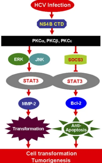
In conclusion, we have revealed a novel mechanism in which
HCV infection activates STAT3 signaling through multiple
cellu-lar signaling pathways. During HCV infection, the viral protein
NS4B activates the expression of several members of the PKC
superfamily, stimulates the ERK/JNK signaling cascades, and
re-presses SOCS3 expression, resulting in the activation of STAT3
activity by enhancing the phosphorylation and translocation of
STAT3. Activated STAT3 then stimulates MMP-2 and Bcl-2
ex-pression, thereby resulting in the regulation of cell
transforma-tion, apoptosis, and possibly, tumorigenesis in response to HCV
infection (
Fig. 6
).
ACKNOWLEDGMENTS
This work was supported by research grants from the Major State Basic Research Development Program (973 Program; 2012CB518900 and 2009CB522506), the National Natural Science Foundation of China (81171525), the National Mega Project on Major Infectious Diseases Pre-vention (2012ZX10002006-003 and 2012ZX10004-207), the National Mega Project on Major Drug Development (2011ZX09401-302-1), Fun-damental Research Funds for the Central Universities (1102001), the In-ternational Science and Technology Cooperation Program of China (2011DFA31030), and the Specialized Research Fund for the Doctoral Program of Higher Education (20090141110033).
We thank Charles M. Rice of Rockefeller University for kindly provid-ing the HCV plasmid pFL-J6/JFH5=C19Rluc2Aubi, Takaji Wakita of the National Institute of Infectious Diseases of Japan for providing HCV strain JFH-1, Francis Chisari of the Scripps Research Institute for provid-ing the Huh7.5.1 cell line, Melanie Cobb of the University of Texas South-western Medical Center for providing the ERK1 and ERK2 mutants, Mi-chael Karin of the University of California at San Diego for providing JNK mutants, and Li Liu of Tsinghua University in China for providing the plasmid pGL3-APRE-luc.
REFERENCES
1.Ali N, Tardif KD, Siddiqui A.2002. Cell-free replication of the hepatitis C virus subgenomic replicon. J. Virol.76:12001–12007.
2.Aligo J, Jia SAZ, Manna D, Konan KV.2009. Formation and function of FIG 6A novel mechanism in which HCV infection activates STAT3 through
multiple cellular signaling pathways. During HCV infection, the viral protein NS4B activates the expression of several members of the PKC family, stimu-lates the JNK/ERK signaling cascade, represses the SOCS3 expression, and enhances STAT3 activity, which in turn stimulates MMP-2 and Bcl-2 expres-sion, resulting in the control of cell transformation, apoptosis, and
tumorigen-esis in response to HCV infection.
on November 7, 2019 by guest
http://jvi.asm.org/
[image:11.585.81.244.72.332.2]hepatitis C virus replication complexes require residues in the carboxy-terminal domain of NS4B protein. Virology393:68 – 83.
3.Alter MJ.1997. Epidemiology of hepatitis C. Hepatology26:62S– 65S. 4.Bates SH, et al.2003. STAT3 signalling is required for leptin regulation of
energy balance but not reproduction. Nature421:856 – 859.
5.Bhattacharya S, Ray RM, Johnson LR.2005. STAT3-mediated transcrip-tion of Bcl-2, Mcl-1 and c-IAP2 prevents apoptosis in polyamine-depleted cells. Biochem. J.392:335–344.
6.Bouffard P, Hayashi PH, Acevedo R, Levy N, Zeldis JB.1992. Hepatitis C virus is detected in a monocyte/macrophage subpopulation of periph-eral blood mononuclear cells of infected patients. J. Infect. Dis.166:1276 – 1280.
7.Choi HJ, Lee JH, Park SY, Cho JH, Han JS.2009. STAT3 is involved in phosphatidic acid-induced Bcl-2 expression in HeLa cells. Exp. Mol. Med. 41:94 –101.
8.Choo QL, et al.1989. Isolation of a cDNA clone derived from a blood-borne non-A, non-B viral hepatitis genome. Science244:359 –362. 9.Chung J, Uchida E, Grammer TC, Blenis J. 1997. STAT3 serine
phosphorylation by ERK-dependent and-independent pathways neg-atively modulates its tyrosine phosphorylation. Mol. Cell. Biol. 17: 6508 – 6516.
10. Chung YL, Sheu ML, Yen SH.2003. Hepatitis C virus NS5A as a potential viral Bcl-2 homologue interacts with Bax and inhibits apoptosis in hepa-tocellular carcinoma. Int. J. Cancer107:65–73.
11. Crovatto M, et al.2000. Peripheral blood neutrophils from hepatitis C virus-infected patients are replication sites of the virus. Haematologica 85:356 –361.
12. Di Bisceglie AM.1997. Hepatitis C and hepatocellular carcinoma. Hepa-tology26:34S–38S.
13. Egger D, et al.2002. Expression of hepatitis C virus proteins induces distinct membrane alterations including a candidate viral replication complex. J. Virol.76:5974 –5984.
14. Einav S, et al.2008. The nucleotide binding motif of hepatitis C virus NS4B can mediate cellular transformation and tumor formation without ha-ras co-transfection. Hepatology47:827– 835.
15. El-Gindy I, El Rahman A, El-Alim MA, Zaki S.2003. Diagnostic poten-tial of serum matrix metalloproteinase-2 and tissue inhibitor of metallo-proteinase-1 as non-invasive markers of hepatic fibrosis in patients with HCV related chronic liver disease. Egypt. J. Immunol.10:27–35. 16. Feng X, et al.2011. Hepatitis C virus core protein promotes the migration
and invasion of hepatocyte via activating transcription of extracellular matrix metalloproteinase inducer. Virus Res.158:146 –153.
17. Ferri C, La Civita L, Zignego AL, Pasero G.1997. Hepatitis-C-virus infection and cancer. Int. J. Cancer71:1113–1115.
18. Ferri C, La Civita L, Zignego AL, Pasero G.1997. Viruses and cancers: possible role of hepatitis C virus. Eur. J. Clin. Invest.27:711–718. 19. Ferri C, et al.1995. Etiopathogenetic role of hepatitis C virus in mixed
cryoglobulinemia, chronic liver diseases and lymphomas. Clin. Exp. Rheumatol.13(Suppl. 13):S135–S140.
20. Giraud S, et al.2002. Functional interaction of STAT3 transcription factor with the coactivator NcoA/SRC1a. J. Biol. Chem.277:8004 – 8011.
21. Gong G, Waris G, Tanveer R, Siddiqui A.2001. Human hepatitis C virus NS5A protein alters intracellular calcium levels, induces oxidative stress, and activates STAT-3 and NF-B. Proc. Natl. Acad. Sci. U. S. A.98:9599 – 9604.
22. Gosert R, et al.2003. Identification of the hepatitis C virus RNA replica-tion complex in Huh-7 cells harboring subgenomic replicons. J. Virol. 77:5487–5492.
23. Guo XZ, Shao XD, Liu MP.2002. Effect of bax, bcl-2 and bcl-xL on regulating apoptosis in tissues of normal liver and hepatocellular carci-noma. World J. Gastroenterol.8:1059 –1062.
24. Hellings JA, van der Veen-du Prie J, Snelting-van Densen R, Stute R. 1985. Preliminary results of transmission experiments of non-A, non-B hepatitis by mononuclear leucocytes from a chronic patient. J. Virol. Methods10:321–326.
25. Hill CS, Treisman R.1995. Transcriptional regulation by extracellular signals: mechanisms and specificity. Cell80:199 –211.
26. Houghton M, Weiner A, Han J, Kuo G, Choo QL. 1991. Molecular biology of the hepatitis C viruses: implications for diagnosis, development and control of viral disease. Hepatology14:381–388.
27. Hsieh TT, Yao DS, Sheen IS, Liaw YF, Pao CC.1992. Hepatitis C
virus in peripheral blood mononuclear cells. Am. J. Clin. Pathol.98: 392–396.
28. Iwata A, et al.2011. Extracellular administration of Bcl2 protein reduces apoptosis and improves survival in a murine model of sepsis. PLoS One 6:e14729. doi:10.1371/journal.pone.0014729.
29. Jiang RQ, et al.2011. Interleukin-22 promotes human hepatocellular carcinoma by activation of STAT3. Hepatology54:900 –909.
30. Jones DM, Patel AH, Targett-Adams P, McLauchlan J.2009. The hep-atitis C virus NS4B protein can trans-complement viral RNA replication and modulates production of infectious virus. J. Virol.83:2163–2177. 31. Kadoya H, Nagano-Fujii M, Deng L, Nakazono N, Hotta H. 2005.
Nonstructural proteins 4A and 4B of hepatitis C virus transactivate the interleukin 8 promoter. Microbiol. Immunol.49:265–273.
32. Kato N, et al.2000. Activation of intracellular signaling by hepatitis B and C viruses: C-viral core is the most potent signal inducer. Hepatology32: 405– 412.
33. Khokhlatchev AV, et al.1998. Phosphorylation of the MAP kinase ERK2 promotes its homodimerization and nuclear translocation. Cell93:605– 615.
34. Koch JO, Bartenschlager R.1999. Modulation of hepatitis C virus NS5A hyperphosphorylation by nonstructural proteins NS3, NS4A, and NS4B. J. Virol.73:7138 –7146.
35. Kortylewski M, Jove R, Yu H.2005. Targeting STAT3 affects melanoma on multiple fronts. Cancer Metastasis Rev.24:315–327.
36. Kuo G, et al.1989. An assay for circulating antibodies to a major etiologic virus of human non-A, non-B hepatitis. Science244:362–364.
37. Lamelin JP, Zoulim F, Trepo C.1995. Lymphotropism of hepatitis B and C viruses: an update and a newcomer. Int. J. Clin. Lab. Res.25:1– 6. 38. Lauer GM, Walker BD.2001. Hepatitis C virus infection. N. Engl. J. Med.
345:41–52.
39. Li S, et al.2009. Hepatitis C virus NS4B induces unfolded protein re-sponse and endoplasmic reticulum overload rere-sponse-dependent NF-B activation. Virology391:257–264.
40. Lin Q, et al.2005. Constitutive activation of JAK3/STAT3 in colon car-cinoma tumors and cell lines: inhibition of JAK3/STAT3 signaling induces apoptosis and cell cycle arrest of colon carcinoma cells. Am. J. Pathol. 167:969 –980.
41. Liu M, et al.2007. Spike protein of SARS-CoV stimulates cyclooxygen-ase-2 expression via both calcium-dependent and calcium-independent protein kinase C pathways. FASEB J.21:1586 –1596.
42. Liu S, et al.2012. Major vault protein: a virus-induced host factor against viral replication through the induction of type-I interferon. Hepatology 56:57– 66.
43. Lu LL, et al.2008. NS3 protein of hepatitis C virus regulates cyclooxy-genase-2 expression through multiple signaling pathways. Virology371: 61–70.
44. Lundin M, Monne M, Widell A, von Heijne G, Persson MAA.2003. Topology of the membrane-associated hepatitis C virus protein NS4B. J. Virol.77:5428 –5438.
45. Mauro A, et al.2002. PKC␣-mediated ERK, JNK and p38 activation regulates the myogenic program in human rhabdomyosarcoma cells. J. Cell Sci.115:3587–3599.
46. Mellor H, Parker PJ.1998. The extended protein kinase C superfamily. Biochem. J.332(Pt 2):281–292.
47. Müller H, et al.1994. B-lymphocytes are predominantly involved in viral propagation of hepatitis C virus (HCV). Arch. Virol. Suppl. 9:307–316.
48. Muratori L, et al.1994. Testing for hepatitis C virus sequences in periph-eral blood mononuclear cells of patients with chronic hepatitis C in the absence of serum hepatitis C virus RNA. Liver14:124 –128.
49. Nunez O, et al.2004. Increased intrahepatic cyclooxygenase 2, matrix metalloproteinase 2, and matrix metalloproteinase 9 expression is associ-ated with progressive liver disease in chronic hepatitis C virus infection: role of viral core and NS5A proteins. Gut53:1665–1672.
50. Paredes AM, Blight KJ.2008. A genetic interaction between hepatitis C virus NS4B and NS3 is important for RNA replication. J. Virol.82:10671– 10683.
51. Peña LR, Nand S, De Maria N, Van Thiel DH.2000. REVIEW: Hepatitis C virus infection and lymphoproliferative disorders. Dig. Dis. Sci.45: 1854 –1860.
52. Penin F, Dubuisson J, Rey FA, Moradpour D, Pawlotsky JM. 2004. Structural biology of hepatitis C virus. Hepatology39:5–19.
53. Price MA, Cruzalegui FH, Treisman R.1996. The p38 and ERK MAP
on November 7, 2019 by guest
http://jvi.asm.org/
kinase pathways cooperate to activate ternary complex factors and c-fos transcription in response to UV light. EMBO J.15:6552– 6563. 54. Santini G, et al.1995. HCV and lymphoproliferative diseases. Clin. Exp.
Rheumatol.13(Suppl 13):S51–S57.
55. Sarcar B, Ghosh AK, Steele R, Ray R, Ray RB.2004. Hepatitis C virus NS5A mediated STAT3 activation requires co-operation of Jak1 kinase. Virology322:51– 60.
56. Schmidt WN, et al.1998. Effect of interferon therapy on hepatitis C virus RNA in whole blood, plasma, and peripheral blood mononuclear cells. Hepatology28:1110 –1116.
57. Schmidt WN, et al.1997. Distribution of hepatitis C virus (HCV) RNA in whole blood and blood cell fractions: plasma HCV RNA analysis under-estimates circulating virus load. J. Infect. Dis.176:20 –26.
58. Shimizu YK, Iwamoto A, Hijikata M, Purcell RH, Yoshikura H.1992. Evidence for in vitro replication of hepatitis C virus genome in a human T-cell line. Proc. Natl. Acad. Sci. U. S. A.89:5477–5481.
59. Simon AR, et al.2000. Regulation of STAT3 by direct binding to the Rac1 GTPase. Science290:144 –147.
60. Tong WY, et al.2002. Physical interaction between hepatitis C virus NS4B protein and CREB-RP/ATF6 beta. Biochem. Biophys. Res. Com-mun.299:366 –372.
61. Turkson J, et al.1999. Requirement for Ras/Rac1-mediated p38 and c-Jun N-terminal kinase signaling in Stat3 transcriptional activity induced by the Src oncoprotein. Mol. Cell. Biol.19:7519 –7528.
62. Wheeler DL, Li Y, Verma AK.2005. Protein kinase C epsilon signals
ultraviolet light-induced cutaneous damage and development of squa-mous cell carcinoma possibly through induction of specific cytokines in a paracrine mechanism. Photochem. Photobiol.81:9 –18.
63. Yang Y, et al.Clinical significance of Cox-2, Survivin and Bcl-2 expres-sion in hepatocellular carcinoma (HCC). Med. Oncol.28:796 – 803. 64. Yu GY, Lee KJ, Gao L, Lai MMC.2006. Palmitoylation and
polymeriza-tion of hepatitis C virus NS4B protein. J. Virol.80:6013– 6023.
65. Yu L, Wang L, Chen S.2012. Exogenous or endogenous Toll-like recep-tor ligands: which is the MVP in tumorigenesis? Cell. Mol. Life Sci.69: 935–949.
66. Yu XW, Kennedy RH, Liu SJ.2003. JAK2/STAT3, not ERK1/2 pathway mediates interleukin-6-elicited inducible NOS activation and decrease in contractility in adult ventricular myocytes. J. Biol. Chem.278:16304 – 16309.
67. Zhang Q, et al.2012. Activation of the Ras/Raf/MEK pathway facilitates hepatitis C virus replication via attenuation of the interferon-JAK-STAT pathway. J. Virol.86:1544 –1554.
68. Zheng Y, et al.2005. Hepatitis C virus non-structural protein NS4B can modulate an unfolded protein response. J. Microbiol.43:529 –536. 69. Zheng Y, et al.2005. Gene expression profiles of HeLa cells impacted by
hepatitis C virus non-structural protein NS4B. J. Biochem. Mol. Biol.38: 151–160.
70. Zignego AL, et al.1992. Infection of peripheral mononuclear blood cells by hepatitis C virus. J. Hepatol.15:382–386.
on November 7, 2019 by guest
http://jvi.asm.org/