Factor 1 in IFN-
␥
-Mediated Inhibition of Norovirus Replication in
Macrophages
Nicole S. Maloney,aLarissa B. Thackray,aGautam Goel,b,cSeungmin Hwang,aErning Duan,aPunit Vachharajani,aRamnik Xavier,b,c and Herbert W. Virgina
Departments of Pathology and Immunology and Molecular Microbiology, Washington University School of Medicine, St. Louis, Missouri, USAa; Center for Computational
and Integrative Biology, Massachusetts General Hospital, Harvard Medical School, Boston, Massachusetts, USAb; and Center for the Study of Inflammatory Bowel Disease,
Massachusetts General Hospital, Harvard Medical School, Boston, Massachusetts, USAc
Noroviruses (NVs) cause the majority of cases of epidemic nonbacterial gastroenteritis worldwide and contribute to endemic enteric disease. However, the molecular mechanisms responsible for immune control of their replication are not completely un-derstood. Here we report that the transcription factor interferon regulatory factor 1 (IRF-1) is required for control of murine NV (MNV) replication and pathogenesisin vivo. This led us to studies documenting a cell-autonomous role for IRF-1 in gamma in-terferon (IFN-␥)-mediated inhibition of MNV replication in primary macrophages. This role of IRF-1 in the inhibition of MNV replication by IFN-␥is independent of IFN-␣signaling. While the signal transducer and activator of transcription STAT-1 was also required for IFN-␥-mediated inhibition of MNV replicationin vitro, class II transactivator (CIITA), interferon regulatory factor 3 (IRF-3), and interferon regulatory factor 7 (IRF-7) were not required. We therefore hypothesized that there must be a subset of IFN-stimulated genes (ISGs) regulated by IFN-␥in a manner dependent only on STAT-1 and IRF-1. Analysis of tran-scriptional profiles of macrophages lacking various transcription factors confirmed this hypothesis. These studies identify a key role for IRF-1 in IFN-␥-dependent control of norovirus infection in mice and macrophages.
N
oroviruses (NVs) are a genus of positive-sense RNA virusesthat are the major etiologic agent of epidemic nonbacterial
gastroenteritis worldwide (11,12). Symptoms of human
norovi-rus infection are generally self-limiting and typically resolve
within 72 h (12), which is consistent with a role for innate antiviral
mechanisms such as interferons (IFNs) in control of symptoms
and replication. Both type I (IFN-␣) and II (IFN-␥) IFNs
de-crease viral RNA and protein expression from a human norovirus
replicon (6,7). Murine noroviruses (MNVs) are culturable viruses
that share structural, biochemical, and genetic features with hu-man noroviruses and provide a tractable small-animal model for investigation of NV immunity, pathogenesis, and IFN-mediated
control of NV replication (4,8,17,41,42).
MNVs replicate in myeloid cells in culture andin vivo(40,41).
Both IFN-␥and IFN-␣play key roles in control of MNV
infec-tionin vivo, since mice deleted of both the IFN-␥receptor
(IFN-␥R⫺/⫺) and the IFN-␣receptor (IFN-␣R⫺/⫺) are more
suscep-tible to lethal infection than mice deleted of either IFN receptor
individually (15,17). Pretreatment with either IFN-␣or IFN-␥
limits MNV replication in cultured macrophages and dendritic
cells (8,15). These studies show, together with studies of human
norovirus replicons (6,7), that IFNs exert direct antiviral effects
that inhibit NV replication in permissive cells. Furthermore,
IFN-␣exerts autocrine control of MNV replication in primary
cell cultures, as MNV replicates to a higher titer in cells lacking the
IFN-␣R than in control cells (41).
IFNs exert direct antiviral effects via induction of the transcrip-tion of multiple potentially antiviral genes (IFN-stimulated genes
[ISGs]). After binding to their respective receptors, IFN-␣and
IFN-␥induce cellular transcription via Janus kinase (JAK)-signal
transducer andactivator oftranscription (STAT) signaling
path-ways (1,25,33). STAT-1 is an essential component of both the
IFN-␣and IFN-␥signaling pathways, and a key role for STAT-1
in control of MNV replication is well established.
STAT-1-defi-cient mice (STAT-1⫺/⫺), like mice lacking both the IFN-␣R and
the IFN-␥R (IFN-␣␥R⫺/⫺), succumb to MNV infection and
have elevated viral titers in multiple tissues (17,27). Moreover,
similar to cells lacking the IFN-␣R, cells lacking STAT-1
repli-cate MNV to a higher level than wild-type (WT) cells (41). In
addition to STAT-1, the autophagy proteins Atg5, Atg16, and Atg7, but not the degradative function of autophagy, are required
for IFN-␥-dependent control of MNV replication in macrophages
(15). However, factors other than STAT-1 and specific autophagy
proteins that are involved in IFN-mediated control of MNV rep-lication are less well defined.
Interferon regulatory factor 1 (IRF-1) is induced by IFNs via
STAT-1 signaling (1). IRF-1 is a transcription factor that has a role
in the stimulation of cytokines, chemokines, and some ISGs in
response to IFN-␥(18–20). Importantly, IRF-1 is capable of
acti-vating antiviral programs against a diverse range of RNA viruses
(16,29,31) and is required for IFN-␥-mediated inhibition of
en-cephalomyocarditis virus (EMCV) and West Nile virus (WNV)
replication in cultured cells (2,18). In addition, IRF-1 has been
implicated in IFN-␣-mediated direct antiviral effects against
EMCV (18) and is important for IFN-␣production in response
to poly(I)-poly(C)in vitrobut not in response to Newcastle
dis-Received20 June 2012 Accepted4 September 2012
Published ahead of print12 September 2012
Address correspondence to Herbert W. Virgin, virgin@wustl.edu.
Copyright © 2012, American Society for Microbiology. All Rights Reserved.
doi:10.1128/JVI.01564-12
on November 7, 2019 by guest
http://jvi.asm.org/
ease virus (24), WNV (2), or poly(I)-poly(C) (30)in vivo. IRF-1 plays additional roles in innate and adaptive immunity.
IRF-1-deficient (IRF-1⫺/⫺) mice exhibit defects in CD8 T cell and
natu-ral killer (NK) cells (24,28) and exhibit increased TH2 skewing
and decreased production of IFN-␥by CD4 T cells as a result of
dysregulation of interleukin-12 (22). Thus, the in vivorole of
IRF-1 during a given viral infection could reflect a combination of direct effects on viral replication in permissive cells and effects on the cellular innate and adaptive immune responses to infec-tion.
Here we demonstrate that IRF-1 has importantin vivoeffects
on MNV replication and pathogenesis that are independent of
effects of IFN-␣and are consistent with a direct antiviral role for
IRF-1 in IFN-␥-mediated inhibition of MNV replication in
pri-mary macrophages. Studies in cultured macrophages confirm a
key cell-autonomous role for IRF-1 in IFN-␥-mediated inhibition
of MNV replication, and this role does not require autocrine
se-cretion of IFN-␣. Importantly, the role for IRF-1 is highly
spe-cific, since IFN-␥does not require other signaling pathways,
in-cluding those dependent on class II transactivator (CIITA), interferon regulatory factor 3 (IRF-3), or interferon regulatory factor 7 (IRF-7), to elicit an antiviral state for MNV. Confirming
this specificity, IFN-␥induces changes in the expression of many
genes, but only a subset of these depends on both STAT-1 and IRF-1 signaling.
MATERIALS AND METHODS
Virus stocks and plaque assay.Plaque-purified MNV-1.CW3 was used for all experiments (38). Concentrated virus stocks were prepared by in-fecting RAW 264.7 cells (ATCC, Manassas, VA) (4). Viral titers were determined by a previously described plaque assay (41), with the follow-ing modifications. Suspension-adapted RAW 264.7 cells were plated at a density of 2⫻106cells per well in 6-well plates and allowed to adhere overnight at 37°C and 5% CO2. Cells were inoculated for 1 h at room temperature on a rocker, after which the inoculum was removed and cells were overlaid with 2 ml/well of Eagle minimum essential medium (Medi-atech, Manassas, VA) containing 10% fetal bovine serum (FBS), 2 mM
L-glutamine, 100g/ml penicillin, 100 g/ml streptomycin, 10 mM HEPES, and 1% methylcellulose (viscosity, 1,500 centipoise; Sigma, St. Louis, MO). After 2 to 4 days of incubation at 37°C and 5% CO2, the overlay was removed and plaques were visualized with 1% crystal violet in 20% ethanol.
Mice, inoculations, and tissue preparation.Mice were bred and housed under specific-pathogen-free conditions in accordance with fed-eral and university regulations (3). All mice used were either derived from C57/BL6 embryonic stem (ES) cells or extensively backcrossed to C57/ BL6 mice. IRF-1⫺/⫺and CIITA-deficient (CIITA⫺/⫺) mice were pur-chased from The Jackson Laboratory, Bar Harbor, ME. IFN-␣R⫺/⫺and IFN-␥R⫺/⫺(17,26) mice were originally obtained from Michel Aguet (Swiss Institute for Experimental Cancer Research, Lausanne, Switzer-land). IRF-3 and IRF-7 doubly deficient (IRF3⫻7⫺/⫺) mice (10) were a generous gift from Michael Diamond (Washington University, St. Louis, MO). Seven- to 9-week-old mice were orally inoculated with 3⫻105PFU of MNV-1.CW3 (38). Five days after inoculation, tissues were harvested into 2-ml sterile, screw-top tubes containing 250l of 1-mm zirconia-silica beads (BioSpec Products, Bartlesville, OK) and stored at⫺80°C. Tissues were homogenized in 1 ml of complete Dulbecco modified Eagle medium (DMEM) by use of a MagNA Lyser machine (Roche Applied Science, Hague Road, IN), and virus titers were determined by plaque assay. For lethality andin vivotiter experiments involving blocking of IFN-␣signaling, 2.5 mg of purified endotoxin-free IFNAR-1-blocking antibody (MAR1-5A3) (32) or isotype-matched MOPC-21 antibody (BioXcell, West Lebanon, NH) was administered intraperitoneally 1 day
prior to infection. For lethality experiments, a second dose of 0.5 mg was administered intraperitoneally 4 days after infection.
Cells, infections, and IFN treatment.Primary bone marrow-derived macrophages were prepared as described previously (15). Briefly, bone marrow (43) was incubated in culture medium (DMEM supplemented with 10% FBS, 5% defined equine serum, 10% CMG14-12 supernatant [37], 1% MEM nonessential amino acids, 1 mM sodium pyruvate, 2 mM
L-glutamine) for 7 days, after which adherent cells were harvested and replated on fresh culture medium. Forin vitroinfections, cells were plated at a density of 1⫻105cells/well in 24-well dishes or 1⫻106cells/well in 6-well dishes. Infections were performed at day 10 ofin vitroculture after 12 to 16 h of treatment with recombinant IFN-␥or IFN-(R&D Systems, Minneapolis, MN) at the indicated doses (15,41). After 30 min of absorp-tion at 4°C, cells were washed and IFNs added back into treated cultures. Cells were harvested by freezing at⫺80°C.
[image:2.585.298.544.79.404.2]Microarray assay and meta-analysis of differentially expressed genes.To identify genes that are regulated by IFN-␥in a STAT-1- and IRF-1-dependent manner, RNAs collected from IFN-␥-treated and un-treated C57BL/6 (WT), IRF-1⫺/⫺, STAT-1⫺/⫺, IRF3⫻7⫺/⫺, and CIITA⫺/⫺ bone marrow-derived macrophages were profiled using Affymetrix Mouse 1.0 Gene ST arrays. Briefly, bone marrow-derived mac-rophages were prepared as described above. After 7 days ofin vitroculture, adherent cells were harvested and replated at a density of 5⫻106cells/well in 10-cm dishes. RNA was collected at day 10 ofin vitroculture after 12 h of treatment with 10 U/ml of recombinant IFN-␥. The microarray assays were performed in three independent experiments. Microarray data were normalized using the robust multiarray average normalization routine in Affymetrix’s Expression Console software. Differential expression analy-sis was carried out in Matlab. Three different comparison schemes were TABLE 1Primers used in this study
Gene Orientation Primer sequence (5=–3=)
EG634650 Forward AGTGGAGCAGTGCCTTGTCTGC Reverse TGGAGCGTTTCTGTGGGAAGCC
CH25H Forward ACGTCCTGATCTGCCTGCTGC Reverse TATTGGGTCGCCAGCGCGAAG
PXMP2 Forward TGCTGGATCGCCTGTTCTTTGCC Reverse TTTGCAGTGCGGGCCAGAAG
COLEC12 Forward ACCACCTGGTCCAACTGGCAAC Reverse ATCCTTTGCTGCCACGCTCTCC
FABP4 Forward TGCTGCAGCCTTTCTCACCTGG Reverse TTGTGGCAAAGCCCACTCCC
GPR33 Forward CATCAGCAACGGCCTCTATCT Reverse AGACCCAGTGATTGTCCTGAA
GBP2 Forward CTGCACTATGTGACGGAGCTA Reverse GAGTCCACACAAAGGTTGGAAA
CCL3 Forward TTCTCTGTACCATGACACTCTGC Reverse CGTGGAATCTTCCGGCTGTAG
GBP1 Forward ACAACTCAGCTAACTTTGTGGG Reverse TGATACACAGGCGAGGCATATTA
STAP1 Forward TCACTGCTCTGCCCCTGTA Reverse AGTAAGGCACACGAGGTCTATTA
SELL Forward TACATTGCCCAAAAGCCCTTAT Reverse CATCGTTCCATTTCCCAGAGTC
Maloney et al.
on November 7, 2019 by guest
http://jvi.asm.org/
employed. First, the gene expression profile of untreated WT bone mar-row-derived macrophages was compared to the profiles of untreated IRF-1⫺/⫺, STAT-1⫺/⫺, IRF3⫻7⫺/⫺, and CIITA⫺/⫺bone marrow-derived macrophages. Second, the gene expression profile of IFN-␥treated WT bone marrow-derived macrophages was directly contrasted with that of untreated WT bone marrow-derived macrophages. Lastly, the gene ex-pression profiles of the various IFN-␥-treated macrophages were normal-ized to the respective control profiles from untreated macrophages and then evaluated for significant differences in fold change compared to the similarly normalized profile of IFN-␥-treated WT macrophages. The sta-tistical significance of the difference in mean expression levels for each gene was assessed by performing a standard two-tailed, two-sample un-pairedttest, assuming unknown and unequal variances between the sam-ples. Genes were also independently ranked based on their significance in separating two labeled groups, which was assessed by computing the area between the empirical receiver operating characteristic (ROC) curve and a random binary classifier slope. Gene expression differences were con-sidered significant if thettest-based nominalPvalue was⬍0.05 and the area between the ROC curves was⬎0.25. Differentially expressed genes were evaluated for pathway enrichment by gene set overlap analysis using mSigDB (34a).
Validation of microarray analyses.To validate our microarray find-ings, a subset of genes was selected for analysis by quantitative reverse transcription-PCR (qRT-PCR) (primer sequences are given inTable 1). Total RNAs were isolated from IFN-␥-treated and untreated C57BL/6, IRF-1⫺/⫺, STAT-1⫺/⫺, IRF3⫻7⫺/⫺, and CIITA⫺/⫺bone marrow-de-rived macrophages, and cDNAs were synthesized (13,14). qRT-PCR was performed with SYBR green (Invitrogen, Carlsbad, CA). Viral transcript levels were normalized to the level of transcription of
glyceraldehyde-3-phosphate dehydrogenase (GAPDH) within each sample, using the⌬⌬CT method (whereCTis the threshold cycle) (21).
Statistical analysis.All data other than microarray data were analyzed using GraphPad Prism software (GraphPad Software, San Diego, CA). Forin vitroexperiments, statistical analyses were performed by one-way analysis of variance (ANOVA) (with Tukey’s posttest), unless specifically stated otherwise.In vivoviral titer data were analyzed with the nonpara-metric Mann-Whitney test. Survival curves were analyzed using the log rank test. All differences not specifically stated to be significant were in-significant (P⬎0.05).
RESULTS
IRF-1 restricts MNV replicationin vivo.Previous studies
dem-onstrated that MNV is controlledin vivo by a combination of
redundant and unique IFN-␣-dependent and IFN-␥-dependent
mechanisms (15, 17). IFN-␥-mediated antiviral effects are
re-vealed and can be studied when IFN-␣signaling is inhibited (15,
17).To assess the possible contribution of IRF-1 to both IFN-␣
-and IFN-␥-dependent control of MNVin vivo, we compared viral
titers in IRF-1⫺/⫺, IFN-␥R⫺/⫺, and C57BL/6 (WT) control mice
treated with either a monoclonal antibody (MAb) against the
IFNAR-1 subunit of the IFN-␣R (anti-IFNAR) (32) or an
iso-type control MAb prior to oral (p.o.) inoculation with MNV. It has been shown previously that MNV replication is restricted to the intestine, spleen, and mesenteric lymph nodes (MLN) in WT mice but is disseminated and present at increased levels in mice with various immune deficiencies, including deficiencies in IFN
FIG 1IRF-1 and the IFN-␥R restrict MNV replication in mice with compromised IFN-␣signaling. WT, IRF1⫺/⫺, and IFN-␥R⫺/⫺mice were treated with a control or anti-IFNAR MAb before inoculation with 3⫻105PFU of MNV. Viral titers were determined in the distal ileum (A), mesenteric lymph nodes (B),
spleen (C), liver (D), lung (E), and brain (F). Seven to 10 mice were used per group, and data represent at least three independent experiments. Statistically significant differences in the anti-IFNAR-treated IRF1⫺/⫺and IFN-␥R⫺/⫺mice relative to WT mice are indicated by asterisks (**,P⬍0.01; ***,P⬍0.001). Other significant differences are discussed in the text. The limit of detection is indicated by a dashed line. Bars indicate arithmetic means.
on November 7, 2019 by guest
http://jvi.asm.org/
[image:3.585.111.474.65.364.2]signaling (17). IRF-1⫺/⫺mice treated with control MAb had
sig-nificantly increased viral titers in the intestine (P⬍0.05), spleen
(P⬍0.01), MLN (P⬍0.01), liver (P⬍0.01), and lung (P⬍0.05)
compared to WT and IFN-␥R⫺/⫺mice treated with control MAb
(Fig. 1AtoF) at day 5 postinoculation. Furthermore, untreated
IRF-1⫺/⫺mice had significantly increased virus titers (P⬍0.001)
in the intestine, spleen, MLN, and liver compared to WT and
IFN-␥R⫺/⫺mice at day 3 postinoculation (data not shown).
Ad-ditionally, WT mice treated with the anti-IFNAR MAb had detect-able levels of MNV in the brain, lung, and liver, in addition to
increased replication in the distal ileum, spleen, and MLN (Fig. 1A
toF). Notably, however, deficiency of either IRF-1 or the IFN-␥R
resulted in significant enhancement of viral replication in all or-gans compared to treatment of WT mice with the anti-IFNAR
MAb (Fig. 1AtoF). Together, these data demonstrate that IRF-1 is
important for restriction of MNV replicationin vivo.
Further-more, when compensatory IFN-␣signaling is inhibited by
ad-ministration of blocking MAb to reveal potential IFN-␥effects,
both IRF-1 and the IFN-␥R are critical for control of MNV
repli-cation.
IRF-1 is critical for protection against lethal MNV infection when IFN-␣signaling is inhibited.In addition to elevated viral
titers, mice deficient in IFN signaling (STAT-1⫺/⫺ and
IFN-␣␥R⫺/⫺mice) succumb to MNV infection (17,27). To evaluate
whether IRF-1 deficiency also results in increased susceptibility to lethal MNV infection, we monitored the survival of
MNV-in-fected WT, IRF-1⫺/⫺, and IFN-␥R⫺/⫺mice that had been treated
with control or anti-IFNAR MAb. None of the mice treated with
control MAb succumbed to MNV infection (Fig. 2). Unlike
IFN-␣␥R⫺/⫺mice (17), anti-IFNAR-treated IFN-␥R⫺/⫺ mice did
not display significantly increased lethality after MNV infection (Fig. 2). Together with the data inFig. 1indicating significant effects of anti-IFNAR antibody treatment on MNV pathogenesis, these data indicate that treatment with this antibody only partially
blocks thein vivoeffects of type 1 IFNs. Nevertheless, IRF-1⫺/⫺
mice treated with anti-IFNAR MAb displayed significant lethality
after MNV infection compared to control MAb-treated IRF-1⫺/⫺
mice and control MAb or anti-IFNAR MAb-treated WT or
IFN-␥R⫺/⫺mice (Fig. 2). Therefore, when IFN-␣signaling is
inhib-ited, there is an essential requirement for IRF-1 for survival after MNV infection.
While IRF-1 plays a role in multiple aspects of viral immunity
(18–20,22,24,28), our observationsin vivothat IRF-1 is
impor-tant for control of MNV both with and without inhibition of
IFN-␣signaling raise the possibility that IRF-1 is important for
IFN-␣- as well as IFN-␥-mediated control of MNV replication in
permissive cells.
IRF-1 is required for IFN-␥- but not IFN-␣-mediated inhi-bition of MNV replication in primary macrophages.Based on
these results and the tropism of MNV for macrophages (41), we
hypothesized that IRF-1 would be required for IFN-mediated control of MNV in macrophages. To address this hypothesis, we generated bone marrow-derived macrophages from WT
and IRF-1⫺/⫺mice and inoculated them with MNV at a high
(5) or low (0.05) multiplicity of infection (MOI). There was no statistically significant difference in MNV replication in
un-treated WT and IRF-1⫺/⫺macrophages over 36 h (Fig. 3Aand
B). However, while treatment of WT macrophages with IFN-␥
significantly inhibited MNV replication, in a dose-dependent
manner (P⬍0.05 for 100 and 10 U/ml IFN-␥at an MOI of 5
and for 100, 10, and 1 U/ml IFN-␥at an MOI of 0.05), there was
a significant defect in IFN-␥-mediated inhibition of MNV
rep-lication in IRF-1⫺/⫺macrophages (Fig. 4AandB). In contrast,
treatment with IFN-inhibited MNV comparably in IRF-1⫺/⫺
and WT macrophages (Fig. 4CandD). Thus, IRF-1 is required
for IFN-␥to control MNV in macrophages but is dispensable
for its control by IFN-.
Inhibition of MNV replication occurs in IFN-␥-activated macrophages independent of the IFN-␣receptor.According
FIG 2IRF-1 is critical for resistance to lethal infection with MNV when IFN-ɑ signaling is inhibited. WT, IRF1⫺/⫺, and IFN-␥R⫺/⫺ mice were treated with a control or anti-IFNAR MAb, inoculated with 3⫻105PFU of
MNV, and monitored for survival for 21 days. Seven mice were used per group, and data represent at least three independent experiments. Survival differences between anti-IFNAR MAb-treated WT and IRF-1⫺/⫺mice were significant (P ⫽0.0003).
FIG 3Replication of MNV in WT and IRF-1⫺/⫺macrophages is similar. Macrophages were inoculated with MNV at an MOI of 5 (A) or 0.05 (B). Viral titers shown represent data from three independent experiments and are means⫾standard errors of the means (SEM). No significant differences be-tween the growth curves were detected by the unpairedttest.
Maloney et al.
on November 7, 2019 by guest
http://jvi.asm.org/
[image:4.585.60.267.64.223.2] [image:4.585.343.500.66.295.2]to some reports, the antiviral effect of IFN-␥can require the
IFN-␣R (36), and IRF-1 is important for production of IFN-␣in
response to certain stimuli (24). Therefore, we examined the
po-tential involvement of autocrine production of IFN-␣in IFN-␥
-induced, IRF-1-dependent inhibition of MNV replication in
mac-rophages. Macrophages were generated from IFN-␣R⫺/⫺mice
and treated with increasing doses of IFN-␥or medium alone
be-fore inoculation with MNV at high and low MOIs. As observed for
WT macrophages (Fig. 4), treatment of IFN-␣R⫺/⫺
macro-phages with IFN-␥resulted in significant inhibition of MNV
rep-lication (Fig. 5AandB), in a dose-dependent manner (P⬍0.05
for 100 and 10 U/ml IFN-␥at an MOI of 5 and for 100, 10 and 1
U/ml IFN-␥at an MOI of 0.05). The effects of IFN-␥were
com-parable between WT and IFN-␣R⫺/⫺macrophages. These data
demonstrate that the IFN-␣R is not required for IFN-␥
-medi-ated inhibition of MNV replication in macrophages. This is
con-sistent with our findingin vivothat IFN-␥and IRF-1 have
func-tions independent of the effects of IFN-ɑ(Fig. 1).
IFN-␥-mediated control of MNV replication requires STAT-1 but not IRF-3, IRF-7, or CIITA.The transcription factors STAT-1, IRF-1, and CIITA are important for induction of specific
ISGs (1) in response to IFN-␥. To investigate the involvement of
other IFN-␥-related signaling molecules in IFN-␥-mediated
con-trol of MNV replication, we generated macrophages from
STAT-1⫺/⫺and CIITA⫺/⫺mice and evaluated their ability to control
MNV replication after treatment with IFN-␥. As controls, we also
analyzed IFN-␥-mediated control of MNV in macrophages
gen-erated from mice deficient in the IRF-1-related transcription
fac-tors IRF-3 and IRF-7 (IRF3⫻7⫺/⫺mice). Treatment of CIITA⫺/⫺
FIG 4IRF-1 is required for IFN-␥-mediated control of MNV replication in primary macrophages. WT and IRF-1⫺/⫺macrophages were treated with increasing doses of IFN-␥(A and B), IFN-(C and D), or medium alone, inoculated with MNV at an MOI of 5 (A and C) or 0.05 (B and C), and harvested at 12 or 24 h postinfection, respectively. Data were collected from three to eight independent experiments and are presented as mean titers⫾SEM. Asterisks indicate significant differences in viral titer between WT and IRF-1⫺/⫺cells (**,P⬍0.01; ***,P⬍0.001). No significant differences were determined between titers in WT and IRF-1⫺/⫺cells for any dose of IFN-used.
FIG 5Inhibition of MNV replication in IFN-␥-activated macrophages is STAT-1 dependent but IFN-␣R, CIITA, IRF-3, and IRF-7 independent. IFN-␣R⫺/⫺, CIITA⫺/⫺, IRF3⫻7⫺/⫺, and STAT-1⫺/⫺macrophages were treated with increasing doses of IFN-␥or medium alone before inoculation at an MOI of 5 (A) or 0.05 (B). These experiments were conducted concurrently with the experiments forFig. 4; therefore, the data for WT macrophages are repeated in the figure for comparison. Data were collected from at least three independent experiments. Asterisks indicate significant differences in viral titers relative to those in WT cells (*,P⬍0.05; **,P⬍0.01; ***,P⬍0.001).
on November 7, 2019 by guest
http://jvi.asm.org/
[image:5.585.135.453.65.335.2] [image:5.585.335.507.412.640.2]and IRF3⫻7⫺/⫺macrophages with IFN-␥significantly inhibited
MNV replication, in a dose-dependent manner, at both high and
low MOIs (Fig. 5AandB). However, STAT1⫺/⫺macrophages
were unable to control MNV replication even after stimulation
with 100 U/ml of IFN-␥. These data demonstrate that the
signal-ing molecules IRF-1 and STAT-1, but not CIITA, IRF-3, or IRF-7,
are required for IFN-␥-mediated inhibition of MNV replication
in macrophages.
Identification of STAT-1- and IRF-1-dependent subset of IFN-␥-regulated genes.Based on the above observations, we
hy-pothesized that there would be a subset of IFN-␥-regulated genes
that depend on STAT-1 and IRF-1 but not on IRF-3, IRF-7, or CIITA for their expression. To address this hypothesis, we per-formed microarray analyses comparing steady-state transcript
levels of 28,853 mouse gene transcripts in WT, IRF-1⫺/⫺,
STAT-1⫺/⫺, CIITA⫺/⫺, and IRF3⫻7⫺/⫺macrophages after 12 h of
treat-ment with IFN-␥, using three independent biological replicates.
The 12-h time point was selected because this is sufficient to
in-duce an antiviral state that limits MNV replication (Fig. 4and5).
We identified 359 genes whose expression was changed⬎2-fold in
WT macrophages by IFN-␥treatment (216 were upregulated and
143 were downregulated). We then determined which of these genes depend on both STAT-1 and IRF-1 but not on CIITA or
IRF-3/7 for regulation by IFN-␥. We found that 38 genes were
differentially regulated by IFN-␥when either CIITA or IRF-3/
IRF-7 was missing. We focused on the remaining CIITA- and
IRF-3/IRF-7-independent genes (Table 2; Fig. 6). Ninety-seven
genes required STAT-1 but not IRF-1 for upregulation by IFN-␥
(group A) (Table 2;Fig. 6). One hundred genes were
downregu-lated in a manner dependent on STAT-1 but not IRF-1 (group B) (Table 2;Fig. 6). Consistent with our hypothesis, a set of genes did
depend on both STAT-1 and IRF-1 (groups C and D) (Table 2;
Fig. 6) for their regulation by IFN-␥. Ten genes required just IRF-1
for downregulation by IFN-␥. Eighteen genes did not meet
statis-tical criteria for assignment to any of the designated groups in
Table 2.
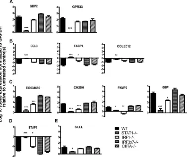
Validation of microarray analysis by qRT-PCR. To assess whether the gene expression changes we detected by microarray analysis could be verified by an independent methodology, we selected 11 genes with various patterns of transcription factor de-pendence and measured their transcript levels by qRT-PCR. To
this end, we isolated RNAs from untreated and IFN-␥-treated
WT, IRF-1⫺/⫺, IRF3⫻7⫺/⫺, STAT-1⫺/⫺, and CIITA⫺/⫺
macro-phages for analysis (Fig. 7). When we compared the fold changes
in expression induced by IFN-␥in the various gene-deficient
mac-rophages to those observed in WT macmac-rophages, we found that for
82% (9 of 11 genes) of the genes, our microarray analyses accu-rately predicted the gene’s dependence on the various
transcrip-tion factors for responsiveness to IFN-␥(Fig. 7). These data
sup-port the bioinformatic analysis of microarray data indicating
patterns of transcription factor dependence for IFN-␥-regulated
genes.
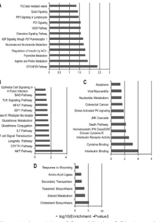
Pathway analysis of gene sets identified by microarray anal-ysis.To identify the gene pathways that these STAT-1- and
IRF-1-dependent genes contribute to in IFN-␥-activated
macro-phages, we analyzed groups A to D (Table 2) separately by gene set
overlap analysis using mSigDB. We found enrichment for several metabolic pathways, such as pyrimidine metabolism, arginine and proline metabolism, and nicotinate and nicotinamide
metabo-lism, among genes that were regulated by IFN-␥in a manner
de-pendent on STAT-1 but not on IRF-1, IRF-3, IRF-7, or CIITA (Fig. 8). Conversely, we found enrichment of several immune
response pathways in genes that were inhibited by STAT-1 (Fig.
8). These included genes involved in the Toll-like receptor
(TLR) signaling pathway, the IL-7 pathway, the NFAT path-way, and glutathione metabolism. The genes that required both
STAT-1 and IRF-1 for upregulation in response to IFN-␥were
enriched for stress-activated protein kinase signaling, JNK cas-cade, and interleukin/cytokine binding pathways, while the
genes that were inhibited by IFN-␥in a manner dependent on
both STAT-1 and IRF-1 were enriched for lipid metabolism, steroid metabolism, cholesterol biosynthesis, and terpenoid biosynthesis.
DISCUSSION
IFNs are likely vital to the host defense against human
norovi-ruses and are important for inhibition of MNV (8,15,17,41).
However, the antiviral mechanisms that IFNs induce to combat NVs and the transcription factors involved in these responses are not well defined. In this report, we demonstrate a critical role for the transcription factor IRF-1 in control of MNV
rep-licationin vivo. Furthermore, we show that in the absence of
IFN-␣signaling, IRF-1 is essential for survival after MNV
infection. We also demonstrate that IRF-1 is required for the
control of MNV by IFN-␥in primary macrophages. Despite
previous studies that suggest a role for IRF-1 in IFN-␣
-in-duced antiviral responses, we found that IRF-1 is dispensable
for control of MNV by IFN-in cultured macrophages.
Fi-nally, we demonstrated an additional requirement for STAT-1,
but not autocrine IFN-␣, CIITA, IRF-3, or IRF-7, in IFN-␥
[image:6.585.39.547.78.186.2]-induced control of MNV in macrophages. Consistent with the importance of STAT-1 and IRF-1 in the antiviral effects of
TABLE 2Transcription factor requirements of genes that were responsive to IFN-␥in macrophagesa
Parameter
Requirement or value for gene category
Group A Group B Group C Group D Group E Group F
Change in gene expression after treatment with IFN-␥
Requires STAT-1 for change ⫹ ⫹ ⫹ ⫹ ⫺ ⫺
Requires IRF-1 for change ⫺ ⫺ ⫹ ⫹ ⫺ ⫺
Requires IRF-3/IRF-7 for change ⫺ ⫺ ⫺ ⫺ ⫺ ⫺
Requires CIITA for change ⫺ ⫺ ⫺ ⫺ ⫺ ⫺
No. of genes in category 97 100 49 26 21 0
aGenes whose expression profiles were changed by IFN-␥were identified and grouped based on the requirement of STAT-1, IRF-1, CIITA, IRF-3, or IRF-7 for this change. Maloney et al.
on November 7, 2019 by guest
http://jvi.asm.org/
FIG 6Heat maps of the genes assigned to group A (A), group B (B), group C (C), and group D (D) by microarray analyses (seeTable 2). Group A represents genes that are upregulated by IFN-␥in a STAT-1-dependent manner, group B represents genes that are downregulated by IFN-␥in a STAT-1-dependent manner, group C represents genes that are upregulated by IFN-␥in an IRF-1- and STAT-1-dependent manner, and group D represents genes that are downregulated by IFN-␥in an IRF-1-and STAT-1-dependent manner. The heat maps represent row-normalizedZ-score values per gene. Expression values of⬎1 standard deviation above the expression level in untreated WT macrophages are indicated in red, and values of⬎1 standard deviation below the WT expression level are indicated in blue.
on November 7, 2019 by guest
http://jvi.asm.org/
[image:7.585.43.545.61.674.2]IFN-␥, we used microarrays and bioinformatics to show that
there is a defined subset of genes whose regulation by IFN-␥
requires these two transcription factors.
IRF-1 in IFN-␥-mediated responsesin vivo.Previous studies
with IRF-1⫺/⫺mice have confirmed the importance of IRF-1 for
defense against both EMCV and WNVin vivo(2,18). However,
due to the complex nature of IRF-1 actions in host defense, thein
vivocontribution of IRF-1 as a direct effector of IFN-␥signaling in
infected cells is unclear. For example, studies of immune
re-sponses to WNV found that IRF-1 is required for IFN-␥-mediated
control of viral replication in macrophages and for shaping the
antigen-specific CD8 T cell response (2). Furthermore, since
IRF-1 contributes to both IFN-␣and IFN-␥responses to EMCV
infectionin vitro, it is difficult to ascertain the specific
contribu-tion of IRF-1 to IFN-␥-dependent responsesin vivoin this model
(18). Our finding that mice deficient in either IRF-1 or IFN-␥R
have elevated viral titers when IFN-␣signaling is blocked is
con-sistent with the idea of shared functions for IRF-1 and IFN-␥in
vivoand strongly suggests that the antiviral effects of IFN-␥are
mediated at least in part by IRF-1in vivo.
IFN-␥-independent functions of IRF-1. Our studies with
blocking antibody (Fig. 1) revealed a role for IRF-1 that is
consis-tent with a role in mediating IFN-␥responsesin vivo.
Interest-ingly, our studies of viral replication in WT, IRF-1⫺/⫺, and
IFN-␥R⫺/⫺mice and our survival studies have also revealed functions
for IRF-1 in restricting MNV replication that cannot be explained
by its role in IFN-␥signaling. IRF-1⫺/⫺mice are highly
suscepti-ble to lethal MNV infection under conditions where IFN-␥R⫺/⫺
mice are not. Moreover, IRF-1⫺/⫺mice treated with control MAb
displayed significantly enhanced replication and viral dissemina-tion of MNV (but not increased lethality) compared to
IFN-␥R⫺/⫺and WT mice treated with control MAb (Fig. 1) for every
organ tested except the brain. This enhanced replication, while not of the same magnitude, paralleled the increased replication and viral dissemination induced in WT mice treated with anti-IFNAR and suggests additional functions of IRF-1 in mediating at
least some of the effects of IFN-␣. However, in primary
macro-phages, we observed that IRF-1 is dispensable for inhibition of
MNV by IFN-(Fig. 4). Furthermore, unlike what is seen for
WNV infection, we did not observe a significant increase in
repli-cation in untreated IRF-1⫺/⫺ macrophages compared to WT
macrophages (Fig. 3) that could explain elevated viral replication
in vivo. Further studies may reveal cell type- and tissue-specific
requirements for IRF-1 in IFN-␥-independent but IFN-␣
-de-pendent control of MNV replication.
STAT-1- and IRF-1-dependent gene pathways. Although STAT-1 and IRF-1 have well-established activities against a di-verse range of RNA viruses, few STAT-1- and IRF-1-dependent individual antiviral effectors have been identified to date. One
such effector is viperin (9), whose activity has been demonstrated
FIG 7Validation of microarray analyses by qRT-PCR. WT, IRF-1⫺/⫺, CIITA⫺/⫺, IRF3⫻7⫺/⫺, and STAT-1⫺/⫺macrophages were treated with 10 U of IFN-␥ or with medium alone. Transcription levels of select genes were examined after 12 h of treatment. Data were collected from three independent experiments. Genes assigned to group A (A), group B (B), group C (C), group D (D), and group E (E) by microarray analyses (seeTable 2) were examined. Asterisks indicate significant differences in transcript levels relative to those in WT cells (*,P⬍0.05; **,P⬍0.01; ***,P⬍0.001).
Maloney et al.
on November 7, 2019 by guest
http://jvi.asm.org/
[image:8.585.101.486.67.386.2]against several families of viruses, including orthomyxoviruses
(39), rhabdoviruses (34), and flaviviruses (35). However, viperin
is not required to control MNV replication in primary
macro-phages orin vivo(L. B. Thackray and H. W. Virgin, unpublished
data). Clearly, there is a need to identify STAT-1- and IRF-1-regulated processes that control MNV replication control. Inter-estingly, we showed that the Atg5-Atg12/Atg16L1 autophagosome elongation protein complex, but not the degradative activity of the
autophagy pathway, is required for IFN-␥-mediated inhibition of
MNV replication in macrophages (15). This property of the
Atg5-Atg12/Atg16L1 complex is shared with IRF-1, as shown here, in
that both are required for the effects of IFN-␥but not IFN-␣on
MNV replication in macrophages. Therefore, it is intriguing to
speculate that IRF-1 regulates a key early step in an IFN-␥/IRF-1/
STAT-1/Atg5-Atg12/Atg16L1-dependent antiviral mechanism.
Our recent work showed that IFN-␥blocks the formation of the
membranous norovirus replication complex in an Atg5-Atg12/
Atg16L1-dependent manner (15). Interestingly, the microarray
analyses in this study revealed that IFN-␥downregulates
compo-nents of lipid and cholesterol biogenesis pathways via an IRF-1-and STAT-1-dependent mechanism. Upregulation of compo-nents of cholesterol biogenesis pathways occurs in cells stably
ex-pressing a human norovirus replicon or infected with WNV (5,
23). We speculate that changes in cellular lipids mediated by
IFN-␥alter membrane rearrangements and trafficking, in an
au-tophagy protein-dependent manner, so as to inhibit formation of the replication complex. Future studies of the IRF-1- and/or
STAT-1-dependent IFN-␥-mediated transcriptional regulation
identified here may uncover novel antiviral mechanisms that can be harnessed as antimicrobial treatments.
FIG 8Gene overlap analyses of the genes assigned to group A (A), group B (B), group C (C), and group D (D) by microarray analyses (seeTable 2). Thexaxis represents thePvalue, indicating the significance of enrichment for any given gene set. The values are plotted on a negative log10scale. All gene sets with values
of⬎1.3 on thexaxis were significantly enriched.
on November 7, 2019 by guest
http://jvi.asm.org/
[image:9.585.138.449.62.502.2]ACKNOWLEDGMENTS
The MAR1-5A3 antibody was a generous gift from R. Schreiber and K. Sheehan. The mM-CSF-producing cell line CMG14-12 was a generous gift from A. Kudo (Tokyo Institute of Technology). We thank the Ge-nome Technology Access Center in the Department of Genetics at Wash-ington University School of Medicine for help with genomic analysis. We thank members of the Virgin lab for their comments on the manuscript and D. Kreamalmeyer for managing mouse colonies.
This work was supported by National Institutes of Health grants ROI AI054483 and U54 AI057160, project 3, to H.W.V. N.S.M. was supported by institutional training grant T32-AI007172. Washington University and H.W.V. receive income based on licenses for MNV technology. The Ge-nome Technology Access Center is partially supported by NCI Cancer Center support grant P30 CA91842 to the Siteman Cancer Center and by ICTS/CTSA grant UL1RR024992 from the National Center for Research Resources (NCRR), a component of the National Institutes of Health (NIH), and the NIH Roadmap for Medical Research.
This publication is solely the responsibility of the authors and does not necessarily represent the official view of the NCRR or the NIH.
REFERENCES
1.Boehm U, Klamp T, Groot M, Howard JC.1997. Cellular responses to interferon-gamma. Annu. Rev. Immunol.15:749 –795.
2.Brien JD, et al.2011. Interferon regulatory factor-1 (IRF-1) shapes both innate and CD8 T cell immune responses against West Nile virus infec-tion. PLoS Pathog.7:e1002230. doi:10.1371/journal.ppat.1002230. 3.Cadwell K, et al.2010. Virus-plus-susceptibility gene interaction
deter-mines Crohn’s disease gene Atg16L1 phenotypes in intestine. Cell141: 1135–1145.
4.Chachu KA, LoBue AD, Strong DW, Baric RS, Virgin HW. 2008. Immune mechanisms responsible for vaccination against and clearance of mucosal and lymphatic norovirus infection. PLoS Pathog.4:e1000236. doi:10.1371/journal.ppat.1000236.
5.Chang KO.2009. Role of cholesterol pathways in norovirus replication. J. Virol.83:8587– 8595.
6.Chang KO, George DW.2007. Interferons and ribavirin effectively in-hibit Norwalk virus replication in replicon-bearing cells. J. Virol.81: 12111–12118.
7.Chang KO, Sosnovtsev SV, Belliot G, King AD, Green KY.2006. Stable expression of a Norwalk virus RNA replicon in a human hepatoma cell line. Virology353:463– 473.
8.Changotra H, et al. 2009. Type I and type II interferons inhibit the translation of murine norovirus proteins. J. Virol.83:5683–5692. 9.Chin KC, Cresswell P.2001. Viperin (cig5), an IFN-inducible antiviral
protein directly induced by human cytomegalovirus. Proc. Natl. Acad. Sci. U. S. A.98:15125–15130.
10. Daffis S, Suthar MS, Szretter KJ, Gale M, Jr, Diamond MS. 2009. Induction of IFN-beta and the innate antiviral response in myeloid cells occurs through an IPS-1-dependent signal that does not require IRF-3 and IRF-7. PLoS Pathog.5:e1000607. doi:10.1371/journal.ppat.1000607. 11. Estes MK, Prasad BV, Atmar RL.2006. Noroviruses everywhere: has
something changed? Curr. Opin. Infect. Dis.19:467– 474.
12. Glass RI, Parashar UD, Estes MK.2009. Norovirus gastroenteritis. N. Engl. J. Med.361:1776 –1785.
13. Goodwin MM, Canny S, Steed A, Virgin HW.2010. Murine gamma-herpesvirus 68 has evolved gamma interferon and stat1-repressible pro-moters for the lytic switch gene 50. J. Virol.84:3711–3717.
14. Goodwin MM, et al.2010. Histone deacetylases and the corepressor NCoR regulate the lytic-latent switch gene 50 in murine gammaherpesvi-rus 68-infected macrophages. J. Virol.84:12039 –12047.
15. Hwang S, et al.2012. Nondegradative role of Atg5-Atg12/Atg16L1 au-tophagy protein complex in antiviral activity of interferon gamma. Cell Host Microbe11:397– 409.
16. Kanazawa N, et al.2004. Regulation of hepatitis C virus replication by interferon regulatory factor 1. J. Virol.78:9713–9720.
17. Karst SM, Wobus CE, Lay M, Davidson J, Virgin HW.2003. STAT1-dependent innate immunity to a Norwalk-like virus. Science299:1575– 1578.
18. Kimura T, et al.1994. Involvement of the IRF-1 transcription factor in antiviral responses to interferons. Science264:1921–1924.
19. Liu J, Cao S, Herman LM, Ma X.2003. Differential regulation of inter-leukin (IL)-12 p35 and p40 gene expression and interferon (IFN)-gamma-primed IL-12 production by IFN regulatory factor 1. J. Exp. Med.198: 1265–1276.
20. Liu J, Guan X, Ma X.2005. Interferon regulatory factor 1 is an essential and direct transcriptional activator for interferon gamma-induced RANTES/ CCL5 expression in macrophages. J. Biol. Chem.280:24347–24355. 21. Livak KJ, Schmittgen TD.2001. Analysis of relative gene expression data
using real-time quantitative PCR and the 2(⫺Delta Delta C(T)) method. Methods25:402– 408.
22. Lohoff M, et al.1997. Interferon regulatory factor-1 is required for a T helper 1 immune response in vivo. Immunity6:681– 689.
23. Mackenzie JM, Khromykh AA, Parton RG.2007. Cholesterol manipu-lation by West Nile virus perturbs the cellular immune response. Cell Host Microbe2:229 –239.
24. Matsuyama T, et al.1993. Targeted disruption of IRF-1 or IRF-2 results in abnormal type I IFN gene induction and aberrant lymphocyte develop-ment. Cell75:83–97.
25. Meraz MA, et al.1996. Targeted disruption of the Stat 1 gene in mice reveals unexpected physiologic specificity of the JAK-STAT signalling pathway. Cell84:431– 442.
26. Muller U, et al.1994. Functional role of type I and type II interferons in antiviral defense. Science264:1918 –1921.
27. Mumphrey SM, et al.2007. Murine norovirus 1 infection is associated with histopathological changes in immunocompetent hosts, but clinical disease is prevented by STAT1-dependent interferon responses. J. Virol. 81:3251–3263.
28. Ogasawara K, et al.1998. Requirement for IRF-1 in the microenviron-ment supporting developmicroenviron-ment of natural killer cells. Nature391:700 –703. 29. Pine R.1992. Constitutive expression of an ISGF2/IRF1 transgene leads to interferon-independent activation of interferon-inducible genes and re-sistance to virus infection. J. Virol.66:4470 – 4478.
30. Reis LFL, Ruffner H, Stark G, Aguet M, Weissman C.1994. Mice devoid of interferon regulatory factor 1 (IRF-1) show normal expression of type I interferon genes. EMBO J.13:4798 – 4806.
31. Schoggins JW, et al.2011. A diverse range of gene products are effectors of the type I interferon antiviral response. Nature472:481– 485. 32. Sheehan KC, et al.2006. Blocking monoclonal antibodies specific for
mouse IFN-alpha/beta receptor subunit 1 (IFNAR-1) from mice immu-nized by in vivo hydrodynamic transfection 1. J. Interferon Cytokine Res. 26:804 – 819.
33. Stark GR, Kerr IM, Williams BR, Silverman RH, Schreiber RD.1998. How cells respond to interferons. Annu. Rev. Biochem.67:227–264. 34. Stirnweiss A, et al.2010. IFN regulatory factor-1 bypasses IFN-mediated
antiviral effects through viperin gene induction. J. Immunol.184:5179 – 5185.
34a.Subramanian A, et al.2005. Gene set enrichment analysis: a knowledge-based approach for interpreting genome-wide expression profiles. Proc. Natl. Acad. Sci. U. S. A.102:15545–15550.
35. Szretter KJ, et al.2011. The interferon-inducible gene viperin restricts West Nile virus pathogenesis. J. Virol.85:11557–11566.
36. Takaoka A, et al.2000. Cross talk between interferon-gamma and -alpha/ beta signaling components in caveolar membrane domains. Science288: 2357–2360.
37. Takeshita S, Kaji K, Kudo A.2000. Identification and characterization of the new osteoclast progenitor with macrophage phenotypes being able to differentiate into mature osteoclasts. J. Bone Miner. Res.15:1477–1488. 38. Thackray LB, et al.2007. Murine noroviruses comprising a single
geno-group exhibit biological diversity despite limited sequence divergence. J. Virol.81:10460 –10473.
39. Wang XY, Hinson ER, Cresswell P. 2007. The interferon-inducible protein viperin inhibits influenza virus release by perturbing lipid rafts. Cell Host Microbe2:96 –105.
40. Ward JM, et al.2006. Pathology of immunodeficient mice with naturally occurring murine norovirus infection. Toxicol. Pathol.34:708 –715. 41. Wobus CE, et al.2004. Replication of norovirus in cell culture reveals a
tropism for dendritic cells and macrophages. PLoS Biol.2:e432. doi: 10.1371/journal.pbio.0020432.
42. Wobus CE, Thackray LB, Virgin HW.2006. Murine norovirus: a model system to study norovirus biology and pathogenesis. J. Virol.80:5104 – 5112.
43. Zhao Z, et al.2007. Coronavirus replication does not require the au-tophagy gene ATG5. Auau-tophagy3:581–585.
Maloney et al.