sarily due to differences in their secondary structures and/or three-dimensional (3D) sizes. For example, the heterologous RNA1 of brome mosaic virus (BMV) is packaged three times more efficiently by CCMV CP than is RNA1 of CCMV, even though the two RNAs have virtually identical lengths. Finally, we show that in an assembly mixture at neutral pH, CP binds reversibly to the RNA and there is a reversible equilibrium between all the various RNA/CP complexes. At acidic pH, excess protein unbinds from RNA/CP complexes and nucleocapsids form irreversibly.
T
he remarkable capacity of the capsid protein (CP) of cowpea chlorotic mottle virus (CCMV) to self-assemblein vitrointo nanometer-size capsids around a broad range of anionic materi-als—its own single-stranded RNA (ssRNA) genome (2,6), heter-ologous ssRNAs (3,4,9,24), organic polymers such as polystyrene sulfonate (11,16,23), metal oxide particles (19,28), functional-ized gold nanoparticles (5), and negatively charged nanoemulsion droplets (13)—as well as into a wide range of structures in the absence of any polyanions (1,8,26) has spurred interest in the mechanism of assembly. But a fundamental question has rarely or only inadequately been addressed: how efficient are the assembly processes?To answer this question, it is first necessary to formulate a precise definition of efficiency of assembly, along with a procedure for determining it. In some cases, efficiency has been quantified as the fraction of filled capsids in the mixture of filled and empty capsids observed in electron micrographs of the assembly mixture (14), but this depends on the formation of empty capsids under the conditions of filled-capsid assembly. In other cases, the relative efficiency of packaging an RNA molecule has been defined as the ratio of the number ofin vivocapsids containing it to the number formed with the genomic RNA (5), when both are present in the same CP-expressing cell; however, this requires additional knowl-edge about the relative levels of replication of both RNAs.
Several assembly experiments, bothin vivoandin vitro, have been reported (29,31,32,34) in which there have been qualitative measurements of the relative amounts of different RNAs packaged by a CP, often with the goal of determining the location of a pack-aging signal. In particular, Qu and Morris (32) studied the length dependence of packaging efficiency by carrying out head-to-head competitions in which protoplasts were coinfected with the wild-type (WT) RNA of turnip crinkle virus along with a series of mu-tant and chimeric RNAs. Also, it has been shown byin vitro head-to-head competitions that hepatitis B virus capsid protein has a
slightly higher preference for its pregenomic RNA than for a het-erologous one (29).
While head-to-head comparisons are informative, they do not by themselves allow an unambiguous determination of efficiency. It is necessary that an additional constraint be imposed: the CP has to be a limiting reagent. The way in which limiting-reagent con-ditions can be precisely achieved has become clear from recent assembly studies on CCMV carried out in our laboratory (12). More explicitly, we have demonstrated that for RNAs ranging in length from 140 to 12,000 nucleotides (nt), a 6:1 mass ratio of CCMV CP to RNA is the minimal amount of CP necessary to package all the RNA (we have called this the “magic ratio”). Thus, if equal masses of two RNAs are mixed with CP at an overall ratio of 3:1, the RNAs must compete for CP because there is sufficient CP to package only one of them completely. The results of such competition experiments can be expressed in terms of a relative packaging efficiencyEof an RNAx, which is defined asE⫽wx/wR, the ratio of the mass of RNAx(competitor) packaged to the mass of the reference RNA; this is equivalent to the ratio of the number of packaged nucleotides ofxto the number of packaged nucleo-tides of the reference molecule.
In this study, we used competition experiments of this kind to quantitatively compare the packaging efficiencies of a wide variety of RNAs by CCMV CP with respect to that of the brome mosaic virus (BMV) RNA1 (B1), which is 3.2 kb in length. We found that RNAs shorter than 2.0 kb cannot compete at all with B1, i.e., have
Received4 July 2012 Accepted27 August 2012
Published ahead of print5 September 2012
Address correspondence to William M. Gelbart, [email protected].
Copyright © 2012, American Society for Microbiology. All Rights Reserved.
doi:10.1128/JVI.01695-12
on November 7, 2019 by guest
relative packaging efficiencies of zero; sufficiently short RNAs, e.g., 0.5-kb molecules, are copackaged with B1; RNAs slightly shorter than 3.2 kb (e.g., 2.0 and 2.8 kb) and slightly longer (3.5 and 4.0 kb) are able to compete, but with relative efficiencies of only 30 to 80%, and involve the formation of “pseudo T⫽2” and T⫽4 capsids, respectively; and homologous RNA (i.e., CCMV RNA1) does not necessarily have the highest packaging efficiency of equal-length RNAs. By carrying out the two-stepin vitro assem-bly process with different orders of mixing of the competing RNAs, we also found that at neutral pH the RNAs are bound reversibly by CP, whereas the formation of mature, RNase-resis-tant nucleocapsids occurs irreversibly under acidic pH conditions.
MATERIALS AND METHODS
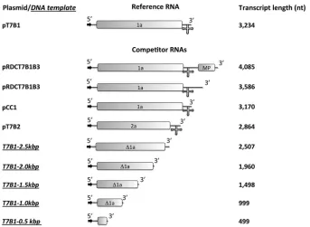
Materials.Restriction enzymes were obtained from New England Bio-Labs. T7 RNA polymerase was a gift from Feng Guo (Department of Biological Chemistry, University of California, Los Angeles). Enzymes were used as recommended by the manufacturer. The plasmids were grown, purified, and analyzed by standard methods (36). Plasmids pT7B1, pT7B2, and pCC1 contain full-length cDNA clones correspond-ing to BMV RNA 1 (B1) (3,234 nt), BMV RNA 2 (B2) (2,864 nt), and CCMV RNA 1 (C1) (3,170 nt) (20); plasmid pRDCT7B1B3 is a cDNA clone corresponding to the ligated product of B1 and B3 (12). All other chemicals used were DNase, RNase, and protease free.
PCR amplification of DNA templates to transcribe RNAs.DNA tem-plates shorter than B1 were prepared by PCR of pT7B1 plasmid; all trun-cations were carried out from the 3=end. We used a universal 5=primer, d(TAATACGACTCACTATAGGTAGACCACGGAACGAGGTTC) (T7 promoter is underlined), and, as reverse primers, d(CACATCCTCTCCT CATGTC), d(GTCTTCAAACCATACACAGTG), d(CTTGCTCAAATT CTTCAACG), d(GGATACAACCAGTTACCGTTG), and d(GTGAACA ACGTCACGGCG) to obtain the corresponding DNA templates for transcribing RNAs with lengths of 499, 999, 1,498, 1,960, and 2,507 nt, respectively. RNAs longer than B1 were transcribed from DNA templates obtained by PCR of pRDCT7B1B3 plasmid. For the 3,586- and 4,065-bp DNA templates the reverse primers d(CGTTGTAAAACGACGGCCA GTG) and d(CGCGCGGTCATCTTACCAGTT), respectively, were used. The forward primer was the same as for the pT7B1 plasmid-derived tem-plates. All the templates were purified by standard procedures (36).
RNA transcription and fluorescent labeling.Plasmids pT7B1 and pT7B2 were linearized with BamHI, pCC1 was linearized with XbaI, and pRDCT7B1B3 was linearized with EcoRI. All DNA templates and plas-mids were transcribed with T7 RNA polymerase. For the quantitative competition experiments, RNAs were labeled with either ChromaTide Alexa Fluor 488-5-UTP (AF488) or ChromaTide Alexa Fluor 546-13-UTP (AF546) (henceforth referred to as modified r546-13-UTPs) from Invitro-gen. These modified rUTPs are randomly incorporated during transcrip-tion. To achieve an average label density between 2 and 5 modified rUTPs per RNA, the molar ratios of rATP to rGTP to rCTP to rUTP to modified rUTP during transcription were modified to 600:600:600:5.32:1. The label density for each labeled RNA was calculated by linear regression of a calibration curve of pure modified rUTPs. The fluorescence measure-ments were carried out with a QuantaMaster spectrofluorimeter (Photon Technology International) in a 40-l quartz cuvette (Sterna Cells). Fluo-rescent RNAs were always kept in the dark in amber tubes, and room light was minimized when possible during handling.
CCMV CP purification.CCMV was purified from infected California cowpea plant (Vigna ungiculatacv. Black Eye) (9), and CP was isolated largely as described previously (6,12). SDS-PAGE and matrix-assisted laser desorption ionization–time of flight mass spectrometry (MALDI-TOF) were employed to ascertain that the purified protein was intact.
In vitroassemblies for the qualitative competition experiments.In carrying out ourin vitropackaging competition assays, we distinguish between qualitative and quantitative measurements of relative
efficien-cies. In the qualitative case, we extract RNA from RNase-treated assembly mixes and simply run the RNA in native and denaturing gels to determine whether one or both are present. In the quantitative case, the RNAs are labeled with different fluorophores so that their relative numbers in RNase-treated assembly mixes can be assayed quantitatively by measuring the fluorescence spectra of the purified nucleocapsids.
Five controls were always used in each qualitative competition exper-iment: reference RNA plus CCMV CP, competitor RNA plus CCMV CP, WT CCMV, reference RNA, and competitor RNA. The RNA concentra-tion in every control was 30 ng/l, and the CP/RNA ratio (wt/wt) for the assemblies was 6:1; the WT CCMV concentration was 120 ng/l (corre-sponding to about 30 ng/l RNA). In every competition experiment, equal masses of the competitor and reference RNAs— each at a concen-tration of 30 ng/l—were mixed in buffer B (BB; 1 M NaCl, 20 mM Tris-HCl [pH 7.2], 1 mM EDTA, 1 mM dithiothreitol [DTT], and 1 mM phenylmethanesulfonylfluoride [PMSF]), with dissociated CCMV CP subunits at a CP/total RNA ratio (wt/wt) of 3:1 (here total RNA is com-petitor RNA plus reference RNA). Samples and controls were dialyzed for 20 h at 4°C against RNA assembly buffer (RAB; 50 mM NaCl, 10 mM KCl, 5 mM MgCl2, 1 mM DTT, 50 mM Tris-HCl [pH 7.2]), followed by a 4-h dialysis against virus suspension buffer (VSB; 50 mM sodium acetate, 8 mM magnesium acetate [pH 4.5]). All controls and samples were processed identically. Aliquots (8l) of each sample and control were set aside for electrophoretic mobility assays, while the rest was treated with RNase A (Invitrogen) (4 ng/l) for 45 min. RNase A was inactivated by incubating the samples with RNase inhibitor (Roche) for 15 min. All samples and controls were purified and concentrated by washing with VSB using a 100-kDa Amicon centrifuge filter (0.5 ml; Millipore) at 3,000⫻gfor 5 min; this step was repeated 4 times. Two 6-l aliquots of each sample and control were set aside for UV-visible (UV-Vis) spectros-copy and transmission electron microsspectros-copy (TEM), and the rest was used to analyze the RNA content by gel electrophoresis. All the procedures were carried out at 4°C.
RNA extraction from VLPs.Prior to RNA extraction, samples were incubated for 15 min with 10 U of RNase inhibitor (Roche) at 4°C. Virus-like particles (VLPs) (⬃750 ng of RNA) were disrupted by adding 10⫻ RNA extraction buffer (0.5 M glycine, 0.5 M NaCl, 0.1 M EDTA [pH 9.0]) and 5l of proteinase K (New England BioLabs); samples were incubated for 30 min at 37°C prior to addition of 5l of 20% SDS. The samples were then vortexed, microcentrifuged, incubated for 1 h at 37°C, diluted to 150 l with TE buffer (10 mM Tris [pH 8.0], 1 mM EDTA), heated at 98°C for 4 min, and chilled on ice for 5 min. Finally, the RNA was extracted by phenol-chloroform and ethanol precipitation (33). The recovered RNA was analyzed by native (in Tris-acetate-EDTA [TAE]) and denaturing (8 M urea in TAE) gel electrophoresis in 1% agarose.
In vitroassemblies for quantitative competition experiments.Each quantitative competition consisted of two sets of studies, each performed in triplicate or quadruplicate. In the first, samples of the reference and competitor RNA were labeled with AF488 and AF546, respectively. In the second set, the labeling was reversed. Nine controls were included in each quantitative competition experiment: AF488-reference RNA plus CCMV CP, AF546-reference RNA plus CCMV CP, AF488-competitor RNA plus CCMV CP, AF546-competitor RNA plus CCMV CP, WT CCMV, and all four kinds of RNAs. The RNA concentration in every control was 30 ng/l, the CP/RNA ratio (wt/wt) was 6:1, and the concentration of WT CCMV was 120 ng/l. As in the qualitative experiments, equal masses of the competitor and reference RNAs at a CP/total RNA ratio (wt/wt) of 3:1 were employed. The assembly was also carried out by a 20-h dialysis against RAB followed by a 4-h dialysis against VSB; we then treated the sample with RNase and purified it exactly as in the qualitative experi-ments. All controls were treated exactly as in the competition samples.
Quantitative measurements by UV-Vis and fluorescence spectros-copy.After purification of the VLPs containing fluorescent rUTPs, all samples were diluted to 52l. A 2-l aliquot was used to measure the UV-Vis absorbance (Fisher Nanodrop spectrophotometer). It has been
Comas-Garcia et al.
on November 7, 2019 by guest
http://jvi.asm.org/
observed that the ratio of the absorbances at 260 and 280 nm for concen-trated virus solutions may be strongly affected by light scattering (30), requiring a correction. Given the low concentration of our samples (⬃ng/ l), the uncorrected UV spectrum differed by less than 1% from the corrected one. The samples containing AF488 and AF546 were excited at 490 and 555 nm, respectively, and the fluorescence spectra were recorded between 500 and 550 nm (AF488) and 560 and 610 nm (AF546). The fluorescence intensity of each sample was normalized with respect to its absorbance at 260 nm and the label density of the excited RNA species.
Electrophoretic mobility analysis (EMA).After the two-step dialysis, 8-l aliquots of each sample and control (before and after RNase A treat-ment) were mixed with 3l of 100% glycerol (RNase, DNase, and pro-tease free) and loaded into a 1% agarose gel (EMD OmniPur) in virus electrophoresis buffer (0.1 M sodium acetate, 1 mM EDTA [pH 6]). The samples were electrophoresed for 1.25 h at 60 V in a horizontal gel appa-ratus (Fisher) at 4°C. For the qualitative experiments, the gels were stained in a solution of 5g/ml ethidium bromide and visualized with an Al-phaimager system (ProteinSimple). For the quantitative experiments, electrophoresis was carried out in complete absence of light. The gels were visualized in an FX Pro plus Fluorimager/PhosphorImager (Bio-Rad) by exciting AF546, followed by staining with ethidium bromide and visual-ization as already described.
TEM analysis of VLPs.For negative staining, purified VLPs were ap-plied to glow-discharged copper grids (400 mesh) that previously had been coated with Parlodion and carbon. A 6l-aliquot of VLPs was spread onto the grid for 1 min, blotted with Whatman filter paper, and then stained with 6l of 1% uranyl acetate for 1 min. Excess stain was removed by blotting with filter paper. The samples were stored overnight in a des-iccator and analyzed with a JEM 1200-EX transmission electron micro-scope equipped with a wide-angle (top mount) BioScan 600 W 1⫻1K pixel digital camera operated at 80 keV. The reported average diameter of VLPs is the geometric mean of two orthogonal measurements determined with ImageJ (U.S. National Institutes of Health) software from recorded images.
RESULTS
We chose to minimize any possible specific RNA-CP interactions by using BMV RNA1 (B1) instead of CCMV RNA1 (C1) as the reference. We designed a series of small competitor RNAs by de-riving them from the 5=end of B1; the lengths for the truncated RNAs were 0.5, 1.0, 1.5, 2.0, and 2.5 kb. For competitor RNAs longer than B1, we used 3.6- and 4.0-kb RNAs that are trunca-tions—maintaining the 5=end— of the product of the ligation of BMV RNAs 1 and 3 (5.3 kb) (12). Finally, we also studied two viral RNAs: BMV RNA 2 (2.8 kb [B2]) and CCMV RNA1 (3.1 kb [C1]). A schematic representation of the plasmids and DNA templates used for the transcription of the RNAs employed can be found in
Fig. 1.
Briefly, a competition experiment consists of mixing equal masses of the competitor and reference RNA with CCMV CP at a CP/total RNA mass ratio of 3:1 in buffer B (BB). The samples are then dialyzed against RNA assembly buffer (RAB) for 20 h, fol-lowed by 4-h dialysis against virus suspension buffer (VSB). Sam-ples are kept at 4°C throughout the experiment.
Both qualitative and quantitative assays were performed. The goal of the qualitative assay was simply to identify which RNA species was packaged. This was achieved by analyzing the reaction mixture in an electrophoretic mobility assay (EMA) before and after RNase A treatment; an EMA under denaturing conditions was also employed to examine RNA extracted from purified VLPs after treatment with RNase A. The resolution of these qualitative experiments was sufficiently high for cases in which only one of the RNAs was packaged. In principle, when both species were packaged, the relative amount of each RNA could also be deter-mined by the latter procedure, but it is not reliable because of
FIG 1Schematic representation of the plasmids and DNA templates used for RNA syntheses. The arrows indicate the T7 transcription promoter, and the cross at the 3=end represents the highly conserved tRNA-like structure in the corresponding RNA transcripts. The boxes represent the open reading frames (ORFs) of the RNAs, 1a and 2a are viral replicases, and MP is the movement protein. “⌬1a” indicates that the viral replicase ORF was truncated. The representations of the plasmid templates are scaled to their relative lengths.
on November 7, 2019 by guest
[image:3.585.122.465.64.315.2]possible degradation of RNA during the extraction process. Quantitative assays were performed by labeling the RNAs with fluorescent rUTPs (AF488 and AF546), which were incorporated duringin vitrotranscription.
Fluorescent labeling has no effect on packaging efficiency.To determine if the incorporation of the modified rUTPs had any effect on the packaging process, we assembled CCMV CP (at the magic ratio) with different ratios of unlabeled to AF488-labeled B1 (0:1, 1:1, and 1:0). As shown inFig. 2a, the EMAs of the three samples are identical. The magic ratio was also not altered; i.e., the minimum CP/RNA ratio required for complete packaging was 6:1 in each case (data not shown). The normalized fluorescence in-tensity of the VLPs that were assembled in a 1:1 unlabeled-labeled
RNA mixture was precisely half that of the VLPs assembled in a 0:1 unlabeled-labeled RNA mixture (Fig. 2b). Finally, negative-stain transmission electron microscopy (TEM) showed that there are no discernible differences between VLPs containing different amounts of labeled and unlabeled RNA (data not shown).
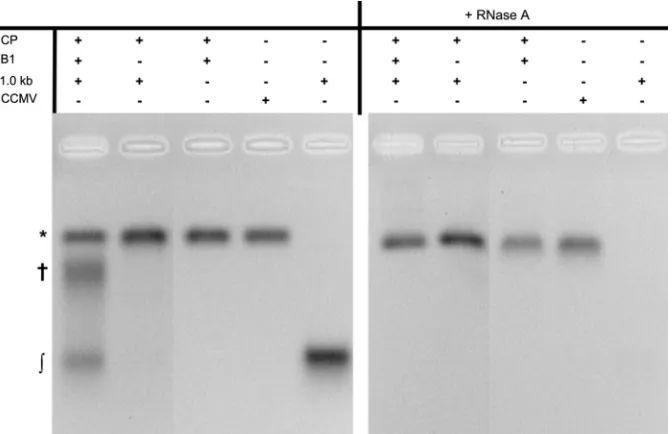
RNAs of 1.0 kb and 1.5 kb cannot compete with B1, but 0.5-kb RNA is copackaged.The EMA for competition experi-ments between B1 and the 1.0- and 1.5-kb RNAs showed similar behavior.Figure 3shows that in a competition between 1.0-kb and B1 there are three main species: VLPs, high-molecular-mass RNA/CP complexes, and free RNA. Denaturing gels of the RNA extracted from purified VLPs after RNase A treatment revealed no RNA other than B1 (Fig. 4). The size distributions determined by
FIG 2The incorporation of AF488 into B1 RNA (B1-488) has no effect on the assembly process. (a) The electrophoretic mobility analysis (EMA) shows that the electrophoretic mobilities of VLPs containing different ratios of unlabeled to labeled B1 are identical. (b) The fluorescence intensity of VLPs in which half the B1 is labeled is half that of VLPs containing only labeled B1.
FIG 3EMA of a competition experiment between B1 and a 1.0-kb RNA. The left panel shows the three main species in a competition experiment: virus-like-particles (VLPs) (*), high-molecular-mass RNA/CP complexes (†), and free RNA (兰). The right panel shows the samples after treatment with RNase A; only the bands corresponding to the VLPs remain. The gel was stained with ethidium bromide.
Comas-Garcia et al.
on November 7, 2019 by guest
http://jvi.asm.org/
[image:4.585.46.539.67.248.2] [image:4.585.126.462.475.692.2]TEM for VLPs containing a 1.0-kb RNA and that from the com-petition between this RNA and B1 are shown inFig. 5e. The dis-tributions for VLPs containing only 1.0-kb RNAs show mixed populations of capsids with diameters between 23 and 26 nm. We have previously argued that it is most likely that capsids with a diameter around 23 nm correspond to “pseudo T⫽2” (henceforth referred to simply as “T⫽2”) and that those with diameters close to 26 nm correspond to T⫽3 capsids (12,23). Here, and through-out this article, we assign T numbers to VLPs on the basis of comparing the VLP radii to that of WT CCMV T⫽3 virions (26 nm). More explicitly, from the fact that radii of Caspar-Klug structures scale as T1/2, we associate radii of 22 and 30 nm with
T⫽2 and -4 VLPs, respectively (23).
On the other hand, the size distribution of capsids containing only B1 is centered around 26 nm (dotted curves inFig. 5). The capsids obtained from the competition experiments involving B1 and 1.0-kb RNA have diameter distributions that are indistin-guishable from those for capsids containing only B1 (Fig. 5e). Similarly, the competition with 1.5-kb RNAs leads to diameter distributions around 26 nm, while the distributions for each of the shorter RNAs alone are bimodal (Fig. 5atod). Along with the data from the RNA extraction (Fig. 4BandC), these observations in-dicate that essentially all of the competitor RNAs were excluded from packaging; hence, their relative packaging efficiency is zero. Finally, it is worth noticing that the amount of high-molecular-mass complexes is small compared to the situation in which the competitor RNA is packaged, which is possible only if most of the competitor RNA is excluded from forming stable RNA/CP com-plexes.
Although the EMA for the competition experiments with a 0.5-kb RNA was almost identical to those for the 1.0- and 1.5-kb competitions, the denaturing EMA of the RNAs extracted from the purified capsids (after RNase A treatment) showed two bands, corresponding to B1 and the 0.5-kb RNA (Fig. 4A).Figure 6a
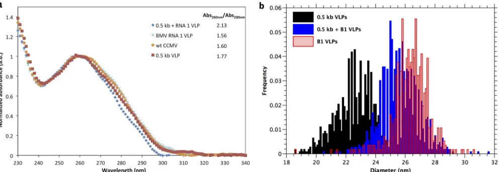
compares the normalized UV-Vis spectra of WT CCMV and VLPs containing either only B1, only 0.5-kb RNA, and both (from this competition experiment). The spectra of VLPs containing a single RNA species overlap that of WT CCMV, but the spectrum of the VLPs from the competition experiment has a significantly higher
ratio of absorbances at 260 and 280 nm (2.13 versus 1.74), dem-onstrating that the concentration of nucleotides per protein is higher than for the other samples. The capsid diameter distribu-tion for VLPs containing only 0.5-kb RNA strongly suggests that they are predominately T⫽2 (average diameter, 23 nm), but the capsids from the competition between B1 and 0.5-kb RNA have a diameter distribution centered at 27 nm (Fig. 6b). Based on the UV-Vis spectra and the capsid diameter distribution, we can con-clude that the 0.5-kb RNA is copackaged with B1. The copackag-ing cannot be attributed to dimerization of the RNAs as shown by native gels of mixtures of the two RNAs, which showed two dis-tinct bands under assembly conditions in the absence of CP (data not shown).
RNAs of 2 kb and longer compete with B1.We found that the shortest RNA that competes with B1 but is not copackaged is 2.0 kb in length (Fig. 4D). The EMA of the assembly mix in this case showed that the RNA was present in the form of either VLPs or high-molecular-mass RNA/CP complexes; essentially no free RNA was detected (data not shown). From an examination of the fluorescence from AF546-labeled RNA in the EMA it is obvious that both RNA species were assembled into VLPs. As we have noted, this technique does not allow us to reliably determine the relative populations of each RNA species. To obtain more quan-titative estimates, the VLPs were purified after RNase A treatment and their fluorescence intensity was then measured and normal-ized with respect to the total RNA content in each and the average label density in each RNA species. To calculate the relative pack-aging efficiency, the normalized intensity of VLPs containing AF546-labeled 2.0-kb RNA was divided by the normalized inten-sity of VLPs labeled with AF546-labeled B1; the same was done for the AF488-labeled RNAs. Because the reaction mixture had equal masses of the 2.0-kb RNA and B1, there were 1.6 times more moles of the smaller RNA than of the full-length RNA, and accordingly, the measured normalized intensities were corrected for this dif-ference. The results for both fluorophores agree: the relative pack-aging efficiency of the 2.0-kb RNA was estimated to be 0.74⫾ 0.16. The size distribution of VLPs with a 2.0-kb RNA indicated a mixed population of T⫽2 and T⫽3 capsids. This is the first of our competition experiments in which a capsid population was not
FIG 4EMAs of RNAs extracted from competitions between B1 and 0.5-kb RNA (A), 1.0-kb RNA (B), 1.5-kb RNA (C), and 2.0-kb RNA (D). Panels A and D show that both competitor RNAs were packaged in the presence of B1, while panels B and C show that neither the 1.0- nor 1.5-kb RNA was packaged. These EMAs were carried out under denaturing conditions: 8 M urea in TAE.
on November 7, 2019 by guest
[image:5.585.52.542.66.235.2]FIG 5(a, b, c, and d) Diameter distributions of VLPs containing a single RNA species: RNAs of 1.0 (a), 1.5 (b), 2.0 (c), and 2.5 (d) kb. (e, f, g, and h) Same as panels a to d, but for VLPs from competition experiments between B1 and RNAs of 1.0 (e), 1.5 (f), 2.0 (g), and 2.5 (h) kb. The dotted line in each panel corresponds to the diameter distribution of VLPs containing B1 RNA. Except for the competition between B1 and a 2.0-kb RNA, which contains a higher population of VLPs with a diameter smaller than 26 nm, the diameter distributions for all competition experiments are almost identical to those for VLPs containing only B1 RNA.
on November 7, 2019 by guest
http://jvi.asm.org/
exclusively associated with T⫽3 capsids (Fig. 5g). We conclude that the T⫽2 capsids contain 2.0-kb RNA, while only about 2/3 of the T⫽3 capsids contain B1 (the other 1/3 contain 2.0-kb RNA).
Figure 5dshows that when 2.5-kb RNA alone is packaged, roughly equal numbers of T⫽2 and T⫽3 capsids are formed; in contrast, the diameter distribution for the capsids arising from the competition between B1 and 2.5-kb RNA showed a single popu-lation centered at 27 nm, corresponding to T⫽3 (Fig. 5h). And yet the corresponding EMA gel (not shown) looks qualitatively simi-lar to that for the competition with 2.0-kb RNA plus B1 (Fig. 4D), so we know that the 2.5-kb RNA is able to compete with B1, even though B1 is preferred. More quantitatively, we found from bulk fluorescence measurements that the relative packaging efficiency of the 2.5-kb RNA is 0.33⫾0.06 (seeFig. 9). It is not clear why we did not observe any T⫽2 capsids in this competition experiment but rather only when the 2.5-kb is at the magic ratio in the absence of B1.
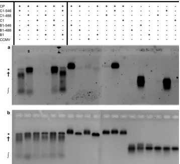
More biologically relevant competition experiments test B1 versus B2 (2.8 kb) and B1 versus C1 (3.1 kb). The EMAs for these samples are almost indistinguishable from those of the 2.0- and 2.5-kb competitions.Figure 7shows the EMA for the competition between B1 and C1; almost all of the unpackaged RNA was found as RNA/CP complexes, and most of the fluorescence signal of the VLPs came from B1. B2 and C1 alone each form T⫽3 capsids with an average diameter of 27 nm; the VLPs resulting from the B1 versus B2 and B1 versus C1 competitions have identical diameter distributions (data not shown). The bulk fluorescence measure-ments showed that the relative packaging efficiencies of B2 and C1 are 0.56⫾0.11 and 0.35⫾0.09, respectively, corresponding to about 1/3 and 1/4 of the VLPs containing B2 and C1, respectively. Finally,Fig. 7shows that the electrophoretic mobilities of C1 and B1 are the same and that the presence of either fluorophore does not affect the result of the competitions.
Longer (3.6- and 4.0-kb) RNAs can also compete.The last two competition experiments involved RNA molecules that are 12.5 and 25% longer than B1. As mentioned earlier, these two mole-cules (3.6- and 4.0-kb long) are truncations of the ligation product
of B1 and B3 and, as in the case of the short RNAs, the 5=end of B1 was conserved. The 3.6-kb molecule was the RNA with the third-highest relative packaging efficiency (0.65⫾0.18) among all of those examined. TEM micrographs show that VLPs containing only a 3.6-kb RNA have the same average diameter (26 nm) as VLPs containing B1 and those arising from the competition ex-periment between B1 and this RNA. The 4.0-kb RNA has a relative packaging efficiency of 0.81⫾0.24; 95% of the VLPs are spherical (with a small fraction of them having a diameter consistent with T⫽4) and the remaining 5% are asymmetric. These asymmetric capsids are ellipsoidal, with an axial ratio ofⱖ1.5, and constitute as much as 25% of all particles when the 4.0-kb RNA is packaged alone (Fig. 8). While ellipsoidal CCMV VLPs of this size have not been reported before, Bancroft et al. (8) observed smaller (28-nm-long) ellipsoidal particles with a similar axial ratio when CCMV was digested with pancreatic RNase at pH 7.3. A summary of the relative packaging efficiencies for RNAs between 1.0 and 4.0 kb in length is shown inFig. 9. The curve drawn there, which is a guide to the eye, pertains only to cases in which T⫽3 capsids form (tri-angles) and shows that there is a maximum packaging efficiency, which, not surprisingly, occurs at the length corresponding to B1. Reversibility of RNA/CP binding at neutral pH; irreversibil-ity of nucleocapsid formation at low pH.To examine if the rela-tive efficiencies of the RNAs that we have observed reflect differ-ences in the kinetics of assembly or differdiffer-ences in stability, we have carried out a series of experiments in which we examined how the order of addition of the RNAs affects the assembly products. The experiments probe the degree to which reversibility exists under assembly conditions.
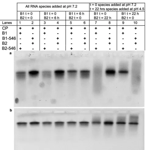
We performed a series of competition experiments between B1 and B2. In the first set of reactions both B1 and B2 were mixed with CP at the same time (in BB) and then dialyzed at neutral pH just as in the previous experiments, with a CP/total RNA ratio of 3:1. In the second set, either B1 or B2 was added to the CP solution and dialyzed against RAB for 6 h before the other RNA was added, followed by dialysis against RAB for 12 h more before dialysis against VSB for 4 h. The result of this experiment is shown in the
FIG 6(a) The normalized UV-Vis spectra of VLPs from a competition experiment between 0.5-kb RNA and B1 show that the ratio of absorbances at 260 and 280 nm (Abs260 nm/Abs280 nm) is considerably higher than it is for WT CCMV or VLPs containing either 0.5-kb RNA or B1. This ratio reflects the relative
concentration of nucleotides in a capsid. (b) The size distribution of VLPs from this competition experiment is almost identical to that for VLPs containing only B1, while the distribution for those containing 0.5-kb RNA is considerably smaller. The higher RNA content in T⫽3 VLPs indicates that the 0.5-kb RNA was copackaged with B1.
on November 7, 2019 by guest
[image:7.585.46.544.66.237.2]EMAs inFig. 10. In the upper gel the signal comes only from RNA labeled with AF546, while the lower EMA shows the total RNA (ethidium bromide staining). In lanes 1 and 2 (left to right), B1 and B2 were added at the same time. Lanes 3 and 4 show the result when B2 was added 6 h after B1, and lanes 5 and 6 show the result for the reverse order. It is evident from the gels that qualitatively
the result is independent of the order of addition or the time between the additions; i.e., B1 is preferentially packaged over B2. We carried out similar studies for a case in which the compe-tition for the CP was markedly different, i.e., involving as a com-petitor the 1.0-kb RNA that is not packaged at all when going head-to-head against B1 at a CP/total RNA ratio of 3:1. As with the case with B1 plus B2, the results of these RNA experiments with B1 plus 1.0-kb RNA were found to be independent of the order of the mixing of the two RNA molecules in the neutral-pH assembly reaction; only B1 was packaged.
The effect of pH on the reversibility of binding was examined in another two sets of experiments, and the results are shown in the four right-most lanes of the EMA inFig. 10. In these experi-ments, one of the RNA species and CP were dialyzed against RAB for 20 h and against VSB for 2 h, after which the other RNA was added and the mixture dialyzed against VSB for an additional 2 h. In contradistinction to the experiments described above in which changes of order are carried out entirely at neutral pH, it is evident that if the second RNA—regardless of whether it is the less or more efficiently packaged one—is added after the pH has been lowered to pH 4.5 (VSB), it is not incorporated into capsids; i.e., the reversibility is lost once the pH is lowered.
These results demonstrate that the assembly process of CCMV CP around RNA at neutral pH is reversible: CP, RNA, and
FIG 7Competition between B1 and C1. (a) This EMA, visualized by exciting Alexa Fluor-546 UTPs, shows that B1 is packaged more efficiently than C1 into VLPs (*). Most of C1 is in the form of high-molecular-mass RNA/CP complexes (†), and there is a small fraction of free RNA (兰). B1-488 (C1-488) and B1-546 (C1-546) correspond to BMV RNA 1 (CCMV RNA 1) labeled with AF488 and AF546. (b) The same gel stained with ethidium bromide showing total RNA content. This EMA also confirms that neither the presence nor the nature of the fluorophore perturbs the assembly process.
FIG 8Negative-stain electron micrograph of VLPs containing a 4.0-kb-long RNA. There are two different sizes of spherical capsids and elongated particles. In competition experiments between B1 and a 4.0-kb RNA, about 5% of the particles have axial ratios ofⱖ1.5; in the absence of B1, 25% of the particles have ratios ofⱖ1.5. The particle at the right end of the white scale bar in the right panel has an axial ratio of 1.63.
Comas-Garcia et al.
on November 7, 2019 by guest
http://jvi.asm.org/
[image:8.585.116.474.66.394.2] [image:8.585.43.284.544.667.2]RNA/CP complexes are in equilibrium with their surroundings. It is only upon pH acidification that true—RNase-resistant— cap-sids are formed, which are no longer at equilibrium, e.g., whose CPs cannot be exchanged upon addition of new RNA. Similar
conclusions were reached recently by Tsvetkova et al. (37), who studied the assembly of BMV CP around gold nanoparticles func-tionalized with carboxylates. They concluded that at neutral pH the CP saturated the anionic particles, adsorbing noncoopera-tively and forming noncompact poorly organized structures. The formation of a well-defined, compact structure occurred in a co-operative fashion only after the pH was lowered to 4.5.
DISCUSSION
A key to interpreting the results we have found for the relative packaging efficiencies of different RNA molecules— by means of their head-to-head competition for CP with a reference RNA un-der conditions where the protein is in short supply— can best be presented in terms of the two-step assembly reaction involved in nucleocapsid formation. More explicitly, it is useful to treat the reversible and irreversible behavior characterizing, respectively, the RNA/CP complex formation in the first (neutral-pH) step and the RNase-resistant nucleocapsid formation in the second (acidic-pH) step.
Our picture of RNA/CP complex formation at neutral pH is consistent with the “en masse” scenario suggested on the basis of simulations by Elrad and Hagan (21) in which strong, nonspecific, CP/RNA interactions (25) lead to RNA molecules “saturated” with capsid proteins. A similar scenario was proposed by Devkota et al. (18) in their molecular mechanics interpretation of high-resolution structural data for Pariacoto virus, and earlier in a gen-eral “micellization” discussion by McPherson (27) of simple self-assembling RNA viruses. The basic idea is that nonspecific electrostatic interactions between the basic residues of the pro-teins and the phosphate backbone drive the binding of the CP to the RNA and induce its condensation into a template for nucleo-capsid formation.
At neutral pH, in the case of CCMV CP, we propose that in a competition experiment between two different RNA species at a CP/total RNA mass ratio of 3:1, essentially all the CP is bound—
FIG 9Relative packaging efficiency of ssRNAs as a function of length. The vertical bars represent the standard deviations. All measurements are relative to B1 (3,224 nt). The triangles represent competitions where the competitor RNAs form T⫽3 capsids only. The circles correspond to cases where T⫽2 and T⫽3 capsids form, and the square corresponds to the case involving T⫽3 and T⫽4. The curve, which is drawn to guide the eye, illustrates that when the RNAs can only form T⫽3 capsids, the relative packaging efficiency has a maximum at 3.2 kb.
FIG 10EMAs of competition experiments between B1 and B2 at different times and for different buffer and gel-staining conditions. (a) The signal comes only from AF546-labeled RNA. (b) Same gel stained with ethidium bromide showing the total RNA content. From left to right: the first two lanes show competition experiments in which both RNAs were added at the same time to RAB (pH 7.2); lanes 3 and 4 show competition experiments in which B2 was added after B1, at pH 7.2; and lanes 5 and 6 show the opposite case, again at pH 7.2. The last four lanes show the effect of adding a second RNA after lowering the pH to 4.5 (VSB).
on November 7, 2019 by guest
[image:9.585.137.448.67.272.2] [image:9.585.45.286.400.647.2]roughly half on each of the RNAs. On average, however, the (smaller number of) longer RNAs will have more CPs bound to them, at the same density as on the shorter RNAs; this will lead, upon pH lowering, to a higher rate of nucleation on the longer molecules. At neutral pH, thermal fluctuations drive the CPs from one RNA molecule to another, and the structure of the RNA/CP complexes will be polydisperse, including various intermediates that involve some cooperative lateral interactions between CPs but that remain in thermal equilibrium with their surroundings. These “procapsid” states are therefore still labile and will be or-dered (“fixed”) into RNase-resistant, “mature” capsids only upon pH lowering.
The kinetic and thermodynamic factors affecting the forma-tion of a mature VLP from a reversibly bound procapsid can be largely understood using the framework of classical homogeneous nucleation theory (17) (homogeneous rather than heterogeneous nucleation applies to the extent that no specific origin of assembly plays a dominant role in the formation of CCMV capsids around RNA). The classical nucleation rate is the number of critical nuclei formed per unit time per unit system volume, with the critical size varying inversely with the free-energy difference (⌬) per mole-cule between the initial and final states. The nucleation rate is proportional to exp(⫺⌬G*/kT), wherekT is the thermal energy and⌬G*—the barrier to forming a critical nucleus—is a decreas-ing function of⌬. In this context, then, we can immediately identify two factors that will affect the nucleation rate of VLPs and hence the relative efficiency of packaging. First, the number of nucleation events per unit time increases with system size: all other things being equal, nucleation is more probable in longer than in shorter RNAs. Second, the more stable the VLP formed the larger will be the magnitude of⌬(i.e., the energy difference per molecule between the procapsid and mature capsid) and the greater the nucleation rate as well. We apply these considerations in discussing the measured relative packaging efficiencies.
Consider, for example, the case of competition between 3.2-kb (B1) and 2.5-kb RNA (or some other RNA, e.g., 2.0-kb RNA, that can compete at least a bit with B1). As discussed above, to a good approximation each molecule competes equally well for binding of CPs and is, on average, half-covered with protein, the 2.5-kb RNA with about 125 CPs and B1 with about 160 CPs. At neutral pH, the RNA/CP complexes arising from 2.5-kb RNA and B1 mixtures are in thermal equilibrium in the sense that they have the same composition and structures—whatever those structures may be (e.g., RNA decorated with bound CP, or loosely formed procapsids, or anything in between)—independent of the order in which the RNAs are added to CP. When the pH is lowered, the protein-protein interactions become stronger, leading to a greatly enhanced nucleation rate for capsid formation, with the longer molecules enjoying a higher rate of nucleation than the shorter ones. As a result, unlike the case at neutral pH where the CP bind-ing and unbindbind-ing rates are essentially identical for all of the RNAs, once nucleation occurs, the longer RNA has a smaller off-rate for protein unbinding and fluctuations will lead to increas-ingly complete, partially formed, mature capsids. Nucleation can also occur on the shorter molecules, of course, but at a lower rate than for the longer ones, thereby giving rise to their smaller pack-aging efficiency.
Recall that molecules shorter than 2.0 kb, when mixed with CP at a 6:1 mass ratio in the absence of competitor, are packaged completely but only in multiple copies, e.g., two 1.0-kb RNAs in a
T⫽2 VLP or two 1.5-kb RNAs in a T⫽3 VLP. A short molecule like this cannot compete effectively against B1 in being packaged because, in addition to the nucleation rate being lower on a shorter molecule, it has to wait—after a critical nucleus begins to grow— for another short-RNA/CP complex to come along in order for VLP formation to be complete. (We cannot yet rule out the pos-sibility that the shorter molecules have already come together at neutral pH.) In the case of the 1.0-kb RNA, nucleation rates are still smaller because T⫽2 capsids have higher energy (per CP) than T⫽3; i.e.,⌬is smaller than it is for T⫽3 capsids with longer RNA.
While the order of adding RNAs at high pH is not of conse-quence for competition outcomes because of thermal equilibra-tion at neutral pH, the order does matter when one of the two RNAs is added at acidic pH. For example, consider the case where only 1.0-kb molecules are mixed with CP and the solution dia-lyzed first against neutral pH (RAB) for 20 h and then against acidic pH (VSB) for 2 h. Even though the nucleation rate for fully saturated 1.0-kb molecules at acidic pH is lower than that for fully saturated 3.2-kb RNAs, and even though a second 1.0-kb RNA must find the nucleated complex, these processes occur well be-fore the 2-h point when the 3.2-kb molecule is added. Accord-ingly, the short RNA is completely packaged.
The packaging efficiency of a 3.6-kb RNA differs little from that of a 2.8-kb RNA, indicating that the CCMV CP is not very dis-criminating when packaging RNAs close to the average viral length (⬇3,000 nt). This agrees with the results of Porterfield et al. (29), who found that hepatitis B virus capsid protein has no strong preference for its viral RNA when the assembly is done in the presence of a heterologous RNA of comparable length. The sur-prising result is that a 4.0-kb RNA has the highest relative packag-ing efficiency of all the competitor RNAs tested. When B1 com-petes against a 4.0-kb RNA at an overall CP/RNA mass ratio of 3:1, both RNAs have, on average, half the amount of protein bound at the magic ratio. The longer RNA complex will have a higher rate of nucleation of CPs into T⫽3 capsids, but on the other hand, the resulting VLP has a higher energy (a smaller⌬) than that of the 3.0-kb molecule when it is confined in the T⫽3 capsid, thereby partially offsetting the effect of length.
In all samples and control assemblies, with the exception of a small fraction of elongated particles that arise when CCMV CP is assembled around a 4.0-kb RNA, only spherical VLPs were ob-served. We have shown earlier that for RNAsⱖ4.5 kb, one RNA is shared by two or more capsids (multiplets) (12); accordingly, it is not surprising that, when approaching this length (e.g., for 4.0-kb RNA), intermediate structures between doublets (two capsids sharing one RNA) and singlets (one RNA per capsid) can be formed. A simple calculation shows that if two T⫽3 half-capsids are connected by a ring of hexamers, the axial ratio of the resulting particle will be 1.5, in agreement with that observed. Moreover, the absence of elongated capsids when B1 competes against a 4.0-kb RNA shows that their free energy is considerably higher (hence smaller⌬) than those of T⫽3 and T⫽4 capsids.
So far, all of the head-to-head competition experiments we have discussed involve two RNA molecules of different length. The competition between RNAs of identical length but different sequences revealed a striking result: even though C1 is the homol-ogous RNA to CCMV CP, its packaging efficiency is lower than that for B1. The two RNAs share 80% sequence homology and B1 is only 67 nt longer, so they are essentially of identical length. They Comas-Garcia et al.
on November 7, 2019 by guest
http://jvi.asm.org/
cies of RNAs of identical lengths but different sequences will be necessary in order to separate these effects. Finally, we remark that one should be able to identify an equal (3.2-kb)-length nonviral sequence RNA that competes even more efficiently against C1 RNA for CCMV capsid protein than does B1. After all, the viral genome has a large number of evolutionary pressures on it beyond needing to be efficiently packaged by its CP; e.g., it needs to be replicated and transcribed efficiently to code properly for its sev-eral gene products and to be able to disassemble efficiently from the capsid once it has penetrated into the cell. A competitor for CP, on the other hand, needs only to have secondary and tertiary structure and overall size and shape that are better optimized for packaging that those of the viral genome.
Mostin vitroassemblies of RNA viruses are performed at neu-tral pH, and CCMV is no exception. What we have shown is that under these conditions RNA and CP are in equilibrium and RNA/CP complex formation is affected by the addition of more RNA (even 6 h after they were initially mixed). It is well known that CCMV CP-CP interactions increase as pH decreases (2); moreover, as pH is lowered sufficiently and in the absence of RNA, CCMV CP can self-assemble into tubes, multishells, and empty capsids (1,8,26). So by taking into account that RNA and CP strongly interact at neutral pH (12) and that CP-CP interactions at acidic pH are stronger than at neutral pH, it is natural for us to propose that CCMV CP self-assembly around RNA involves at least two steps.
While the focus of this research has been onin vitroassembly, it is appropriate to ask if a pH-driven two-step assembly has any relevance forin vivo assembly. It is known that in the case of flaviviruses (35),in vivocapsid maturation is driven by pH acidi-fication. For these viruses, immature capsids are assembled at the endoplasmic reticulum (pH 7.2) and are transported through the secretory pathway to the trans-Golgi network, where maturation occurs upon pH lowering (pH 5.7) (35). The assembly pathway of bromovirusesin vivohas been less well studied, but recent fluo-rescence experiments (7) have pointed to the role of subcellular localization of CP in vesicles in which there is a colocalization with sites of replication; the relative amounts of CP and RNA in these sites are unknown. We cannot rule out the possibility that cytopa-thology induced by virus infection could alter the normal pH in these sites, making the environment more congenial for virus as-sembly.
In summary, we have shown that thein vitropackaging effi-ciency of CCMV CP depends nonmonotonically on the length of ssRNA. When a competitor RNA is significantly shorter than the
packaging efficiency than C1, which is the homologous RNA for CCMV CP. Finally, we have shown that at neutral pH, RNA and CCMV CP form complexes that are in thermal equilibrium with respect to exchange of CP, while after lowering of the pH to acidic conditions, RNase-resistant nucleocapsids arise irreversibly from these complexes.
ACKNOWLEDGMENTS
We thank Feng Guo for providing the T7 polymerase, Martin Phillips for all his help at the UCLA-DOE Biochemistry Instrumentation Facility, Venus Vakhshori for her help purifying CCMV, and Avinoam Ben-Shaul, Robijn Bruisma, and Rees F. Garmann for many helpful discussions.
This study was supported by NSF grants CHE 0714411 and CHE 1051507 to C.M.K. and W.M.G. R.D.C.-N. and M.C.-G. received partial support from CONACyT and UC-Mexus.
REFERENCES
1.Adolph KW, Butler PJG.1974. Studies on the assembly of a spherical plant virus. I. States of aggregation of the isolated protein. J. Mol. Biol.
88:327–338.
2.Adolph KW, Butler PJG.1976. Assembly of a spherical plant virus. Phi-los. Trans. R. Soc. Lond. B Biol. Sci.276:113–122.
3.Adolph KW, Butler PJG.1977. Studies on the assembly of a spherical plant virus. III. Reassembly of infectious virus under mild conditions. J. Mol. Biol.109:345–357.
4.Allison R, Janda M, Ahlquist P.1988. Infectious in vitro transcripts from cowpea chlorotic mottle virus cDNA clones and exchange of individual RNA components with brome mosaic virus. J. Virol.62:3581–3588. 5.Aniagyei SE, et al.2009. Synergistic effects of mutations and nanoparticle
templating in the self-assembly of cowpea chlorotic mottle virus capsids. Nano Lett.9:393–398.
6.Annamalai P, Rao ALN.2005. Dispensability of 3=tRNA-like sequence for packaging cowpea chlorotic mottle virus genomics RNAs. Virology
332:650 – 658.
7.Bamunusinghe D, Seo J-K, Rao ALN.2011. Subcellular localization and rearrangement of endoplasmic reticulum by brome mosaic virus capsid protein. J. Virol.85:2953–2963.
8.Bancroft JB, Hills GJ, Markham R.1967. A study of the self-assembly process in a small spherical virus formation of organized structures from protein subunits in vitro. Virology31:354 –379.
9.Bancroft JB.1970. The self-assembly of spherical plant viruses. Adv. Virus Res.16:99 –134.
10. Belyi VA, Muthukumar M. 2006. Electrostatic origin of the genome packaging in viruses. Proc. Natl. Acad. Sci. U. S. A.103:17174 –17178. 11. Cadena-Nava RD, et al.2011. Exploiting fluorescent polymers to probe
the self-assembly of virus-like particles. J. Phys. Chem. B115:2386 –2391. 12. Cadena-Nava RD, et al.2012. Self-assembly of viral capsid protein and RNA molecules of different sizes: requirement for a specific high protein/ RNA mass ratio. J. Virol.86:3318 –3326.
13. Chang CB, Knobler CM, Gelbart WM, Mason TG.2008. Curvature dependence of viral protein structures on encapsidated nanoemulsion droplets. ACS Nano2:281–286.
on November 7, 2019 by guest
14. Chen C, Kwak Stein B, Kao CC, Dragnea B.2005. Packaging of gold particles in viral capsids. J. Nanosci. Nanotechnol.5:2029 –2033. 15. Choi YG, Rao ALN.2010. Molecular studies on bromovirus capsid
pro-tein. VII. Selective packaging of BMV RNA4 by specific N-terminal argi-nine residue. Virology275:207–217.
16. Comellas-Aragonès M, et al.2009. Controlled integration of polymers into viral capsids. Biomacromolecules10:3141–3147.
17. Debenedetti PG.1996. Metastable liquids: concepts and principles, p 148 –159. Princeton University Press, Princeton, NJ.
18. Devkota B, et al.2009. Structural and electrostatic characterization of Pariacoto virus: implications for viral assembly. Biopolymers91:530 –537. 19. Douglas T, Young MJ.1998. Host-guest encapsulation of materials by
assembled virus protein cages. Nature393:152–155.
20. Dreher TW, Rao ALN, Hall TC.1989. Replicationin vivoof mutant brome mosaic virus RNAs defective in aminoacylation. J. Mol. Biol.206: 425– 438.
21. Elrad OM, Hagan MF.2010. Encapsulation of a polymer by an icosahe-dral virus. Phys. Biol.7:045003– 045020.
22. Gopal A, Zhou HZ, Knobler CM, Gelbart WM.2012. Visualizing large RNA molecules in solution. RNA18:284 –299.
23. Hu Y, Zandi R, Anavitarte A, Knobler CM, Gelbart WM.2008. Pack-aging of a polymer by a viral capsid: the interplay between polymer length and capsid size. Biophys. J.94:1428 –1436.
24. Johnson JM, Willits DA, Young MJ, Zlotnick A.2004. Interaction with capsid protein alters RNA structure and the pathway for in vitro assembly of cowpea chlorotic mottle virus. J. Mol. Biol.335:455– 464.
25. Katen S, Zlotnick A.2009. The thermodynamics of virus capsid assembly. Methods Enzymol.455:395– 417.
26. Lavelle L, et al.2009. Phase diagram of self-assembled viral capsid protein polymorphs. J. Phys. Chem. B113:3813–3820.
27. McPherson A.2005. Micelle formation and crystallization as paradigms for virus assembly. Bioessays27:447– 458.
28. Minten IJ, et al.2011. Metal-ion-induced formation and stabilization of protein cages based on the cowpea chlorotic mottle virus. Small7:911– 919.
29. Porterfield JZ, et al.2010. Full-length hepatitis B virus core protein packages viral and heterologous RNA with similarly high levels of coop-erativity. J. Virol.84:7174 –7184.
30. Porterfield JZ, Zlotnick A.2010. A simple and general method for deter-mining the protein and nucleic acid content of viruses by UV absorbance. Virology407:281–288.
31. Pyne JW, Hall TC.1979. Efficient ribosome binding of brome mosaic virus (BMV) RNA4 contributes to its ability to outcompete the other BMV RNAs for translation. Intervirology11:23–29.
32. Qu F, Morris TJ.1997. Encapsidation of turnip crinkle virus is defined by a specific packaging signal and RNA size. J. Virol.71:1428 –1435. 33. Rao ALN.2006. Genome packaging by spherical plant RNA viruses.
Annu. Rev. Phytopathol.44:3.1–3.27.
34. Reusken CBEM, Neeleman L, Bol JF.1994. The 3=-untranslated region of alfalfa mosaic virus RNA 3 contains at least two independent binding sites for viral coat protein. Nucleic Acids Res.22:1346 –1353.
35. Rodenhuis-Zybert IA, Wilschut J, Smit JM.2011. Partial maturation: an immune-evasion strategy of dengue virus? Trends Microbiol.19:248 –254. 36. Sambrook J, Fritsch EF, Maniatis T.1989. Molecular cloning: a labora-tory manual, 2nd ed. Cold Spring Harbor Laboralabora-tory Press, Cold Spring Harbor, NY.
37. Tsvetkova I, et al.2012. Pathway switching in templated virus-like par-ticle assembly. Soft Matter8:4571– 4577.
38. Yoffe AM, et al.2008. Predicting the sizes of large RNA molecules. Proc. Natl. Acad. Sci. U. S. A.105:16153–16158.
Comas-Garcia et al.