Human Parvovirus B19 DNA Replication Induces a DNA Damage
Response That Is Dispensable for Cell Cycle Arrest at Phase G
2
/M
Sai Lou,a,bYong Luo,bFang Cheng,bQinfeng Huang,bWeiran Shen,bSteve Kleiboeker,cJohn F. Tisdale,dZhengwen Liu,aand Jianming Qiub
Department of Infectious Diseases, First Affiliated Hospital, School of Medicine, Xi’an Jiaotong University, Xi’an, Chinaa
; Department of Microbiology, Molecular Genetics and Immunology, University of Kansas Medical Center, Kansas City, Kansas, USAb
; ViraCor-IBT Laboratories, Lee’s Summit, Missouri, USAc
; and Molecular and Clinical Hematology Branch, National Institute of Diabetes and Digestive and Kidney Diseases, National Heart, Lung, and Blood Institute, National Institutes of Health, Bethesda, Maryland, USAd
Human parvovirus B19 (B19V) infection is highly restricted to human erythroid progenitor cells, in which it induces a DNA
damage response (DDR). The DDR signaling is mainly mediated by the ATR (ataxia telangiectasia-mutated and Rad3-related)
pathway, which promotes replication of the viral genome; however, the exact mechanisms employed by B19V to take advantage
of the DDR for virus replication remain unclear. In this study, we focused on the initiators of the DDR and the role of the DDR
in cell cycle arrest during B19V infection. We examined the role of individual viral proteins, which were delivered by
lentivi-ruses, in triggering a DDR in
ex vivo
-expanded primary human erythroid progenitor cells and the role of DNA replication of the
B19V double-stranded DNA (dsDNA) genome in a human megakaryoblastoid cell line, UT7/Epo-S1 (S1). All the cells were
cul-tured under hypoxic conditions. The results showed that none of the viral proteins induced phosphorylation of H2AX or
replica-tion protein A32 (RPA32), both hallmarks of a DDR. However, replicareplica-tion of the B19V dsDNA genome was capable of inducing
the DDR. Moreover, the DDR
per se
did not arrest the cell cycle at the G
2/M phase in cells with replicating B19V dsDNA
ge-nomes. Instead, the B19V nonstructural 1 (NS1) protein was the key factor in disrupting the cell cycle via a putative
transactiva-tion domain operating through a p53-independent pathway. Taken together, the results suggest that the replicatransactiva-tion of the B19V
genome is largely responsible for triggering a DDR, which does not perturb cell cycle progression at G
2/M significantly, during
B19V infection.
H
uman parvovirus B19 (B19V) is a small nonenveloped virus
with a single-stranded DNA (ssDNA) genome of 5.6 kb (
18
)
and belongs to the genus
Erythrovirus
in the family
Parvoviridae
(
68
). B19V infection in healthy adults is self-limiting, but in
im-munocompromised individuals, those with inherited hemolytic
anemia, and pregnant women, B19V infection can cause aplastic
crisis and hydrops fetalis, which can be fatal (
74
). B19V infection
is restricted to human erythroid progenitor cells (EPCs) (
2
,
44
,
51
,
63
). During B19V infection, nine major mRNA transcripts are
generated by alternative processing of a single precursor mRNA
(
50
) and encode one large nonstructural 1 (NS1) protein, two
small nonstructural proteins (11-kDa and 7.5-kDa proteins), and
two capsid proteins (VP1 and VP2) (
37
,
64
,
78
). The NS1 protein
is essential for B19V DNA replication (
78
) and is a transactivator
for viral gene expression (
22
,
25
,
55
), as well as for the expression
of several cellular genes (
24
,
41
,
48
). NS1 is also thought to induce
cell cycle arrest (
45
,
70
) and apoptosis (
42
) of infected EPCs. The
11-kDa protein plays a role in the viral DNA replication (
78
) and
apoptosis induced during infection (
13
); however, the function of
the 7.5-kDa protein remains unknown. Apart from serving as a
minor structural protein (
30
), VP1 also contains a unique region
that is essential for the intracellular trafficking of the virus into the
nucleus (
75
). VP2 is the major structural protein involved in
vi-rion formation (
30
,
31
).
Ex vivo-expanded EPCs are highly permissive for B19V
infec-tion (
62
,
72
) and support an efficient productive B19V infection
when EPCs are cultured under hypoxic conditions (
10
,
53
). A few
erythropoietin-dependent cell lines, e.g., the megakaryoblastoid
cell line UT7/Epo-S1 (S1) (
47
), support B19V infection with
lim-ited efficiency; however, replication of the B19V genome is
in-creased approximately 100-fold when S1 cells are cultured under
hypoxic conditions (
10
). After B19V infection, host cells show
remarkable responses to viral infection. B19V induces a DNA
damage response (DDR) (
39
), cell cycle arrest (
45
–
47
,
70
), and cell
death (
13
,
42
,
46
,
73
), among which the B19V-induced DDR is
hijacked by the virus to promote viral DNA replication (
39
). Three
phosphatidylinositol 3-kinase-like kinases (PI3Ks), ataxia
telangi-ectasia-mutated (ATM), ataxia telangitelangi-ectasia-mutated and
Rad3-related (ATR), and the DNA-dependent protein kinase catalytic
subunit (DNA-PKcs) are activated; however, the ATR is the major
determinant for the DDR induced during B19V infection, and
both the ATR and DNA-PKcs are essential for promoting viral
genome replication (
39
). Which viral components are responsible
for triggering the DDR and whether the induced DDR is
detri-mental to host cells during B19V infection are currently unknown.
DDR involves biochemical pathways that arrest cell cycle
pro-gression in response to DNA damage or replication stress.
Activa-tion of ATR or ATM phosphorylates downstream factors to
in-duce cell cycle arrest and facilitate DNA repair (
7
,
35
). As B19V
infection activates the ATR and ATM signaling pathways, they
presumably contribute to G
2/M arrest during B19V infection.
Received23 April 2012Accepted17 July 2012 Published ahead of print25 July 2012
Address correspondence to Jianming Qiu, jqiu@kumc.edu, or Zhengwen Liu, liuzhengwen2011@gmail.com.
Copyright © 2012, American Society for Microbiology. All Rights Reserved.
doi:10.1128/JVI.01007-12
on November 7, 2019 by guest
http://jvi.asm.org/
However, pharmacological inhibitors of DDR do not rescue G
2/M
arrest in infected cells, although viral DNA replication is
signifi-cantly decreased (
39
). In contrast, the B19V NS1 plays a key role in
inducing G
2/M arrest during B19V infection in EPCs through the
interaction between its nuclear localization signal (NLS) domain
and the cellular transcription factors E2F4/E2F5 (
70
). It is
argu-able that DDR signaling and the interaction between NS1 and
E2F4/E2F5 are redundant for arresting B19V-infected cells at the
G
2/M phase; nevertheless, the role of the DDR in cell cycle
pertur-bation during B19V infection needs to be confirmed.
We recently demonstrated that hypoxic conditions promote
efficient B19V infection in both EPCs and S1 cells (
10
). B19V
infection of EPCs under hypoxic conditions
in vitro
mimics B19V
infection of EPCs in the human bone marrow and fetal liver,
where such hypoxic conditions exist (
16
,
52
,
58
,
67
). To
under-stand the cause of the DDR and to differentiate the role of the
DDR from that of NS1 in inducing G
2/M arrest, in this study we
cultured both S1 cells and EPCs under hypoxic conditions; EPCs
were transduced with lentiviruses expressing individual viral
pro-teins, and S1 cells were transfected with the double-stranded DNA
(dsDNA) form of the B19V ssDNA genome.
MATERIALS AND METHODS
Cells and virus infection.Primary human CD34⫹cells were isolated from granulocyte colony-stimulating factor (G-CSF)-mobilized peripheral blood stem cells from healthy donors according to a protocol (02-H-0160) approved by the National Heart, Lung, and Blood Institute institu-tional review board. EPCs were expandedex vivofrom the primary human CD34⫹cells in Wong medium as previously described (9,72). Briefly, cells frozen on day 4 of culture were thawed and cultured under normoxic conditions until day 7. The cells were then transferred to hypoxic condi-tions (1% O2and 5% CO2) for 48 h before infection or transduction (10).
The S1 cells were cultured as described previously (26) and kept under hypoxic conditions for 48 h before electroporation, B19V infection, or lentiviral transduction.
The B19V plasma sample (no. P158,⬃1⫻1011genome copies [gc]/
ml) was supplied by the ViraCor-IBT Laboratories (Lee’s Summit, MO). B19V infection was carried out at a multiplicity of infection (MOI) of 1,000 gc/cell by following the methods previously described (10).
Transfection.The electroporation of S1 cells was performed as previ-ously described (26). The B19V dsDNA genome (M20) and its derivative mutants were recovered from SalI-digested pIB19-M20 and its derivative mutants.
Plasmid construction. (i) Lentiviral vectors.The DNA coding se-quences for the B19V NS1, 11-kDa, 7.5-kDa, VP1, and VP2 proteins were optimized at GenScript USA Inc. (Piscataway, NJ) to enhance protein expression efficiency (79). The pLenti-CMV-IRES-GFP-WPRE vector (36) was used for inserting C-terminally Flag-tagged optimized (opt) NS1-, 11-kDa protein-, 7.5-kDa protein-, VP1-, and VP2-coding se-quences at BamHI and BsrGI sites, resulting in the following constructs: pLenti-optNS1, pLenti-opt11-kDa, pLenti-opt7.5-kDa, pLenti-optVP1, and pLenti-optVP2.
The B19V P6 promoter-based NS1-expressing lentiviral vector, pLenti-½ITR-P6-NS1, was constructed as follows: a DNA fragment from nucleotides 187 to 2628 of the B19V genome, which spans the left half of the inverted terminal repeat (ITR), namely, ½ITR, the P6 pro-moter, and the NS1-encoding sequence, with a Flag epitope at the C terminus, was inserted into ClaI/SalI-digested pLenti-CMV-GFP-WPRE, which was made by removing the puromycin gene expression cassette from pLenti-GFP-Puro (Addgene Inc., Cambridge, MA). The amino acid sequence of B19V NS1 (GenBank accession no.
AAQ91878.1) was aligned with that of adeno-associated virus 2 (AAV2) Rep78 (GenBank accession no.AAC03775.1), which locates
the conserved endonuclease domain (69) and helicase Walker A box site (43,69) in the B19V NS1 (seeFig. 2C). Putative transactivation domains (TADs) at the C terminus of the B19V NS1 were predicted by the 9aaTAD program (54) (seeFig. 3A). Then, mutations of the putative endonuclease and helicase-A domains and the predicted TAD2 and TAD3 were intro-duced into the NS1-encoding region of pLenti-½ITR-P6-NS1, construct-ing ½ITR-P6-NS1(endo-), ½ITR-P6-NS1(heli-), pLenti-½ITR-P6-NS1(mTAD2), and pLenti-½ITR-P6-NS1(mTAD3), respectively. pLenti-½ITR-P6-RFP was made by replacing the NS1-encoding sequence with a red fluorescent protein (RFP) open reading frame (ORF) (13).
(ii) Mutants of pIB19-M20.The B19V infectious clone pIB19-M20 (80), NS1 knockout mutant [pIB19-M20(NS1⫺)], and VP2 knockout mutant [pIB19-M20(VP⫺)] (78) were gifts from Kevin Brown and Ning Zhi at the NIH. The M20 DNA is a dsDNA form of the B19V ssDNA genome, which presumably is an intermediate replicative form (RF) of the B19V genome during virus replication (27). Mutations of the putative endonuclease and helicase-A domains and the predicted TAD1, TAD2, and TAD3 of NS1 (seeFig. 2Cand3A) were introduced into pIB19-M20, creating pIB19-M20(endo⫺), pIB19-M20(heli⫺), and pIB19-M20(mTAD1, mTAD2, and mTAD3).
(iii) pLKO-shRNA vectors.The validated coding sequence for p53 short hairpin RNA (shRNA) was obtained from Sigma (St. Louis, MO) as follows: 5=-CCG GTC GGC GCA CAG AGG AAG AGA ATC TCG AGA TTC TCT TCC TCT GTG CGC CGT TTT TC-3= (clone identifier, NM_000546.x-1095s1c1). pLKO-shRNA-p53 was made by inserting a DNA fragment of this sequence into the AgeI/EcoRI-digested pLKO-GFP-scramble-shRNA vector (Addgene Inc.) as described previously (9). All the nucleotide numbers of the B19V genome refer to GenBank accession no.AY386330.
First antibodies used.Rat anti-B19V NS1 (anti-NS1) and anti-11-kDa protein polyclonal antibodies were previously reported (13, 39). Mouse anti-7.5-kDa protein polyclonal antibody was made by immuniz-ing mice with purified glutathioneS-transferase-fused 7.5-kDa protein by following the method previously described (36). The following antibodies were obtained commercially: anti-VP1/VP2 antibody (MAB8292, Milli-pore, Billerica, MA), anti-phosphorylated H2AX (␥-H2AX) antibodies (05-636, Millipore; 2212-1, Epitomics, Burlingame, CA), anti-phosphor-ylated replication protein A32 (p-RPA32) (3237-1, Epitomics), anti-p53 antibody (GTX102965, GeneTex, Irvine, CA), anti-Flag antibody (200472, Agilent Technologies, Santa Clara, CA), and anti--actin anti-body (A5441, Sigma-Aldrich, St. Louis, MO).
Lentivirus production and transduction.Lentiviruses were gener-ated as previously described (36). EPCs were transduced at an MOI of approximately 10/cell as previously described (9), whereas S1 cells were transduced at an MOI of 1/cell. S1 cells were preincubated with p53-shRNA lentivirus by slow rotation at 4°C for 1.5 h before culture at 37°C.
Immunofluorescence analysis.Immunofluorescence (IF) staining was performed as previously described (39). Texas Red-conjugated don-key anti-rat antibody (catalog no. 712-076-153) was used for NS1 or 11-kDa protein detection, and fluorescein isothiocyanate (FITC)-conjugated donkey anti-mouse antibody (catalog no. 715-095-151) was used for ␥-H2AX (anti-␥-H2AX, Millipore) or Alexa Fluor 488-conjugated don-key anti-rabbit antibody (catalog no. 711-546-152) was used for p-RPA32. Rhodamine red-X-conjugated donkey anti-mouse antibody (catalog no. 715-295-150) was used for 7.5-kDa protein or VP1/VP2 detection, and Alexa Fluor 488-conjugated donkey anti-rabbit antibody (catalog no. 711-546-152) was used for ␥-H2AX (anti-␥-H2AX, Epitomics) or p-RPA32. All the secondary antibodies were purchased from Jackson Im-munoResearch Laboratories (West Grove, PA).
Images were taken under a confocal microscope (Eclipse C1 Plus, Nikon) with Nikon EZ-C1 software.
Flow cytometry analysis.Cells were stained and analyzed by flow cytometry as previously described (11,36). For cell cycle analysis of the B19V-infected S1 cells and EPCs and of S1 cells transfected with M20 and its mutants, cells were fixed, permeabilized, and costained 48 h
on November 7, 2019 by guest
http://jvi.asm.org/
tion (p.i.) or posttransfection (p.tx.) in succession with the rat anti-NS1 antibody, a Cy5-conjugated goat anti-rat antibody (catalog no. 112-176-143) for detection of NS1, and 4=,6-diamidino-2-phenylindole (DAPI). For cell cycle analysis of the lentivirus-transduced S1 cells and EPCs, cells were stained 48 h posttransduction (p.td.) with the mouse Flag anti-body, followed by a Dylight 649-conjugated goat anti-mouse antibody (catalog no. 115-496-003) and DAPI.
To determine the level of a DDR, S1 cells were fixed, permeabilized, and costained 48 h p.i./p.tx. with the mouse anti-␥-H2AX (Millipore) and rat NS1 antibodies, followed by FITC-conjugated donkey anti-mouse (catalog no. 715-095-151) and Alexa Fluor 647-conjugated donkey anti-rat (catalog no. 712-606-153) secondary antibodies for the detection of␥-H2AX and NS1, respectively. All of the secondary antibodies were purchased from Jackson ImmunoResearch Laboratories.
All of the samples were analyzed on a three-laser flow cytometer (LSR II, BD Biosciences) at the Flow Cytometry Core of the University of Kansas Medical Center. All flow cytometry data were analyzed using FACSDiva software (BD Biosciences).
Southern blot analysis.Low-molecular-weight (Hirt) DNA was ex-tracted from treated cells, followed by DpnI digestion and Southern blot-ting as previously described (26) with M20 DNA as a probe template. Images were developed and analyzed with a Typhoon 9700 phosphor-imager (GE Healthcare).
SDS-PAGE and Western blot analysis.Sodium dodecyl sulfate-poly-acrylamide gel electrophoresis (SDS-PAGE) was performed as previously described (13). For Western blotting, cell lysates were prepared and ana-lyzed as previously described (36).
RESULTS
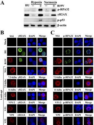
Individual B19V viral protein does not induce a DDR in EPCs.
We previously reported that B19V infection induces a DDR in
infected EPCs cultured under normoxic conditions (
39
). Since
hypoxic conditions promote the infection of EPCs by B19V (
10
),
we cultured EPCs under hypoxic conditions and infected them
with B19V. B19V infection of EPCs cultured under hypoxic
con-ditions also induced a DDR, as evidenced by the phosphorylation
of both H2AX and RPA32 (which are hallmarks of the DDR [
5
,
40
,
65
]), at levels approximately three times higher than those in
in-fected cells cultured under normoxic conditions (
Fig. 1A
).
Nota-bly, p53 was phosphorylated in B19V-infected EPCs under
nor-moxic conditions and phosphorylated further under hypoxic
conditions.
Since B19V expresses the nonstructural proteins (NS1,
7.5-kDa protein, and 11-7.5-kDa protein) in addition to the structural
proteins (VP1 and VP2) during infection (
37
,
64
,
78
), we
exam-ined whether the expression of the individual viral proteins
in-duced a DDR in EPCs. We expressed the nonstructural proteins
and structural proteins in EPCs via lentiviral transduction. As a
positive control for the DDR, EPCs were treated with hydroxyurea
(HU). At 48 h p.td., EPCs were analyzed for the expression of
phosphorylated H2AX and RPA32 by IF staining. B19V-infected
EPCs showed pan-distribution of
␥
-H2AX (
Fig. 1B
) and
colocal-ized foci of NS1 with p-RPA32 (
Fig. 1C
) in the nuclei, which was
consistent with that in infected EPCs cultured under normoxic
conditions (
39
). However, we observed that NS1 was expressed in
the cytoplasm to a greater extent than in the nucleus, likely due to
the high infectivity of B19V in EPCs cultured under hypoxic
con-ditions (
10
). In contrast to the formation of nuclear foci during
B19V infection, NS1-transduced EPCs showed neither
phos-phorylation of H2AX nor colocalization of NS1 and p-RPA32
(
Fig. 1B
and
C
). A proportion of the NS1 in transduced EPCs
appeared to be in the cytoplasm, mimicking the NS1 expression
pattern observed during viral infection. The difference in
␥
-H2AX and p-RPA32 expression between B19V-infected and
NS1-transduced EPCs suggests that B19V NS1 alone is not
suf-ficient to induce the DDR observed during B19V infection.
Similarly, when the 7.5-kDa, 11-kDa, VP1, and VP2 proteins
were expressed in EPCs by their respective lentiviruses, neither
H2AX nor RPA32 was phosphorylated in transduced EPCs
(
Fig. 1B
and
C
), whereas HU treatment of control cells induced
the phosphorylation of both H2AX and RPA32.
Taken together, these results confirm that B19V viral proteins,
including NS1, the 7.5-kDa protein, the 11-kDa protein, VP1, and
VP2, alone do not induce the DDR in EPCs that is observed during
B19V infection.
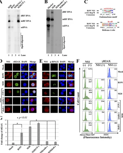
Replication of the B19V dsDNA genome induces a DDR in S1
cells.
Hypoxic conditions support the efficient replication of the
B19V dsDNA genome (M20 DNA) in S1 cells at a level
approxi-mately 100 times greater than that observed under normoxic
con-ditions (
10
). Since B19V replication in EPCs requires a DDR (
39
),
we hypothesized that the replication of B19V M20 DNA in S1 cells
induces a DDR as well. To this end, S1 cells were transfected with
M20 and a mutant [M20(VP
⫺)] in which the VP2 ORF was
knocked out and neither VP1 nor VP2 was expressed (data not
shown). Cells were infected with B19V as a control. We found that
both wild-type M20-transfected and B19V-infected S1 cells
gen-erated DpnI digestion-resistant bands of ssDNA, monomer RF
(mRF) DNA, and double RF (dRF) DNA on a Southern blot (
Fig.
2A
). This indicated that B19V M20 DNA replicated in the
transfected cells. The negative control, M20(NS1
⫺)-trans-fected cells, did not generate any DpnI digestion-resistant DNA
bands, confirming that NS1 is essential for B19V DNA
replica-tion. Transfection of mutant M20(VP
⫺) yielded clear mRF and
dRF DNA bands but not ssDNA bands (
Fig. 2A
, lane 4).
Pro-duction of ssDNA was significantly inhibited in the absence of
capsid proteins during B19V DNA replication. When the
pu-tative endonuclease domain or helicase-A site was mutated, the
M20(endo
⫺) and M20(heli
⫺) mutants did not replicate in
transfected cells, as evidenced by the lack of DpnI
digestion-resistant DNA bands (
Fig. 2B
, lanes 3 and 4).
The transfection of M20 induced phosphorylation of both
H2AX and RPA32 and induced the formation of NS1 and
p-RPA32 foci in the nuclei of transfected cells, similar to that
observed in B19V-infected S1 cells (
Fig. 2D
and
E
). Moreover,
H2AX and RPA32 were phosphorylated and colocalized with NS1
in the nuclei of M20(VP
⫺)-transfected S1 cells. Notably,
M20(endo
⫺)- and M20(heli
⫺)-transfected NS1-positive (NS1
⫹)
cells did not express
␥
-H2AX (
Fig. 2D
) or p-RPA32 (
Fig. 2E
).
Next, we carefully quantified the DDR induced in S1 cells infected
with B19V or transfected with M20 and its mutants by flow
cy-tometry. Replicative B19V M20 DNAs [M20 and M20(VP
⫺)] and
B19V infection induced significant expression of
␥
-H2AX in
NS1-expressing cells (
Fig. 2F
and
G
), which was significantly higher
than those induced by nonreplicative M20 DNAs [M20(endo
⫺)
and M20(heli
⫺)].
Taken together, these results show that replication of B19V M20
DNAper se, without ssDNA production, is sufficient to induce a DDR
at a level similar to that induced during B19V infection.
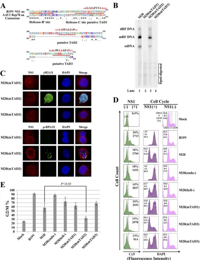
B19V DNA replication-induced DDR does not induce G
2/M
arrest after transfection.
B19V infection of EPCs triggers a DDR
cascade with the activation of all three PI 3-kinases (ATM, ATR,
and DNA-PKcs) (
39
). It also arrests infected cells at the G
2/M
Lou et al.
on November 7, 2019 by guest
http://jvi.asm.org/
phase (
70
). ATM- and ATR-mediated signaling is thought to be
the major pathway leading to cell cycle arrest at the G
2/M
check-point (
35
,
61
). However, in EPCs the expression of B19V NS1
alone can induce G
2/M arrest (
70
). Hence, we examined the role of
the B19V DNA replication-induced DDR in G
2/M arrest.
We first tried to identify an M20 mutant that replicates but does
not induce significant G
2/M arrest in transfected cells. The DNA
binding (endonuclease) and helicase domains of NS1 were critical for
B19V DNA replication (
Fig. 2B
), and mutations in the NLS site
abol-ished DNA replication (data not shown). We focused on the C
ter-minus of NS1, particularly the predicted TADs, since the TAD of
minute virus of mice (MVM) NS1 is dispensable for MVM DNA
replication (
33
). Three TADs were predicted by analyzing the C
ter-minus of NS1 using a TAD prediction program (
54
) (
Fig. 3A
). We
mutated each of the predicted TADs in M20 and assessed their
repli-cation capability. Only the M20(mTAD2), which harbors mutations
in TAD2, replicated in transfected S1 cells at a level comparable with
that in the wild type (
Fig. 3B
, lane 3). Furthermore, we examined the
ability of the M20-based TAD mutants to trigger a DDR in
trans-fected cells. The results showed that in only the
M20(mTAD2)-transfected (NS1
⫹) cells were both H2AX and RPA32 clearly
phosphorylated but not in cells transfected with M20(mTAD1)
and M20(mTAD3) (
Fig. 3C
), supporting the notion that B19V
DNA replication is critical for inducing a DDR.
We next tested the ability of wild-type M20 and its mutants to
arrest the cell cycle at G
2/M by transfecting them into S1 cells.
FIG 1B19V viral proteins do not induce a DDR in EPCs cultured under hypoxic conditions. (A) B19V infection induces a stronger DDR in EPCs cultured under hypoxic conditions than in EPCs cultured under normoxic conditions. EPCs cultured at either normoxic or hypoxic conditions were mock infected (⫺) or infected with B19V (⫹) at the same MOI. At 48 h p.i., cells were harvested and analyzed by Western blotting using the indicated antibodies. (B and C) B19V viral proteins do not elicit a DDR individually in EPCs cultured under hypoxic conditions. EPCs cultured under hypoxic conditions were mock infected, infected with B19V, or transduced with the indicated lentiviruses. (B) At 48 h p.i. or p.td., cells were fixed and costained with an antiviral protein antibody (red), an anti-␥-H2AX antibody (green), and DAPI (blue). (C) At 48 h p.i. or p.td., cells were fixed and costained with an antiviral protein antibody (red), an anti-p-RPA32 antibody (green), and DAPI (blue). Confocal images were taken at a magnification of⫻100. Hydroxyurea (HU)-treated EPCs served as a positive control for DDR detection.
on November 7, 2019 by guest
http://jvi.asm.org/
[image:4.585.135.451.66.487.2]FIG 2Replication of B19V dsDNA genome induces a DDR in S1 cells. S1 cells, which were precultured under hypoxic conditions for 48 h, were infected with B19V or electroporated with B19V dsDNA genome (M20) or its mutants, as indicated. At 48 h p.i. or p.tx., cells were harvested and analyzed as follows. (A and B) Southern blot analysis. Hirt DNA was extracted from infected or transfected S1 cells and digested with DpnI followed by Southern blotting. In panel B, M20 DNA (10 ng) was used as the size marker. The smaller band detected coincidentally at the site of the ssDNA is likely degraded M20 DNA. mRF, monomer replicative form; dRF, double replicative form. (C) Diagram of the mutations in the endonuclease motif and helicase-A site of the NS1 protein. Amino acid sequences of B19V NS1 (GenBank accession no.AAQ91878.1) and AAV2 Rep78 (AAC03775.1) were aligned using ClustalW2 (32). Asterisks show the consensus sequences in the endonuclease and helicase-A domains, and mutations are shown in red. (D and E) IF staining of the DDR markers. Cells were costained with anti-NS1 (red), anti-␥-H2AX (green) (D) or anti-p-RPA32 (green) (E) antibodies, and DAPI (blue). Confocal images were taken at a magnification of⫻100. (F) Flow cytometry analysis. Cells were costained with anti-NS1 antibodies, anti-␥-H2AX antibodies, and their respective secondary antibodies and analyzed by flow cytometry. Thexaxes of all the plots are in logarithmic scale, while theyaxes are in arithmetic scale. For NS1 staining (left-hand histograms), a reference line was drawn based on the secondary antibody-only control (mock) for a cutoff between the NS1-negative (NS1⫺) and -positive (NS1⫹) populations. Both the NS1-positive rate (%) and the mean fluorescence intensity (MFI) of NS1 expression in the NS1⫹population are presented in each plot. For␥-H2AX staining, levels of␥-H2AX are shown as the MFI both in the NS1⫹and NS1⫺cell populations of each group in the middle and right-hand histograms, respectively. The MFI in the mock groups was read from the whole population. (G) Statistical analysis. The fold change of␥-H2AX represents the ratio of the MFI of␥-H2AX for the NS1⫹to that for the NS1⫺population and is represented as the mean plus or minus the standard deviation from at least three independent experiments.P
values were determined using Student’sttest.
on November 7, 2019 by guest
http://jvi.asm.org/
[image:5.585.86.501.36.552.2]FIG 3B19V DNA replication-induced DDR is dispensable for G2/M arrest induced after transfection. (A) Diagram of predicted transactivation domains (TADs)
within the C terminus of B19V NS1. The TADs within B19V NS1 were predicted by analyzing the NS1 amino acid sequence using the 9aaTAD program (54). Three predicted TADs are shown with the mutated amino acids indicated in red above the TADs. (B) Southern blot analysis of transfected DNA replication. S1 cells cultured under hypoxic conditions were transfected with M20, M20(mTAD1), M20(mTAD2), or M20(mTAD3). At 48 h p.tx., cells were collected for Hirt DNA preparation followed by Southern blotting. (C) IF staining of the DDR markers. At 48 h p.tx., the transfected cells were costained with anti-NS1 (red), anti-␥-H2AX, or anti-p-RPA32 (green) antibodies and DAPI (blue). Confocal images were taken at a magnification of⫻100. (D) Flow cytometry analysis of the cell cycle. M20 and its indicated mutants were electroporated into S1 cells. B19V-infected S1 cells served as a positive control for NS1 detection and cell cycle analysis. At 48 h p.i. or p.tx., cells were costained with an anti-NS1 antibody and a Cy5-conjugated secondary antibody for NS1 detection and DAPI for cell cycle analysis by flow cytometry. Theyaxes for all the plots are in arithmetic scale. Thexaxes for the NS1-staining plots are in logarithmic scale, while those for the remaining DAPI-staining plots are in arithmetic scale. The methods for gating of the NS1⫺and NS1⫹cell populations are the same as those described inFig. 2F. The cell cycle was analyzed in both NS1⫹and NS1⫺populations in each group, as indicated in the middle and right-hand histograms, respectively. The percentage (%) of the cells in each phase of the cell cycle is shown. (E) Statistical analysis. Data for the percentage of cells at the G2/M phase (G2/M%) were obtained from
at least three independent experiments and are presented as the mean plus or minus the standard deviation. ThePvalue was determined using Student’sttest.
on November 7, 2019 by guest
http://jvi.asm.org/
[image:6.585.87.498.63.597.2]Notably, the M20 mutants that did not replicate induced clear
G
2/M arrest at levels similar to or higher than those observed in
the wild type, with the percentage of cells in G
2/M phase ranging
from 63.1% to 88.6% in NS1
⫹cells (
Fig. 3D
and
E
). More
impor-tantly, the replicative TAD mutant, M20(mTAD2), significantly
decreased the number of cells at the G
2/M phase compared with
that of the wild-type M20 control (32.8% vs. 57.7%, respectively)
(
Fig. 3E
).
Collectively, these results show that replication of the B19V
dsDNA genome is not required to induce G
2/M arrest in
trans-fected S1 cells. Since replication of the B19V dsDNA genome is
essential for inducing a DDR, we conclude that the DDR is
dis-pensable for G
2/M arrest of the cells in which B19V DNA
repli-cates. The results also suggest that B19V NS1 alone is sufficient to
arrest the cell cycle at G
2/M during infection.
Expression of NS1 alone is sufficient to induce G
2/M arrest in
S1 cells and EPCs.
To examine the sole role of NS1 in inducing
G
2/M arrest, we expressed only wild-type NS1 and the NS1
mu-tants, which originated from the M20 mumu-tants, by lentiviral
trans-duction in S1 cells and EPCs. Transtrans-duction of
Lenti-½ITR-P6-NS1, in which expression of wild-type NS1 is driven by the native
B19V P6 promoter (
55
), recapitulated G
2/M arrest to the level
induced by B19V infection in S1 cells, in which over 90% of NS1
⫹cells were arrested at the G
2/M phase. This was also observed after
the transduction of lentiviruses expressing the NS1 mutants
ex-cept for NS1(mTAD2) (
Fig. 4A
). The expression of NS1(mTAD2)
increased the number of NS1
⫹cells at G
2/M only to 36.6%, a level
similar to that induced by the expression of the RFP control
(42.4%) (
Fig. 4C
). In EPCs, in agreement with the results observed
in S1 cells, both B19V infection and transduction of lentiviruses
expressing wild-type NS1 and the NS1 mutants, NS1(endo
⫺),
NS1(heli
⫺), and NS1(mTAD3), induced over 90% of NS1
⫹cells
at the G
2/M phase (
Fig. 4B
). Notably, the expression of
NS1(mTAD2) in EPCs resulted in 42.2% of NS1
⫹cells to arrest at
G
2/M, a level close to that induced by expression of the RFP
con-trol (35.0%) (
Fig. 4D
). We noticed that the expression of RFP in
the context of Lenti-½ITR-P6-RFP induced a higher number of
cells at G
2/M (approximately 40%) than G
2/M-phased cells
(ap-proximately 20%) in the mock group in both S1 cells and EPCs. It
has been reported that a consensus Toll-like receptor (TLR) ligand
sequence (5
=
-GTTTTGT-3
=
) exits in the B19V P6 promoter and
inhibits proliferation of EPCs by arresting the cell cycle at the S
and G
2/M phases (
28
). We also observed that a lentiviral vector
bearing a B19V sequence of nucleotides 187 to 615 (contains
½ITR and the core P6 promoter) arrested the cell cycle at G
2/M
(data not shown).
Taken together, these results confirm that the expression of
B19V NS1 alone is sufficient to induce nearly complete G
2/M
ar-rest in the context of the B19V P6 promoter and that the predicted
TAD2 of NS1 is critical for G
2/M arrest induced by NS1 in both S1
cells and EPCs.
The p53-mediated pathway is not required for G
2/M arrest
induced by B19V infection or transfection of the B19V dsDNA
genome.
ATM and ATR activation during a DDR phosphorylates
p53 at Ser
15(
3
,
60
). The cyclin-dependent kinase Cdk1, which
controls the G
2/M transition, is inhibited by the product of the
p53-targeting gene, p21 (
6
). Since B19V NS1 transactivates the
expression of p21 (
45
) and p53 is phosphorylated at Ser
15during
B19V infection of EPCs (
70
) (
Fig. 1A
), we decided to examine the
role of p53 in B19V-induced G
2/M arrest.
We first attempted to knock out p53 in EPCs using p53-specific
shRNAs, which proved to be very difficult (data not shown).
For-tunately, nearly perfect p53 knockout was obtained in S1 cells
transduced with a p53-shRNA-expressing lentivirus (
Fig. 5A
). We
found that the percentage of p53-deficient NS1
⫹cells (either
in-fected with B19V or transin-fected with M20) at the G
2/M phase did
not decrease compared with that in scramble (scr)-shRNA-treated
cells (
Fig. 5B
and
C
), suggesting that the p53-mediated pathway is
not used to induce G
2/M arrest during infection or transfection.
We also observed that the transfection of M20 caused fewer cells to
arrest at G
2/M than B19V infection (56.8% vs. 90.6%) (
Fig. 5B
);
however, knockout of p53 did not ameliorate G
2/M arrest
signif-icantly under either condition (
Fig. 5C
).
DISCUSSION
In this study, we report that the replication of the B19V dsDNA
genome, but not the individual viral proteins, induces a DDR. We
identified an M20 mutant, M20(mTAD2), containing a mutated
transactivation domain within NS1, which replicated well in S1
cells but did not arrest the cell cycle at the G
2/M phase. In
agree-ment with this result, the NS1 harboring the TAD2 mutation did
not induce significant G
2/M arrest in either EPCs or S1 cells by
lentiviral transduction. Finally, we showed that the p53-mediated
pathway is not involved in G
2/M arrest during B19V infection.
Thus, our results provide evidence that the DDR induced during
B19V infection does not contribute to G
2/M arrest and confirm
that NS1 alone is sufficient to induce the G
2/M arrest in the
con-text of the B19V P6 promoter.
A DDR is a response by the cellular surveillance network that
senses and repairs damaged DNA and is required to maintain
genome integrity (
14
). During viral infection, a DDR plays a role
in the intrinsic antiviral mechanism to eliminate the invasion of
the viral genome; however, in some cases, the virus exploits the
DDR signaling mechanism to promote its own replication (
71
).
One of the major consequences of a DDR is cell cycle arrest, which
ensures that there is time for DNA repair after DNA damage (
57
).
B19V infection of both EPCs and S1 cells typically arrests the cell
cycle at G
2/M (
39
,
47
,
70
). Preventing replication of the B19V
dsDNA genome by mutating the endonuclease domain or
heli-case-A motif within NS1, which prohibits the DDR, did not
re-duce the percentage of cells arrested at G
2/M; however, mutating
TAD2 could result in a replication-competent M20 mutant but
with reduced G
2/M arrest. NS1 alone is sufficient to cause G
2/M
arrest, which requires a predicted TAD2. Therefore, B19V utilizes
the mechanism of a DDR only to promote viral DNA replication
(
39
), and the NS1-induced G
2/M arrest is dispensable for viral
DNA replication. The role of the NS1-induced G
2/M arrest during
B19V infection warrants further investigation. The fact that the
p53 pathway is not used by B19V NS1 to arrest the cell cycle
indi-cates that the TAD2 of NS1 does not transactivate p53-regulating
genes. NS1 expression in EPCs upregulates 44 genes and
down-regulates 28 genes that are involved in cell cycle regulation (
70
).
Therefore, we hypothesize that TAD2 of NS1 may play an
impor-tant role in regulating one or several of these genes. The NLS
domain of NS1 is thought to be critical for inducing G
2/M arrest,
which is mediated by the interaction between NS1 and E2F4/E2F5
(
70
); however, mutation of the NLS, which prevents NS1
trans-port into the nucleus (
70
), may also prevent the function of the
TAD. Clearly, our results highlight a novel mechanism underlying
Lou et al.on November 7, 2019 by guest
http://jvi.asm.org/
G
2/M arrest induced by NS1 during B19V infection, which is
in-dependent of the DDR and p53 activation.
Viral proteins often play an important role in triggering a DDR
during infection (
8
,
34
,
71
), particularly in the large T antigen of
polyomaviruses (
21
,
49
,
77
) and the Vpr of HIV (
76
). In
parvovi-ruses, the replication of the AAV2 genome during coinfection
with adenovirus elicits an ATM-, ATR-, and DNA-PKcs-mediated
DDR (
17
,
59
). We reported that DNA replication of the minute
FIG 4NS1 alone is sufficient to induce G2/M arrest in S1 cells and EPCs. (A and B) Flow cytometry analyses of S1 cells and EPCs transduced with lentiviruses.S1 cells (A) or EPCs (B) cultured under hypoxic conditions were either infected with B19V or transduced with the indicated lentiviruses. The Lenti-½ITR-P6-RFP serves as a lentiviral control. At 48 h p.i. or p.td., cells were costained with anti-NS1 and Cy5-conjugated secondary antibodies (for B19V-infected cells) or anti-Flag and Dylight 649-conjugated secondary antibodies (for lentivirus-transduced cells), followed by DAPI for cell cycle analysis. NS1 staining and cell cycle analyses were performed as described inFig. 3D. (C and D) Statistical analysis. Statistical data of the G2/M% were obtained from at least three independent
experiments in S1 cells (C) and EPCs (D), indicated as the mean plus or minus the standard deviation. ThePvalues were determined using Student’sttest.
on November 7, 2019 by guest
http://jvi.asm.org/
[image:8.585.89.500.60.599.2]virus of canines (MVC) genome is responsible for an
MVC-in-duced DDR rather than the viral proteins (
38
). The MVM also
induces a DDR during infection (
1
,
56
), but the small
nonstruc-tural protein NS2 is dispensable (
56
). The AAV2 Rep78 induces a
DDR by introducing nicks in the cellular chromatin (
4
), but it is
only a minor contributor to the DDR induced during AAV2
in-fection (
59
). The present study shows that B19V NS1, the small
nonstructural proteins (7.5-kDa and 11-kDa proteins), and the
capsid proteins failed to induce an obvious DDR in EPCs when
they were expressed individually. However, we have not ruled out
the possibility of the combined expression of B19V proteins,
which is the case during B19V infection, and transfection of the
replication-competent M20 DNAs inducing a DDR. Knockout of
the 11-kDa protein in M20 DNA showed a decreased level of DNA
replication in S1 cells (data not shown). We speculate that a
com-bined expression of B19V proteins supports B19V DNA
replica-tion to the highest extent, which induces a maximum DDR. This
actually is true for the NS1 protein. NS1 is essential for B19V
DNA replication (
Fig. 2A
), but NS1 alone could not induce a
DDR (
Fig. 1B
and
C
). Taken together, our results support the
conclusion that the DDR induced during parvovirus infection
is mediated mainly through replication of the viral genome, for
which NS1 is essential.
Because EPCs are difficult to transfect (
13
), we delivered the
B19V dsDNA genome into S1 cells by electroporation. Replication
of the B19V dsDNA genome, even in the absence of ssDNA
pro-duction, was sufficient to trigger a DDR in transfected S1 cells at a
level similar to that observed during B19V infection. These results
indicate that replication of the B19V dsDNA genome during
in-fection of EPCs initiates the DDR independently of the
produc-tion of the ssDNA viral genome. However, the ssDNA genomes of
B19V and other parvoviruses have hairpin termini at both ends
(
20
). The gap between the termini displays a structure of a
single-strand break (SSB) that is recognized by ATR (
15
). Since it was
FIG 5The p53 pathway is not necessary for B19V-induced G2/M arrest. S1 cells cultured under normoxic conditions were transduced with p53-shRNA- andscramble-shRNA (scr-shRNA)-expressing lentiviruses, which also expressed GFP (9), and then cultured under hypoxic conditions for 48 h. The cells then were mock infected, infected with B19V, or transfected with M20. (A) Western blot analysis of p53 expression. At 48 h p.i. or p.tx., cells were analyzed for the expression of p53 by Western blotting. The blot was probed with an anti-p53 antibody, stripped, and reprobed to examine the-actin levels. “No treatment” indicates cells not transduced with lentivirus. The arrow indicates the p53 band. (B) Flow cytometry analysis of the cell cycle. At 48 h p.i. or p.tx., cells were costained with the anti-NS1 antibody and Cy5-conjugated secondary antibodies for NS1 detection and DAPI for cell cycle analysis by flow cytometry as described inFig. 3D. In the shRNA-treated groups, shRNA-expressing cells were selected based on the GFP signal expressed by the lentiviral vector and shown in the left-hand histograms. (C) Statistical analysis of the cell cycle results. The ratio of the percentage of cells at G2/M (G2/M%) in the NS1⫹population to that in the NS1⫺population in
each group is named as the fold change of the G2/M%. Statistical data of the fold change of the G2/M% were obtained from at least three independent experiments,
indicated as the mean plus or minus the standard deviation. ThePvalues were determined using Student’sttest.
Lou et al.
on November 7, 2019 by guest
http://jvi.asm.org/
[image:9.585.84.504.67.417.2]difficult to detect the ssDNA viral genome in the nucleus upon
B19V infection (data not shown), we have not provided direct
evidence that the B19V ssDNA genome elicits a DDR. However,
when UV-inactivated B19V was used to infect EPCs at an MOI of
1,000 gc/cell, the same as that for the wild-type B19V in our study,
neither
␥
-H2AX nor p-RPA32 was detected in infected cells (data
not shown). It has been reported that infection of UV-inactivated
AAV at a high MOI (5,000 gc/cell) elicited a DDR by mimicking
the stalled replication fork (
29
). Further studies are warranted to
determine the nature of the B19V ssDNA genome as an SSB
sub-strate for ATR recognition.
B19V induces a DDR via replication of the RF genome, which
involves replication intermediates (
27
) rather than via NS1
ex-pression alone. The replication of parvovirus DNA follows the
rolling-hairpin model of DNA replication, which involves a strand
displacement step (
19
). Therefore, the formation of a replication
origin complex at the replication origin, or other steps, such as
nicking at the terminal resolution site by NS1 followed by
un-winding of the RF DNA or hairpin by the helicase activity of NS1
(which resembles a stalled replication fork), is likely the key for
triggering a DDR during virus infection. It is possible that ATR
recognizes the nicked site within the terminal sequence of the
genome and that the ssDNA binding protein RPA32 is recruited to
the displaced ssDNA, which mimics a stalled replication fork.
Thus, our observations have laid a solid foundation for further
study of how replication of the ssDNA genome of autonomous
parvovirus induces a DDR during virus infection.
Regulation of cell cycle progression, symbolized by arrest at the
S or G
2/M phase checkpoint, is a classic strategy for infecting
vi-ruses to exploit cellular components/mechanisms to aid viral
ge-nome replication (
12
,
23
). MVM and MVC infections activate
ATM-mediated signaling, which arrests infected cells at the S and
G
2/M phases; the DDR then directly leads to cell cycle arrest (
1
,
38
). The Vpr protein of HIV is responsible for a DDR during HIV
infection, which results in G
2/M arrest through ATR-checkpoint
kinase 1 (Chk1) signaling (
66
). It is intriguing that B19V DNA
replication-induced DDR does not result in G
2/M arrest, even
though Chk1 and Chk2 are activated during infection (
39
).
There-fore, our observations suggest multiple roles for virus
infection-triggered DDR. In some cases, the DDR induces cell cycle arrest,
which in turn facilitates virus replication or kills infected cells. In
other cases, the DDR is directly involved in promoting DNA
rep-lication without arresting the cell cycle.
In summary, we have demonstrated that replication of the
B19V dsDNA genome induces DDR signaling and that the DDR is
dispensable for triggering cell cycle arrest during B19V DNA
rep-lication. However, the NS1 protein was instrumental in G
2/M
ar-rest, which did not require the putative activity of endonuclease
and helicase but did require TAD2 function. Furthermore, this
process was independent of the p53 pathway. Further studies are
needed to examine how TAD2 functions to induce G
2/M arrest
and how this function coordinates with the function of the NLS to
induce G
2/M arrest. Thus, B19V may have evolved a unique
strat-egy employing NS1 to overcome difficulties in arresting the cell
cycle in rapidly proliferating EPCs. The reason why the induced
DDR during B19V infection of EPCs, particularly the activation of
ATR and ATM signaling, did not significantly result in G
2/M
ar-rest will be an intear-resting topic for future study.
ACKNOWLEDGMENTS
This work was supported in full by PHS grant R01 AI070723 from the NIAID and grant R21 HL106299 from the NHLBI to J.Q.
We are indebted to Susan Wong at the Hematology Branch, NHLBI, NIH, for helping culture CD34⫹human hematopoietic stem cells.
REFERENCES
1.Adeyemi RO, Landry S, Davis ME, Weitzman MD, Pintel DJ.2010. Parvovirus minute virus of mice induces a DNA damage response that facilitates viral repli-cation. PLoS Pathog.6:e1001141. doi:10.1371/journal.ppat.1001141.
2.Anderson MJ, et al.1988. Human parvovirus B19 and hydrops fetalis. Lanceti:535.
3.Banin S, et al.1998. Enhanced phosphorylation of p53 by ATM in re-sponse to DNA damage. Science281:1674 –1677.
4.Berthet C, Raj K, Saudan P, Beard P.2005. How adeno-associated virus Rep78 protein arrests cells completely in S phase. Proc. Natl. Acad. Sci. U. S. A.102:13634 –13639.
5.Block WD, Yu Y, Lees-Miller SP.2004. Phosphatidyl inositol 3-kinase-like serine/threonine protein kinases (PIKKs) are required for DNA dam-age-induced phosphorylation of the 32 kDa subunit of replication protein A at threonine 21. Nucleic Acids Res.32:997–1005.
6.Bunz F, et al.1998. Requirement for p53 and p21 to sustain G2 arrest after DNA damage. Science282:1497–1501.
7.Chaturvedi P, et al.1999. Mammalian Chk2 is a downstream effector of the ATM-dependent DNA damage checkpoint pathway. Oncogene18: 4047– 4054.
8.Chaurushiya MS, Weitzman MD. 2009. Viral manipulation of DNA repair and cell cycle checkpoints. DNA Repair (Amst.)8:1166 –1176. 9.Chen AY, et al.2010. Role of erythropoietin receptor signaling in
parvo-virus B19 replication in human erythroid progenitor cells. J. Virol.84: 12385–12396.
10. Chen AY, Kleiboeker S, Qiu J.2011. Productive parvovirus B19 infection of primary human erythroid progenitor cells at hypoxia is regulated by STAT5A and MEK signaling but not HIF alpha. PLoS Pathog.7:e1002088. doi:10.1371/journal.ppat.1002088.
11. Chen AY, Luo Y, Cheng F, Sun Y, Qiu J.2010. Bocavirus infection induces a mitochondrion-mediated apoptosis and cell cycle arrest at G2/M phase. J. Virol.84:5615–5626.
12. Chen AY, Qiu J.2010. Parvovirus infection-induced cell death and cell cycle arrest. Future Virol.5:731–741.
13. Chen AY, et al.2010. The small 11kDa non-structural protein of human parvovirus B19 plays a key role in inducing apoptosis during B19 virus infection of primary erythroid progenitor cells. Blood115:1070 –1080. 14. Ciccia A, Elledge SJ.2010. The DNA damage response: making it safe to
play with knives. Mol. Cell40:179 –204.
15. Cimprich KA, Cortez D.2008. ATR: an essential regulator of genome integrity. Nat. Rev. Mol. Cell Biol.9:616 – 627.
16. Cipolleschi MG, Dello SP, Olivotto M.1993. The role of hypoxia in the maintenance of hematopoietic stem cells. Blood82:2031–2037. 17. Collaco RF, Bevington JM, Bhrigu V, Kalman-Maltese V, Trempe JP.
2009. Adeno-associated virus and adenovirus coinfection induces a cellu-lar DNA damage and repair response via redundant phosphatidylinositol 3-like kinase pathways. Virology392:24 –33.
18. Cotmore SF, Tattersall P.1984. Characterization and molecular cloning of a human parvovirus genome. Science226:1161–1165.
19. Cotmore SF, Tattersall P.2005. A rolling-haipin strategy: basic mecha-nisms of DNA replication in the parvoviruses, p 171–181.InKerr J, Cot-more SF, Bloom ME, Linden RM, and Parrish CR (ed), Parvoviruses. Hodder Arnold, London, England.
20. Cotmore SF, Tattersall P.2005. Structure and organization of the viral genome, p 73–94.InKerr J, Cotmore SF, Bloom ME, Linden RM, and Parrish CR (ed), Parvoviruses. Hodder Arnold, London, England. 21. Dahl J, You J, Benjamin TL.2005. Induction and utilization of an ATM
signaling pathway by polyomavirus. J. Virol.79:13007–13017.
22. Doerig C, Hirt B, Antonietti JP, Beard P.1990. Nonstructural protein of parvoviruses B19 and minute virus of mice controls transcription. J. Virol. 64:387–396.
23. Emmett SR, Dove B, Mahoney L, Wurm T, Hiscox JA.2005. The cell cycle and virus infection. Methods Mol. Biol.296:197–218.
24. Fu Y, et al.2002. Regulation of tumor necrosis factor alpha promoter by human parvovirus B19 NS1 through activation of AP-1 and AP-2. J. Virol. 76:5395–5403.
on November 7, 2019 by guest
http://jvi.asm.org/
25. Gareus R, et al.1998. Characterization of cis-acting and NS1 protein-responsive elements in the p6 promoter of parvovirus B19. J. Virol.72:609–616. 26. Guan W, et al.2008. Block to the production of full-length B19 virus
transcripts by internal polyadenylation is overcome by replication of the viral genome. J. Virol.82:9951–9963.
27. Guan W, Wong S, Zhi N, Qiu J.2009. The genome of human parvovirus B19 virus can replicate in non-permissive cells with the help of adenovirus genes and produces infectious virus. J. Virol.83:9541–9553.
28. Guo YM, et al.2010. CpG-ODN 2006 and human parvovirus B19 ge-nome consensus sequences selectively inhibit growth and development of erythroid progenitor cells. Blood115:4569 – 4579.
29. Jurvansuu J, Raj K, Stasiak A, Beard P.2005. Viral transport of DNA damage that mimics a stalled replication fork. J. Virol.79:569 –580. 30. Kaufmann B, Chipman PR, Kostyuchenko VA, Modrow S, Rossmann
MG.2008. Visualization of the externalized VP2 N termini of infectious human parvovirus B19. J. Virol.82:7306 –7312.
31. Kaufmann B, Simpson AA, Rossmann MG. 2004. The structure of human parvovirus B19. Proc. Natl. Acad. Sci. U. S. A.101:11628 –11633. 32. Larkin MA, et al.2007. Clustal W and Clustal X version 2.0.
Bioinfor-matics23:2947–2948.
33. Legendre D, Rommelaere J.1994. Targeting of promoters for trans acti-vation by a carboxy-terminal domain of the NS-1 protein of the parvovi-rus minute viparvovi-rus of mice. J. Virol.68:7974 –7985.
34. Lilley CE, Schwartz RA, Weitzman MD.2007. Using or abusing: viruses and the cellular DNA damage response. Trends Microbiol.15:119 –126. 35. Liu Q, et al.2000. Chk1 is an essential kinase that is regulated by Atr and
required for the G(2)/M DNA damage checkpoint. Genes Dev.14:1448 – 1459.
36. Lou S, et al.2012. Molecular characterization of the newly identified human parvovirus 4 in the family Parvoviridae. Virology422:59 – 69. 37. Luo W, Astell CR.1993. A novel protein encoded by small RNAs of
parvovirus B19. Virology195:448 – 455.
38. Luo Y, Chen AY, Qiu J.2011. Bocavirus infection induces a DNA damage response that facilitates viral DNA replication and mediates cell death. J. Virol.85:133–145.
39. Luo Y, et al.2011. Parvovirus B19 infection of human primary erythroid progenitor cells triggers ATR-Chk1 signaling, which promotes B19 virus replication. J. Virol.85:8046 – 8055.
40. Mah LJ, El-Osta A, Karagiannis TC.2010. gammaH2AX: a sensitive molecular marker of DNA damage and repair. Leukemia24:679 – 686. 41. Moffatt S, et al.1996. A cytotoxic nonstructural protein, NS1, of human
parvovirus B19 induces activation of interleukin-6 gene expression. J. Vi-rol.70:8485– 8491.
42. Moffatt S, Yaegashi N, Tada K, Tanaka N, Sugamura K.1998. Human parvovirus B19 nonstructural (NS1) protein induces apoptosis in ery-throid lineage cells. J. Virol.72:3018 –3028.
43. Momoeda M, Wong S, Kawase M, Young NS, Kajigaya S.1994. A putative nucleoside triphosphate-binding domain in the nonstructural protein of B19 parvovirus is required for cytotoxicity. J. Virol.68:8443– 8446.
44. Morey AL, Fleming KA.1992. Immunophenotyping of fetal haemopoi-etic cells permissive for human parvovirus B19 replication in vitro. Br. J. Haematol.82:302–309.
45. Morita E, Nakashima A, Asao H, Sato H, Sugamura K.2003. Human parvovirus B19 nonstructural protein (NS1) induces cell cycle arrest at G(1) phase. J. Virol.77:2915–2921.
46. Morita E, Sugamura K.2002. Human parvovirus B19-induced cell cycle arrest and apoptosis. Springer Semin. Immunopathol.24:187–199. 47. Morita E, et al.2001. Human parvovirus B19 induces cell cycle arrest at
G(2) phase with accumulation of mitotic cyclins. J. Virol.75:7555–7563. 48. Nakashima A, Morita E, Saito S, Sugamura K.2004. Human parvovirus B19 nonstructural protein transactivates the p21/WAF1 through Sp1. Vi-rology329:493–504.
49. Orba Y, et al.2010. Large T antigen promotes JC virus replication in G2-arrested cells by inducing ATM- and ATR-mediated G2 checkpoint signaling. J. Biol. Chem.285:1544 –1554.
50. Ozawa K, et al.1987. Novel transcription map for the B19 (human) pathogenic parvovirus. J. Virol.61:2395–2406.
51. Ozawa K, Kurtzman G, Young N.1986. Replication of the B19 parvovi-rus in human bone marrow cell cultures. Science233:883– 886. 52. Parmar K, Mauch P, Vergilio JA, Sackstein R, Down JD.2007.
Distri-bution of hematopoietic stem cells in the bone marrow according to re-gional hypoxia. Proc. Natl. Acad. Sci. U. S. A.104:5431–5436.
53. Pillet S, et al.2004. Hypoxia enhances human B19 erythrovirus gene expression in primary erythroid cells. Virology327:1–7.
54. Piskacek S, et al.2007. Nine-amino-acid transactivation domain: estab-lishment and prediction utilities. Genomics89:756 –768.
55. Raab U, et al.2002. NS1 protein of parvovirus B19 interacts directly with DNA sequences of the p6 promoter and with the cellular transcription factors Sp1/Sp3. Virology293:86 –93.
56. Ruiz Z, Mihaylov IS, Cotmore SF, Tattersall P.2011. Recruitment of DNA replication and damage response proteins to viral replication centers during infection with NS2 mutants of Minute Virus of Mice (MVM). Virology410:375–384.
57. Sancar A, Lindsey-Boltz LA, Unsal-Kacmaz K, Linn S.2004. Molecular mechanisms of mammalian DNA repair and the DNA damage check-points. Annu. Rev. Biochem.73:39 – 85.
58. Schneider H.2011. Oxygenation of the placental-fetal unit in humans. Respir. Physiol. Neurobiol.178:51–58.
59. Schwartz RA, Carson CT, Schuberth C, Weitzman MD.2009. Adeno-associated virus replication induces a DNA damage response coordinated by DNA-dependent protein kinase. J. Virol.83:6269 – 6278.
60. Shieh SY, Ikeda M, Taya Y, Prives C. 1997. DNA damage-induced phosphorylation of p53 alleviates inhibition by MDM2. Cell91:325–334. 61. Smith J, Tho LM, Xu N, Gillespie DA. 2010. The ATM-Chk2 and ATR-Chk1 pathways in DNA damage signaling and cancer. Adv. Cancer Res.108:73–112.
62. Sol N, et al.1999. Possible interactions between the NS-1 protein and tumor necrosis factor alpha pathways in erythroid cell apoptosis induced by human parvovirus B19. J. Virol.73:8762– 8770.
63. Srivastava A, Lu L.1988. Replication of B19 parvovirus in highly enriched hematopoietic progenitor cells from normal human bone marrow. J. Vi-rol.62:3059 –3063.
64. St Amand AJ, Astell CR.1993. Identification and characterization of a family of 11-kDa proteins encoded by the human parvovirus B19. Virol-ogy192:121–131.
65. Stucki M, Jackson SP.2006. gammaH2AX and MDC1: anchoring the DNA-damage-response machinery to broken chromosomes. DNA Repair (Amst.)5:534 –543.
66. Tachiwana H, et al.2006. HIV-1 Vpr induces DNA double-strand breaks. Cancer Res.66:627– 631.
67. Takubo K, et al.2010. Regulation of the HIF-1alpha level is essential for hematopoietic stem cells. Cell Stem Cell7:391– 402.
68. Tijssen P, et al.2012. Family Parvoviridae, p 405– 425.InKing AM, Lefkowitz E, Adams MJ, Carstens EB (ed), Virus taxonomy: ninth report of the International Committee on Taxonomy of Viruses. Elsevier, Lon-don, United Kingdom.
69. Walker SL, Wonderling RS, Owens RA.1997. Mutational analysis of the adeno-associated virus type 2 Rep68 protein helicase motifs. J. Virol.71: 6996 –7004.
70. Wan Z, et al.2010. Human parvovirus B19 causes cell cycle arrest of human erythroid progenitors via deregulation of the E2F family of tran-scription factors. J. Clin. Invest.120:3530 –3544.
71. Weitzman MD, Lilley CE, Chaurushiya MS.2010. Genomes in conflict: maintaining genome integrity during virus infection. Annu. Rev. Micro-biol.64:61– 81.
72. Wong S, et al.2008. Ex vivo-generated CD36⫹erythroid progenitors are highly permissive to human parvovirus B19 replication. J. Virol.82:2470–2476. 73. Yaegashi N, et al.1999. Parvovirus B19 infection induces apoptosis of
erythroid cells in vitro and in vivo. J. Infect.39:68 –76.
74. Young NS, Brown KE.2004. Parvovirus B19. N. Engl. J. Med.350:586 –597. 75. Zadori Z, et al.2001. A viral phospholipase A2 is required for parvovirus
infectivity. Dev. Cell1:291–302.
76. Zhao RY, Elder RT.2005. Viral infections and cell cycle G2/M regulation. Cell Res.15:143–149.
77. Zhao X, et al.2008. Ataxia telangiectasia-mutated damage-signaling ki-nase- and proteasome-dependent destruction of Mre11-Rad50-Nbs1 sub-units in simian virus 40-infected primate cells. J. Virol.82:5316 –5328. 78. Zhi N, et al.2006. Molecular and functional analyses of a human parvovirus
B19 infectious clone demonstrates essential roles for NS1, VP1, and the 11-kilodalton protein in virus replication and infectivity. J. Virol.80:5941–5950. 79. Zhi N, et al.2010. Codon optimization of human parvovirus B19 capsid genes greatly increases their expression in nonpermissive cells. J. Virol.84:13059–13062. 80. Zhi N, Zadori Z, Brown KE, Tijssen P.2004. Construction and sequencing of an infectious clone of the human parvovirus B19. Virology318:142–152.
Lou et al.
on November 7, 2019 by guest
http://jvi.asm.org/