Inactivation of
Burkholderia cepacia
Complex Phage KS9 gp41
Identifies the Phage Repressor and Generates Lytic Virions
䌤
†
Karlene H. Lynch,
1Kimberley D. Seed,
1Paul Stothard,
2and Jonathan J. Dennis
1*
Department of Biological Sciences, University of Alberta, Edmonton, Alberta, Canada,1and Department of Agricultural,
Food and Nutritional Science, University of Alberta, Edmonton, Alberta, Canada2
Received 31 August 2009/Accepted 13 November 2009
TheBurkholderia cepaciacomplex (BCC) is made up of at least 17 species of Gram-negative opportunistic
bacterial pathogens that cause fatal infections in patients with cystic fibrosis and chronic granulomatous disease. KS9 (vB_BcenS_KS9), one of a number of temperate phages isolated from BCC species, is a prophage
ofBurkholderia pyrrociniaLMG 21824. Transmission electron micrographs indicate that KS9 belongs to the
familySiphoviridaeand exhibits the B1 morphotype. The 39,896-bp KS9 genome, comprised of 50 predicted
genes, integrates into the 3ⴕend of the LMG 21824 GTP cyclohydrolase II open reading frame. The KS9 genome
is most similar to uncharacterized prophage elements in the genome ofB. cenocepaciaPC184 (vB_BcenZ_
PC184), as well asBurkholderia thailandensisphageE125 andBurkholderia pseudomalleiphage1026b. Using
molecular techniques, we have disrupted KS9 gene41, which exhibits similarity to genes encoding phage
repressors, producing a lytic mutant named KS9c. This phage is incapable of stable lysogeny in either LMG
21824 orB. cenocepaciastrain K56-2 and rescues aGalleria mellonella infection model from experimentalB.
cenocepaciaK56-2 infections at relatively low multiplicities of infection. These results readily demonstrate that
temperate phages can be genetically engineered to lytic form and that these modified phages can be used to
treat bacterial infectionsin vivo.
The Burkholderia cepacia complex (BCC) is a group of at
least 17 Gram-negative species, the first identified strains of which were characterized as onion pathogens by W. H. Burk-holder (9). Although these bacteria have a number of benefi-cial activities, including the promotion of crop growth and the degradation of organic pollutants, they have gained notoriety in the last two decades as serious opportunistic pathogens (19, 21, 25). BCC species, particularlyB. multivoransandB.
ceno-cepacia, cause serious respiratory infections in patients with
cystic fibrosis and chronic granulomatous disease (42, 7). These infections are especially problematic due to symptom severity, the inherent antibiotic resistance of Bcc species, and the potential for rapid spread through susceptible patient pop-ulations (25, 23). Difficulties in treating these infections have led to the unfortunate practice of segregating patients, which has high economic, social, and psychological costs (18).
Because of these clinical difficulties, interest in the isolation and characterization of Burkholderia-specific bacteriophages (or phages) has increased in recent years, with the apparent potential for using phages as therapeutic agents. Phage therapy is the clinical application of phages to prevent and/or to treat infections, which offers a promising alternative to antibiotic treatment for resistant bacteria such as those of the BCC (33, 39). A second benefit of these phage studies is that they may provide insight into the possible mechanisms of BCC virulence. For example, BcepMu, a transposable phage that specifically
infects strains of B. cenocepacia, was found to carry genes similar to exeA, involved in toxin secretion, and mdmB and
oafA, two acyltransferases (44). Finally, asBurkholderiaphages tend to be underrepresented in comparative studies with re-spect toEscherichia coliand lactic acid bacteria phages, BCC-specific phage studies provide novel information about a rela-tively uncharacterized group of viruses.
Although phage therapy using temperate virions can be ef-fective (39), there are several reasons why lytic phages are generally considered the most appropriate candidates for use in phage therapy. One of the concerns is that phage integration can lead to lysogenic conversion and enhanced virulence (8). A second concern is that integration of temperate phages results in superinfection immunity due to expression of the phage repressor from the prophage. This protein binds to the oper-ators of infecting phage DNA and represses gene expression, preventing both the initiation of the lytic cycle and the estab-lishment of lysogeny (14). A third concern is that lysogeny affects the kinetics of infection. When a phage infects a cell and undergoes lysogeny instead of entering the lytic cycle, the cell survives, and no new phage particles are released (27). A final problem is that prophages can lead to specialized transduction after induction. Specialized transduction occurs after inexact excision of a prophage from the bacterial chromosome. Bac-terial DNA flanking the prophage is packaged into the capsid, and this sequence, which can potentially encode virulence fac-tors, can subsequently recombine into the chromosome of a new host (14).
It has been estimated that more than half of tailed phages have evolved a temperate lifestyle, although some estimates have been greater than 90% (1, 22). This situation makes the isolation of naturally lytic phages extremely difficult, particu-larly when they must have a specific host range that includes
* Corresponding author. Mailing address: Department of Biolog-ical Sciences, University of Alberta, Edmonton, Alberta T6G 2E9, Canada. Phone: (780) 492-2529. Fax: (780) 492-9234. E-mail: jon .dennis@ualberta.ca.
† Supplemental material for this article may be found at http://jvi .asm.org/.
䌤Published ahead of print on 25 November 2009.
1276
on November 8, 2019 by guest
http://jvi.asm.org/
ing for clear plaquevirmutants, is complicated by the fact that such mutations are undefined.
This report describes the characterization of KS9 (vB_BcenS_ KS9), a prophage ofBurkholderia pyrrociniaLMG 21824 (41), and its conversion to a lytic phage through specific molecular modification of gene41encoding its putative lytic phase re-pressor. Preliminary characterization of short sequences by Seed and Dennis (41) indicated that the genome of KS9, whose host range includes BccB. cenocepaciaK56-2, shows similarity to the genomes of two non-BCCBurkholderiaphages:E125, a prophage of Burkholderia thailandensis E125 (47), and
1026b, a prophage ofBurkholderia pseudomallei1026b (17). However, no phages closely related to KS9 have been func-tionally tested to demonstrate that proteins similar to gp41 function as true phage repressors. In the present study, we have used the BCC infection model ofGalleria mellonella(40) to assess both the contribution of the KS9 prophage to BCC host virulence and the ability of a genetically modified KS9 to treatB. cenocepaciainfections without stably integrating into the host bacterial chromosome as a prophage.
MATERIALS AND METHODS
Bacterial strains and growth conditions.Burkholderia cenocepaciaK56-2 and
B. pyrrociniaLMG 21824 are members of the BCC experimental strain panel
(32) and the updated BCC strain panel (13), obtained from Belgium Coordi-nated Collection of Microorganisms LMG Bacteria Collection (Ghent, Belgium) and the CanadianBurkholderia cepaciaComplex Research and Referral Repos-itory (Vancouver, BC, Canada). K56-2 lipopolysaccharide (LPS) mutants were kindly provided by Miguel Valvano (University of Western Ontario, London, Ontario, Canada). These strains were grown aerobically overnight at 30°C on half-strength Luria-Bertani solid medium or broth. Transformations were per-formed with chemically competent DH5␣ (Invitrogen, Carlsbad, CA). Trans-formed DH5␣were plated on LB medium containing 100g of ampicillin/ml or 100g of trimethoprim/ml and 15g of tetracycline/ml and grown overnight at 37°C. Electroporations were performed by using a Bio-Rad MicroPulser (Bio-Rad, Hercules, CA) and were plated on half-strength LB medium containing 300
g of trimethoprim/ml and 300g of tetracycline/ml. Strains were stored at
⫺80°C in LB medium containing 20% glycerol.
Electron microscopy. Overnight cultures of LMG 21824 were pelleted by centrifugation at 10,000⫻gfor 2 min. The supernatant was filter sterilized using a 0.45-m-pore-size filter. This solution was incubated on a carbon grid for 5 min at room temperature and then stained with 2% phosphotungstate. A Philips/FEI (Morgagni) transmission electron microscope with charge-coupled device cam-era was used to take the KS9 micrograph, with the assistance of the University of Alberta Department of Biological Sciences Microscopy Service Unit.
KS9 propagation and DNA isolation.KS9 was isolated by Seed and Dennis (41) from a single plaque on a lawn ofB. pyrrociniaLMG 21824. Phage stocks were prepared in 1.5 ml of suspension medium (50 mM Tris-HCl [pH 7.5], 100 mM NaCl, 10 mM MgSO4, 0.01% gelatin solution) containing KS9 plaques
isolated using a sterile Pasteur pipette. After the addition of CHCl3and
incu-bation for 1 h at room temperature, stocks were stored at 4°C. Propagation and plaque assays of KS9 were performed as described previously (41). A 100-l portion of phage stock was incubated for 20 min at room temperature with 100
l ofB. cenocepaciaK56-2 overnight culture. After the addition of 3 ml of soft
nutrient agar, this mixture was poured onto half-strength LB medium and grown overnight at 30°C. For efficiency of plating (EOP) determinations, this procedure was repeated in three separate trials. For DNA isolation, plates of half-strength LB medium showing confluent KS9 lysis were prepared as described above using 100l of a KS9 high titer stock, 100l of K56-2, and 3 ml of soft nutrient agarose. Phages were isolated from these plates by overlaying with suspension medium, and the DNA was extracted by using the Wizard Lambda Preps DNA purification system (Promega, Madison, WI).
Library construction and sequence analysis. KS9 DNA was digested with EcoRI and SalI (Invitrogen), and the restriction fragments were separated on
␣
were isolated by using a QIAprep miniprep kit (Qiagen, Hilden, Germany). The presence of an insert in pUC19 was verified by restriction digest and agarose gel electrophoresis. The sequencing of plasmid inserts was performed on an ABI 3100 genetic analyzer (Applied Biosystems, Foster City, CA) by the University of Alberta Department of Biological Sciences Molecular Biology Service Unit.
Sequences were edited with EditView (Perkin-Elmer, Waltham, MA) and aligned into a single contig by using AutoAssembler (Perkin-Elmer). A total of 327 sequences were used, with an assembled length of 39,896 bp. Primers (Sigma-Genosys, Oakville, Ontario, Canada) designed to amplify internal regions of the plasmid inserts were used for primer walking. Gaps between sequences were filled by PCR amplification and cloning and sequencing of the resulting amplicons using the TOPO TA kit (Invitrogen). This protocol was also used to sequence the KS9 insertion sites (LMG 21824 forward primer, 21824F [CCCACGCGCTACGGTACG]; KS9 reverse primer, KS9R [CCGA TGTAGTCCAGGCACACC]; KS9 forward primer, KS9F [CACTGGGCGC CCGTTGAG]; LMG 21824 reverse primer, 21824R [AGGTTGGTGTCGG CCGTCC]). PCR was performed with an Eppendorf MasterCycler gradient DNA thermal cycler (Eppendorf, Hamburg, Germany) using TopTaq DNA polymerase (Qiagen) and the recommended reaction composition and cycling conditions.
To identify the putative overlap region of the KS9attP site, the sequence between gene30and the integrase gene,31, was analyzed by using BLASTN. This region showed 71% identity with a 207-bp region (positions 1155342 to 1155547) ofBurkholderia lata383 chromosome 2 (also known as ATCC 17760/ LMG 22485). This region of similarity is longer thanattLbecause it includes both attLand a downstream region similar to KS9. TheBurkholderia lata383 DNA sequence flanking the similar region was used to design primers for amplification of the junction between the LMG 21824 genome sequence and the 5⬘end of the KS9 prophage. The site of integration was then verified by sequencing this amplicon and a second amplicon from the 3⬘KS9/LMG 21824 junction. The K56-2/5⬘KS9 junction in K56-2::KS9 was also identified by using this procedure. The assembled sequences were analyzed by using the NCBI suite of programs. Annotation was performed using this suite, including BLASTX (3; http://blast .ncbi.nlm.nih.gov/) and ORF Finder (http://www.ncbi.nlm.nih.gov/gorf/gorf .html), and GeneMark.hmm-P (30; http://exon.biology.gatech.edu/) (Table 1). In all cases (except genes12[expressed by a translational frameshift] and24⬘[a gene embedded in24]), the manual assignments matched the GeneMark results. Gene and gene product numbers were assigned based on the numbering system ofE125/1026b. The predicted KS9 protein most similar to gp2 of these phages was assigned the name gp2 and the gene encoding this product was named gene 2. Subsequent proteins were named gp3, etc. Protein transmembrane domains were identified by using OCTOPUS (46; http://octopus.cbr.su.se/). Stem-loop structures were identified by using mfold (50; http://mfold.bioinfo.rpi.edu/). Sig-nal peptide cleavage sites were identified by using LipoP (26; http://www.cbs.dtu .dk/services/LipoP/). The sequences of KS9,1026b, andB. cenocepaciaPC184 were compared by using the Artemis Comparison Tool (11). The KS9 genome map was prepared by using GenVision (DNASTAR, Madison, WI).
Mutagenesis andGalleria mellonellainfection.Plasmid pKL1 was created by ligating pALTER-1 (Promega) to an EcoRI/KpnI PCR amplicon of KS9 bp 5054 to 6078 (EcoRIF, AAGAATTCCAGCGCGGCATCG; KpnIR, TTGGTACCC GCCGTGTGCTTG), the 630-bp KpnI fragment of p34S-Tp2 (15, 16), and a KpnI/BamHI PCR amplicon of KS9 bp 6481 to 7580 (KpnIF, AAGGTACCGTC TGCAATTCAATAGC; BamHIR, TTGGATCCTTGGTGCTTTCTCG). LMG 21824 was electroporated with pKL1 and mutants with a single crossover (LMG 21824::pKL1) were selected for on LB medium containing 300g of tri-methoprim/ml and 300g of tetracycline/ml. Construction of LMG 21824 (KS9 32) has been described previously (31). Briefly, an internal gene32segment was amplified and ligated into theoriR/TpR
of pTnMod-OTp⬘(15). This construct was electroporated into LMG 21824, and transformants were selected on LB medium containing 300g of trimethoprim/ml. To isolate phages from LMG 21824::pKL1, overnight cultures of these mutants were pelleted by centrifugation at 10,000⫻gfor 2 min, and the supernatant was filter sterilized using a
0.45-m-pore-size filter. Plaque assays were performed as described above, and single plaques to be screened were added to 500l of suspension medium. PCR screening was performed by using the primers 40F (TCGTGACTGGCTGTTT TCGGAC) and 42R (GCGGCCAATTTCACGAGTCG). The TopTaq DNA polymerase reaction composition and cycling protocol were used with the re-placement of template DNA by 1l of phage suspension.
To isolate KS9- and KS9c-insensitive K56-2 (including K56-2::KS9), plates of
on November 8, 2019 by guest
http://jvi.asm.org/
TABLE 1. KS9 genome annotation a Gene Coding region Putative function Strand Predicted RBS and start codon Length (no. of aa residues) Closest relative Alignment region (no. of aa residues) % ID Source GenBank accession 31 241–1515 Integrase Plus TAGTtttgcATG 424 Phage integrase 1–424/424 91 Burkholderia cenocepacia MC0-3 YP_001779169.1 32 1658–2962 Hypothetical protein Plus ATTTGGGcatctcatGTG 434 Hypothetical protein PROVALCAL_01964 3–396/396 28 Providencia alcalifaciens DSM 30120 ZP_03319024.1 33 2955–3710 Hypothetical protein Plus GGAGGccttggccaATG 251 Hypothetical protein CPS_0494 1–203/247 33 Colwellia psychrerythraea 34H YP_267252.1 34 3823–4098 Transcriptional regulator Minus GGGAgaatcaATG 91 Prophage CP4-57 regulatory protein (AlpA) family 1–76/80 59 Burkholderia thailandensis E264 YP_442445.1 35 4123–4947 Chromosome partitioning protein Minus AACAAAAtataaccATG 274 Hypothetical protein BB1674 1–256/274 65 Bordetella bronchiseptica RB50 NP_888219.1 36 4999–5064 Hypothetical protein Minus GAAGGAATgatcgATG 21 NA NA NA NA NA 37 5068–5274 Hypothetical protein Minus GTGAGcacgccATG 68 Hypothetical protein Bmul_4870 10–67/67 53 Burkholderia multivorans ATCC 17616 YP_001584833.1 38 5282–5407 Hypothetical protein Minus ATTTGAGGtgcacATG 41 NA NA NA NA NA 39 5415–5555 Hypothetical protein Minus AGACGAGGccctgATG 46 NA NA NA NA NA 40 5824–6111 Hypothetical protein Plus GAGGGTggggctgttgATG 95 Hypothetical protein BthaB_33341 1–77/77 71 Burkholderia thailandensis Bt4 ZP_02389868.1 41 6079–6480 Repressor protein Minus TGAAttgcagacATG 133 gp52 1–133/133 75 Burkholderia thailandensis Bt4 ZP_02389867.1 42 6566–6805 Hypothetical protein Plus AAAGttgcggcATG 79 Hypothetical protein BthaT_11693 1–77/80 71 Burkholderia thailandensis TXDOH ZP_02371673.1 43 6986–7186 Hypothetical protein Minus GGAGccggatttATG 66 Hypothetical protein OsJ_023465 384–438/613 40 Oryza sativa (japonica cultivar-group) EAZ39982.1 44 7276–7782 Hypothetical protein Plus AAGGGtaaaaATG 168 Hypothetical protein Bpse9_06757 5–175/176 46 Burkholderia pseudomallei 91 ZP_02446505.1 45 7810–9177 Helicase Plus GAAGAATctggggaaacaATG 455 Replicative DNA helicase 1–455/455 60 Burkholderia cenocepacia PC184 YP_002091324.1 46 9223–10263 Replication protein Plus TGGCGccgcgcaATG 346 Phage O protein 16–289/319 23 Salmonella enterica subsp . enterica serovar Javiana strain GA_MM04042433 EDN74656 47 10274–10672 Hypothetical protein Plus GAAGAATGAcggggaaattATG 132 gp66, conserved hypothetical protein 5–133/139 41 Burkholderia phage 644–2 YP_001111145.1 48 10844–11419 Hypothetical protein Plus GGGGAGttcgttaATG 191 Hypothetical protein syc0482_c 48–180/182 27 Synechococcus elongatus PCC 6301 YP_171192.1 49 11449–11817 Homing endonuclease Plus GGAGAgcgtaATG 122 gp82 phage protein 38–148/159 92 Burkholderia cenocepacia PC184 YP_002091325.1 1 12013–12516 Hypothetical protein Plus GGAAGcctcacATG 167 Hypothetical protein BCPG_00011 1–167/167 91 Burkholderia cenocepacia PC184 YP_002091326.1 2 12561–14237 Terminase Plus GGGTATGGcctgcccGTG 558 Phage terminase-like protein 62–615/615 89 Burkholderia cenocepacia PC184 YP_002091327.1 3 14234–15523 Portal protein Plus GGAAATctacatcctATG 429 Phage-related protein 1–429/429 85 Burkholderia cenocepacia PC184 YP_002091328.1 4 15552–16307 Protease/scaf fold protein Plus GTGccttcagcGTG 251 Protease subunit of ATP-dependent Clp proteases 1–251/251 93 Burkholderia cenocepacia PC184 YP_002091329.1 5 16376–17641 Major capsid protein Plus GATGGAGactgtATG 421 Phage -c31 gp36-like protein 1–418/418 58 Ralstonia solanacearum MolK2 YP_002254489.1 6 17692–18276 Hypothetical protein Plus GTGAGGaataaATG 194 Hypothetical protein RSMK01647 4–176/184 40 Ralstonia solanacearum MolK2 YP_002254490.1 7 18287–18613 Head-tail adaptor Plus GAGGaccgtATG 108 Phage head-tail adaptor, putative 1–108/108 84 Burkholderia oklahomensis C6786 ZP_02367135.1 8 18606–19028 Hypothetical protein Plus GAAGGTGggggagaagtATG 140 gp9 1–140/140 85 Burkholderia phage 1026b NP_945039.1 9 19025–19369 Hypothetical protein Plus GGGGGtggccgGTG 114 Hypothetical protein BCPG_00017 1–114/114 86 Burkholderia cenocepacia PC184 YP_002091332.1 10 19429–19893 Major tail subunit Plus ATGAGGggcttATG 154 gp70 1–154/154 83 Burkholderia phage Bcep176 YP_355406.1 11 19922–20389 Tail assembly chaperone Plus GAAAGGAAAgagtgATG 155 gp69’ 8–133, 142–155/246 76, 78 Burkholderia cepacia phage Bcep176 YP_355404.1
on November 8, 2019 by guest
http://jvi.asm.org/
12 19922–20664 Minor tail protein Plus GAAAGGAAAgagtgATG 247 gp69’ 8–246/246 78 Burkholderia cepacia phage Bcep176 YP_355404.1 13 20678–24778 Tail length tape measure protein Plus AGGTGGAtagaaaatATG 1366 Phage-related minor tail protein 1–1366/1366 97 Burkholderia cenocepacia PC184 YP_002091337.1 14 24778–25116 Minor tail protein Plus GGGATGgggtgATG 112 gp67 1–112/112 88 Burkholderia phage Bcep176 YP_355402.1 15 25126–26934 Hypothetical protein Plus ATAGGGttagaaaATG 602 gp66 1–193/335 52 Burkholderia cepacia phage Bcep176 YP_355401.1 16 26939–27622 Minor tail protein Plus GAGTTAgccatATG 227 gp65 phage protein 1–227/227 92 Burkholderia cenocepacia PC184 YP_002091339.1 17 27672–28424 Tail component protein Plus GGGTTtttttATG 250 Hypothetical protein BCPG_00025 1–250/250 95 Burkholderia cenocepacia PC184 YP_002091340.1 18 28421–28984 Tail component protein Plus GGGGGTgcgaaGTG 187 gp63 phage protein 1–187/187 97 Burkholderia cenocepacia PC184 YP_002091341.1 19 28981–32889 Tail tip fiber protein Plus AGAGGAtcaagtGTG 1302 Phage–related protein, tail component 1–1300/1301 92 Burkholderia cenocepacia PC184 YP_002091342.1 20 32886–33194 Hypothetical protein Plus GGGGGAGGtgggcGTG 102 Hypothetical protein BTH_II1063 3–104/104 95 Burkholderia thailandensis E264 YP_439260.1 21 33194–33910 Hypothetical protein Plus GGAGTGtattaATG 238 Hypothetical protein BTH_II1064 9–246/246 93 Burkholderia thailandensis E264 YP_439261.1 22 33910–34248 Holin Plus GGGAATTgtgtgATG 112 holin 36–147/147 94 Burkholderia thailandensis E264 YP_439262.1 23 34251–34700 Endolysin Plus GGAGAAAAAtaaacATG 149 Hypothetical protein BTH_II1066 1–149/149 92 Burkholderia thailandensis E264 YP_439263.1 24 34697–35179 Rz Plus CGGAGgggctATG 160 gp23 phage protein 1–160/160 81 Burkholderia cenocepacia PC184 YP_002091345.1 24“ 34896–35159 Rz1 Plus AGGGGGAAcgctgaagATG 87 Hypothetical protein Bamb_1851 6–92/180 65 Burkholderia cepacia AMMD YP_773741.1 25 35318–36109 DNA adenine methylase Plus AAATACGGctttcacaATG 263 DNA adenine methylase 1–262/262 87 Burkholderia pseudomallei 1106b ZP_02107289.1 26 36211–37107 Hypothetical protein Plus TGAGAacacaATG 298 Hypothetical protein BTH_II1069 1–298/298 93 Burkholderia thailandensis E264 YP_439266.1 27 37131–37622 Hypothetical protein Minus GGGAtacactGTG 163 Hypothetical protein BCPG_00032 1–162/163 75 Burkholderia cenocepacia PC184 YP_002091347.1 28 37677–38312 Hypothetical protein Plus AGGGGAGcggtctATG 211 Hypothetical protein BthaT_29201 1–210/214 89 Burkholderia thailandensis TXDOH ZP_02375127.1 29 38440–39201 Hypothetical protein Minus AGAGGCTcattcccATG 253 Hypothetical protein SCP1.182 118–238/251 49 Streptomyces coelicolor A3(2) NP_639763.1 30 39194–39565 Hypothetical protein Minus TGCGGAAcgaacATG 123 RNase G/E 296–393/611 28 Prochlorococcus marinus subsp . marinus strain CCMP1375 NP_876048.1 aAbbreviations: RBS, ribosome-binding site; aa, amino acid; ID, identity; NA, not applicable Putative functions were assigned based on BLASTX resul ts.
on November 8, 2019 by guest
http://jvi.asm.org/
K56-2 and KS9 or K56-2 and KS9c exhibiting confluent lysis were prepared as described above and incubated overnight at 30°C. Plates were overlaid with 3 ml of water and placed on a rocking platform at 4°C for 4 h. The water was recovered, and the cells were pelleted by centrifugation at 10,000⫻gfor 2 min. The supernatant was removed, and the cells were resuspended in water and plated on half-strength LB medium. Single colonies were isolated and colony PCR was used to screen for lysogeny using primers 21824F and KS9R. In this procedure, prior to addition ofTaqin the TopTaq protocol, a colony is added to the reaction mixture, incubated at 99.9°C for 5 min, and cooled on ice.
We followed the procedure forG. mellonellainfection and treatment outlined by Seed and Dennis (39, 40). Briefly, 1 ml of an overnight culture of K56-2 or K56-2::KS9 was pelleted by centrifugation at 10,000⫻gfor 2 min and resus-pended in 1 ml of 10 mM MgSO4supplemented with 1.2 mg of ampicillin/ml. To
compare the virulence of K56-2 and K56-2::KS9, serial dilutions from 101to 107
(bacterial concentrations of⬃106
to 100
cells/5l, respectively) were made in MgSO4-ampicillin solution.G. mellonellalarvae were stored at 4°C and warmed
to room temperature prior to infection. Infections were performed using a 250-l syringe fitted with a reproducibility adapter (Hamilton, Reno, NV). For each larva, 5l of serially diluted bacteria was injected into the hindmost left proleg. This procedure was repeated with 10 larvae for each dilution. Ten control larvae were injected with MgSO4-ampicillin solution. Larvae were incubated at 30°C,
and mortality was recorded at 48 h postinfection. This protocol was repeated three times for each strain. Standard deviations were calculated by using Mi-crosoft Excel. To assess the activity of KS9 and KS9c inG. mellonella, phage lysates were first passaged through Pierce Detoxi-Gel endotoxin removal gel (Thermo Scientific, Rockford, IL), and dilutions were made in MgSO4-ampicillin
solution. Larvae were injected with 5l of a 1:104
dilution of K56-2 into the hindmost left proleg and 5l of a 1:100, 1:101, or 1:102dilution of phage into the
second hindmost left proleg. Ten control larvae were injected with K56-2 and MgSO4-ampicillin solution, and ten control larvae were injected with undiluted
phage and MgSO4-ampicillin solution. Larvae were incubated at 30°C, and
mor-tality was recorded 48 h postinfection. This protocol was repeated three times for each multiplicity of infection (MOI) tested. To isolate K56-2 from KS9- and KS9c-treated larvae, hemolymph was extracted from larvae surviving 48 h postin-fection by using a 20-gauge needle. Serial dilutions were made in MgSO4
-ampicillin solution, and cells were plated on half-strength LB medium containing 100g of ampicillin/ml.
RESULTS AND DISCUSSION
Plaque and virion morphology.Seed and Dennis (41)
orig-inally isolated KS9 from a culture ofB. pyrrociniaLMG 21824. Induction with UV light or mitomycin C was not necessary. When propagated onB. cenocepaciaK56-2, KS9 forms small clear plaques, 0.3 to 1.0 mm in diameter. To the best of our knowledge, KS9 is unable to form plaques on B. mallei (10 strains tested),B. pseudomallei(10 strains tested), orB.
thai-landensis(4 strains tested). Transmission electron microscopy
indicates that KS9 has the B1 morphotype of the family
Sipho-viridae in the order Caudovirales (Fig. 1) (2). The virion is
comprised of an icosahedral head (with a diameter of 75 nm) and a long, noncontractile tail (with a length of 250 nm). The phage particles were highly fragile, since a large number of intact capsids were visible in which the tail had broken off close to or at the head/tail adaptor. The KS9 virion is larger than that of bothE125 (63-nm head diameter, 203-nm tail length) and
1026b (56-nm head diameter, 200-nm tail length) (47, 17).
Receptor binding.To determine whether LPS is involved in
[image:5.585.112.477.69.361.2]KS9 adhesion, K56-2 LPS mutant strains were tested in a plaque assay with high-titer stocks of KS9 and KS5 (a second BCC phage isolated by Seed and Dennis [41]). Although KS5 produced confluent lysis on strains XOA7 (waaL::pGP⍀Tp, EOP⫽0.8), XOA15 (wabR::pGP⍀Tp, EOP⫽1.3), XOA17 (wabS::pGP⍀Tp, EOP ⫽ 1.1), RSF19 (wbxE::pGP⍀Tp,
FIG. 1. Transmission electron micrograph of KS9. The sample was negatively stained with 2% phosphotungstic acid and viewed at 180,000-fold magnification with a Philips/FEI (Morgagni) transmission electron microscope.
on November 8, 2019 by guest
http://jvi.asm.org/
EOP⫽0.5), and K56-2 (EOP⫽1), KS9 was unable to form plaques on any of these strains (29, 35). Both phages are likely to use the LPS as a receptor because neither phage was able to lyse the LPS mutants XOA8 (wabO::pGP⍀Tp) or CCB1 (waaC::pGP⍀Tp) (35). These results suggest that KS9 requires a relatively complete LPS structure in order to infect K56-2, likely binding to an LPS component located distal to lipid A, whereas KS5 likely binds to a receptor located deeper within the LPS, proximal to lipid A. These results must be interpreted cautiously because, since the mutations have caused significant deficits in LPS structure, the overall organization of the outer membrane may have been altered as well (29). Although these results are consistent with both KS9 and KS5 using LPS com-ponents as receptors, further experiments are required in or-der to exclude the possibility of adhesion to other outer mem-brane structures.
Characterization of the KS9 genome.The KS9 genome is
39,896 bp (bp) in length and encodes 50 putative protein-coding genes (Table 1). The G⫹C content of the genome is 60.7%, which is identical to that of1026b and slightly lower than that ofE125 (61.2%) (17, 47). The majority of the start codons are ATG (42 of 50), with fewer GTG (8) present. Most stop codons are TGA (30 of 50), with equal numbers of both TAA and TAG codons (10 each). Each of the predicted KS9 proteins has some degree of similarity to other proteins, as determined by a BLASTX search, except for gp36, gp38, and gp39. The protein with the lowest detectable similarity to oth-ers is gp46, putatively involved in replication, which has 23% identity with a phage O protein ofSalmonella entericasubsp.
enterica serovar Javiana strain GA_MM04042433. The
pre-dicted proteins most similar to database entries are gp13, the tail length tape measure protein, and gp18, a tail component protein. Both of these proteins have 97% identity with proteins encoded by theB. cenocepaciaPC184 genome.
The KS9 genome exhibits a modular organization (Fig. 2).
Module boundaries were assigned based on BLASTX predic-tions of gene function, so KS9 genes encoding hypothetical proteins were not grouped as part of a module unless the flanking genes encoded two proteins with similar functions (as is the case for genes6and15). The smallest module, which encodes proteins involved in replication, is made up of two genes,45 and 46. The DNA packaging/head morphogenesis module is made up of genes2to7. Although the predicted product of gene6has not been assigned a putative function, it is included as part of this module because of its location. Similarly, genes10to19comprise the tail morphogenesis mod-ule. Gene15, encoding a hypothetical protein, has been in-cluded here because of its position. The fourth module in the genome, involved in lysis, includes genes 22 to 24⬘, which encode the putative holin, endolysin, Rz and Rz1 proteins. The sequence has been deposited in GenBank, with accession no. NC_013055.1.
Similarity to B. cenocepacia PC184. The predicted gene
products of KS9 are most similar to those of a prophage from
B. cenocepaciaPC184 (vB_BcenZ_PC184; Table 1 and Fig. 3).
As shown in Table S1 in the supplemental material, the genes encoding gp1-4, gp7-19, gp22-24⬘, gp26-28, gp45, and gp49 all have similar genes in a single locus in PC184, spanning open reading frames (ORFs) BCPG_00009 to BCPG_00033. An exception is KS9 gene15, which encodes a protein similar to PC184 BCPG_01060. For instances in which the PC184 pro-teins are more closely related than predicted propro-teins from any other source, the percent identities of the proteins range from 60% (gp45) to 97% (gp13 and gp18) (Table 1). These proteins are involved in DNA packaging/head assembly, tail assembly, lysis, replication, and endonuclease activity. Casjens (12) out-lines several standards by which one can predict whether a bacterial genomic sequence contains a prophage. These in-clude (i) the presence of phage-related genes (especially those required for morphogenesis), (ii) continuous organization
un-FIG. 2. Map of the KS9 prophage. Genes transcribed in the forward direction are displayed above those transcribed in the reverse direction. Gene names are listed above, and the scale (in base pairs) is shown below. The vertical extension of gene12indicates the ORF after a translational frameshift. Gene24⬘is shown embedded in gene24. HEG, homing endonuclease gene.
on November 8, 2019 by guest
http://jvi.asm.org/
disturbed by non-phage related genes, (iii) characteristic prophage gene order, and (iv) the presence of genes encoding hypothetical proteins (especially if phage related). Using the standards put forward by Casjens (12) for identification of prophage sequences, this locus in PC184 appears to contain an uncharacterized prophage or prophage element: it contains multiple phage genes (including morphogenesis genes), the organization is continuous, the genes encoding proteins similar to those in KS9 are present in the same order as the genes in the KS9 prophage, and genes encoding proteins similar to phage-related hypothetical proteins (such as KS9 gp1) are present.
Characterization of the KS9 prophage insertion site.
Be-cause of the similarities between KS9,E125, and1026b, it was predicted that the overlap region of the KS9attPwould be located at a similar position in the chromosome (upstream of the integrase gene) and that it would facilitate integration into a similar locus, likely the 3⬘end of a tRNAPro-3coding region
(47, 17). Although the position of the KS9attP is similar to those of bothE125 and1026b (upstream of the integrase gene), it facilitates recombination into a different host target gene. Using a 20-bpattPoverlap region (Fig. 4), KS9 integrates into the 3⬘end of a gene encoding the LMG 21824 or K56-2 GTP cyclohydrolase II (GCHII) enzyme. GCHII is responsible for the production of 2,5-diamino-6--ribosyl-4(3H )-pyrimidi-none 5⬘-phosphate from GTP. This product is the first inter-mediate in the synthesis of riboflavin, which is necessary for metabolism (38). The GCHII ORF lies immediately upstream
of an ORF encoding a GCN5-related N-acetyltransferase (GNAT). That phage integration does not disrupt the se-quence of the predicted GCHII ORF (with theattP overlap region replacing the last 19 bp of the gene, including the stop codon) suggests that there is no loss of GCHII function in a KS9 lysogen.
To our knowledge, there are no other published reports of prophage integration into a bacterial GCHII gene. However, we have identified a sequence in GenBank that strongly sug-gests that prophage insertion can occur at this site in other BCC genomes. In chromosome 2 ofB. cenocepaciaAU 1054,
ORF Bcen_3636 (encoding GCHII) is located 5⬘ to ORF
Bcen_3637(encoding GNAT), separated by 125 bp. In
con-trast, the distance separating these genes inBurkholderia lata
383 (Bcep18194_B1027 GCHII [1154713 to 1155363] and
Bcep18194_B1047[1174320 to 1174886] GNAT) is 18,957 bp.
Between these two genes inBurkholderia lata 383, the annotated sequence contains an additional 19 genes including a phage integrase
(Bcep18194_B1028), a terminase (Bcep18194_B1042), a lytic
transg-lycosylase (Bcep18194_B1044), and 16 other genes without assigned functions.
[image:7.585.112.483.67.367.2]In order to characterize this region further, we analyzed the sequence of this region beginning with the GCHII start codon and ending with the GNAT stop codon using GeneMark (30). Proteins similar to the proteins encoded by GeneMark-pre-dicted ORFs were found by using BLASTX. Thirty-six ORFs were found by GeneMark, including the GCHII (ORF 1) and GNAT (ORF 36). According to BLASTX analysis, proteins
FIG. 3. Artemis comparison tool analysis ofE125,1026b, KS9, and the similar locus ofBurkholderia cenocepaciaPC184. Comparison of
1026b (above) andE125 (below); comparison of1026b (above) and KS9 (below); comparison of KS9 (above) and PC184 (below).
on November 8, 2019 by guest
http://jvi.asm.org/
encoded by 16 of the ORFs predicted by GeneMark are similar to phage-related proteins (see Table S2 in the supplemental material). These include proteins from phages that infect both Gram-positive (such asLactococcus) and Gram-negative (such
asBurkholderia) bacteria. Interestingly, predicted products of
four of these ORFs (3, 17, 19, and 32) are similar toE125 and
1026b proteins, respectively: gp33/gp32 (ORF 32), gp36 of
1026b (ORF 3), gp60/gp71 (ORF 17), and gp65/gp76 (ORF 19). Only one of these sets, gp33/gp32, is similar to a gene present in KS9, gp28. These data suggest that an uncharacter-ized prophage has integrated intoBurkholderia lata383 at the same (or at a similar) site as KS9 and that this phage is at least partially related to KS9. Because of its small size, it is not known whether this prophage is defective or whether it has retained all of the elements necessary for a productive lytic cycle. Again, using Casjens (12) parameters for prophage lo-calization, the region we have identified inBurkholderia lata
383 meets these requirements: (i) genes encoding such phage proteins as integrase, terminase, and virion morphogenesis factors are present; (ii) there are no obvious nonphage genes in the almost 19-kbp sequence; (iii) the gene order is consistent with prophage organization, with the genes encoding the pro-teins similar to1026b gp36-gp76 to the 5⬘of the gene encod-ing the protein similar to gp32 (as in the1026b prophage); and (iv) genes encoding proteins similar to phage-related hy-pothetical proteins are present.
Analysis of KS9 morphogenesis genes.The predicted DNA
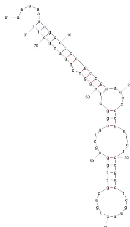
packaging/head assembly proteins of KS9 are similar to those encoded by PC184,E125, and1026b, but the major capsid protein itself is dissimilar (see Table 1 and Table S2 in the supplemental material). Each putative KS9 tail protein has a similar protein encoded by PC184,E125, and 1026b. As discussed above, although KS9 gene15is assumed to be part of the tail morphogenesis module because of its location, its pre-dicted product does not have an assigned function and so may not be involved in tail production. In many phage genomes, two proteins are encoded between the major tail protein and tail tape measure genes by way of a⫺1 translational frameshift (48). It is thought that frameshifting allows phages to control the relative expression levels of two proteins during infection (48). In KS9, these two proteins are gp11, a tail assembly chaperone, and gp12, a minor tail protein. These proteins are predicted to have the same N-terminal sequence, but gp12 (expressed via the frameshift) is predicted to be longer and have a different C terminus. We have found a putative frame-shift site between bases 20,347 and 20,353. This site contains a stretch of seven adenine residues that can cause the ribosome to slip backwards by one position, resulting in a⫺1 frameshift. Although AAAAAAA is not the canonical frameshift se-quence (XXXYYYZ, where Y is A or T), it is the same
sequence that was identified for phages such as c2 of
Lacto-coccusand PSA ofListeria monocytogenes(48). Providing
fur-ther support that this is the correct frameshift sequence, we have used the mfold program to identify a potential stem-loop structure formed by 65 downstream bases (50) (Fig. 5). Al-though such structures are not necessarily found at frameshift sites, their presence suggests a mechanism by which the ribo-some may stall and subsequently change its reading frame (48).
Analysis of KS9 lysis genes.KS9 encodes four proteins
pu-tatively involved in lysis, all of which are similar to proteins from PC184, but notE125 or 1026b (see Table S1 in the supplemental material). The genes encoding these proteins are part of a single module (Fig. 2). The first gene in the module, gene22, encodes a putative holin. Class I holins, such asS, have three transmembrane domains, whereas class II holins only have two (49). Because OCTOPUS analysis (46) indicates that gp22 has three transmembrane domains, we predict that this protein is a class I holin. Phage lysins may be one of three major types: endo--N-acetylglucosaminidases or
N-acetylmuramidases, endopeptidases, orN
-acetylmuramyl-L-alanine amidases (20). We predict that the KS9 lysin is an
[image:8.585.311.520.63.427.2]FIG. 4. Sequence of the KS9 attPoverlap region and attL/R in LMG 21824. The overlapping sequence present inattLandattRof the KS9 prophage and in the chromosome of the vegetative phage (virion) is underlined.
FIG. 5. Predicted stem-loop structure downstream of the putative KS9 gp11 translational frameshift sequence. Structure was determined by using mfold analysis of the 65 bases downstream of the AAAAAAA site (50).
on November 8, 2019 by guest
http://jvi.asm.org/
superfamily (E-value⫽2e ). The KS9 Rz/Rz1 pair is the last gene segment in the lysis module. Rz/Rz1 proteins function in lysis by joining the inner and outer membranes following holin insertion and lysin activity (5). The Rz1 gene,24⬘, is located out of frame in the Rz gene,24. Using the LipoP program (26), it is predicted that a signal peptidase II cleavage site is present between residues 22 (alanine) and 23 (cysteine) of gp24⬘. Cleavage at this site would produce a 65-amino-acid lipoprotein. Of these 65 amino acids, 7 are proline (10.8%), a value consistent with the proportion of proline found in the processed BcepMu (23.1%), SalMu (15.5%), and PhotoMu (13.4%) Rz1 lipoproteins (44).
Depending on the organism, the proteins similar to KS9 gp49 in the GenBank database have been annotated as either putative class I holins or as HNH homing endonucleases. If gp49 were to act as a class I holin, it would likely function with gp22 as part of a holin-antiholin pair. These systems are used to control when the onset of lysis occurs (28). However, OCTOPUS analysis of the gp49 sequence suggests that it has no transmembrane domains, so we predict that this protein is not a holin and that some other protein encoded by KS9 may be involved in controlling lysis timing. A Conserved Domain Search of the predicted gp49 sequence indicates that it belongs to the HNHc superfamily (E-value ⫽ 8e⫺6) found in HNH
homing endonucleases. Homing endonuclease genes (HEGs) are common in phage genomes. For example, T4 encodes 15 homing endonucleases, 6 of which (I-TevIII and MobA-E) belong to this family (34). HEGs are referred to as selfish genetic elements. They use a mechanism called homing to copy their sequence from one place in one gene to the same place in the same gene at a different locus (10). In order to complete this process, endonucleases (such as those in the HNH family) are used to cut the DNA at a specific 15- to 30-bp site, which is then fixed by using a double-strand break repair. Further experiments are required to determine whether gp49 has hom-ing endonuclease activity or whether it performs some other function during infection.
Contribution of the KS9 prophage to bacterial virulence.In
many bacterial species, prophage genes make significant con-tributions to the virulence of the organism. Although examples have been documented in a wide variety of Gram-negative organisms (includingVibrio cholerae,Escherichia coli,
Pseudo-monas aeruginosa, Neisseria meningitidis, Salmonella enterica,
andShigella flexneri), there is limited evidence that prophage
genes contribute to virulence inBurkholderiaspecies (8, 43). It has been suggested that becauseBurkholderiaare not strictly pathogenic and can instead survive in a variety of both terres-trial and aquatic environments, classical toxin genes may not have provided a strong evolutionary advantage to these species (43). Instead, lysogenic conversion genes of Burkholderia
prophages would be more likely to encode proteins that would increase the viability of the cell both in the environment andin vivo(43).
In the KS9 genome, we identified one gene, gene32, whose product may have an effect on the pathogenicity of LMG 21824 or a K56-2 KS9 lysogen. When the gp32 sequence was sub-jected to a Conserved Domain Search, the conserved domain “COG3950: predicted ATP-binding protein involved in
viru-nicity ofB. cenocepaciaK56-2, we compared the virulence of wild-type and KS9-lysogenized K56-2 (K56-2::KS9) in the
Gal-leria mellonellawax moth model. In this infection model, the
50% lethal dose (LD50) for K56-2 is 900 CFU (40). For both
K56-2 and K56-2::KS9, infection with between 104 and 106
CFU resulted in 100% mortality. There was no significant difference between the mortality of larvae infected with either K56-2 or K56-2::KS9 at any of the doses tested. Although gene
32contains a domain putatively involved in virulence, these data suggest that it has no effect on the pathogenicity of KS9 lysogens.
Construction and analysis of a KS9 lytic variant. The
method used to convert KS9 into a lytic phage was to inser-tionally inactivate the putative KS9 phage repressor, gene41. It is well established that repressor mutant phages cannot stably lysogenize (45). Platt et al. (37) constructed a strain ofE. coliexpressingcI from the chromosome and lysogenized this strain with a cI mutant, W30 (4). When this strain was cocultured withE. coli that was sensitive toinfection, the numbers of sensitiveE. colipresent decreased. They proposed that this system could be used to provide continuous delivery of lytic phagesin vivo. We wanted to expand on this concept in two ways, first by showing that a lytic mutant could be con-structed by using genetic engineering and second by showing that this mutant phage could be activein vivo.
Gene 41encodes a 133-amino-acid protein. According to BLASTP analysis, this protein shows 55% identity with gp52, the putative repressor protein ofE125 (E-value⫽4e⫺30), as
well as similarity to cI-like proteins of phages BP-4795, H-19B, SfV, cdtI, and a putative transcriptional repressor of phage
V10. A nonreplicating knockout plasmid (pKL1) was con-structed containing DNA flanking the KS9 gene41interrupted by a trimethoprim resistance cassette in a pALTER-1 (Tcr)
backbone. After transformation of this construct intoB.
pyrro-cinia LMG 21824, a Tcr TpR mutant (LMG 21824::pKL1,
generated by a single crossover) was isolated.
If a double-crossover event occurs that replaces the repressor gene with a TpR cassette, the cells should no longer be viable
because the prophage will be induced in the absence of the re-pressor protein, resulting in lysis. We used two different assays to assess if this induction was occurring in LMG 21824::pKL1. First, we screened 1800 TcrTpRLMG 21824::pKL1 colonies for loss of
tetracycline resistance and maintenance of trimethoprim resis-tance (i.e., a double crossover without loss of viability). No Tcs
TpR colonies were found, suggesting that the double-crossover
event was lethal due to prophage induction. Second, we com-pared the release of K56-2-infecting phages from LMG 21824, LMG 21824::pKL1, and LMG 21824 with a mutation in KS9 gene
32(LMG 21824 [KS932]). The number of phages released from LMG 21824::pKL1 was more than 10 times greater than the number released from the other two strains (Fig. 6), a finding consistent with phage induction occurring in LMG 21824::pKL1 following a double-crossover event. These two experiments pro-vide further epro-vidence that gene41 is the repressor, since it is required to maintain stable integration of the KS9 prophage.
Phages isolated from culture supernatants of LMG 21824:: pKL1 were screened for the presence of the TpR cassette.
Using primers flanking gene41, a PCR product of 603 bp was
on November 8, 2019 by guest
http://jvi.asm.org/
amplified with wild-type KS9, whereas a product of 832 bp was amplified from the mutated repressor phage with the TpR
cassette. A representative phage isolate that tested positive during PCR screening was named KS9c. This isolate showed no defects in activity and was able to produce confluent lysis of
B. cenocepacia K56-2 in an agar overlay at titers of 107/ml,
similar to that of wild-type KS9. The host range of KS9c was tested, and it was found to be identical to that of wild-type KS9, suggesting that there was no change in the susceptibility of Bcc strains to this phage.
To assess whether KS9c is able to stably integrate into K56-2, plates of K56-2 and KS9 or K56-2 and KS9c exhibiting confluent lysis were overlaid with water, and the surviving cells were isolated. Then, 150 bacterial isolates from KS9c lysates were replica plated onto solid medium with or with-out trimethoprim. These isolates were unable to grow on trimethoprim medium, suggesting that KS9c had not stably integrated into any of these bacteria. A total of 50 KS9-insen-sitive and 50 KS9C-insenKS9-insen-sitive K56-2 isolates were recovered from the phage lysates and were assayed for their (i) growth phenotype, (ii) trimethoprim resistance, (iii) susceptibility to KS9 and KS9c, and (iv) ability to amplify with primers de-signed to the K56-2/5⬘KS9 prophage junction. The majority of KS9-insensitive isolates (41 isolates, 82%) were not lysog-enized. These strains grew normally, were TpS, KS9RKS9cR
and were PCR negative for KS9 integration. Eighteen percent (9 isolates) were stably lysogenized. These strains grew nor-mally, were TpSKS9RKS9cR, and were PCR positive. A
dif-ferent distribution was observed for the KS9c-insensitive iso-lates. In this case, all 50 isolates (100%) were nonlysogenized. Like the nonlysogenized KS9-insensitive isolates, these strains grew normally, were TpSKS9RKS9cR, and were PCR negative
for KS9 integration. Although almost one-fifth of KS9-insen-sitive isolates were stably lysogenized, none of the KS9c-insen-sitive isolates showed this phenotype. Therefore, we have shown that inactivating prophage KS9 gene41results in the
loss of lysogen viability and increased prophage induction from LMG 21824 and prevention of stable lysogeny after K56-2 infection. Consequently, we conclude that gene41encodes the KS9 repressor protein and that KS9 is converted from a tem-perate to a lytic phage following the loss of this gene.
We chose to test the efficacy of KS9c in anin vivoalternative infection model. Seed and Dennis (39) showed thatG.
mel-lonellalarvae infected with twice the lethal dose of K56-2 could
be rescued after administration of phage KS12 at an MOI of 5,000. We tested the efficacy of treatment with endotoxin-free KS9c lysates in K56-2-infected larvae at MOIs between 0.5 and 100. Treatment with KS9c at MOIs of 50 or 100 increased the survival of infected larvae to that of uninfected controls (Fig. 7). This result is in sharp contrast to untreated infected larvae that had survival rates near zero. MOIs lower than 50 were ineffective, since the survival of larvae after treatment was comparable to that of untreated infected controls. In contrast to larvae treated with higher MOIs, those treated with MOIs lower than 50 that remained alive 48 h postinfection exhibited both reduced movement and signs of melanization, suggesting that the K56-2 infection had progressed but was not yet fatal. As a result, we conclude that phage treatment had little or no effect at the lower MOIs. This result is expected, since higher MOIs of lytic phages are generally more effective for thera-peutic purposes (6). Our results indicate that KS9c is active in
an in vivo model and can effectively treat experimental B.
cenocepaciaK56-2 infections when administered at an MOI of
50 or greater.
Because KS9c is unable to stably lysogenize K56-2 and fa-cilitate superinfection immunity, we predicted that KS9c might be a more effective therapeutic agent than KS9. We treated K56-2-infected larvae with either KS9 or KS9c at MOIs of 50, 5, and 0.5 and compared their ability to increase larval survival. In contrast to our prediction, we found no significant differ-ences between the survival of larvae treated with the two dif-ferent phages (Fig. 8). In order to assess why there was no
FIG. 6. Enumeration of K56-2-infecting phages released into the supernatant from 16-h overnight cultures of LMG 21824, LMG 21824 (KS9 32), and LMG 21824::pKL1. Cultures were pelleted by centrifugation at 10,000⫻gfor 2 min, and the supernatant was filter sterilized using a 0.45-m-pore-size filter and tested in a plaque assay with K56-2. This procedure was repeated three times for each strain.
on November 8, 2019 by guest
http://jvi.asm.org/
difference observed between the two treatments, we isolated K56-2 from the hemolymph of infected, KS9-treated larvae and screened these isolates for lysogeny. In contrast to thein
vitroscreening presented above where nearly 20% of
insensi-tive isolates were stably lysogenized, 0 of 50 KS9R KS9cR
K56-2in vivoisolates analyzed were lysogenized based on PCR screening. Because lysogeny does not appear to make a signif-icant contribution to KS9 resistance in the infected larvae at the MOI tested, this result explains why there was no signifi-cant difference in thein vivoefficacy of KS9 and KS9c.
[image:11.585.83.501.68.295.2]Differ-FIG. 7. Survival of K56-2-infected KS9c-treatedGalleria mellonellaat 48 h postinfection. Larvae were infected with twice the LD50of K56-2 and treated with KS9c at MOIs between 0.5 and 100. Control larvae were injected with either twice the LD50of K56-2 and MgSO4-ampicillin solution or undiluted KS9c and MgSO4-ampicillin solution. For each MOI, 10 larvae were infected and treated in three separate experiments.
FIG. 8. Survival of K56-2-infected and KS9- or KS9c-treatedGalleria mellonellaat 48 h postinfection. Larvae were infected with twice the LD50 of K56-2 and treated with KS9 or KS9c at an MOI of 50, 5, or 0.5. Control larvae were injected with either twice the LD50of K56-2 and MgSO4-ampicillin solution or undiluted KS9 or KS9c and MgSO4-ampicillin solution. For each MOI, 10 larvae were infected and treated in three separate experiments.
on November 8, 2019 by guest
http://jvi.asm.org/
[image:11.585.83.502.427.684.2]screening (100in vivoversus 0.1in vitro), which would tend to favor lysogeny. However, rapid infection, lysis and release of progeny phage would be predicted to increase the effective
MOIin vitromore so than in vivo, leading to a greater
pro-portion of lysogenized cells in the lysate than in the larvae (36). In summary, by knocking out the putative KS9 repressor gene, we have created a functionally lytic phage named KS9c. This phage does not stably lysogenize B. cenocepacia K56-2 and it is effective in treating experimental infections in theG.
mellonellamodel. Although it is unexpected that this phage is
only as effective as the wild-type phage in this model (as op-posed to more effective), we suggest that this result is due to the low level of lysogeny observed at the MOIs testedin vivo. We have determined the genome sequence of the siphovirus KS9, a prophage ofB. pyrrociniaLMG 21824. The KS9 proph-age integrates into both LMG 21824 andB. cenocepaciaK56-2 in the 3⬘end of the GTP cyclohydrolase II gene. It is 39,896 bp in length and encodes 50 genes, many of which show similarity to genes of the Burkholderia prophages E125 and 1026b and an uncharacterized prophage element ofB. cenocepacia
PC184. Although one gene was identified with a putative role in pathogenicity, KS9 integration was shown to have no effect on the virulence ofB. cenocepaciaK56-2 in theG. mellonella
model. We were able to show that gp41 encodes the phage lysogenic repressor by developing a lytic mutant of KS9, named KS9c. Unlike KS9, KS9c was unable to stably lysogenize K56-2. Treatment of K56-2-infectedG. mellonellalarvae with KS9c at MOIs of 50 or greater resulted in larva survival comparable to uninfected controls, indicating that KS9c is an effective anti-bacterial agentin vivo. As a proof-of-principle, we have shown that temperate phages can be genetically engineered to lytic form and that these engineered phages are activein vivo.
ACKNOWLEDGMENTS
This study was funded by an operating research grant to J.J.D. from the Canadian Cystic Fibrosis Foundation and funds from the Canadian Institutes of Health Research for the “CIHR Team on Aerosol Phage Therapy.” K.H.L. and K.D.S. are indebted to the Natural Sciences and Engineering Research Council of Canada for CGS-D and PGS-D scholarships, respectively.
We thank David DeShazer (U.S. Army Medical Research Institute for Infectious Diseases) for testing theB. pseudomalleiandB. mallei host ranges and Miguel Valvano (University of Western Ontario) for providing the B. cenocepaciaLPS mutants. We also thank Randy Mandryk, University of Alberta Microscopy Service Unit, and Pat Murray and Lisa Ostafichuk, University of Alberta Molecular Biology Service Unit, for their assistance.
REFERENCES
1.Ackermann, H.-W.2005. Bacteriophage classification, p. 67–89.InE. Kutter and A. Sulakvelidze (ed.), Bacteriophages: biology and applications. CRC Press, Boca Raton, FL.
2.Ackermann, H.-W.2001. Frequency of morphological phage descriptions in the year 2000. Arch. Virol.146:843–857.
3.Altschul, S. F., T. L. Madden, A. A. Scha¨ffer, J. Zhang, Z. Zhang, W. Miller, and D. J. Lipman.1997. Gapped BLAST and PSI-BLAST: a new generation of protein database search programs. Nucleic Acids Res.25:3389–3402. 4.Bassford, Jr., P. J., C. Bradbeer, R. J. Kadner, and C. A. Schnaitman.1976.
Transport of vitamin B12intonBmutants ofEscherichia coli. J. Bacteriol.
128:242–247.
5.Berry, J., E. J. Summer, D. K. Struck, and R. Young.2008. The final step in the phage infection cycle: the Rz and Rz1 lysis proteins link the inner and outer membranes. Mol. Microbiol.70:341–351.
7.Bottone, E. J., S. D. Douglas, A. R. Rausen, and G. T. Keusch. 1975. Association ofPseudomonas cepaciawith chronic granulomatous disease. J. Clin. Microbiol.1:425–428.
8.Boyd, E. F., and H. Bru¨ssow.2002. Common themes among bacteriophage-encoded virulence factors and diversity among the bacteriophages involved. Trends Microbiol.10:521–529.
9.Burkholder, W. H.1950. Sour skin, a bacterial rot of onion bulbs. Phytopa-thology40:115–117.
10.Burt, A., and V. Koufopanou.2004. Homing endonuclease genes: the rise and fall and rise again of a selfish element. Curr. Opin. Genet. Dev.14:609– 615.
11.Carver, T. J., K. M. Rutherford, M. Berriman, M.-A. Rajandream, B. G. Barrell, and J. Parkhill.2005. ACT: the Artemis comparison tool. Bioinfor-matics21:3422–3423.
12.Casjens, S.2003. Prophages and bacterial genomics: what have we learned thus far? Mol. Microbiol.49:277–300.
13.Coenye, T., P. Vandamme, J. J. LiPuma, J. R. W. Govan, and E. Ma-henthiralingam.2003. Updated version of theBurkholderia cepaciacomplex experimental strain panel. J. Clin. Microbiol.41:2797–2798.
14.Dale, J., and S. F. Park.2004. Molecular genetics of bacteria, 4th ed. John Wiley & Sons, Inc., Hoboken, NJ.
15.Dennis, J. J., and G. J. Zylstra. 1998. Plasposons: modular self-cloning minitransposon derivatives for rapid genetic analysis of gram-negative bac-terial genomes. Appl. Environ. Microbiol.64:2710–2715.
16.Dennis, J. J., and G. J. Zylstra.1998. Improved antibiotic-resistance cas-settes through restriction site elimination usingPfuDNA polymerase PCR. Biotechniques25:772–776.
17.DeShazer, D.2004. Genomic diversity ofBurkholderia pseudomalleiclinical isolates: subtractive hybridization reveals a Burkholderia mallei-specific prophage inB. pseudomallei1026b. J. Bacteriol.186:3938–3950.
18.Duff, A. J. A.2002. Psychological consequences of segregation resulting from chronicBurkholderia cepaciainfection in adults with CF. Thorax57:756–758. 19.Estrada-De Los Santos, P., R. Bustillos-Cristales, and J. Caballero-Mellado.
2001.Burkholderia, a genus rich in plant-associated nitrogen fixers with wide environmental and geographic distribution. Appl. Environ. Microbiol.67:
2790–2798.
20.Fischetti, V. A.2005. The use of phage lytic enzymes to control bacterial infections, p. 321–334.InE. Kutter and A. Sulakvelidze (ed.), Bacterio-phages: biology and applications, CRC Press, Boca Raton, FL.
21.Folsom, B. R., P. J. Chapman, and P. H. Pritchard.1990. Phenol and trichloroethylene degradation byPseudomonas cepaciaG4: kinetics and in-teractions between substrates. Appl. Environ. Microbiol.56:1279–1285. 22.Freifelder, D. M.1987. Molecular biology, 2nd ed. Jones and Bartlett
Pub-lishers, Portola Valley, CA.
23.Govan, J. R. W., P. H. Brown, J. Maddison, C. J. Doherty, J. W. Nelson, M. Dodd, A. P. Greening, and A. K. Webb.1993. Evidence for transmission of
Pseudomonas cepaciaby social contact in cystic fibrosis. Lancet342:15–19.
24.Hanlon, G. W.2007. Bacteriophages: an appraisal of their role in the treat-ment of bacterial infections. Int. J. Antimicrob. Agents30:118–128. 25.Isles, A., I. Maclusky, and M. Corey.1984.Pseudomonas cepaciainfection in
cystic fibrosis: an emerging problem. J. Pediatr.104:206–210.
26.Juncker, A. S., H. Willenbrock, G. Von Heijne, S. Brunak, H. Nielsen, and A. Krogh.2003. Prediction of lipoprotein signal peptides in gram-negative bac-teria. Protein Sci.12:1652–1662.
27.Kropinski, A. M.2006. Phage therapy: everything old is new again. Can. J. Infect. Dis. Med. Microbiol.17:297–306.
28.Kutter, E., R. Raya, and K. Carlson.2005. Molecular mechanisms of phage infection, p. 165–222.InE. Kutter and A. Sulakvelidze (ed.), Bacterio-phages: biology and applications, CRC Press, Boca Raton, FL.
29.Loutet, S. A., R. S. Flannagan, C. Kooi, P. A. Sokol, and M. A. Valvano.2006. A complete lipopolysaccharide inner core oligosaccharide is required for resistance ofBurkholderia cenocepaciato antimicrobial peptides and bacte-rial survival in vivo. J. Bacteriol.188:2073–2080.
30.Lukashin, A. V., and M. Borodovsky.1998. GeneMark.hmm: new solutions for gene finding. Nucleic Acids Res.26:1107–1115.
31.Lynch, K. H., and J. J. Dennis.2008. Assessment of the contribution of prophage KS9 protein gp32 to the virulence ofBurkholderia pyrrociniaLMG 21824, abstr. W14. Banff Conference on Infectious Diseases, Banff, Alberta. 32.Mahenthiralingam, E., T. Coenye, J. W. Chung, D. P. Speert, J. R. W. Govan, P. Taylor, and P. Vandamme.2000. Diagnostically and experimentally useful panel of strains from theBurkholderia cepaciacomplex. J. Clin. Microbiol.
38:910–913.
33.Merril, C. R., D. Scholl, and S. L. Adhya.2003. The prospect for bacterio-phage therapy in Western medicine. Nat. Rev. Drug Discov.2:489–497. 34.Miller, E. S., E. Kutter, G. Mosig, F. Arisaka, T. Kunisawa, and W. Ruger.
2003. Bacteriophage T4 genome. Microbiol. Mol. Biol. Rev.67:86–156. 35.Ortega, X., A. Silipo, M. S. Saldías, C. C. Bates, A. Molinaro, and M. A.
Valvano.2009. Biosynthesis and structure of theBurkholderia cenocepacia
on November 8, 2019 by guest
http://jvi.asm.org/
36.Payne, R. J. H., and V. A. A. Jansen.2001. Understanding bacteriophage therapy as a density-dependent kinetic process. J. Theor. Biol.208:37–48. 37.Platt, R., D. L. Reynolds, and G. J. Phillips.2003. Development of a novel
method of lytic phage delivery by use of a bacteriophage P22 site-specific recombination system. FEMS Microbiol. Lett.223:259–265.
38.Ren, J., M. Kotaka, M. Lockyer, H. K. Lamb, A. R. Hawkins, and D. K. Stammers.2005. GTP cyclohydrolase II structure and mechanism. J. Biol. Chem.280:36912–36919.
39.Seed, K. D., and J. J. Dennis.2009. Experimental bacteriophage therapy increases survival ofGalleria mellonellalarvae infected with clinically rele-vant strains of theBurkholderia cepaciacomplex. Antimicrob. Agents Che-mother.53:2205–2208.
40.Seed, K. D., and J. J. Dennis.2008. Development ofGalleria mellonellaas an alternative infection model for theBurkholderia cepacia complex. Infect. Immun.76:1267–1275.
41.Seed, K. D., and J. J. Dennis.2005. Isolation and characterization of bacte-riophages of theBurkholderia cepaciacomplex. FEMS Microbiol. Lett.251:
273–280.
42.Speert, D. P., D. Henry, P. Vandamme, M. Corey, and E. Mahenthiralingam.
of phages in the pathogenesis ofBurkholderia, or “Where are the toxin genes
inBurkholderiaphages?” Curr. Opin. Microbiol.10:410–417.
44.Summer, E. J., C. F. Gonzalez, T. Carlisle, L. M. Mebane, A. M. Cass, C. G. Savva, J. LiPuma, and R. Young. 2004.Burkholderia cenocepaciaphage BcepMu and a family of Mu-like phages encoding potential pathogenesis factors. J. Mol. Biol.340:49–65.
45.Susskind, M. M., and D. Botstein.1978. Molecular genetics of bacteriophage P22. Microbiol. Rev.42:385–413.
46.Viklund, H., and A. Elofsson.2008. OCTOPUS: improving topology predic-tion by two-track ANN-based preference scores and an extended topological grammar. Bioinformatics24:1662–1668.
47.Woods, D. E., J. A. Jeddeloh, D. L. Fritz, and D. DeShazer.2002.
Burkhold-eria thailandensisE125 harbors a temperate bacteriophage specific for
Burk-holderia mallei. J. Bacteriol.184:4003–4017.
48.Xu, J., R. W. Hendrix, and R. L. Duda.2004. Conserved translational frame-shift in dsDNA bacteriophage tail assembly genes. Mol. Cell16:11–21. 49.Young, R., I. N. Wang, and W. D. Roof.2000. Phages will out: strategies of
host cell lysis. Trends Microbiol.8:120–128.
50.Zucker, M.2003. Mfold web server for nucleic acid folding and hybridization prediction. Nucleic Acids Res.31:3406–3415.