MICRO COMPUTED TOMOGRAPHIC EVALUATION OF
CANAL TRANSPORTATION AND CENTERING ABILITY OF
PROTAPER UNIVERSAL, HYFLEX EDM AND WAVEONE
GOLD - AN
INVITRO STUDY
Dissertation submitted to
THE TAMILNADU Dr. M.G.R. MEDICAL UNIVERSITY
In partial fulfillment for the Degree of
MASTER OF DENTAL SURGERY
BRANCH IV
ACKNOWLEDGEMENT
I take this opportunity to sincerely thank my post graduate teacher and my guide Dr. B. Veni Ashok, M.D.S., Professor, Department of Conservative Dentistry and Endodontics, Ragas Dental College and Hospital, for his perseverance in motivating, guiding and supporting me throughout my study period.
My sincere thanks to Dr. R. Anil Kumar, M.D.S., Professor and Head, Department of Conservative Dentistry and Endodontics, Ragas Dental College and Hospital, who has helped me with his advice and immense support throughout my post graduate curriculum.
My sincere thanks to Dr. S. Ramachandran, M.D.S., Professor & former Principal and Dr. R. Indira, M.D.S.,Professor and former HOD, Department of Conservative Dentistry and Endodontics, Ragas Dental College and Hospital, who helped me with their advice and immense support throughout their tenure.
I extend my sincere thanks to Dr. P. Shankar, M.D.S., Professor,
Ragas Dental College and Hospital, for his guidance, and constant encouragement during the completion of my study.
I extend my sincere thanks to Dr. C.S. Karumaran, M.D.S., Professor,
I extend my sincere thanks to Dr. M. Rajasekaran, M.D.S., Professor,
for his guidance and constant encouragement throughout the completion of this work.
My sincere thanks toDr. Shankar Narayan, M.D.S., Dr. S.M. Venkatesan, M.D.S., Readers Department of Conservative Dentistry and Endodontics, Ragas Dental College and Hospital, who have helped me with their guidance, support and constant encouragement throughout my study period wherever and whenever needed.
I would like to solemnly thank Dr. Sabari, M.D.S., Dr. Arrvind Vikram, M.D.S., Dr. B. Venkatesh M.D.S, Readers for all the help during my study period.
I would also like to thank Dr. C. Nirmala M.D.S, Senior lecturer for her friendly guidance and support.
I also wish to thank the management of Ragas Dental College and Hospital, Chennai for their help and support.
My sincere thanks to SASTRA UNIVERSITY for providing me with Micro-CT facility.
My sincere thanks to Dr. Bijivin for his guidance in biostatistics and also for the moral support.
I sincerely thank, Dr. Aravind for his constant support and encouragement throughout my study.
I remain ever grateful to all my batchmates, juniors and friends for their support.
I would like to thank my parents, for their support and encouragement throughout these years without which, I would not have never reached so far.
My sincere thanks to Mr.K.Thavamani for his guidance and support in DTP and Binding works.
CONTENTS
S. NO. INDEX PAGE NO.
1. INTRODUCTION 1
2. AIM AND OBJECTIVES 7
3. REVIEW OF LITERATURE 8
4. MATERIALS AND METHODS 27
5. RESULTS 33
6. DISCUSSION 36
7. SUMMARY 58
8. CONCLUSION 59
9. BIBLIOGRAPHY 60
LIST OF TABLES
TABLE
No TITLE
1 PROTAPER GROUP: READING AT 3MM, 6MM AND 9MM- PRE-INSTRUMENTATION AND POST-PRE-INSTRUMENTATION
2 HYFLEX EDM GROUP: READING AT 3MM, 6MM AND 9MM- PRE-INSTRUMENTATION AND POST-INSTRUMENTATION
3
WAVEONE GOLD GROUP: READING AT 3MM, 6MM AND 9MM- PRE- INSTRUMENTATION AND
POST-INSTRUMENTATION
4 CANAL TRANSPORTATION AND CENTERING RATIO VALUES
OF PROTAPER GROUP AT 3MM, 6MM AND 9MM
5 CANAL TRANSPORTATION AND CENTERING RATIO VALUES
OF HYFLEX EDM GROUP AT 3MM, 6MM AND 9MM
6 CANAL TRANSPORTATION AND CENTERING RATIO VALUES
OF WAVEONE GOLD GROUP AT 3MM, 6MM AND 9MM
7 COMPARISON OF CANAL TRANSPORTATION AT 3MM
AMONG PROTAPER GROUP, HYFLEX EDM GROUP AND WAVEONE GOLD GROUP
8
COMPARISON OF CANAL TRANSPORTATION AT 6MM AMONG PROTAPER GROUP, HYFLEX EDM GROUP AND WAVEONE GOLD GROUP
9
[image:9.612.86.517.149.680.2]10
PAIRWISE COMPARISON OF CANAL TRANSPORTATION WITH ALL GROUPS AT 3 MM, 6 MM AND 9 MM USING POST HOC TESTS – TUKEY HSD
11
COMPARISON OF CANAL CENTERING ABILITY AT 3MM AMONG PROTAPER GROUP, HYFLEX EDM GROUP AND WAVEONE GOLD GROUP
12
COMPARISON OF CANAL CENTERING ABILITY AT 6MM PROTAPER GROUP, HYFLEX EDM GROUP AND WAVEONE GOLD GROUP
13
COMPARISON OF CANAL CENTERING ABILITY AT 9MM PROTAPER GROUP, HYFLEX EDM GROUP AND WAVEONE GOLD GROUP
14
LIST OF GRAPHS
GRAPH NO.
TITLE
1 COMPARISON OF THREE GROUPS WITH RESPECT TO CANAL
TRANSPORTATION AT 3MM
2 COMPARISON OF THREE GROUPS WITH RESPECT TO CANAL
TRANSPORTATION AT 6MM
3 COMPARISON OF THREE GROUPS WITH RESPECT TO CANAL
TRANSPORTATION AT 9MM
4 COMPARISON OF THREE GROUPS WITH RESPECT TO CANAL CENTERING RATIO AT 3MM
5 COMPARISON OF THREE GROUPS WITH RESPECT TO CANAL CENTERING RATIO AT 6MM
LIST OF FIGURES
FIGURE
NO. TITLE
1 EXTRACTED MANDIBULAR MOLAR TEETH
2 DECORONATED MANDIBULAR MESIAL ROOTS
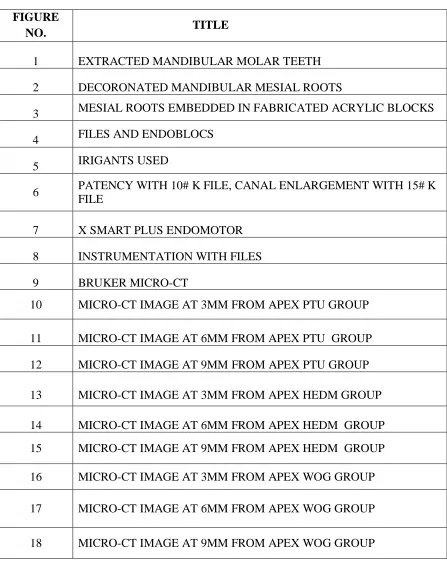
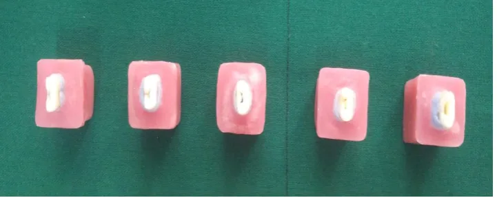
3 MESIAL ROOTS EMBEDDED IN FABRICATED ACRYLIC BLOCKS
4 FILES AND ENDOBLOCS
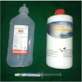
5 IRIGANTS USED
6 PATENCY WITH 10# K FILE, CANAL ENLARGEMENT WITH 15# K FILE
7 X SMART PLUS ENDOMOTOR
8 INSTRUMENTATION WITH FILES
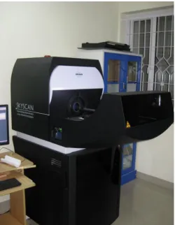
9 BRUKER MICRO-CT
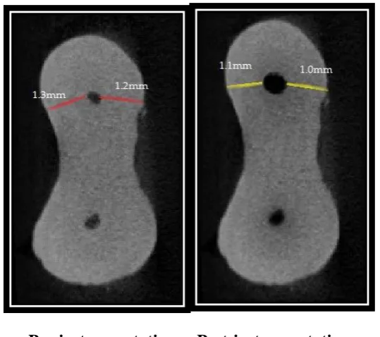
10 MICRO-CT IMAGE AT 3MM FROM APEX PTU GROUP
11 MICRO-CT IMAGE AT 6MM FROM APEX PTU GROUP
12 MICRO-CT IMAGE AT 9MM FROM APEX PTU GROUP
13 MICRO-CT IMAGE AT 3MM FROM APEX HEDM GROUP
14 MICRO-CT IMAGE AT 6MM FROM APEX HEDM GROUP
15 MICRO-CT IMAGE AT 9MM FROM APEX HEDM GROUP
16 MICRO-CT IMAGE AT 3MM FROM APEX WOG GROUP
17 MICRO-CT IMAGE AT 6MM FROM APEX WOG GROUP
ABBREVIATIONS USED
S.NO ABBREVIATIONS
1 NiTi Nickel Titanium
2 HEDM Hyflex EDM
3 WOG WaveOne GOLD
4 PTU ProTaper Universal
5 NaOCL Sodium hypochlorite
6 EDTA Ethylenediamine tetraacetic acid
7 CM Control memory
8 > Greater than
9 < Less than
10 mm Millimeter
11 Micro-CT Micro-Computed Tomography
12 # Size
13 % Percentage
14 EDM Electric discharge machining
15 CT Canal transportation
1
INTRODUCTION
Success of endodontics is mainly attributed by the following three
basic steps- mechanical preparation, disinfection and a three dimensional
obturation of root canal system. Chemomechanical preparation is a critical
step in endodontic procedure. The main purpose of root canal instrumentation
is to clean and shape the root canal system and to allow the placement of a
fluid tight seal, hence influencing the outcome of the treatment.91
Schilder emphasized that the root canal should have a continuously
tapering conical form, from access cavity till the apex, with the narrowest
cross sectional diameter at the apex and the largest at the orifice preserving the
apical foramen and not altering the original canal curvature as this is the most
appropriate canal shape for irrigation and obturation.92
However, these objectives are difficult to achieve because of the highly
variable root canal anatomy. Achieving a proper taper in a curved canal
becomes difficult as there are chances of canal transportation and loss of
centering ability. Excessive dentin removal in single direction within the canal
rather than in all directions equidistantly from the main tooth axis causes what
is known as canal transportation which is an undesirable deviation from
natural canal path and centering ability is the ability of the instrument to stay
2
Deviation from the original canal curvature can lead to: 31
1. Excessive and inappropriate dentin removal.
2. Straightening of the canal and creation of a ledge in the dentinal wall.
3. A defect known as an elbow which forms coronal to the elliptical –
shaped apical seal.
4. Canal with hourglass appearance.
5. Over – preparation that weakens the tooth, resulting in fracture of the
root.
Traditionally, inflexible stainless steel instruments have been used for
root canal instrumentation. But, such instruments have a tendency to cause
number of procedural errors like apical transportation, zip and ledge formation
can occur along with loss of working length.3
To overcome these, significant amount of research have taken place
which resulted in the introduction of NiTi instruments by Walia et al. (1988)
who employed a nickel–titanium arch wire to fabricate a root canal file. NiTi
is well known for its superior characteristics of super elasticity and shape
memory.The mechanical properties of NiTi endodontic instruments including
flexibility, torsional resistance, and flexural fatigue are fundamental
requirements of endodontic instruments for successful outcome. NiTi
instruments have progressively undergone several changes over the decades
3
NiTi systems differ from one another in the design of cutting blades, taper, tip
configuration.70
Despite these improvements, file separation during root canal
preparation in continuous rotation motion (Barbakow & Lutz1997) is one of
main drawback. Although many factors play a role in NiTi file failures, two of
the most important reasons are cyclic and torsional fatigue.82
Cyclic fatigue failure occurs because of the alternating
tension/compression cycles that instruments are subjected to when flexed in
the region of maximum curvature of the canal and torsional fatigue occurs in
narrow straight canal when file tip binds to the canal and shaft continues to
rotate.88
To avoid or decrease the incidence of instrument separation newer
kinds of NiTi endodontic files fabricated by proprietary thermomechanical
processes such as M-wire files, Controlled memory (CM) files , and R-phase
wire have been introduced and have shown improved flexibility and cyclic
fatigue resistance compared to traditional NITI files like ProTaper Universal.86
Several researchers advocated reciprocating motion instead of the
continuous rotation motion to increase cyclic fatigue resistance.52
Reciprocating motion has an advantage for the preparation of curved canal and
maintaining the original form of the canal while shaping the root canal.52 Two
of these single-file systems, Reciproc (VDW, Munich, Germany) and
4
reciprocating motion and are made of a special NiTi alloy (M-Wire) to
increase flexibility and improve cyclic fatigue resistance of the instrument.48
Furthermore to increase the durability and strength of the instrument
has led to the introduction of novel technologies like Electric discharge
machining (EDM) and Gold treatment in endodontic field for the first time.
HyFlex EDM (HEDM; Coltene/Whaledent AG, Altstaetten,
Switzerland) is the recently introduced file system in the market which is
manufactured by EDM process. The spark erosion caused by this EDM
process hardens the surface of NiTi file, which results in superior fracture
resistance and improved cutting efficiency. HEDM NiTi files are
manufactured from Controlled Memory alloy technology like the HyFlex CM
(Coltene/Whaledent AG) NiTi files. HEDM has file shaft with 3 different
cross sections: triangular in the coronal third, trapezoidal in the middle third,
and almost quadratic in the apical third.48
WaveOne GOLD is the first file system to manufacture by GOLD
treatment process. WaveOne GOLD (WOG, Dentsply Maillefer) is modified
from WaveOne and as same movement kinematics like Wave One, but the
cross section of the file has been modified to the parallelogram structure with
two cutting edges in order to make the file more flexible. The most
importantly WaveOne GOLD is made from GOLD alloy technology, which is
based on heating the file and then slowly cooling it whereas WaveOne is made
5
manufacturer claims that the flexibility of files is improved through this new
heat treatment method.48
Shaping ability of endodontic files can be evaluated by various
methods such as reassembly technique (Bramante et al. 1987, hulsmann et al
1999, kuttler et al 2001), radiographic comparisons (southward et al. 1987,
jardine and gulabivala 2000) and silicone impressions of canals (goldman et al
1989). These methods are limited as they are two dimensional, not accurate
and invasive. Newer mode of investigation like Computed tomography, spiral
CT AND Cone Beam Computed tomography allow a noninvasive and
reproducible three dimensional evaluation of the external and internal
morphology of the tooth67.
Emergence of Micro-computed Tomography (Micro-Ct) has opened
up new possibility in the field of endodontics. The micro-CT is a
non-destructive and noninvasive method to obtain two- and three-dimensional
images. This technology has been widely applied to evaluate root canal
anatomy, techniques and materials related to the endodontic treatment. Classic
feature of micro-CT is that it allow the use of the same sample for different
tests without destruction of the sample which is very important particularly
when is required to evaluate volume pre and post instrumentation, quality of
root canal obturation or removal of the material from root canal (retreatment)
in high resolution than clinical scanners. Other advantages of micro-CT are the
possibility of repeated scanning and the manipulation of image using specific
6
The purpose of this invitro study was to evaluate the canal
transportation and centering ability of three rotary systems namely ProTaper
Universal (full sequence rotary system), Hyflex EDM (continuous rotation
motion, control memory wire and EDM technology), and WaveOne GOLD
(single file system, reciprocating motion, Gold heat treatment procedure) in
the preparation of curved mesial root canals of mandibular molars using
micro-CT imaging.
7
AIM AND OBJECTIVES
AIM:
The aim of the present invitro study was to determine and compare the
canal transportation and centering ability of ProTaper Universal, Hyflex EDM
and WaveOne GOLD using Micro-CT.
OBJECTIVES:
1. To determine and compare the canal transportation in curved
mesiobuccal canal of mandibular first molars of ProTaper Universal, Hyflex
EDM and WaveOne GOLD using micro-CT.
2. To determine and compare the centering ability in curved
mesiobuccal canal of mandibular first molars of ProTaper Universal, Hyflex
8
REVIEW OF LITERATURE
Schilder(1974)92 discussed the cleaning and shaping of root canals in
the symposium on endodontics. He suggested that the apical portion of curved
canals is best manipulated with in and out strokes of the files that have been
precurved to simulate the curve of the root canals. Reaming action in the
apical third of curved canals should be avoided as it can cause undesirable
reverse flow to that portion, in the canals and tend to cause instrument
breakage. He pointed out that a tapering funnel preparation is required for
cleaning the root canal system effectively and to permit the compaction of
gutta percha with either vertical or lateral forces.
Weine et al (1975)76 studied the effect of preparation procedures on
original canal shape and on apical foramen. Simulated canals in acrylic blocks
were used for first time in his study. The study showed that following routine
preparation in a curved canal, S shape which was not a tapering funnel from
orifice to the apex was produced. Instead the prepared canal funnelled down
to a site short of the apex, which they called the elbow, and then widened
towards the apex, which was called the zip.
Weine et al (1976)77 studied the effect of the preparation with hand
instrumentation and endodontic hand pieces on original canal shape simulated
resin blocks. The more that any instrument was rotated within a canal with a
9
between the curve and the tip of the preparation. According to them, the
canals in resin blocks were exactly the same, though the preparation was not
identical to in vivo preparation. Further the study showed that canals with
sharp curvatures, mechanical hand pieces and reaming action with hand
instruments created wider zips, than hand instrumentation with flaring and
removal of flutes.
Gambill JM et al (1996)20compared the nickel titanium and stainless
steel instrumentation using computed tomography in thirty six single rooted
teeth. They found that Ni-Ti instruments used in a reaming technique caused
significantly less canal transportation(p< 0.05), removed significantly less
volume of dentin (p<0.05), required less instrumentation time (p<0.05), and
produced more centered and rounder canal preparations than k-flex stainless
steel files used in a quarter turn/pull technique.
Wu et al (2000)78 studied the occurrence of apical transportation (AT) complicating the root filling procedure and resulting in a compromised seal.
In part I of their study, human mandibular premolars with single, curved or
straight canals were prepared by Lightspeed or a step-back hand filing
technique. An AT index was determined using a double exposure radiographic
technique. The prepared canals were obturated using lateral condensation of
gutta percha. Leakage along the apical 3 mm of root filling was measured with
a fluid transport model. After hand filing, AT and perforation occurred in
10
Lightspeed preparation, AT occurred in only 19% of the curved canals. The
hand filing/curved group leaked statistically significant more than the hand
filing/straight and Lightspeed/curved groups (p = 0.002). It was concluded that
the occurrence of AT was a factor that negatively influences
the apical seal when curved canals were obturated by lateral condensation of
gutta-percha
Peters OA et al (2001)53 assessed effects of four Ni–Ti preparation techniques on root canal geometry by using micro computed tomography. It
was found that all instrumentation techniques left 35% or more of the canals
surface area unchanged.
Peters OA et al (2003)52 evaluated the performance of ProTaper instruments in shaping root canals of extracted human maxillary molars using
micro-CT. Teeth samples were scanned, before and after shaping with
ProTaper. Canals were three-dimensionally reconstructed and evaluated for
volume, surface area, canal transportation and prepared surface. Authors
found that volume and surface area increased significantly and gross
preparation errors were found infrequently. Apical canal transportation
ranged from 0.02 to 0.40 mm and was independent of canal type and ‘wide’
canals had a significantly higher (P < 0.05) proportion of unprepared surfaces
than ‘constricted’ canals. It was concluded that canals in maxillary molars
were prepared invitro using ProTaper instruments without major procedural
11
Paque et al (2005)49 evaluated the performance of Endo-Eze Anatomic Endodontic Technology (AET) stainless steel instruments in
shaping maxillary molar root canals employing micro-CT. They found that
Endo-Eze AET instruments shaped root canals in maxillary molars with
substantial canal transportation, particularly in mesiobuccal root canals.
Preparation with this instrument removed high volumes of dentine, even
though apical preparation was size 30. Based on the results, it was concluded
that Endo-Eze AET cannot be recommended for the preparation of teeth with
curved root canals.
Javerhi et al (2007)29 compared the apical transportation and changes in canal curvature of Hero 642, RaCe, and ProTaper in mesiobuccal canals of
60 maxillary first molars using a radiographic platform. Statistically
significant difference was found in ProTaper group. They suggested that
ProTaper file system should be implemented in combination with other less
tapered more flexible systems, like RaCe, in preparing curved canals.
Bonaccorso et al (2008)7 compared the shaping ability of ProTaper, Mtwo, BioRaCe, and BioRaCe + S-Apex instruments in simulated canals with
an S-shaped curvature. It was found that ProTaper instruments caused more
pronounced canal transportation in the apical curvature (P < .01) than all other
instruments. They concluded by saying that NiTi systems including less
tapered and more flexible instruments like S-Apex seem to be useful when
12
Versiani MA et al (2008)74 evaluated the influence of shaft design on the shaping ability of ProTaper, ProFile, and ProSystem GT rotary
instruments in sixty curved mesial canals of mandibular molars. It was
concluded that all instruments were able to shape curved mesial canals in
mandibular molars to size 30 without significant errors. The differences in
shaft designs seemed not to affect their shaping capabilities.
Lopez et al (2008)38 studied the level of apical transportation in mesiobuccal roots of upper molars after manual instrumentation with stainless steel files, preparation with the K3 system, and with a reciprocating NSK handpiece. They found that the stainless steel file sizes #35 and #40 caused significant apical transportation, and K3 system proved safe for apical preparation, with little deviation.
Vaudt J et al (2009)72 investigated instrumentation time, working safety and the shaping ability of two rotary nickel– titanium (NiTi) systems
(Alpha System and ProTaper Universal) in comparison to stainless steel hand
instruments in mesial root canals of mandibular molars. They found that
instrumentation time of the Alpha System was significantly reduced compared
with ProTaper Universal and hand instrumentation. The Alpha System
showed significantly less apical straightening compared with the other
instruments. In the apical cross-sections, Alpha System resulted in
significantly less uninstrumented canal walls compared with stainless steel
13
systems, an apical straightening effect could not be prevented; areas of
uninstrumented root canal wall were left in all regions using the various
systems.
Yin X et al (2010)81 assessed the efficacy of ProTaper rotary system and traditional instruments by using micro–computed tomography (micro-CT)
in mandibular molars with C-shaped canals. They observed that ProTaper
rotary system maintained the canal curvature with few procedural errors,
whereas traditional instrumentation can clean more canal surface.
Gergi R et al (2010)21 compared canal transportation and centering ability of 2 rotary nickel-titanium (NiTi) systems (Twisted Files [TF] and
Pathfile-ProTaper [PP]) with conventional stainless steel K-files in ninety root
canals with severe curvature and short radius. Amount of transportation and
centering ability were assessed. They found that less transportation and better
centering ability occurred with TF rotary instruments (P < .0001). K-files
showed the highest transportation followed by PP system. PP system showed
significant transportation when compared with TF (P < .0001).
Yang G et al (2011)79 studied the effects of Mtwo and ProTaper Universal (PTU) on root canal geometry three-dimensionally by using micro–
CT in 20 canals. The parameters evaluated were canal surface area, volume,
structure model index, thickness, straightening, canal transportation, and
uninstrumented surface area. They found that the preparation significantly
changed canal surface area, volume, structure model index, thickness, and
14
types concerning these parameters and uninstrumented surface area. The
canals prepared with PTU showed larger values of transportation compared
with those in Mtwo group at the apical third. It was concluded that both of the
instrumentation systems produced canal preparations with adequate geometry.
PTU produced larger transportation at apical third.
Ounsi HF et al (2011)46 studied the shaping capacity of NiTi rotary instruments by photographic or micro–computed tomography (micro-CT)
measurements. Ten new sets of ProTaper Universal instruments were used in
60 resin blocks simulating curved root canals. Groups 1 to 6 (n = 10)
represented the first to sixth use of the instrument, respectively. Digitized
images of the prepared blocks were taken in both mesiodistal (MD) and
buccolingual (BL) directions and area measurements were calculated using
AutoCAD (Autodesk Inc, San Rafael, CA). They found significant differences
between groups (P < .001). Regarding measurement type, there were no
significant differences between BL and MD measurements, but there were
significant differences between micro-CT and BL measurements (P < .001)
and micro-CT and MD measurements(P = .001). Significant differences were
also noted between uses sand concluded that canal preparations are
significantly smaller after the third use of the same instrument.
You SY et al (2011)83 studied the shaping ability of reciprocating motion and continuous rotation motion in curved root canals. The
mesiobuccal and distobuccal canals of maxillary molars were instrumented
15
group were prepared by using continuous rotation with pecking motion,
whereas the canals in the reciprocating motion (RM) group were prepared
with reciprocating motion (clockwise 140 degrees and counter clockwise 45
degrees).They found that the application of reciprocating motion during
instrumentation did not result in increased apical transportation when
compared with continuous rotation motion, even in the apical part of curved
canals. It was concluded that reciprocating motion might be an attractive
alternative method to prevent procedural errors during root canal shaping.
Stern et al (2012)67 compared the centering ability and the shaping ability of ProTaper (PT) files used in reciprocating motion and PT and
Twisted Files (TF) used in continuous rotary motion using micro-computed
tomography in sixty mesial canals of thirty mandibular molars. They found no
difference in the transportation and centering ratio between the techniques and
no significant difference between the times of instrumentation and concluded
ProTaper files used in reciprocating motion and PT and TF used in continuous
rotary motion were capable of producing centered preparations with no
substantial procedural errors.
Pasqualini D et al (2012)50 compared the ability of manual and mechanical glide path to maintain the original root canal anatomy in first
permanent molars using Micro CT. It was confirmed that NiTi rotary PathFile
instruments preserve the original canal anatomy and caused less canal
16
Alves Vde et al (2012)2 evaluated the occurrences of apical transportation and canal aberrations produced with different instruments used
to create a glide path in the preparation of curved root canals, namely manual
K-files and PathFile and Mtwo nickel-titanium rotary files and concluded
that neither the manual instruments nor the PathFile or Mtwo rotary
instruments used to create a glide path had any influence on the occurrence of
apical transportation or produced any canal aberration.
Zhao D et al (2013)85 studied the canal shaping properties of Hyflex CM, Twisted Files (TF), and K3 rotary nickel-titanium files by using micro–
computed tomography in maxillary first molars. They found that Hyflex CM
and TF instruments shaped curved root canals in maxillary first molar without
significant shaping errors.
Elsherief et al (2013)19 compared the effects of ProTaper, Revo-S and Hero Shaper in preparing the curved root canals using cone-beam computed
tomography in human mandibular molars. They found that all instruments
maintained the original canal curvature well with no significant differences
between the different files. It was concluded that all instruments maintained
the original canal curvature well and were safe to use. Areas of
uninstrumented root canal wall were left in all regions using the various
systems.
Talati A et al (2013)69 compared the shaping ability of Mtwo, RaCe and Medin rotary instruments during the preparation of curved root canals in
post-17
instrumentation radiographs, the straightening of the canal and the apical
transportation were determined with AutoCAD software. It was found that
Mtwo instruments to be superior to the other rotary instruments.
Kumar B S et al (2013)33 compared canal transportation and centering ability of Twisted and Hyflex Rotary Files with stainless steel hand
k-flexofiles by using Spiral Computed Tomography in 90 extracted human
mandibular single rooted Premolar teeth. All the teeth were scanned before
and after instrumentation by using Spiral Computed Tomography. K-files
showed highest transportation and less centered when compared to the
Twisted and Hyflex rotary files. No significant difference was found between
TF and Hyflex CM instruments.
Maitin et al (2013)41 analysed the shaping ability of protaper, k3, RaCe and Mtwo using spiral computed tomography (CT) in eighty freshly
extracted human mandibular first molars. They found that canals prepared
with ProTaper had more canal transportation at all the three levels of root
canal (coronal, middle and apical third). Canals prepared with Mtwo were
well centered at coronal and middle third whereas, with RaCe canals were
centered only at the apical third.
Capar et al (2014)10 compared the effects of OneShape, ProTaper Universal F2, ProTaper Next X2, Reciproc (R), Twisted File Adaptive (TFA),
and WaveOne primary rotary systems on transportation, canal curvature,
centering ratio, surface area, and volumetric changes of curved mesial root
18
imaging. They found that the 6 different file systems straightened root canal
curvature similarly and produced similar canal transportation in the
preparation of mesial canals of mandibular molars. Reciproc instrumentation
exhibited superior performance compared with the OS, TFA, and PU systems
with respect to volumetric change.
Nazari Moghadam et al (2014)43 studied the canal transportation and centering ability of Twisted File (TF) to that of Reciproc system using CBCT
and found that both file systems were able to keep the original curvature of the
canal and thus can be considered safe for clinical application.
Elnaghy AM et al (2014)18 evaluated and compared the volume of removed dentin, transportation, and centering ability of ProTaper Next (PTN)
system with and without glide path preparation by using cone beam computed
tomography (CBCT) imaging. They found that there was no significant
difference among the tested groups regarding the volume of removed dentin
and centering ratio. It was concluded that PG/PTN instrumentation method
revealed better performance with fewer canal aberrations when compared with
instrumentation performed with PF/PTN or PTN only.
Zhao D et al (2014)84 studied the canal shaping properties of ProTaper Next, ProTaper Universal, and WaveOne in mandibular first molars by using
micro–computed tomographic (micro-CT) scanning. Canals were prepared
with PTU, PTN, and WaveOne systems under hypochlorite irrigation. The
volume of the untreated canal; the volume of dentin removed after
19
the coronal, middle, and apical thirds of canals were measured. They found
that PTN, PTU, and WaveOne instruments shaped root canals in mandibular
first molars without significant shaping errors. The curved canals prepared
using PTN had less apical transportation than the canals prepared using
WaveOne and PTU.
Hwang YH et al (2014)26 compared the shaping ability of Mtwo, a conventional nickel-titanium file system, and Reciproc, a reciprocating file
system. Root canal shaping was performed on the mesiobuccal and
distobuccal canals of extracted maxillary molars. In the RR group (n = 15),
Reciproc was used in a reciprocating motion; in the MR group, Mtwo was
used in a reciprocating motion; and in the MC group, Mtwo was used in a
continuous rotating motion. Micro–computed tomographic images taken
before and after canal shaping were used to analyse canal volume change and
the degree of transportation at the cervical, middle, and apical levels. The time
required for canal shaping was recorded. It was concluded that in terms of
shaping ability, Mtwo used in a reciprocating motion was not significantly
different from the Reciproc system.
Reddy PJ et al (2014)55 compared the shaping ability of two different rotary Nickel –Titanium (Ni-Ti) files, One shape file and Twisted files in a
simulated artificial canals. It was found that the twisted files shaped the canals
20
Saber et al (2015)57 compared the shaping ability of ProTaper Next, iRaCe and Hyflex CM rotary NiTi files during the preparation of severely
curved root canals in extracted human molar teeth. They found that use of
PTN resulted in significantly greater canal straightening than IR and HF(P <
0.05), with no significant differences between IR and HF (P > 0.05). There
were no significant differences between the three groups with respect to apical
transportation (P > 0.05). IR and HF were significantly faster than PTN (P <
0.05), with no significant differences between IR and HF (P > 0.05). It was
concluded that all three files were safe to use in curved canals.
Burklein et al (2015)8 compared the shaping ability of Mtwo, ProTaper Universal, ProTaper NEXT and BT-RaCe instruments during the
preparation of curved root canals in extracted teeth. Using pre- and
post-instrumentation radiographs, straightening of the canal curvatures and canal
transportation were determined with a computer image analysis programme.
Preparation time and instrument failures were also recorded. It was found that
all instruments maintained root canal curvature well and were safe. However,
care should be taken when using the BT-RaCe instrument due to its unique
cylindrical design.
Marceliano-Alves et al (2015)42 investigated the changes in three-dimensional geometry and the centering ability of root canals prepared with
Reciproc, WaveOne, Twisted File and HyFlex CM systems using
Micro-computed tomographic imaging technology in mesial canals of mandibular
21
maintain the original canal anatomy with less canal transportation than
Reciproc and WaveOne; however, these differences were unlikely to be of
clinical significance.
Deepak J et al (2015)16 compared the effects of fifth generation rotary systems [OneShape (OS), ProTaper Next (PTN), Revo S (RS)] on canal curvature, transportation and centering ratio of curved mesial root canals of
mandibular molar via cone-beam computed tomographic (CBCT) imaging.
They found that all file systems that were used, straightened the root canal
curvature similarly. RS instrumentation exhibited superior performance
compared with the OS and PTN systems with respect to transportation and
centering ratio.
Hiran-us et al (2015)22 studied the shaping ability of ProTaper Universal, ProTaper NEXT and iRace endodontic file systems by comparing
three parameters: canal deviation, apical foramen position and instrumentation
time. Pre- and post-operative images were superimposed to determine any
canal deviation or change in apical foramen position. The instrumentation
times were recorded. They found that iRace system resulted in the least mean
canal deviation and the iRace system also required the least instrumentation
time. The iRace system demonstrated the most favourable shaping ability in
all three parameters.
Agarwal RS et al (2015)1 compared the canal transportation, centering ability, and the time taken for preparation of curved root canals after
22
tomography (CBCT) in sixty mesiobuccal canals of mandibular molars. They
found that there was minor difference between the tested groups. Single file
systems demonstrated average canal transportation and centering ability
comparable to full sequence Protaper system in curved root canals.
Uzunoglu E et al (2015)71 studied apical transportation and centering ratios in curved root canals, which were instrumented with ProTaper Next up
to size X3 and with OS up to OSA 1 in forty-eight mesial canals of
mandibular molars. Apical transportation was assessed pre- and post-
instrumentation using cone-beam computed tomographic (CBCT) scans of 1,
2, 3, 4, and 5 mm sections. No significant difference was found between the
file systems regarding apical transportation and centering ratio values (p >
0.05). Transportation in the mesial direction was greater than the distal
transportation for both file systems.
Stavileci M et al (2015)66 evaluated and compared the root canal shaping efficacy of ProTaper rotary files and standard stainless steel K-files
using micro-computed tomography in sixty extracted upper second premolars.
They found that neither the manual nor the rotary techniques completely
prepared the root canal and both the techniques caused slight straightening of
the root canal.
Silva L et al (2016)64 evaluated canal transportation and centering ability of ProTaper Next (PTN) and Twisted File Adaptive (TFA) systems
using micro-computed tomographic (micro-CT) imaging. They found that the
23
transportation and centering ratios at all levels (P > 0.05) and concluded that PTN and TFA had similar results regarding canal transportation and centering
ability.
Coelho MS et al (2016)13 evaluated the effects of establishing glide path on the centering ability and the preparation time of WaveOne and
Reciproc in mesial root canals of mandibular molars. It was found that the
preparation in groups without glide paths was swifter than the other groups
(P=0.001). However, no difference was observed regarding centering ability. They concluded that establishing a glide path increased the total
instrumentation time for preparing curved canals with WaveOne and Reciproc
instruments. Glide path had no influence on the centering ability of these
systems.
Shivashankar MB et al (2016)62 compared the canal transportation and volumetric changes in the root canal dentin among Mtwo, ProTaper (PT)
and ProTaper Next(PTN) file system using Computed Tomography (CT) in
45 mesiobuccal root canals of extracted first molar teeth. They found that all
the three file systems tested in the study presented similar behavior with
respect to the root canal transportation. Lesser canal transportation was
recorded in Mtwo. But no statistically significant difference was seen in terms
of canal transportation and volume of dentin removed between all three rotary
systems (p>0.05).
24
They found that PTG system produced overall less canal transportation in the
curved portion when compared to PTU system.
Da Silva et al (2016)15 compared the shaping ability of Bio- Race and ProTaper Next during the preparation of curved root canals in mandibular
molars using micro–computed tomographic imaging. The percentage of dentin
removed after preparation, root canal volume increase, untreated canal walls,
structure model index, degree of canal transportation, and centering ability
were measured. It was observed that instrumentation of moderately curved
mesial roots with 2 independent root canals and foramina using the BR and
PTN rotary file systems were equally effective. Both instrumentation systems
caused negligible procedural errors with minimal apical transportation.
Santa-Rosa et al (2016)59 evaluated the preparation of mesiobuccal (MB) root canals of maxillary molars with severe curvatures using WaveOne
with reciprocating motion and OneShape with rotary movement, using
micro-computed tomography (micro-CT). The shaping ability and amount of canal
transportation were assessed by a comparison of the pre- and
post-instrumentation micro-CT scans. He concluded that WaveOne and One Shape
single-file systems were able to shape curved root canals, producing minor
changes in the canal curvature.
Jardine AP et al (2016)28 compared apical transportation, centering ratio during root canal preparation with Wizard Navigator (WN), WaveOne
(WO), ProTaper Universal (PT) in mesiobuccal roots of maxillary molars
25
groups had a similar centering ratio without procedural errors or significant
structural changes. At 5 mm from the apex, the WO group showed the largest
canal transportation towards the furcation and the root canal preparation was
faster than in the WN and PT groups.
Saberi et al (2017)58 compared the centring ability and transportation of ProTaper Next (PTN), ProTaper Universal (PTU), Race 123 and RevoS
using micro-computed tomography in mesial root canals of mandibular
molars. They observed that ProTaper Next prepared more centred root canal
shapes when compared with Race, PTU and RevoS. In the coronal and middle
third of the root canals, the differences in centring between PTN and
PTU/RevoS were significant. PTN root canal preparations were more centred
than those achieved with all other instruments in the apical third.
Venino PM et al (2017)73studied the shaping ability of ProTaper Next (PTN) and the novel HyFlex EDM (HEDM) instruments by means of micro–
computed tomography imaging in forty teeth. Root canal transportation and
centering ratio were evaluated in mesiodistal and buccolingual directions at 5
levels. They found that HEDM and PTN files were similarly effective, and
both safely prepared the root canals, respecting their original anatomies.
HEDM files performed better in terms of buccolingual canal transportation
and centering ratio at the section between the middle and coronal thirds.
Ozyurek T et al (2017)48 compared the shaping ability of Reciproc, HyFlex EDM and WaveOne GOLD nickel-titanium (NiTi) files made of
26
tested NiTi files caused various levels of resin removal. WOG and HEDM
NiTi files were found to cause a lower level of resin removal than RPC NiTi
files.
Shreya gill et al (2017)63compared canal transportation and centering ability of Rotary Hyflex CM, WaveOne, Twisted files using Cone Beam
Computed Tomography (CBCT) in curved mesiobuccal roots of maxillary
molars. They found that Hyflex CM files maintaining the canal curvature with
27
MATERIALS AND METHOD
ARMAMENTARIUM
Sixty extracted mandibular molar teeth
Diamond disc
Straight hand piece (NSK, Japan)
K –files no 10, 15 and 20 ( MANI,INC )
ProTaper Universal ( DentsplyMaillefer, Ballaigues, Switzerland )
Hyflex EDM (Coltene/Whaledent, Switzerland)
WaveOne Gold (DentsplyMaillefer, Ballaigues, Switzerland )
Endo Bloc (DentsplyMaillefer, Switzerland)
2.5% sodium hypochloride irrigating solution (Prime Dental Products
P Ltd)
17% EDTA (RC HELP, Prime Dental Products P Ltd)
Normal Saline (Eurolife healthcare Pvt. Ltd)
Polyether impression material ( Aquasil, DentsplyMaillefer )
Custom made epoxy resin holder
Endodontic motor (X-smartTMplus, Dentsply Tulsa Dental,Tulsa,OK)
Micro-CT (Bruker micro-CT skyscan 1176 DST- Nano Mission SR/
28 SAMPLE COLLECTION
Human mandibular molars extracted for various reasons unrelated to
the study were collected for the study. All the teeth were cleaned, disinfected
and stored in saline at 4ºC until use.
INCLUSION CRITERIA
Mandibular molars with fully formed apex having two separate mesial
canals and apical foramen were included.
EXCLUSION CRITERIA
Teeth with root caries, open/immature apices, previous endodontic
manipulation, calcifications, external resorption, dilacerations, anastomosis
between canals and C-shaped canals were excluded.
SAMPLE SIZE
Sixty human mandibular molar roots were selected from the pool of
collected samples which met the inclusion and exclusion criteria.
PREPARATION OF THE SPECIMENS
To standardize canal instrumentation, diamond disc with water coolant
was used to decoronate all the teeth at cemento-enamel junction. Then the
distal roots were separated leaving the mesial roots of mandibular molars with
approximately 12±1mm in length preventing the introduction of confounding
variables. Only the mesiobuccal canals were taken for instrumentation. Glide
path was created with size #10 K files using 2.5% sodium hypochlorite and
29
into canal until just visible at the apical foramen and then subtracting 1 mm
from it.
Preoperative operative scanning of samples were done using Micro-CT.
Then, sixty extracted teeth were divided into three instrumentation group as
Group1 (Protaper Universal), Group 2 (Hyflex EDM) and group 3 (WaveOne
GOLD) with twenty teeth each (n= 20). Thin layer of polyether impression
material was used to coat the roots to simulate the periodontal ligament and
were placed in acrylic resin holder which steamlines the core registration
process.
All groups, were first enlarged to a size 20 K- file following which, they
were subjected to instrumentation: Rotary files in Group 1 and 2, reciprocating
files in group 3. Each instrument was used to prepare only four canals. Rotary
and reciprocating instruments were used in rotation with a 6:1 reduction hand
piece powered by a torque- limited endo motor (X-smartTMplus, Dentsply Tulsa
Dental). For each file, the individual torque limit and rotational speed
recommended by the manufacturer were used. Reciprocating files were used in
a reciprocating working motion generated by the motor. Canals were prepared
according to the following protocol.
GROUP 1 (N=20): PROTAPER UNIVERSAL (PTU)
ProTaper rotary files were used at 300 rpm speed, 1.8 – 2.2 Ncm torque
to prepare the canals. Sx used to enlarge the coronal portion of the canal, S1and
S2 were used to shape middle third and F1, F2 were used to shape till working
30
In each group, 2.5% sodium hypochlorite and 17% EDTA was used as
irrigant. After preparation, final irrigation was done with normal saline.
GROUP 2 (N=20): HYFLEX EDM
The instrumentation of root canal was done with the pecking motion.
Files were used at 500 rpm and 2.5Ncm, with slightly apical pressure and
brushing motion. The operative sequence was size 25, .12 taper (orifice opener)
at two-third of the WL; size 10, .05 taper (glide path file); and size 25, .08 taper
(one-file) till WL.
GROUP 3 (N=20): WAVEONE GOLD (WOG)
The primary reciprocating WaveOne gold file with size #25 and taper
of .07 was used in the “ WaveOne ALL’’ mode by the endomotor. The files
are used with brushing action and a gentle inward 'stroking' motion of short 2–
3mm amplitude with minimum apical pressure at 350 rpm.
MICRO- COMPUTERTOMOGRAPHY SCANNING
Samples were individually embedded in high-precision impression
material with orifice facing down for precise repositioning during the
acquisition of pre- and post-operative scans. Group of five mesial roots were
positioned in a sample holder and were brought to carbon fiber bed of the
micro-CT scanner (SkyScan 1174v2: Bruker micro-CT, Kontich, Belgium).
Samples were scanned at 50 kV and 800 mA and an isotropic resolution of 19.6
micrometer. The long axes of the mesial roots were adjusted to be
perpendicular to the beam to provide scans in the same sagittal positions. Then,
31
exposure time of 7000 milliseconds. X-rays were filtered with a 1-mm-thick
aluminum filter. the Acquired Images were reconstructed into cross sectional
slices with NRecon v.1.6.9 software (Bruker micro-CT) using 15% beam
hardening and ring artefact correction 5 % and similar contrast limit, resulting
in the acquisition of 700 to 900 (pixel size 18 micron) transverse cross-sections
per tooth.
EVALUATION METHODOLOGY
The recorded images were processed using CTAn v.1.14.4 software
(Bruker micro-CT) to calculate quantitative parameters and construct visual 3D
models. The volume of interest for each specimen, extending from the furcation
region to the apex of the mesial root, was set by integrating regions of interest
in all of the cross sections. The gray scale range was required to recognize the
dentin before and after instrumentation was determined by using a density
histogram with the global threshold method. Comparisons between the original
segmented scan were performed to ensure the accuracy of the segmentation.
Task lists were applied to generate separated binary images of the root canal
space and dentin using a custom-processing tool.
ROOT CANAL TRANSPORTATION ANALYSIS
For root canal transportation analysis, axial sections corresponding to
distances of 3, 6, and 9mm from the anatomic apex were selected. Canal
transportation was calculated in millimetres using the formula
([X1-X2]-[Y1-Y2]) as described by Gambill et al
32 uninstrumented canal,
X2 is the shortest distance between the mesial portions of the root and
instrumented canal, Y1 is the shortest distance between the distal portions of
the root and uninstrumented canal,
And Y2 is the shortest distance between the distal portions of the root and
instrumented canal.
According to this formula,
A result of 0 indicated no canal transportation.
A positive result indicated transportation away from the furcation.
And a negative result indicated transportation toward the furcation.
CENTERING ABILITY
It was calculated for each cross section using the values obtained in the
assessment of root canal transportation with the ratio of (X1-X2) to (Y1-Y2).
If these numbers were not equal, then the lower figure was considered to be
the numerator of the ratio. According to this formula, a result of 1 indicated
the optimal centering ability and closer the result to zero the worse the ability
of the instrument to keep itself in canal central axis. The data obtained was
Teeth decoronated at the level of CEJ and distal roots were separated leaving mesial roots approximately 12±1mm in length
WL was established with a size 15 k-file by withdrawing 1mm when the tip of the instrument was seen through the major foramen
60 Samples were divided into three groups (n=20)
Pre-operative scanning of samples was done using micro-CT and images were reconstructed using NRecon sotware
Instrumentation of mesiobuccal canal was done with each system (2.5%NaOCl and 17% EDTA was used as irrigant during instrumentation)
Post-operative scanning of samples was done using Micro-CT and images were reconstructed using NRecon software
GROUP 2 HEDM
GROUP 1
PTU
GROUP 3
Reconstructed images were processed using CTAn v.1.14.4 software
The shortest distance from the canal wall to the external root surface was measured in the mesio-distal direction at 3mm, 6mm and 9mm from the apex was
obtained from axial section of images.
FIGURE 1: EXTRACTED MANDIBULAR MOLAR TEETH
FIGURE 3: MESIAL ROOTS EMBEDDED IN FABRICATED
ACRYLIC BLOCKS
[image:54.595.137.461.377.650.2]
FIGURE 5: IRRIGANTS USED
FIGURE 6: PATENCY WITH 10# K FILE, CANAL ENLARGEMENT
FIGURE 7: X SMART PLUS ENDOMOTOR
[image:56.595.169.430.383.631.2][image:58.595.165.433.109.335.2]
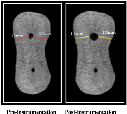
Pre-instrumentation Post-instrumentation
FIGURE 10: MICRO-CT IMAGE AT 3MM FROM APEX PTU GROUP
Pre-instrumentation Post-instrumentation
[image:58.595.163.433.430.669.2]Pre-instrumentation Post-instrumentation
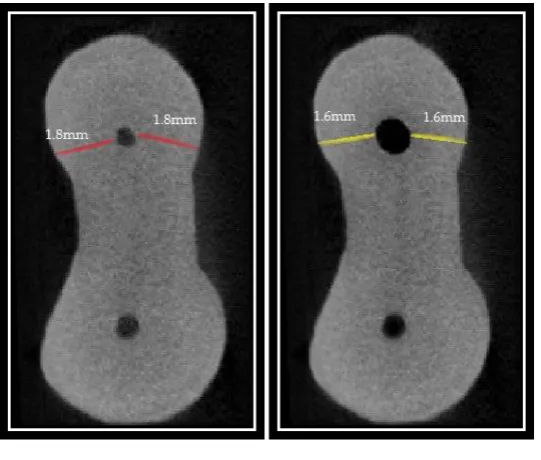
FIGURE 12: MICRO-CT IMAGE AT 9MM FROM APEX PTU GROUP
Pre-instrumentation Post-instrumentation
[image:59.595.163.434.423.659.2]Pre-instrumentation Post-instrumentation
FIGURE 14: MICRO-CT IMAGE AT 6MM FROM APEX HEDM GROUP
Pre-instrumentation Post-instrumentation
[image:60.595.167.435.406.645.2]Pre-instrumentation Post-instrumentation
FIGURE 16: MICRO-CT IMAGE AT 3MM FROM APEX WOG GROUP
Pre-instrumentation Post-instrumentation
[image:61.595.167.434.407.638.2]Pre-instrumentation Post-instrumentation
33
RESULTS
The data obtained were entered in an excel spread sheet and analyzed
using SPSS (Statistical Package for Social Sciences) V.20 software. The
confidence interval was set at 95% and p value was set for 0.05 and any value
equal to or less than was considered to be significant.
Canal transportation was analyzed using kruskal walis test followed by tukeys
post hoc test. Centering ability was analyzed using one way anova followed by
tukeys post hoc test.
Table 1, 2 and 3 shows reading at 3mm, 6mm and 9mm pre- and post-
instrumentation of three groups.
Table 4 shows canal transportation and centering ratio values of
ProTaper Universal group at 3mm, 6mm and 9mm.
Table 5 shows canal transportation and centering ratio values of
Hyflex EDM group at 3mm, 6mm and 9mm.
Table 6 shows canals transportation and centering ratio values of
WaveOne GOLD group at 3mm, 6mm and 9mm.
Table 7, Graph 1 shows comparison of canal transportation at 3mm
among PTU group, HEDM group and WOG group. The mean and SD values
of canal transportation at 3mm of PTU group, HEDM group and WOG group
34
a statistical significant difference between all three groups at 3mm. (p value
<0.05)
Table 8, Graph 2 shows comparison of canal transportation at 6mm
among PTU group, HEDM group and WOG group. The mean and SD values
of canal transportation at 6 mm of PTU group, HEDM group and WOG group
were 0.175(±.1682), 0.055(±.0887) and 0.080(±.1005) respectively. There
was a statistical significant difference between all three groups at 6 mm. (p
value <0.05)
Table 9, Graph 3 shows comparison of canal transportation at 9 mm
among PTU group, HEDM group and WOG group. The mean and SD values
of canal transportation at 9mm of PTU group, HEDM group and WOG group
were -0.20(±.1654), 0.060(±.1095) and 0.085(±.1040) respectively. There was
a statistical significant difference between all three groups at 9 mm. (p value
<0.05)
Table 10 shows pair wise comparison of canal transportation at 3 mm
6 mm and 9mm. There was a significant difference between PTU group and
HEDM group, PTU group and WOG group (p value<0.05). There was no
significant difference between HEDM group and WOG group at 3mm, 6mm
and 9 mm (p value>0.05).
Table 11, Graph 4 shows comparison of canal centering ability at 3
mm among PTU group, HEDM group and WOG group. The mean and SD
35
group were 0.4230(±.20329), 0.8000(±.25131) and 0.7325(±.30893)
respectively. There was a statistical significant difference between all three
groups at 3mm. (p value <0.05).
Table 12, Graph 5 shows comparison of canal centering ability at
6mm among PTU group, HEDM group and WOG group. The mean and SD
values of centering ratio at 6mm of PTU group, HEDM group and group 3
were 0.4405(±.22700), 0.6745(±.27888) and 0.6655(±.31660) respectively.
There was a statistical significant difference between all three groups at 6 mm.
(p value <0.05).
Table 13, Graph 6 shows comparison of canal centering ability at 9
mm among PTU Group, HEDM Group and WOG Group. The mean and SD
values of centering ratio at 9mm of PTU group, HEDM group and WOG
group were 0.4940(±.24446), 0.7070(±.25931) and 0.7250(±.25521)
respectively. There was a statistical significant difference between all three
groups at 9 mm. (p value <0.05).
Table 14 shows pair wise comparison of canal centering ability at 3
mm 6 mm and 9mm. There was significant difference between PTU group and
HEDM group, PTU group and WOG group (p value <0.05). There was no
significant difference between HEDM group and WOG group at 3mm, 6mm
TABLE 1: PROTAPER GROUP: READING AT 3MM, 6MM AND 9MM- PRE-INSTRUMENTATION AND POST- INSTRUMENTATION
Samples No: 3mm (Apical third) 6mm (Middle third) 9mm (Coronal third)
X1 X2 Y1 Y2 X1 X2 Y1 Y2 X1 X2 Y1 Y2
1 0.8 0.3 0.9 0.8 1.3 0.8 1.2 1.1 1.8 1.6 1.8 1.2
2 0.9 0.5 0.8 0.7 1.2 0.8 1.3 1.2 1.7 1.5 1.8 1.6
3 1.1 0.8 1.0 0.9 1.1 0.7 1.2 1.1 1.5 1.3 1.7 1.1
4 1.2 1.1 1.1 1.0 1.3 0.8 1.1 1.0 1.6 1.4 1.5 1.0
5 0.9 0.6 1.0 0.9 1.1 0.7 0.9 0.8 1.7 1.6 1.7 1.4
6 0.7 0.6 0.8 0.7 0.9 0.7 1.1 1.1 1.5 1.4 1.6 1.2
7 0.8 0.5 0.9 0.8 1.3 0.9 0.9 0.8 1.9 1.7 1.8 1.6
8 1.0 0.6 1.1 1.0 1.2 1.1 1.1 1.0 2.1 1.9 2.2 1.9
9 1.2 1.1 0.9 0.8 1.1 1.0 1.2 0.9 2.2 2.0 2.3 1.9
10 1.1 0.8 1.0 0.9 1.3 1.2 1.1 1.0 2.3 2.1 2.0 1.5
11 0.8 0.4 0.9 0.8 1.3 1.2 1.2 1.0 1.8 1.6 1.8 1.2
12 0.9 0.6 0.8 0.7 1.2 0.9 1.1 1.0 1.7 1.5 1.8 1.4
13 1.1 0.6 1.0 0.9 1.1 0.9 1.1 1.0 1.5 1.3 1.7 1.3
14 1.2 0.9 1.1 1.0 1.3 1.2 1.2 1.1 1.6 1.4 1.5 1.0
15 0.9 0.5 1.0 0.9 1.1 0.9 0.9 0.8 1.7 1.3 1.7 1.4
16 0.7 0.6 0.8 0.6 0.9 0.5 1.1 0.9 1.5 1.4 1.6 1.2
17 0.8 0.4 0.9 0.8 1.3 0.9 1.2 1.0 1.9 1.8 1.8 1.3
18 1.0 0.6 1.1 1.0 1.2 0.8 1.2 1.0 2.1 1.9 2.2 2.0
19 1.2 1.1 0.9 0.8 1.1 0.7 0.9 1.0 2.2 2.0 2.3 1.9
TABLE 2: HYFLEX EDM GROUP: READING AT 3MM, 6MM AND 9MM- PRE-INSTRUMENTATION AND POST-INSTRUMENTATION
Samples No: 3mm (Apical third) 6mm (Middle third) 9mm (Coronal third)
X1 X2 Y1 Y2 X1 X2 Y1 Y2 X1 X2 Y1 Y2
1 0.8 0.7 0.7 0.6 1.2 1.1 1.1 1.0 1.8 1.6 1.7 1.6
2 0.7 0.5 0.9 0.8 1.1 1.0 1.2 1.1 1.6 1.4 1.7 1.5
3 1.0 0.9 0.9 0.8 1.3 1.1 1.1 1.0 1.5 1.1 1.6 1.4
4 0.8 0.6 0.8 0.7 0.9 0.7 1.1 1.0 1.3 1.1 1.5 1.4
5 0.7 0.6 0.8 0.7 1.1 1.0 1.2 1.1 1.9 1.6 1.8 1.6
6 1.1 0.9 1.0 0.9 1.0 0.8 1.1 1.0 2.0 1.7 1.8 1.6
7 1.2 1.1 0.9 0.8 1.2 1.0 1.0 0.9 1.4 1.0 1.6 1.4
8 0.7 0.5 0.9 0.8 1.3 1.0 1.2 1.1 1.8 1.6 1.7 1.5
9 1.0 0.9 1.0 0.9 1.0 0.9 0.9 0.7 1.6 1.4 1.7 1.6
10 1.1 0.9 0.7 0.6 0.9 0.8 1.0 0.9 1.5 1.4 1.6 1.5
11 0.8 0.6 0.7 0.6 1.2 1.1 1.1 0.9 1.8 1.6 1.8 1.6
12 0.7 0.6 0.9 0.8 1.1 1.0 1.2 1.1 1.6 1.4 1.6 1.2
13 1.0 0.8 0.9 0.8 1.3 1.1 1.1 1.0 1.5 1.3 1.5 1.3
14 0.8 0.6 0.8 0.7 0.9 0.6 1.1 1.0 1.3 1.0 1.3 1.2
15 0.7 0.6 0.8 0.6 1.1 0.9 1.2 1.1 1.9 1.7 1.9 1.7
16 1.1 0.9 1.0 0.8 1.0 0.7 1.1 1.0 2.0 1.9 2.0 1.9
17 1.1 0.9 0.9 0.7 1.2 1.1 1.0 0.9 1.4 1.1 1.3 1.3
18 0.7 0.6 0.9 0.8 1.3 1.1 1.0 1.1 1.8 1.5 1.8 1.6
19 1.0 0.8 1.0 0.9 1.0 0.9 0.9 0.8 1.6 1.2 1.6 1.4
TABLE 3: WAVEONE GOLD GROUP: READING AT 3MM, 6MM AND 9MM- PRE-INSTRUMENTATION AND POST-
INSTRUMENTATION Samples No: 3mm (Apical third) 6mm (Middle third) 9mm (Coronal third)
X1 X2 Y1 Y2 X1 X2 Y1 Y2 X1 X2 Y1 Y2
1 1.1 0.8 0.8 0.7 1.2 1.1 1.1 1.0 1.4 1.2 1.6 1.2
2 0.8 0.7 0.9 0.8 0.9 0.6 1.2 1.1 1.8 1.6 1.7 1.5
3 1.1 0.8 1.0 0.9 1.1 1.0 1.2 1.0 1.7 1.6 1.7 1.3
4 1.2 1.1 1.1 1.0 1.0 0.7 0.9 0.8 1.5 1.3 1.6 1.4
5 0.9 0.7 1.1 1.0 1.1 0.5 1.2 1.1 1.6 1.5 1.7 1.3
6 1.0 0.7 0.9 0.8 1.2 1.1 1.1 1.0 1.5 1.3 1.7 1.3
7 0.8 0.7 0.9 0.7 1.0 0.7 0.9 0.8 1.4 1.2 1.6 1.2
8 0.9 0.8 1.1 1.0 0.9 0.8 0.9 0.8 1.8 1.6 1.8 1.4
9 1.0 0.9 1.2 1.1 1.2 1.0 1.1 1.0 1.9 1.7 2.0 1.6
10 0.9 0.7 1.1 1.0 1.3 1.2 1.2 1.1 1.6 1.4 1.7 1.3
11 1.1 1.0 0.9 0.8 1.2 1.0 1.2 1.1 1.4 1.2 1.5 1.1
12 0.8 0.7 0.9 0.8 0.9 0.6 0.9 0.8 1.8 1.6 1.7 1.3
13 1.1 1.0 1.0 0.9 1.1 0.8 1.0 0.9 1.7 1.5 1.6 1.4
14 1.2 1.1 1.1 1.0 1.0 0.9 0.9 0.8 1.5 1.3 1.7 1.5
15 0.9 0.6 0.8 0.7 1.1 1.0 1.0 0.9 1.6 1.4 1.8 1.6
16 1.0 0.9 0.9 0.8 1.2 1.1 1.0 0.9 1.5 1.3 1.7 1.5
17 0.8 0.7 0.9 0.8 1.0 0.9 0.8 0.7 1.4 1.2 1.6 1.4
18 0.9 0.6 1.0 0.9 0.9 0.6 0.9 0.8 1.8 1.6 1.8 1.6
19 1.0 0.9 1.1 1.0 1.2 1.1 1.0 0.9 1.9 1.7 1.7 1.5